comparison of four different embolic materials for uterine
TRANSCRIPT
Yale UniversityEliScholar – A Digital Platform for Scholarly Publishing at Yale
Yale Medicine Thesis Digital Library School of Medicine
2009
Comparison of Four Different Embolic MaterialsFor Uterine Artery Embolization In Post-ProcedureMRI EnhancementSteven David AbramowitzYale University
Follow this and additional works at: http://elischolar.library.yale.edu/ymtdl
Part of the Medicine and Health Sciences Commons
This Open Access Thesis is brought to you for free and open access by the School of Medicine at EliScholar – A Digital Platform for ScholarlyPublishing at Yale. It has been accepted for inclusion in Yale Medicine Thesis Digital Library by an authorized administrator of EliScholar – A DigitalPlatform for Scholarly Publishing at Yale. For more information, please contact [email protected].
Recommended CitationAbramowitz, Steven David, "Comparison of Four Different Embolic Materials For Uterine Artery Embolization In Post-ProcedureMRI Enhancement" (2009). Yale Medicine Thesis Digital Library. 108.http://elischolar.library.yale.edu/ymtdl/108
Comparison of Four Different Embolic Materials For Uterine Artery Embolization In
Post-Procedure MRI Enhancement
A Thesis Submitted to the
Yale University School of Medicine
in Partial Fulfillment of the Requirements for the
Degree of Doctor of Medicine
by
Steven David Abramowitz
2009

2
COMPARISON OF FOUR DIFFERENT EMBOLIC MATERIALS FOR UTERINE
ARTERY EMBOLIZATION IN POST-PROCEDURE MRI ENHANCEMENT
Steven D. Abramowitz, Gary M. Israel, Shirley M. McCarthy, Jeffrey S. Pollak, Robert I. White Jr., and
Michael G. Tal. Department of Radiology, Yale University, School of Medicine, New Haven, CT.
The aim of this study was to assess embolic agent equivalency in uterine artery
embolization (UAE) using post-procedure MRI enhancement of uterine fibroids in
patients embolized using Embosphere Microspheres, (EM) Contour SE spheres (CSE),
Poly-Vinyl Alcohol particles (PVA) and Bead Block spheres (BB).
A total of 84 women with 6-month MRI follow-up constituted this retrospective
study. Within this group, 25 women were treated with PVA, 23 were treated with CSE,
19 were treated with EM and 17 were treated with BB. Pre- and post-procedure MRI
exams were analyzed for the total number of fibroids present in the uterus of each patient
and the percentage individual fibroid enhancement of each fibroid was scored in quartile
intervals. The overall percentage change in enhancement was then calculated for each
patient. Bivariate analysis using Generalized Linear Modeling and one-way ANOVA was
used to assess differences in infarction by different embolic materials.
Of patients treated with PVA and EM, there was a mean reduction in
enhancement by 76.60% and 83.07%, respectively, compared to a mean reduction of
52.53% and 49.78% in patients treated with CSE and BB, respectively. There was a
statistically significant difference between CSE or BB and EM or PVA.
Patients treated with BB and CSE demonstrate a reduced degree of infarction on
follow-up MRI than those patients treated with PVA or EM.

3
Acknowledgements
I would like to thank Dr. Michael Tal for his support and guidance over the past
few years. Without his trust and mentorship, this work would not have been possible.
I would also like to thank Drs. Gary Israel and Shirley McCarthy for their role in
publishing this work.
4
Table of Contents
INTRODUCTION................................................................................................................5
BACKGROUND..................................................................................................................5
HISTORY OF ALTERNATIVE TREATMENT METHODS.....................................................6
MEDICAL THERAPY ......................................................................................................6
SURGICAL INTERVENTION .............................................................................................7
HYSTERECTOMY ........................................................................................................8
MYOMECTOMY ..........................................................................................................9
UTERINE ARTERY EMBOLIZATION...............................................................................10
TECHNICAL ASPECTS OF UAE ....................................................................................11
OUTCOMES..................................................................................................................12
COMPARISON TO HYSTERECTOMY ..............................................................................14
COMPARISON TO MYOMECTOMY ................................................................................16
COMPARISON TO ALL SURGICAL INTERVENTIONS .......................................................17
RISKS ..........................................................................................................................18
EMBOLIC AGENT OPTIONS ..........................................................................................19
PURPOSE..........................................................................................................................22
MATERIALS AND METHODS.......................................................................................22
PATIENT POPULATION...................................................................................................22
UAE PROCEDURE..........................................................................................................23
MRI SPECIFICATIONS ...................................................................................................25
IMAGE ANALYSIS...........................................................................................................25
DATA ANALYSIS.............................................................................................................26
RESULTS ..........................................................................................................................27
DISCUSSION ....................................................................................................................27
FIGURES AND TABLES..................................................................................................32
BIBLIOGRAPHY..............................................................................................................36
5
Introduction
Background
First described in 1793 by Matthew Baillie of St. George’s Hospital, London,
uterine leiomyomata are the most common benign tumor of the female reproductive
track. Symptomatic uterine leiomyomata (commonly referred to as “fibroids”) may be
found in up to 25% of all women and up to 40% of all women over the age of 40. (1, 2)
Although uterine fibroids are benign, lack premalignant characteristics, and rarely
damage adjacent anatomic structures, they can cause severe menorrhagia, pain, bulk-
related symptoms and lead to subfertility.
Traditionally, women seeking medical assistance for fibroid management are
treated surgically, as there are no proven effective long-term medical therapies available.
However, for many women, surgical intervention represents an undesirable solution.
Surgical therapies such as hysterectomy or myomectomy are invasive and may render
patients infertile or with questionable fertility. Furthermore, these surgeries both involve
considerable morbidity. (3) Thus, there an increasing need for a minimally invasive
treatment alternative emerged from both patients and physicians.
Encouraged by the observation that there was an improvement in fibroid-related
symptoms after patients underwent uterine artery embolization (UAE) for postpartum
hemorrhage, Rivina et al first described UAE as an acceptable treatment alternative for
symptomatic uterine fibroids in 1995. (4) The demand for UAE for fibroid management
was immediately apparent and numerous publications surfaced touting the new minimally
invasive method of reducing fibroid-related symptom without the need for surgery. (5, 6)
6
It was this subsequent work that has since led to the development of UAE as a tool for
minimally invasive, uterus-preserving fibroid management.
Over the past 14 years, multiple embolic agents have become commercially
available and have been approved by the Food and Drug Administration (FDA) for use in
the procedure. (7, 8)Numerous technical refinements have also been made to the
procedure, such as the use of microcatheters, in order to improve post-procedure
outcomes. While technical improvements to UAE have been carefully researched and
scrutinized, not as much attention has been focused on comparing and assessing the
newly emerging embolic agents responsible for the ultimate success of the procedure. (9)
It is this gap in knowledge that we hope to narrow.
History of Alternative Treatment Methods
Medical Therapy
For decades, physicians have idealized a pill with minimal side effects that would
lead to fibroid regression and symptom resolution without impacting a woman’s fertility
status. To date, no effective medical therapy exists that meets these criteria. However,
there are multiple medical therapies available for the short-term management of fibroid
symptoms. Most of these therapies are hormonal in nature and rely on manipulating the
known mechanisms of fibroid development.
Since the discovery and initial synthesis of gonadotropin-releasing hormone
(GnRH), translational research has sought to apply hormone analogues to the treatment of

7
fibroids. Gonadotropin-releasing hormone analogues (GnRHa) are the mostly widely
used derivatives of this work. GnRHa can cause fibroid regression, limit menorrhagia and
reduce bulk-related symptoms but only after an initial flare and worsening of symptoms.
(10) However, they may only be employed for a short period of time as they cause
significant osteopenia in pre- and peri-menopasal women. (11, 12) Gonadotropin-
releasing hormone antagonists avoid the symptom flare associated with GnRHa, but carry
a similar side-effect profile. Cessation of both GnRH-based therapies causes marked
rebound growth.
Selective estrogen receptor modulators have been studied, as well, but they are
proven efficacious only in post-menopausal women. Thus, they do not benefit the large
majority of fibroid sufferers. This is also true with aromatase inhibitors. (10) In the pre-
menopausal population, progesterone antagonists, such as mifepristone and asoprisnil,
cause significant regression in fibroid size, but they, and similar progesterone therapies
like danazol and gestrinone, are not compatible with reproduction.
As a result, most medical therapies are commonly utilized in the pre-operative
period to reduce leiomyoma size.
Surgical Intervention
Traditionally, two surgical procedures are utilized in fibroid management:
hysterectomy and myomectomy. Although both procedures have considerable morbidity,
they are performed with relative frequency.(3) Annually, there are almost 600,000
hysterectomies performed in the United States, of which 30% to 70% are for fibroid
8
management. (13, 14) In comparison, each year there are approximately 35,000
abdominal, laproscopic and hysteroscopic myomectomies performed for fibroid
management. (15)
Hysterectomy
Hysterectomy is the gold standard treatment of uterine leiomyomata as excision
of the uterus and accompanying fibroids allow for no chance of recurrence. However, as
stated before, it can be an undesirable and an extreme treatment in those patients who
suffer from mild to severe fibroid symptoms as it renders the patients infertile. In many
women, hysterectomy can also result in issues of sexual dysfunction and psychological
side effects including depression and a loss of femininity. (16, 17)
Despite the frequency with which the surgery is performed, it is not without
significant risk of complications. Hysterectomy carries a major morbidity rate of 3%, a
minor morbidity rate of 14% and a mortality rate of 1-2 women per 1000 operations
performed in major medical centers. (18) The average patient will spend 5.1 days in the
hospital after the operation.
For those women who have no complications in the peri-operative period, there
are still long-term risks. There is a considerable risk of urinary stress incontinence as
reported by Altman et al. Altman et al looked at 165260 women who underwent
hysterectomy and compared their long-term outcomes to a control group of 470506
women who did not have the operation. They found that rate of urinary stress
incontinence per 100000 person-years was 179 [95% CI 173-186] in the exposed cohort

9
versus 76 [95% CI 73-76] in the unexposed cohort. (19) There has also been a
documented increased risk for the need for subsequent pelvic prolapse surgery at a later
stage.
Myomectomy
Myomectomy is a surgical alternative to hysterectomy. During myomectomy,
fibroids are surgically removed from the myometrium and the uterus is preserved since
reconstruction is performed. The operation may be performed laproscopically,
abdominally or hysteroscopically, although hysteroscopic myomectomy is reserved for
small, submucosal fibroids. (20)
Depending upon the method of myomectomy, the surgery carries a major
morbidity rate of 2% to 5%, a minor morbidity rate of 15% to 29% and a mortality rate of
2 women per 1000 operations performed in major medical centers. (21) As the uterus is
highly vascular, all methods of myomectomy carry a risk of hemorrhage with up to 20%
of cases requiring transfusion. The vascular nature of the uterus also contributes to its
adhesiogenic properties. Laproscopic or abdominal myomectomy may result in
significant intra-abdominal adhesions leading to short- and long-term complications such
as small bowel obstruction. (22)
Patients undergoing myomectomy have an average hospital length of stay of 3.8
days during which there is a high incidence of fever. Patients are also observed for
infection, continued bleeding, visceral damage from trocar placement, uterine perforation

10
and thromboembolism. These and intra-operative complications lead to an intra-operative
and short-term hysterectomy conversion rate of 2%. (21)
In the long-term, myomectomy patients have a significant rate of fibroid
recurrence. A review of 41 studies found the risk for symptomatic fibroid recurrence was
20% to 50% 5 years from the operation. (23) Women who have had a myomectomy are
also at risk for rupture during gestation. With respect to fertility, research reports mixed
results after myomectomy. However, the majority of literature seems to report that
fertility is improved by myomectomy, as is the risk of miscarriage, when compared to
those women who did not undergo surgical intervention for fibroid management. (24)
Uterine Artery Embolization
Prior to 1995, uterine artery embolization had been successfully used for
refractory postpartum hemorrhage, for bleeding after gynecologic surgery or pelvic
trauma and for embolization of pelvic artiovenous malformation. (6, 25) It was in 1995
that Ravina et al modified the technique for use in fibroid management after noticing
fibroid symptom improvement in patients that had undergone UAE for one of the above
stated reasons. (4) The procedure achieves success without uterine compromise because
blood to leiomyoma comes from end arteries and lack collateral flow supply. Therefore,
fibroids are preferentially affected by flow reduction as compared normal myometrium
that quickly acquires new blood flow from collateral vessels.(26)
Technical Aspects of UAE

11
The goal of UAE for effective fibroid treatment is the complete devascularization
of all fibroids via the introduction of particulate embolic agent into both uterine arteries.
(26) To achieve this goal, the patient is first given analgesia so that they may tolerate the
procedure. The type of analgesia varies depending upon the interventionalists performing
the embolization. Local anesthesia, epidural anesthesia, spinal anesthesia, conscious
sedation, and general anesthesia have all been reported. (27)
Once anesthesia is obtained, access is typically achieved via the catheterization of
a single femoral artery and pelvic arteriography is performed in order to define the
patient’s vasculature. Bilateral femoral access may be achieved with the benefit of
reducing radiation exposure and procedure time, however it is generally more
cumbersome, thus not done. (26) Once femoral access and arteriography is accomplished,
the contralateral uterine artery is then selectively catheterized. The artery is identified
using subtraction angiography and arteriography is performed in order to assess uterine
artery anatomy and the presence of any ovarian collateral vasculature. (25) If the vascular
anatomy is deemed to be suitable, the embolic agent is then introduced until the agent’s
angiogenic endpoint is reached. This procedure is then repeated on the ipsilateral uterine
artery.
The procedure usually lasts approximately 1 hour and will expose the patient to
an average of 20rads of radiation. (5, 28) However, these factors are incredibly operator
dependant. (29)Patients are usually admitted overnight for observation and pain
management using non-steroidal anti-inflammatory drugs or narcotics. Extension of
hospital stay for pain management beyond one night is not uncommon.

12
The standard of care aims for a technical success rate of 96% in uterine artery
embolization. (30) The success rate of the procedure may be adversely affected by artery
tortuosity, small vessel caliber and anatomic variants. However, the introduction of
microcatheters aids the interventionalist in overcoming these obstacles. (31) Some
interventionalists choose to microcatheters exclusively to avoid arterial spasm, which, if
it occurs, may negatively impact technical success rates. If spasm does occur,
antispasmodics, such as glyceryl trinitrate, may be administered.
Outcomes
The ideal treatment for uterine leiomyomata would eliminate symptoms, reduce
tumor size, limit fibroid recurrence and preserve the patient fertility. Uterine artery
embolization accomplishes some, if not all, of these objectives. While the data supporting
uterine artery is overwhelmingly based in case series or prospective oberservational
studies, the results tend to be consistent.
UAE relieves fibroid-related symptoms in the vast majority of patients. (29, 30,
32) In a study of 5 year long-term results, Kim et al found that 85% to 95% of patients
reported a reduction in menorrhagia and 40 to 60% of patients reported a reductions in
bulk-related symptoms. (33) This is consistent with 2 month to 4 month magnetic
resonance (MR) imaging follow-up that show a decrease of uterine volume from 40% to
60%. (6) The same study also indicates that when technical success is achieved, the
reduction of uterine volume continues over time. Further studies have proved that the
reduction of fibroid symptomatology following UAE is unrelated to the number of
13
fibroids presents, their location or their size and solely related to successful achievement
of embolic agent angiogenic endpoint. (34)
Patients treated with UAE have relatively low rates of short-term recurrence.
Approximately 10% of patients treated with UAE will have a recurrence of their
symptoms at 1 year. Of those patients who present with symptom recurrence, 87.5% are
due to new leiomyoma formation and only 12.5% are due to fibroid regrowth. (35) Long-
term studies assessing fibroid recurrence after UAE indicate that 30% of patients will
have new leiomyoma formation detected by MR imaging, with only 13% of patients
reporting symptoms. (33)
With respect to a patient’s sexuality and mental health, studies indicate that over
surgical procedures, those patients who underwent UAE have significantly improved or
maintained body image. (36) Many women report improved pleasure from and increased
habit of sexual intercourse after UAE. (37) Moreover, 3 months after UAE women have a
statistically significant improvement in sexual function and psychological well-being.
Currently, there lacks sufficient date to conclude the impact UAE has on fertility.
(38)Fibroids, especially those that are submucosal, adversely impact fertility via uterine
distortion. (39) There are no published prospective studies regarding fertility in UAE
patients that document women who attempt pregnancy after the procedure, therefore
cycle, conception and fecundity rate are not available. However, the Society of
Interventional Radiology Foundation is in the process of gathering this data as of 1999
with the creation of a national fibroid registry. To date, anecdotal evidence is positive
with many centers reporting normal full-term pregnancy in patients status post UAE.
14
Limited data indicates little to no relation between UAE and impact on gestational term
or complication rates.
Concern does exist that ovarian function may be compromised as a result of
embolic material in the ovarian blood supply. (6) There is also a question as to whether or
not decreased vascular supply of the uterine myometrium and endometrium after UAE
impacts embryo implantation. However, histological evidence at this point does not
indicate any significant impact on the surrounding myometrium or ovary despite the
presence of embolic agent in these tissues. (40)
Comparison to Hysterectomy
Until recently, there was little high quality data comparing UAE to hysterectomy.
However, in the past 5 years, two randomized controlled trials producing Level 1 data
have been published comparing these two treatment modalities.
The first trial was entitled, “Embolization vs. Hysterectomy for Symptomatic
Uterine Fibroids (EMMY).” This prospective, randomized multicenter trial had two year
follow-up for patients in both cohorts. The primary endpoint was the prevention of
hysterectomy in 75% of those cases receiving UAE. Secondary endpoints regarding
length of hospital stay, complication and reintervention rates, volume reduction, quality
of life measures and costs were also assessed.
Of those patients who underwent UAE for fibroid management, only 23.5%
underwent hysterectomy at the 2 year endpoint. (41) Thus, the trial established that UAE
is a non-inferior method of fibroid therapy in comparison to hysterectomy. The
15
reintervention rate for UAE was 28.4% as compared to the reintervention rate of 8% for
the surgical arm of the study. A large component of the reintervention rate in those
patients who underwent UAE was due to technical failure. Many critics of the EMMY
trial argue that this is indicative that the multicenter nature of the trial let to the inclusion
of inexperienced sites leading to technique error.
Secondary endpoints in the EMMY trial indicate that there were no statistically
significant differences in the major and the minor complication rates between UAE and
hysterectomy. However, those patients who underwent UAE had a statistically significant
less hospital length of stay at 2.5 days compared to the 5.1 days spent in the hospital
status post hysterectomy. (41) Patients who underwent UAE also returned to work more
quickly and with less pain in the first 24 hours in comparison to those patients who
underwent hysterectomy.
The second trial was entitled, “Hysterectomy or Percutaneous Embolization for
Uterine Leiomyomata (HOPEFUL).” This retrospective cohort study analyzed outcomes
from patients who underwent either UAE or hysterectomy in the UK. Of those patients
who underwent UAE, 85% reported a reduction in their symptoms as opposed to 95% of
those patients who underwent hysterectomy. (42) However, there were fewer major or
minor complications, 19%, in those patients in the UAE cohort as opposed to 26% in the
hysterectomy cohort. Additionally, economic data from the study suggest UAE was less
expensive at £2536 versus £8283 for hysterectomy even with the cost of subsequently
needed reintervention. (43) Quality of life adjusted years was not statistically significant
until the value of retaining the uterus was factored into the calculation.
16
Comparison to Myomectomy
Currently, there are no large-scale, published randomized controlled trials
evaluating the outcomes of myomectomy alone versus UAE for the management of
fibroid tumors. However, there are extensive publications documenting institutional
results. Broder et al compared long-term outcomes of patients treated with abdominal
myomectomy versus those treated with UAE. They found that women who were
embolized were more likely to require further invasive treatment for management of
leiomyomata, at 29% versus 3%. (13) However, in those patients who underwent UAE
and did not require subsequent reintervention, 92% of emoblization patients and 90% of
myomectomy reported symptomatic resolution. Women who underwent embolization
were more likely to be satisfied with their choice of therapy, though, at 94% versus 79%
for myomectomy.
The best comparisons regarding fibroid recurrence between those patients treated
with myomectomy and those treated with UAE are not head-to-head studies at this point,
but rather large single procedure research. Currently, the accepted recurrence rate of
fibroids in myomectomy patients at 2 years is 15% to 20%. (1) Fedele et al indicated,
though, that after myomectomy, up to 50% of patients show new leiomyoma
development at 5 years. (44) This is more, although statistical significance is not know,
than the accepted recurrence rate of 10% for fibroids after UAE at 1 year and a rate of
30% regrowth at 5 years. (33, 35) Myomectomy, though, tends to be associated with
greater morbitidy, even though the coversion rate to hysterectomy in both myomectomy
and UAE is comparable at about 1% for each. (38)
17
There are also no randomized controlled trials comparing fertility outcomes after
UAE versus myomectomy. Currently, there exists a large body of conflicting evidence
based on institutional experience with both procedures. There, however, limited studies
indicating that patients may have better rates of conception and lesser rates of miscarriage
after myomectomy in comparison to UAE. (6) One small retrospective study suggests
that patients may have higher preterm and malpresentation rates after UAE as opposed to
myomectomy. (45) Much more research needs to be conducted regarding this issue,
though, before any definitive comparisons may be made.
Comparison to all Surgical Interventions
The “REST” trial (Randomized Controlled Trial of Emblization vs. Surgical
Treatment for Fibroids) was a multicenter government funded randomized controlled trial
comparing all surgical interventions to uterine artery embolization. Its endpoint was the
quality of life at 1 year status post intervention based on the Medical Outcomes Study 36-
Item Short-form General Health Survery (SF-36). Although power calculation dictated an
enrollment of 200 patients (90% power to detect a 10 point change in SF-36), only 80%
power was achieved. Nevertheless, there was no statistically significant difference in
mean SF-36 at 1 year between the UAE and surgical cohorts.
UAE had a higher minor complication rate of 34% versus 20% for surgical
outcomes; however, most minor complications for UAE were temporally related and
relatively near the time of the intervention. There was no statistically significant
difference in the major complication rate between the two study cohorts. There was a

18
statistically significant difference in the length of hospitalization and 24 hour pain scores
favoring UAE. Additionally, UAE was cheaper, on average, than surgical intervention at
£1751 versus £2702.
Overall, the study concluded that UAE was a safe and effective alternative to
surgery.
Risks
While UAE has proven to be a safe minimally invasive alternative to surgery for
fibroid management, it is not without its risk. The major complication rate of UAE is
between 1% and 5%. (46, 47) Serious infection resulting in extended hospitalization and
major morbidity has been reported in 1% to 2% of patients with increased frequency with
large fibroid infarction. (48) In 1% of patients, subsequent hysterectomy is needed
usually to combat degenerating leiomyoma infection or severe postembolization
syndrome.
Postembolization syndrome is most often one of the minor complications in the
post-procedure period. Nearly one third of patients post-embolization will have low-
grade pyrexia, discomfort and malaise lasting 3 days to 7 days. (27) These symptoms,
accompanied by leukocytosis secondary to leiomyoma infarction and necrosis as well as
nausea and vomiting represent postembolization syndrome. This occurs in nearly 15% of
patients after UAE and may require readmission for monitoring.
Many patients also complain of chronic vaginal discharge after UAE. Between
4% ad 7% of patients have chronic vaginal discharge as a result of fluid accumulation in
19
the infracted leiomyoma that is able to communicate with the endometrial cavity. (46, 49)
Spontaneous resolution of the discharge occurs in nearly 94% of patients, however in the
remaining population, hysteroscopic resection of the necrotic fibroid is curative and may
be required. In fact, post-embolization, nearly 10% of patients require hysteroscopy for
fibroid extrusion for various reasons. (5, 28, 48)
Ovarian failure leading to premature menopause has also been reported in 1% to
2% of patients. (50) This is most frequently reported in peri-menopausal women. Ovarian
failure is likely due to embolization of the ovary via undetected collateral utero-ovarian
arteries. Therefore, although promising for symptom relief, UAE contains unique
morbidity risks such as postembolization syndrome, infection, primary ovarian failure,
etc. (51)
Embolic Agent Options
Evidence indicates that complete devascularization of all leiomyomata is required
for effective treatment of and symptomatic resolution of uterine fibroids. (52) However,
the choice of embolic agent in achieving this goal has remained a divisive topic. Current
commercially available embolic agents vary widely by size, shape, composition, method
of delivery and accepted angiographic endpoint. Published research may be found both
attesting to the efficacy as well as highlighting the failure of each. As more embolic
agents become available, the quagmire expands.
Until 2002, there were only two FDA approved embolic agents for use in UAE.
The first embolic agent is non-spherical poly-vinyl alcohol (Contour; Target Vascular,
20
Boston Scientific, Marlborough, MA). First used in UAE by Ravina at al, it remains the
industry gold standard and is the most commonly utilized particulate embolic material
used in UAE. (4, 8) PVA is considered to be a permanent embolic against since it is not
biodegradable. (53) Histologic exam after UAE with PVA indicates that the variation in
particle size tends cause more proximal than distal vessel occlusion. Occlusion also
appears to be secondary to platelet aggregation and thrombus formation in the
intraluminal lattice of polyvinyl alcohol particle as the particles were noted not to occupy
the entire lumen of the emoblized vessel. (54, 55) This has raised the theoretical concern
of recanalization of fibroid vasculature via distal non-embolized vessels. (56) However,
the clinic impact appears to be insignificant with complete infarction of uterine
leiomyomata being achieved after UAE with PVA so long as the endpoint of complete
vessel occlusion is achieved. (29)
The second embolic agent used during this time period was Gelfoam, a water-
insoluble hemostatic embolic prepeared from purified skin gelatin. Gelfoam had been
frequently used as a biodegradable, intravascular embolic agent prior to its application in
UAE. Gelfoam promotes hemostasis by providing structural support to thrombus
formation and by causing an acute foreign body reaction. (53) Gelfoam resorption
typically occurs within 7 to 21 days of particulate introduction leading to recanalization
in 3 weeks to 4 months after UAE. (57) However, despite comparable outcomes to PVA,
Gelfoam still is not as widely used as PVA or other embolic agents introduced after 2002.
In November 2002, tris-acryl gelatin microspheres (EM; Embosphere; Biosphere
Medical, Rockland, MA) received UAE indication from the FDA and rapidly gained
market share becoming the embolic material of choice for UAE. (8) Tris-acryl gelatin

21
microsphere are thought to offer a theoretical advantage over non-spherical PVA as they
are more uniform in nature and have been found to have more distal penetration on
histology than non-spherical PVA. (58) This leads to a theoretically greater uniform and
targeted embolization of the perifibroid plexus when the embolic endpoint, or “pruned-
tree appearance,” is reached. (9, 56) However, a prospective randomized study
comparing PVA and EM found no statistically significant difference in the clinical or
imaging outcomes of the two embolic agents. (59)
In effort to recapture lost market share, spherical PVA (CSE; Contour SE; Boston
Scientific, Natick, MA) was developed using the chemical properties of non-spherical
PVA reengineered into the successful spherical shape of EM. In March 2004, after a
preliminary animal study suggested embolic equivalence to non-spherical PVA, the FDA
approved CSE for use in the treatment of uterine fibroids. (60) The same study indicated
that the inflammatory response to CSE was significantly less than to that of other agents
tested. This may correlated to recently published data suggesting that there exists a
statistically significant difference in the clinical outcomes of those patients undergoing
UAE with CSE as opposed to EM. (9) However, there are not randomized controlled
trials comparing CSE to other embolic agents.
The most recent embolic agent approved for use in UAE by the FDA is spherical
poly-vinyl alcohol hydrogel (BB; Bead Block; Biocompatibles Farnham, UK). Similar in
composition and chemical structure to spherical PVA (CSE), there are currently no
clinical studies demonstrating its effectiveness as compared to other materials. There are
also no histologic studies evaluating the means and method of the agent’s embolic
effects. Despite this fact, a Terumo survey of Interventional Radiologists at the 2005

22
Society of Interventional Radiology showed that 72% of interventional radiologists
surveyed believe that BB was a superior embolic agent to PVA and EM. Given concerns
regarding spherical PVA, careful assessment of the extent of fibroid infarction after
embolization using BB with contrast-enhanced MRI is necessary.
Purpose
Given the lack of reliable data regarding newly released embolic agents, the
purpose of our study was to compare the efficacy of four commonly used embolic agents:
Embosphere Microspheres, Contour SE spheres, Poly-Vinyl Alcohol particles and Bead
Block spheres. We tested the hypothesis that not all embolic materials are equivalent,
using post-procedure MRI fibroid enhancement in patients treated by UAE. The
commercially available embolic material evaluated were Embosphere Microspheres
(EM), Contour SE spheres (CSE), Poly-Vinyl Alcohol particles (PVA) and Bead Block
spheres (BB).
Materials and Methods
Patient Population
Upon receiving approval from our institutional human investigations committee
for waiver of consent of medical record review, 84 women were enrolled in our Health
Insurance Portability and Accountability compliant retrospective study. Criteria for study
inclusion were as follows: pre-menopausal females 25-45 years of age, a clinical
23
diagnosis of symptomatic leiomyomas and pre- and 6 month post-UAE MRI at our
institution. All patients included in the study underwent UAE at our institution between
December of 2004 and January of 2006.
Each patient underwent UAE with one of the four embolic agents included in our
study. A total of 84 women had uneventful procedures resulting in bilateral embolization
per embolic agent endpoint. The mean time to 6 month post-UAE follow-up MRI was 6.6
months with a range of 5.2-8.1 months.
UAE Procedure
Patients underwent standard bilateral UAE performed by one of three experienced
interventional radiologists: MT, JP and RW with 8, 15 and 25 years experience
respectively at the time our study was conducted. Prior to catheterization, patients
received an intravenous line as well as a local anesthesia. Pelvic aortography was
acquired using an Omniflush Catheter (Angiodynmaics, Queensbury, NY) from the level
of the renal arteries in order to evaluate collateral blood supply to the fibroids.(61)
Uterine artery access was achieved through a 5-F catheter with the use of a
coaxial 3-F microcatheter (Renegade Hi-Flow, Target Therapeutics/Boston Scientific,
Fremont, CA). The microcatheter was advanced over a micro-guide wire using contrast
as a guide. The microcatheter was maneuvered into the proximal portion of the transverse
segment of the uterine artery. Embolization was then performed. In the event of spasm,
embolization was postponed until resolution.
24
Women received one of four embolic agents: 25 women were treated with Poly-
Vinyl Alcohol particles (Contour; Target Vascular, Boston Scientific, Marlborough,
MA),19 were treated with trisacryl gelatin microspheres (Embosphere; Biosphere
Medical, Rockland, MA), 23 were treated with spherical PVA (Contour SE; Boston
Scientific, Natick, MA) and 19 were treated with compressible microspheres (Bead
Block; Biocompatibles Farnham, UK). The selection of embolic agent was based upon
agent availability and the preferences of the interventional radiologist performing the
procedure. Preparation of the selected embolic agent varied according to the product
specific specifications. PVA (300-500 µm), CSE particles (500-700 and 700-900 µm)
and BB (500-700 and 700-900 µm) were suspended in 6-7mL of normal saline and 6-
7mL of nonionic contrast solution. EM (500-700 µm) were mixed with 10mL of contrast
to obtain a 50% concentration of contrast in the syringe. Embolization endpoint was also
agent dependent. Sluggish forward flow in the main transverse or ascending uterine
artery was the angiogenic endpoint for CSE and PVA, whereas sluggish flow with a
“pruned-tree” appearance was the angiogenic endpoint for BB or EM.
Following embolization of both uterine arteries, pelvic aortography was repeated
in order to evaluate uterine artery perfusion as well as to evaluate for the presence of
collateral blood supply to the uterus. In some cases where ovarian artery collateral flow
was observed, the ovarian arteries were embolized to prevent recurrence of
symptoms.(62)
MRI Specifications
25
Gadolinium-enhanced MR imaging was performed before and approximately 6-
months after UAE for evaluation of fibroids. Imaging was performed using a 1.5 T
scanner (GE Signa, Milwaukee, WI). Sequences obtained were a localizer, fat-suppressed
sagittal fast spin-echo scans (TR 6200/TE 104), coronal fast spin-echo scans (TR
3900/TE 103), axial T1 weighted gradient echo scans (TR 180/TE 2.3, 4.8), and coronal
3D gradient echo (TR 5.9/1.2/40) pre and post IV gadolinium administration
(0.1mmol/kg). A timing bolus was used to obtain the arterial phase and three post
contrast-enhanced scans were obtained during the arterial phase and then at 60, and 120
seconds after gadolinium administration. Subtraction images of the pre- and post-
gadolinium scans were subsequently performed. The field of view varied from 20 to 28
cm and the matrix was 256 x 256.
Image Analysis
Images were evaluated for leiomyoma number and leiomyoma enhancement.
There was no minimal fibroid size established for inclusion in the study and the number
of fibroids found in each patient ranged from 1 to 38.The number of fibroids in the uterus
and their subsequent individual enhancement was determined by a radiologist, GI, with
eight years experience reading pelvic MRI. The radiologist was blinded to the type of
embolic agent used and all measurements were taken at the same reading station utilizing
the same reading methodology to control for multiple measurements. On the pre- and
post-UAE MRI scans, an enhancement score of 0%, 25%, 50%, 75%, or 100% was
assigned to each fibroid based on a qualitative estimate of percentage volume showing
26
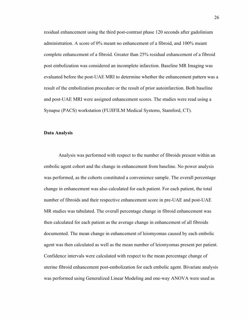
residual enhancement using the third post-contrast phase 120 seconds after gadolinium
administration. A score of 0% meant no enhancement of a fibroid, and 100% meant
complete enhancement of a fibroid. Greater than 25% residual enhancement of a fibroid
post embolization was considered an incomplete infarction. Baseline MR Imaging was
evaluated before the post-UAE MRI to determine whether the enhancement pattern was a
result of the embolization procedure or the result of prior autoinfarction. Both baseline
and post-UAE MRI were assigned enhancement scores. The studies were read using a
Synapse (PACS) workstation (FUJIFILM Medical Systems, Stamford, CT).
Data Analysis
Analysis was performed with respect to the number of fibroids present within an
embolic agent cohort and the change in enhancement from baseline. No power analysis
was performed, as the cohorts constituted a convenience sample. The overall percentage
change in enhancement was also calculated for each patient. For each patient, the total
number of fibroids and their respective enhancement score in pre-UAE and post-UAE
MR studies was tabulated. The overall percentage change in fibroid enhancement was
then calculated for each patient as the average change in enhancement of all fibroids
documented. The mean change in enhancement of leiomyomas caused by each embolic
agent was then calculated as well as the mean number of leiomyomas present per patient.
Confidence intervals were calculated with respect to the mean percentage change of
uterine fibroid enhancement post-embolization for each embolic agent. Bivariate analysis
was performed using Generalized Linear Modeling and one-way ANOVA were used as
27
statistical tools. Compiled data were analyzed using SAS for Windows 9.1 (version 8.0,
SAS Institute Inc, Cary, NC). All P values reflect the result of 2-tailed tests (a = 0.05).
Results
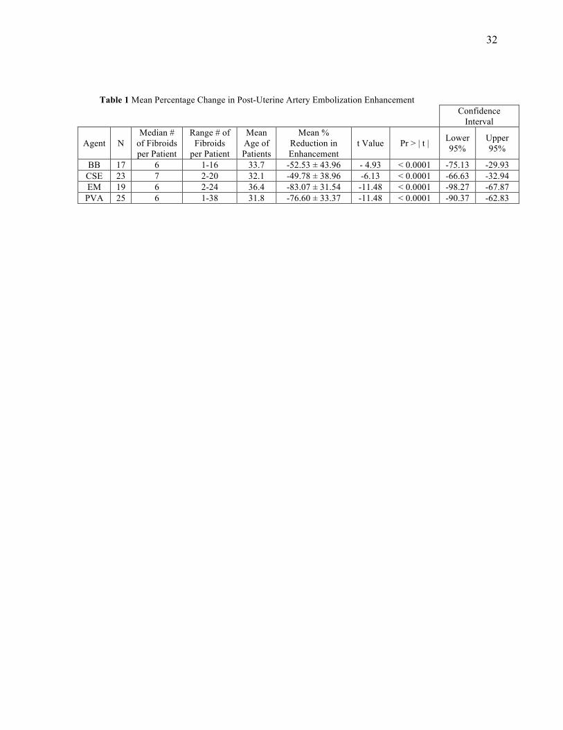
Of patients treated with PVA and EM, there was a mean reduction in
enhancement by 76.60% and 83.07% respectively, compared to a mean reduction of
52.53% and 49.78% of patients treated with CSE and BB, respectively (Table 1).
Bivariate analysis showed the following results when comparing embolic agents
in pairs using 2-tail Fisher’s exact test of least square means: BB to EM resulted in a p =
0.0153, BB to PVA resulted in a p = 0.0412, CSE to EM resulted in a p = 0.0047 and
CSE to PVA resulted in a p = 0.0139 (Table 2). One-way ANOVA indicated that there
was no statistically significant difference in the post-procedural change in enhancement
between CSE and BB. The change in enhancement between PVA and EM was also non
significant. However, a statistically significant difference did exist in the post-
embolization change in fibroid enhancement between BB and EM, BeadBlock and PVA,
CSE and EM and CSE and PVA.
Discussion
Previous studies have sought to compare embolic agents by assessing and
comparing pre and post-embolization enhancement of the dominant leiomyoma.(52, 63)
We chose to assess the percentage change in enhancement of individual fibroids in a

28
patient for two reasons. First, research has indicated that there is an association between
the recurrence of fibroid-related symptoms and the incomplete infarction of fibroids post-
embolization as detected by gadolinium-enhanced MR imaging.(9, 64) Further work has
also implied that incompletely infarcted fibroids are likely to continue growing after
embolization increasing a patient’s risk for future symptom recurrence.(52)Therefore, we
felt that residual enhancement of any fibroid post-embolization would represent a risk for
symptom return post-UAE since evaluating only the dominant fibroid excludes changes
in the numerous other fibroids that impact on clinical success. Second, in evaluating the
total number of fibroids present in each patient, we were also able to determine the mean
number of fibroids per patient per embolic agent. This allowed us to implement a cross-
cohort control for patient comparison.
We found statistically significant differences in the six month post-embolization
mean percentage enhancement reduction between the tested agents, demonstrating that
CSE and BB are less effective embolic agents at infarcting fibroids than EM and PVA
when used according to our protocol. Given previous research, we can infer that those
patients embolized with BB or CSE are at a higher risk for symptom recurrence than
those patients embolized with EM or PVA.
Previous work in embolic agent comparison corroborates some of our findings. A
randomized comparative study conducted by Spies et al. also found that trisacryl gelatin
microspheres were comparable to PVA for post-embolization infarction.(59) Additional
research also performed by Spies et al. found that spherical PVA had a substantially
lower likelihood of causing fibroid infarction than trisarcyl gelatin microspheres.(9)

29
Limitations of our study include possible systemic errors that could have led to
the differences in residual enhancement. For each embolic agent, we used techniques that
had differing, albeit thought to be analogous, endpoints. We also strove to ensure that the
endpoint was stable without collateral flow. The angiogenic endpoint for patients treated
with CSE and PVA was sluggish-flow or near stasis whereas the angiogenic endpoint for
patients treated with BB or EM was sluggish flow with a “pruned-tree” appearance.
Additionally, we used recommended embolic material sizes for each agent. These sizes
did vary from agent to agent. For the embolic agents SPVA, CSE and BB, we used 500-
700 micron particles with an increase to 700-900 microns in some patients. For PVA,
300-500 micron particles were used with an increase to 500-700 micron particles in some
patients.
To reduce other technical errors, we followed many previously established
procedural guidelines as described by Spies et al.(31) Before and after UAE, we
conducted abdominal aortography to evaluate for enlarged ovarian arteries. Such
enlargement may contribute collateral flow to the fibroids and impede UAE fibroid
ablation.(61) We also used micro-catheters during embolization to reduce arterial spasm
in the uterine arteries, thus preventing a false angiographic endpoint.(31) Taking
measures to prevent technical failure was especially important given our evaluation of
efficacy when using various embolic agents. Questions pertaining to the validity of these
preventative measures, however, fell outside the scope of our study.
Another limitation to our study was its size as we described a small, albeit
statistically significant, group of patients. In large part, this was due to the
discontinuation of the use of CSE and BB by interventionalists at our institution as

30
retrospective data seemed to show a higher rate of post-procedure enhancement than
other agents in use. Therefore, there was no opportunity to expand these patient cohorts.
The resulting small cohort groups may have skewed our results, however there was no
statistically significant difference between the cohorts with respect to age or the mean
number of fibroids per patient. Additionally, our assignment of embolic agents to patients
was neither double-blind nor truly random but based on the preferences of the
interventional radiologist performing the procedure.
Despite these limitations, we feel that the failure of CSE and BB to perform as
well as EM and PVA raises questions regarding the continued use of both embolic agents
in UAE. As Spies et al. have stated, it is important to question the quality of evidence
when accepting new embolic agents into general practice.(9) This statement seemed
especially apropos given previous work suggesting discrepancies in adequacy between
CSE and EM and PVA. Our study suggests that BB, an embolic agent recently introduced
and approved for UAE use, also has a substantially lower likelihood of causing effective
fibroid tumor infarction than EM or PVA.
The reasons for the differences in post-procedural fibroid infarction between CSE,
PVA, CSE and BB are open to speculation and should be subject to further investigation.
Evidence has implicated embolic particle migration and particle size and shape in the
ability of the embolic agent to induce fibroid infarction.(8, 60) Further work should be
done to evaluate the ability of embolic agents to infarct fibroid tumors in relation to the
properties of those agents. An understanding of this relationship would further the
development of new embolic agents to be used in the treatment of leiomyomas. Research
must also be done to establish appropriate imaging protocols for evaluating the outcomes
31
of new embolic agents prior to their release and to establish acceptable parameters for
embolic agent infarction efficacy.
Our data suggests that in the treatment of uterine fibroids by UAE, patients treated
with Bead Block and Contour SE demonstrate a statistically significant reduced degree of
infarction on follow-up MRI than those patients treated with classic PVA or Embosphere
microspheres. Further studies must be done to determine whether or not BB and CSE are
embolic agents acceptable for continued use in fibroid ablation via UAE.
32
Table 1 Mean Percentage Change in Post-Uterine Artery Embolization Enhancement
Confidence Interval
Agent N Median #
of Fibroids per Patient
Range # of Fibroids
per Patient
Mean Age of Patients
Mean % Reduction in Enhancement
t Value Pr > | t | Lower 95%
Upper 95%
BB 17 6 1-16 33.7 -52.53 ± 43.96 - 4.93 < 0.0001 -75.13 -29.93 CSE 23 7 2-20 32.1 -49.78 ± 38.96 -6.13 < 0.0001 -66.63 -32.94 EM 19 6 2-24 36.4 -83.07 ± 31.54 -11.48 < 0.0001 -98.27 -67.87
PVA 25 6 1-38 31.8 -76.60 ± 33.37 -11.48 < 0.0001 -90.37 -62.83
33
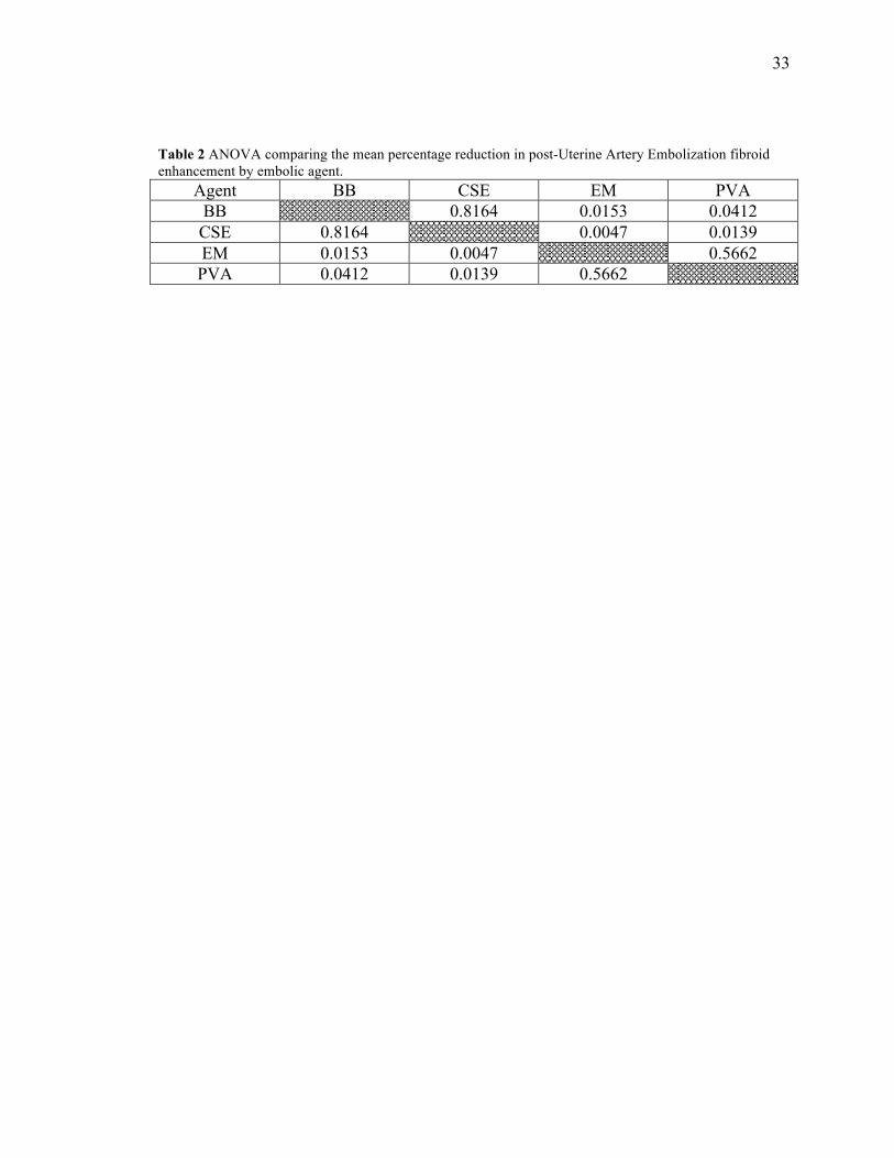
Table 2 ANOVA comparing the mean percentage reduction in post-Uterine Artery Embolization fibroid enhancement by embolic agent.
Agent BB CSE EM PVA BB 0.8164 0.0153 0.0412 CSE 0.8164 0.0047 0.0139 EM 0.0153 0.0047 0.5662
PVA 0.0412 0.0139 0.5662
34
Figures
35
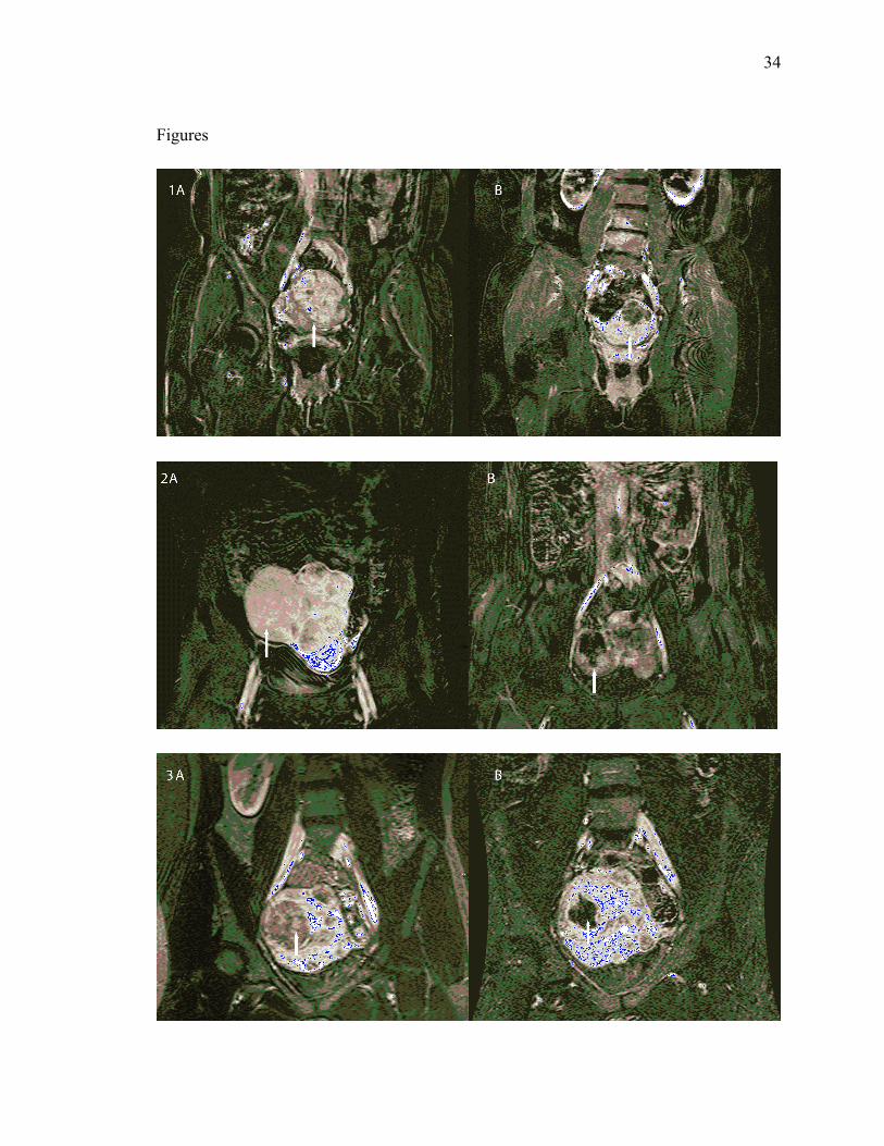
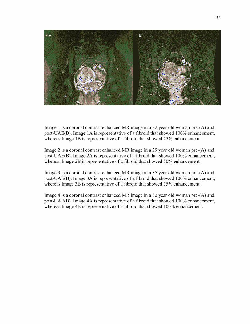
Image 1 is a coronal contrast enhanced MR image in a 32 year old woman pre-(A) and post-UAE(B). Image 1A is representative of a fibroid that showed 100% enhancement, whereas Image 1B is representative of a fibroid that showed 25% enhancement. Image 2 is a coronal contrast enhanced MR image in a 29 year old woman pre-(A) and post-UAE(B). Image 2A is representative of a fibroid that showed 100% enhancement, whereas Image 2B is representative of a fibroid that showed 50% enhancement. Image 3 is a coronal contrast enhanced MR image in a 35 year old woman pre-(A) and post-UAE(B). Image 3A is representative of a fibroid that showed 100% enhancement, whereas Image 3B is representative of a fibroid that showed 75% enhancement. Image 4 is a coronal contrast enhanced MR image in a 32 year old woman pre-(A) and post-UAE(B). Image 4A is representative of a fibroid that showed 100% enhancement, whereas Image 4B is representative of a fibroid that showed 100% enhancement.
36
References 1. Buttram VC,Jr. Uterine leiomyomata--aetiology, symptomatology and management. Prog Clin Biol Res 1986; 225:275-296. 2. Marshall LM, Spiegelman D, Barbieri RL, et al. Variation in the incidence of uterine leiomyoma among premenopausal women by age and race. Obstet Gynecol 1997; 90:967-973. 3. Guarnaccia MM, Rein MS. Traditional surgical approaches to uterine fibroids: abdominal myomectomy and hysterectomy. Clin Obstet Gynecol 2001; 44:385-400. 4. Ravina JH, Herbreteau D, Ciraru-Vigneron N, et al. Arterial embolisation to treat uterine myomata. Lancet 1995; 346:671-672. 5. Goodwin SC, McLucas B, Lee M, et al. Uterine artery embolization for the treatment of uterine leiomyomata midterm results. J Vasc Interv Radiol 1999; 10:1159-1165. 6. Marshburn PB, Matthews ML, Hurst BS. Uterine artery embolization as a treatment option for uterine myomas. Obstet Gynecol Clin North Am 2006; 33:125-144. 7. Katsumori T, Akazawa K, Mihara T. Uterine artery embolization for pedunculated subserosal fibroids. AJR Am J Roentgenol 2005; 184:399-402. 8. Ryu RK. Uterine artery embolization: current implications of embolic agent choice. J Vasc Interv Radiol 2005; 16:1419-1422. 9. Spies JB, Allison S, Flick P, et al. Spherical polyvinyl alcohol versus tris-acryl gelatin microspheres for uterine artery embolization for leiomyomas: results of a limited randomized comparative study. J Vasc Interv Radiol 2005; 16:1431-1437. 10. Wu T, Chen X, Xie L. Selective estrogen receptor modulators (SERMs) for uterine leiomyomas. Cochrane Database Syst Rev 2007; (4):CD005287. 11. Friedman AJ, Hoffman DI, Comite F, Browneller RW, Miller JD. Treatment of leiomyomata uteri with leuprolide acetate depot: a double-blind, placebo-controlled, multicenter study. The Leuprolide Study Group. Obstet Gynecol 1991; 77:720-725. 12. Leather AT, Studd JW, Watson NR, Holland EF. The prevention of bone loss in young women treated with GnRH analogues with "add-back" estrogen therapy. Obstet Gynecol 1993; 81:104-107. 13. Broder MS, Goodwin S, Chen G, et al. Comparison of long-term outcomes of myomectomy and uterine artery embolization. Obstet Gynecol 2002; 100:864-868. 14. Lepine LA, Hillis SD, Marchbanks PA, et al. Hysterectomy surveillance--United States, 1980-1993. MMWR CDC Surveill Summ 1997; 46:1-15. 15. Edwards RD, Moss JG, Lumsden MA, et al. Uterine-artery embolization versus surgery for symptomatic uterine fibroids. N Engl J Med 2007; 356:360-370. 16. Kjerulff KH, Langenberg PW, Rhodes JC, Harvey LA, Guzinski GM, Stolley PD. Effectiveness of hysterectomy. Obstet Gynecol 2000; 95:319-326. 17. McPherson K, Herbert A, Judge A, et al. Psychosexual health 5 years after hysterectomy: population-based comparison with endometrial ablation for dysfunctional uterine bleeding. Health Expect 2005; 8:234-243. 18. McPherson K, Metcalfe MA, Herbert A, et al. Severe complications of hysterectomy: the VALUE study. BJOG 2004; 111:688-694. 19. Altman D, Granath F, Cnattingius S, Falconer C. Hysterectomy and risk of stress-urinary-incontinence surgery: nationwide cohort study. Lancet 2007; 370:1494-1499.

37
20. Taylor A, Sharma M, Tsirkas P, et al. Surgical and radiological management of uterine fibroids--a UK survey of current consultant practice. Acta Obstet Gynecol Scand 2005; 84:478-482. 21. Dubuisson JB, Chapron C, Fauconnier A, Kreiker G. Laparoscopic myomectomy and myolysis. Curr Opin Obstet Gynecol 1997; 9:233-238. 22. LaMorte AI, Lalwani S, Diamond MP. Morbidity associated with abdominal myomectomy. Obstet Gynecol 1993; 82:897-900. 23. Fauconnier A, Chapron C, Babaki-Fard K, Dubuisson JB. Recurrence of leiomyomata after myomectomy. Hum Reprod Update 2000; 6:595-602. 24. Palomba S, Zupi E, Falbo A, et al. A multicenter randomized, controlled study comparing laparoscopic versus minilaparotomic myomectomy: reproductive outcomes. Fertil Steril 2007; 88:933-941. 25. Goodwin SC, Vedantham S, McLucas B, Forno AE, Perrella R. Preliminary experience with uterine artery embolization for uterine fibroids. J Vasc Interv Radiol 1997; 8:517-526. 26. Pelage JP, Soyer P, Le Dref O, et al. Uterine arteries: bilateral catheterization with a single femoral approach and a single 5-F catheter--technical note. Radiology 1999; 210:573-575. 27. Bratby MJ, Belli AM. Radiological treatment of symptomatic uterine fibroids. Best Pract Res Clin Obstet Gynaecol 2008; 22:717-734. 28. Spies JB, Scialli AR, Jha RC, et al. Initial results from uterine fibroid embolization for symptomatic leiomyomata. J Vasc Interv Radiol 1999; 10:1149-1157. 29. Pron G, Bennett J, Common A, et al. Technical results and effects of operator experience on uterine artery embolization for fibroids: the Ontario Uterine Fibroid Embolization Trial. J Vasc Interv Radiol 2003; 14:545-554. 30. Bratby MJ, Hussain FF, Walker WJ. Outcomes after unilateral uterine artery embolization: a retrospective review. Cardiovasc Intervent Radiol 2008; 31:254-259. 31. Spies JB. Uterine artery embolization for fibroids: understanding the technical causes of failure. J Vasc Interv Radiol 2003; 14:11-14. 32. Goodwin SC, Spies JB, Worthington-Kirsch R, et al. Uterine artery embolization for treatment of leiomyomata: long-term outcomes from the FIBROID Registry. Obstet Gynecol 2008; 111:22-33. 33. Kim MD, Lee HS, Lee MH, Kim HJ, Cho JH, Cha SH. Long-term results of symptomatic fibroids treated with uterine artery embolization: In conjunction with MR evaluation. Eur J Radiol 2008; 34. Firouznia K, Ghanaati H, Sanaati M, Jalali AH, Shakiba M. Uterine artery embolization in 101 cases of uterine fibroids: do size, location, and number of fibroids affect therapeutic success and complications? Cardiovasc Intervent Radiol 2008; 31:521-526. 35. Marret H, Alonso AM, Cottier JP, Tranquart F, Herbreteau D, Body G. Leiomyoma recurrence after uterine artery embolization. J Vasc Interv Radiol 2003; 14:1395-1399. 36. Hehenkamp WJ, Volkers NA, Bartholomeus W, et al. Sexuality and body image after uterine artery embolization and hysterectomy in the treatment of uterine fibroids: a randomized comparison. Cardiovasc Intervent Radiol 2007; 30:866-875.

38
37. Voogt MJ, De Vries J, Fonteijn W, Lohle PN, Boekkooi PF. Sexual functioning and psychological well-being after uterine artery embolization in women with symptomatic uterine fibroids. Fertil Steril 2008; 38. Marshburn PB, Matthews ML, Hurst BS. Uterine artery embolization as a treatment option for uterine myomas. Obstet Gynecol Clin North Am 2006; 33:125-144. 39. Eldar-Geva T, Meagher S, Healy DL, MacLachlan V, Breheny S, Wood C. Effect of intramural, subserosal, and submucosal uterine fibroids on the outcome of assisted reproductive technology treatment. Fertil Steril 1998; 70:687-691. 40. Colgan TJ, Pron G, Mocarski EJ, Bennett JD, Asch MR, Common A. Pathologic features of uteri and leiomyomas following uterine artery embolization for leiomyomas. Am J Surg Pathol 2003; 27:167-177. 41. Volkers NA, Hehenkamp WJ, Birnie E, Ankum WM, Reekers JA. Uterine artery embolization versus hysterectomy in the treatment of symptomatic uterine fibroids: 2 years' outcome from the randomized EMMY trial. Am J Obstet Gynecol 2007; 196:519.e1-519.11. 42. Dutton S, Hirst A, McPherson K, Nicholson T, Maresh M. A UK multicentre retrospective cohort study comparing hysterectomy and uterine artery embolisation for the treatment of symptomatic uterine fibroids (HOPEFUL study): main results on medium-term safety and efficacy. BJOG 2007; 114:1340-1351. 43. Wu O, Briggs A, Dutton S, et al. Uterine artery embolisation or hysterectomy for the treatment of symptomatic uterine fibroids: a cost-utility analysis of the HOPEFUL study. BJOG 2007; 114:1352-1362. 44. Fedele L, Parazzini F, Luchini L, Mezzopane R, Tozzi L, Villa L. Recurrence of fibroids after myomectomy: a transvaginal ultrasonographic study. Hum Reprod 1995; 10:1795-1796. 45. Goldberg J, Pereira L, Berghella V, et al. Pregnancy outcomes after treatment for fibromyomata: uterine artery embolization versus laparoscopic myomectomy. Am J Obstet Gynecol 2004; 191:18-21. 46. Walker WJ, Carpenter TT, Kent AS. Persistent vaginal discharge after uterine artery embolization for fibroid tumors: cause of the condition, magnetic resonance imaging appearance, and surgical treatment. Am J Obstet Gynecol 2004; 190:1230-1233. 47. Spies JB, Spector A, Roth AR, Baker CM, Mauro L, Murphy-Skrynarz K. Complications after uterine artery embolization for leiomyomas. Obstet Gynecol 2002; 100:873-880. 48. Godfrey CD, Zbella EA. Uterine necrosis after uterine artery embolization for leiomyoma. Obstet Gynecol 2001; 98:950-952. 49. Walker WJ, Pelage JP. Uterine artery embolisation for symptomatic fibroids: clinical results in 400 women with imaging follow up. BJOG 2002; 109:1262-1272. 50. Hurst BS, Stackhouse DJ, Matthews ML, Marshburn PB. Uterine artery embolization for symptomatic uterine myomas. Fertil Steril 2000; 74:855-869. 51. Spies JB, Warren EH, Mathias SD, Walsh SM, Roth AR, Pentecost MJ. Uterine fibroid embolization: measurement of health-related quality of life before and after therapy. J Vasc Interv Radiol 1999; 10:1293-1303. 52. Pelage JP, Guaou NG, Jha RC, Ascher SM, Spies JB. Uterine fibroid tumors: long-term MR imaging outcome after embolization. Radiology 2004; 230:803-809.
39
53. Siskin GP, Englander M, Stainken BF, Ahn J, Dowling K, Dolen EG. Embolic agents used for uterine fibroid embolization. AJR Am J Roentgenol 2000; 175:767-773. 54. Aziz A, Petrucco OM, Makinoda S, et al. Transarterial embolization of the uterine arteries: patient reactions and effects on uterine vasculature. Acta Obstet Gynecol Scand 1998; 77:334-340. 55. Siskin GP, Eaton LA,Jr, Stainken BF, Dowling K, Herr A, Schwartz J. Pathologic findings in a uterine leiomyoma after bilateral uterine artery embolization. J Vasc Interv Radiol 1999; 10:891-894. 56. Derdeyn CP, Moran CJ, Cross DT, Dietrich HH, Dacey RG,Jr. Polyvinyl alcohol particle size and suspension characteristics. AJNR Am J Neuroradiol 1995; 16:1335-1343. 57. Barth KH, Strandberg JD, White RI,Jr. Long term follow-up of transcatheter embolization with autologous clot, oxycel and gelfoam in domestic swine. Invest Radiol 1977; 12:273-280. 58. Chua GC, Wilsher M, Young MP, Manyonda I, Morgan R, Belli AM. Comparison of particle penetration with non-spherical polyvinyl alcohol versus trisacryl gelatin microspheres in women undergoing premyomectomy uterine artery embolization. Clin Radiol 2005; 60:116-122. 59. Spies JB, Allison S, Flick P, et al. Polyvinyl alcohol particles and tris-acryl gelatin microspheres for uterine artery embolization for leiomyomas: results of a randomized comparative study. J Vasc Interv Radiol 2004; 15:793-800. 60. Siskin GP, Dowling K, Virmani R, Jones R, Todd D. Pathologic evaluation of a spherical polyvinyl alcohol embolic agent in a porcine renal model. J Vasc Interv Radiol 2003; 14:89-98. 61. Binkert CA, Andrews RT, Kaufman JA. Utility of nonselective abdominal aortography in demonstrating ovarian artery collaterals in patients undergoing uterine artery embolization for fibroids. J Vasc Interv Radiol 2001; 12:841-845. 62. Barth MM, Spies JB. Ovarian artery embolization supplementing uterine embolization for leiomyomata. J Vasc Interv Radiol 2003; 14:1177-1182. 63. Katsumori T, Nakajima K, Tokuhiro M. Gadolinium-enhanced MR imaging in the evaluation of uterine fibroids treated with uterine artery embolization. AJR Am J Roentgenol 2001; 177:303-307. 64. deSouza NM, Williams AD. Uterine arterial embolization for leiomyomas: perfusion and volume changes at MR imaging and relation to clinical outcome. Radiology 2002; 222:367-374.