comparative bioreduction of fe(iii) with geobacter ...webs.ucm.es/info/biohidro/publicaciones del...
TRANSCRIPT

Comparative bioreduction of Fe(III) with Geobacter metallireducens and Bacillus infernus
J. Crespoa, J.A. Muñozb, F. Gonzálezc, M. L. Blázquezd and
A. Ballestere
Departamento de Ciencia de los Materiales e Ingeniería Metalúrgica, Facultad de Ciencias Químicas, Universidad Complutense de Madrid, 28040 Madrid, Spain
[email protected], [email protected], [email protected],
[email protected], [email protected] Keywords: Geobacter metallireducens, Bacillus infernus, magnetite, ferric pyrophosphate, vivianite
Abstract. A comparative study on the Fe-reducing ability of pure anaerobic strains of Geobacter
metallireducens and Bacillus infernus was investigated using different sources of Fe(III). Batch
tests were carried out in aqueous solutions containing a reducing agent (lactate or formate) and at a
constant temperature of 37ºC and 50ºC respectively. The formation of biogenic compounds of
Fe(II) was determined using XRD and SEM-EDX techniques.
The bioreduction of magnetite was not affected by the type of bacterial strain used. The kinetics
of the process, initially very fast, stopped rapidly for both types of microorganisms. Vivianite
(Fe3(PO4)2.8H2O) was detected as the main biogenic compound formed during the bioreduction
process.
Introduction
Iron is one of the most abundant elements in the universe and the most abundant in the earth as a
whole. In the Earth’s crust, iron is the fourth element and the second metal most abundant. Despite
its abundance, only a small proportion of iron is available for its biogeochemical recycling [1].
The bioavailability of iron depends on its oxidation state which is a function of Eh and pH. Of
the wide range of microorganisms using ferric ion as electron acceptor in the anaerobic respiration,
Geobacter metallireducens and Bacillus infernus are widely spread in natural environments [2,3].
Some of the more influential variables in the bioreduction process of Fe3+ are: both the electron
acceptor (Fe(III) compounds) and donor (organic compounds) sources, the environmental
conditions (depth, temperature, pH, [Fe3+], etc.) and, of course, the type of microorganism involved.
The growth of microorganisms on insoluble electron acceptors of Fe3+ (such as hematite, α-Fe2O3,
or goethite, α-FeOOH) might proceed according to several strategies [3-6]: whether by producing
chelating molecules or electron-shuttling compounds or by direct contact.
In the present work, the bioreduction of magnetite with or without the addition of ferric
pyrophosphate as electron acceptors for Geobacter metallireducens and Bacillus infernus was
investigated. For each experimental condition, the solution was physicochemically analyzed and the
chemical changes of the magnetite surface examined by SEM-EDX after 170 days.
Materials and Methods
Sources of Fe(III). Iron pyrophosphate (Fe4O21P6) of chemical grade was used as soluble electron
acceptor of Fe(III). A synthetic crystalline magnetite (Fe3O4) with a particle size lower than 0.8 mm
was used as insoluble electron acceptor of Fe(III).
Bacterial strains. Two pure anaerobic bacterial strains of the Oregon Collection of Methanogens
were used: Geobacter metallireducens (OCM 645) and Bacillus infernus Th-23 (OCM 479) (Figure
1). The mesophilic strain of G. metallireducens was grown at 37ºC on MS nutrient medium [7] at
pH 7 with 10 mM of sodium lactate (as electron donor) and 5 mM of ferric pyrophosphate,
Advanced Materials Research Vols. 20-21 (2007) pp. 561-564online at http://www.scientific.net© (2007) Trans Tech Publications, Switzerland
All rights reserved. No part of contents of this paper may be reproduced or transmitted in any form or by any means without thewritten permission of the publisher: Trans Tech Publications Ltd, Switzerland, www.ttp.net. (ID: 147.96.7.60-18/07/07,14:31:13)

Fe4O21P6 (as electron acceptor), complexed with sodium citrate. The moderately thermophilic strain
of B. infernus Th-23 was grown at 50ºC on MS medium at pH 8 with 10 mM of sodium formate (as
electron donor) and 5 mM of ferric pyrophosphate, Fe4O21P6 (as electron acceptor), complexed with
sodium citrate.
Figure 1. SEM micrographs of bacterial cells: G. metallireducens (left) and B. infernus (right).
Bioreduction tests. Bioreduction tests were performed in anaerobic bottles of 100 mL containing
90 mL of MS nutrient medium and 10 mL of a bacterial culture in the exponential growth phase.
The same electron donors and at the same concentrations as described previously were used for
each strain. 5 mM of iron pyrophosphate and/or 1 g of synthetic magnetite were added as sources of
Fe(III). Tests were run at 37ºC and 50ºC for 170 days using pure strains of Geobacter
metallireducens and Bacillus infernus, respectively.
The following parameters were periodically controlled: pH, redox potential (vs. Ag/AgCl), cell
concentration (results not shown) and iron in solution (both FeTotal and Fe2+). Total iron was
determined by atomic sorption spectrometry while ferrous iron was measured by photocolorimetry
using o-phenantroline. The solid residues obtained after 170 days were dried at 80ºC and then
analyzed by XRD and SEM-EDX.
Results and Discussion
The bioreduction process using magnetite as insoluble source of Fe(III) was carried out under two
different experimental conditions for each bacterial strain tested: 1) with magnetite and the
corresponding electron donor (lactate for Geobacter metallireducens and formate for Bacillus
infernus) and 2) with magnetite and the electron donor plus ferric pyrophosphate.
The variation of pH during the test was almost negligible for both types of bacteria (Figure 2a).
Nevertheless, in tests with Bacillus infernus, pH decreased initially. A similar drop in the redox
potential was observed for all the tests, irrespectively of the presence of the iron pyrophosphate
(Figure 2b). The increase of the potential, after a sharp drop in the first six days, could be related
with the removal of Fe(II) from solution and its deposition on mineral surfaces. In this sense, some
authors [8] have reported that the precipitation of Fe2+ would favour the inhibition of the
bioreduction process.
In tests with pyrophosphate, the amount of total iron in solution initially decreased and
simultaneously the amount of Fe(II) increased (Figures 2c and 2d). On the contrary, in tests without
pyrophosphate, the initial increase of Fe(II) in solution coincided with a progressive increase of the
concentration of FeTotal, although the amount of ferrous ion was lower than the total iron.
Nevertheless, the differences in the amount of Fe(II) generated by both types of bacteria were
negligible. In this sense, different researchers [9,10] have shown that the reduction of magnetite
with other kinds of bacteria strongly depends on the pH of the aqueous medium, being
thermodynamically favoured at lower pH (<6.5). Therefore, the recalcitrant behaviour of magnetite
observed in the present study could be explained, at least in part, as due to the unfavourable
themodynamics of the system at neutral pH.
Biohydrometallurgy: From the Single Cell to the Environment562

6.5
7
7.5
8
pH
0 50 100 150
Time (days)
Fe3O4 (G.m.) Fe3O4 (B.i.)
Fe3O4 + Pyrophosphate (G.m.) Fe3O4 + Pyrophosphate (B.i.)
0
50
100
150
200
E (
mV
)
0 50 100 150
Time (days)
0
10
20
30
Fe
(II)
(m
g)
0 50 100 150
Time (days)
0
20
40
60
80
100
Fe
(To
tal)
(m
g)
0 50 100 150
Time (days) Figure 2. Evolution of different parameters in biotic tests using Geobacter metallireducens (G.m.)
or Bacillus infernus (B.i.): pH (a), redox potential (b), ferrous ion (c) and total iron (d).
The parameters controlling the rate and extent of the magnetite reduction and the resulting
products are still on debate. Very often, magnetite has been considered as a final product in the
dissimilatory reduction of ferric oxides [9]. Nevertheless, magnetite is a magnetic mineral with a
spinel structure containing approximately 31% of FeO and 69% of Fe2O3 and, thus, susceptible to
reduction.
The SEM micrographs of both the untreated and treated magnetite are shown in Figure 3. In the
samples attacked with Geobacter metallireducens (Figure 3b) and with Bacillus infernus (Figure
3c) a new mineralogical phase was detected for both systems. The new product was identified by
EDX as vivianite (Fe3(PO4)2.8H2O) (Figure 3d).
Table 1 shows EDX microanalyses of the different samples. The unattacked magnetite resulted
in Fe/O ratios between 0.54 and 0.84, with an arithmetic average of 0.69, slightly lower than the
theorical value of 0.75. Therefore, the slight enrichment of oxygen in the solid surface could be due
to an atmospheric oxidation before the experiment. EDX microanalyses of treated magnetites
confirmed the enrichment of iron on the mineral surface (Table I), with Fe/O ratios higher than
unity, except for vivianite where that ratio notably decreased up to approximately 0.2. In the case of
vivianite, all the microanalysis show the presence of Fe, O and P as major elements, with minor
amounts of Na, Ca, Mg, K and Cl. Therefore, the precipitation of vivianite on the magnetite surface
could explain the inhibition of the reduction of Fe(III) by the bacterial cell.
a) b)
c) d)
Advanced Materials Research Vols. 20-21 563
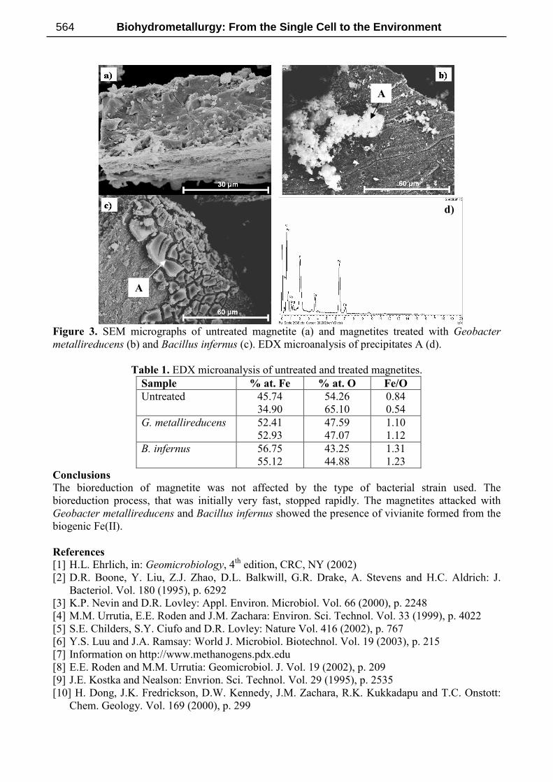
Figure 3. SEM micrographs of untreated magnetite (a) and magnetites treated with Geobacter
metallireducens (b) and Bacillus infernus (c). EDX microanalysis of precipitates A (d).
Table 1. EDX microanalysis of untreated and treated magnetites.
Sample % at. Fe % at. O Fe/O
Untreated 45.74
34.90
54.26
65.10
0.84
0.54
G. metallireducens 52.41
52.93
47.59
47.07
1.10
1.12
B. infernus 56.75
55.12
43.25
44.88
1.31
1.23
Conclusions
The bioreduction of magnetite was not affected by the type of bacterial strain used. The
bioreduction process, that was initially very fast, stopped rapidly. The magnetites attacked with
Geobacter metallireducens and Bacillus infernus showed the presence of vivianite formed from the
biogenic Fe(II).
References
[1] H.L. Ehrlich, in: Geomicrobiology, 4th edition, CRC, NY (2002)
[2] D.R. Boone, Y. Liu, Z.J. Zhao, D.L. Balkwill, G.R. Drake, A. Stevens and H.C. Aldrich: J.
Bacteriol. Vol. 180 (1995), p. 6292
[3] K.P. Nevin and D.R. Lovley: Appl. Environ. Microbiol. Vol. 66 (2000), p. 2248
[4] M.M. Urrutia, E.E. Roden and J.M. Zachara: Environ. Sci. Technol. Vol. 33 (1999), p. 4022
[5] S.E. Childers, S.Y. Ciufo and D.R. Lovley: Nature Vol. 416 (2002), p. 767
[6] Y.S. Luu and J.A. Ramsay: World J. Microbiol. Biotechnol. Vol. 19 (2003), p. 215
[7] Information on http://www.methanogens.pdx.edu
[8] E.E. Roden and M.M. Urrutia: Geomicrobiol. J. Vol. 19 (2002), p. 209
[9] J.E. Kostka and Nealson: Envrion. Sci. Technol. Vol. 29 (1995), p. 2535
[10] H. Dong, J.K. Fredrickson, D.W. Kennedy, J.M. Zachara, R.K. Kukkadapu and T.C. Onstott:
Chem. Geology. Vol. 169 (2000), p. 299
d)
A
A
Biohydrometallurgy: From the Single Cell to the Environment564