commonly missed injuries of the extremities 45 syllabus.pptbonepit.com/syllabi/commonly missed...
TRANSCRIPT
1/15/2012
1
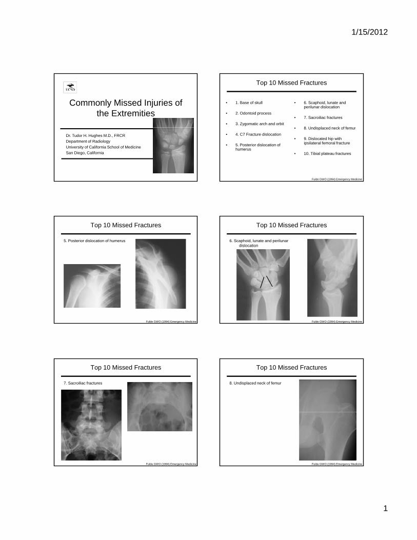
Commonly Missed Injuries of the Extremities
Dr. Tudor H. Hughes M.D., FRCRDepartment of RadiologyUniversity of California School of MedicineSan Diego, California
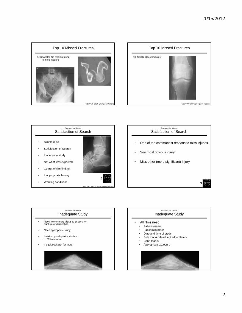
Top 10 Missed Fractures
• 1. Base of skull
• 2. Odontoid process
• 3. Zygomatic arch and orbit
• 6. Scaphoid, lunate and perilunar dislocation
• 7. Sacroiliac fractures
8 Undisplaced neck of femur• 4. C7 Fracture dislocation
• 5. Posterior dislocation of humerus
• 8. Undisplaced neck of femur
• 9. Dislocated hip with ipsilateral femoral fracture
• 10. Tibial plateau fractures
Fulde GWO (1994) Emergency Medicine
Top 10 Missed Fractures
5. Posterior dislocation of humerus
Fulde GWO (1994) Emergency Medicine
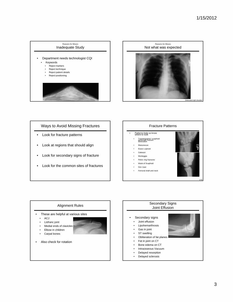
Top 10 Missed Fractures
6. Scaphoid, lunate and perilunar dislocation
Fulde GWO (1994) Emergency Medicine
Top 10 Missed Fractures
7. Sacroiliac fractures
Fulde GWO (1994) Emergency Medicine
Top 10 Missed Fractures
8. Undisplaced neck of femur
Fulde GWO (1994) Emergency Medicine
1/15/2012
2
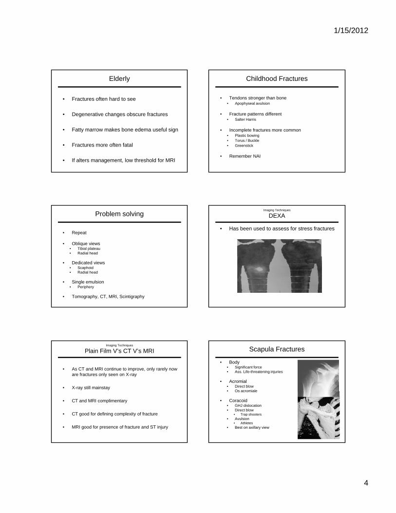
Top 10 Missed Fractures
9. Dislocated hip with ipsilateral femoral fracture
Fulde GWO (1994) Emergency Medicine
Top 10 Missed Fractures
10. Tibial plateau fractures
Fulde GWO (1994) Emergency Medicine
• Simple miss
• Satisfaction of Search
• Inadequate study
Reasons for Misses
Satisfaction of Search
q y
• Not what was expected
• Corner of film finding
• Inappropriate history
• Working conditionsTalar neck fracture with subtalar dislocation
?
Reasons for Misses
Satisfaction of Search
• One of the commonest reasons to miss injuries
• See most obvious injury
• Miss other (more significant) injury
?
Reasons for Misses
Inadequate Study
• Need two or more views to assess for fracture or dislocation
• Need appropriate study
• Insist on good quality studies• With empathy• With empathy
• If equivocal, ask for more
Reasons for Misses
Inadequate Study
• All films need• Patients name• Patients number• Date and time of study• Side marker (lead, not added later)• Cone marks• Appropriate exposure
1/15/2012
3
Reasons for Misses
Inadequate Study
• Department needs technologist CQI• Keywords
• Reject markers• Reject technique• Reject patient details• Reject positioning
Reasons for Misses
Not what was expected
Dislocated right shoulder
Ways to Avoid Missing Fractures
• Look for fracture patterns
• Look at regions that should align
• Look for secondary signs of fracture
• Look for the common sites of fractures
Fracture Patterns
• Patterns help us know where to look
• Transtriquetral / scaphoidperilunate fracture dislocation
• Maisoneuve
• Essex Lopresti
• Galeazzi
• Monteggia
• Pelvic ring fractures
• Waist of Scaphoid
• Don Juan
• Femoral shaft and neck
27M
Alignment Rules
• These are helpful at various sites• ACJ• Lisfranc joint• Medial ends of clavicles• Elbow in childrenElbow in children• Carpal bones
• Also check for rotation
Secondary SignsJoint Effusion
• Secondary signs• Joint effusion• Lipohemarthrosis• Gas in jointj• ST swelling• Obliteration of fat planes• Fat in joint on CT• Bone edema on CT• Intraosseous Vacuum• Delayed resorption• Delayed sclerosis
1/15/2012
4
Elderly
• Fractures often hard to see
• Degenerative changes obscure fractures
• Fatty marrow makes bone edema useful sign
• Fractures more often fatal
• If alters management, low threshold for MRI
Childhood Fractures
• Tendons stronger than bone• Apophyseal avulsion
• Fracture patterns different• Salter Harris
• Incomplete fractures more common• Plastic bowing• Torus / Buckle• Greenstick
• Remember NAI
Problem solving
• Repeat
• Oblique views• Tibial plateau• Radial head
• Dedicated views• Scaphoid• Radial head
• Single emulsion• Periphery
• Tomography, CT, MRI, Scintigraphy
Imaging Techniques
DEXA
• Has been used to assess for stress fractures
Imaging Techniques
Plain Film V’s CT V’s MRI
• As CT and MRI continue to improve, only rarely now are fractures only seen on X-ray
• X-ray still mainstay
• CT and MRI complimentary
• CT good for defining complexity of fracture
• MRI good for presence of fracture and ST injury
Scapula Fractures
• Body• Significant force• Ass. Life-threatening injuries
• Acromial• Direct blow• Os acromialeOs acromiale
• Coracoid• GHJ dislocation• Direct blow
• Trap shooters• Avulsion
• Athletes• Best on axillary view
1/15/2012
5
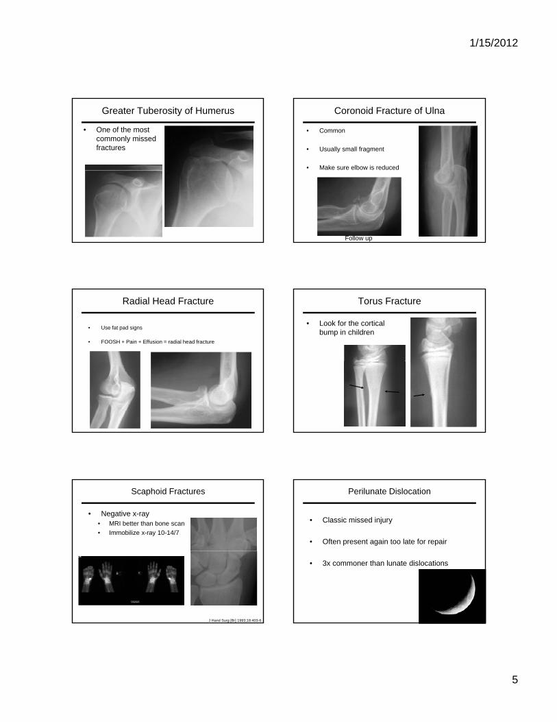
Greater Tuberosity of Humerus
• One of the most commonly missed fractures
Coronoid Fracture of Ulna
• Common
• Usually small fragment
• Make sure elbow is reduced
ref
Follow up
Radial Head Fracture
• Use fat pad signs
• FOOSH + Pain + Effusion = radial head fracture
Torus Fracture
• Look for the cortical bump in children
ref
Scaphoid Fractures
• Negative x-ray• MRI better than bone scan • Immobilize x-ray 10-14/7
. J Hand Surg [Br] 1993;18:403-6.
Perilunate Dislocation
• Classic missed injury
• Often present again too late for repair
• 3x commoner than lunate dislocations
1/15/2012
6
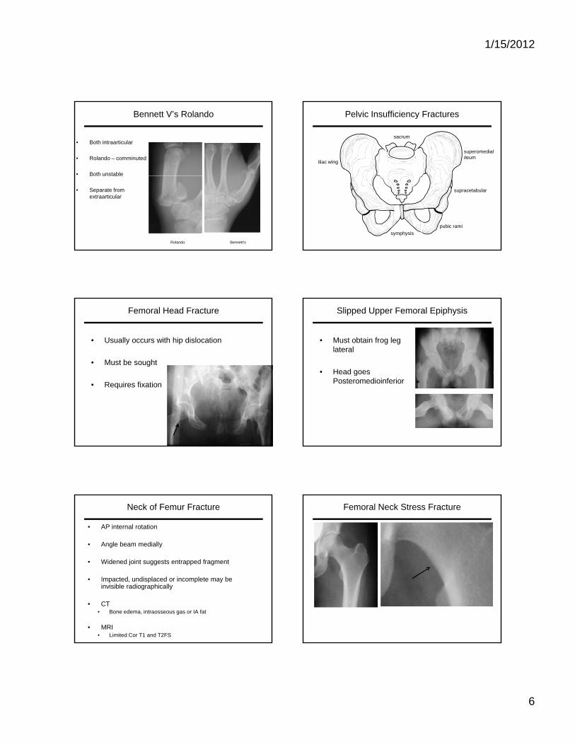
Bennett V’s Rolando
• Both intraarticular
• Rolando – comminuted
• Both unstableBoth unstable
• Separate from extraarticular
Rolando Bennett’s
Pelvic Insufficiency Fractures
sacrum
superomedial ileum
iliac wing
pubic rami
supracetabular
symphysis
Femoral Head Fracture
• Usually occurs with hip dislocation
• Must be sought
• Requires fixation
Slipped Upper Femoral Epiphysis
• Must obtain frog leg lateral
• Head goes• Head goes Posteromedioinferior
Neck of Femur Fracture
• AP internal rotation
• Angle beam medially
• Widened joint suggests entrapped fragment
• Impacted, undisplaced or incomplete may be invisible radiographically
• CT • Bone edema, intraosseous gas or IA fat
• MRI • Limited Cor T1 and T2FS
Femoral Neck Stress Fracture
1/15/2012
7
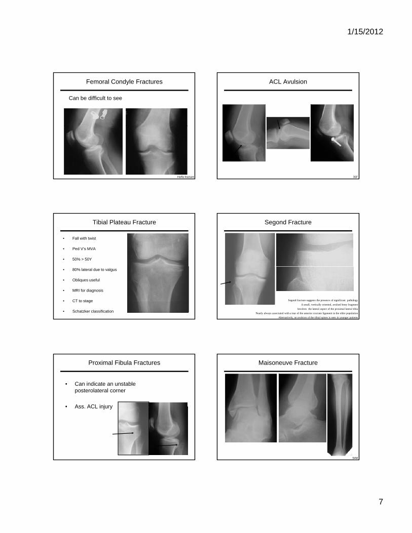
Femoral Condyle Fractures
Can be difficult to see
Hoffa fracture
ACL Avulsion
35F
Tibial Plateau Fracture
• Fall with twist
• Ped V’s MVA
• 50% > 50Y
• 80% lateral due to valgus
• Obliques useful
• MRI for diagnosis
• CT to stage
• Schatzker classification
Segond Fracture
Segond fracture suggests the presence of significant pathologyA small, vertically oriented, avulsed bony fragment
Involves the lateral aspect of the proximal lateral tibiaNearly always associated with a tear of the anterior cruciate ligament in the older population
Alternatively, an avulsion of the tibial spines is seen in younger patients
Proximal Fibula Fractures
• Can indicate an unstable posterolateral corner
• Ass ACL injury• Ass. ACL injury
Maisoneuve Fracture
54M
1/15/2012
8
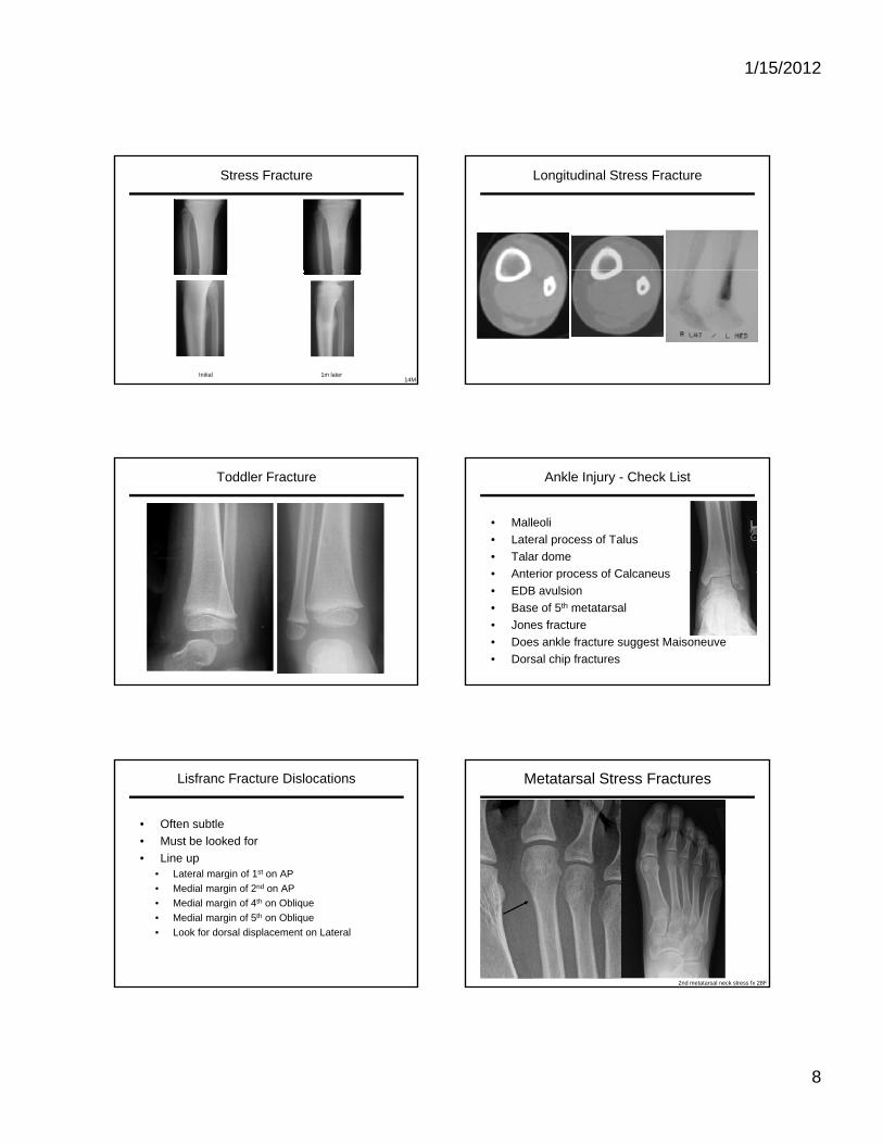
Stress Fracture
Initial 1m later14M
Longitudinal Stress Fracture
Toddler Fracture Ankle Injury - Check List
• Malleoli• Lateral process of Talus• Talar dome
Anterior process of Calcaneus• Anterior process of Calcaneus• EDB avulsion• Base of 5th metatarsal• Jones fracture• Does ankle fracture suggest Maisoneuve• Dorsal chip fractures
Lisfranc Fracture Dislocations
• Often subtle• Must be looked for• Line up
• Lateral margin of 1st on AP• Lateral margin of 1st on AP• Medial margin of 2nd on AP• Medial margin of 4th on Oblique• Medial margin of 5th on Oblique• Look for dorsal displacement on Lateral
Metatarsal Stress Fractures
2nd metatarsal neck stress fx 28F