common pediatric foot...
TRANSCRIPT

Common Pediatric Foot Deformities
• Angular deformities of LL: – Bow legs.
– Knock knees.
• Rotational deformities of LL: – In-toeing.
– Ex-toeing.
• Leg aches.
• CDH.
• Feet problems.
• Irritable hip.
Common Orthopedic Problems
in Children

Angular LL Deformities of LL

Angular Deformities
Nomenclature
Bow legs Knock knees
Genu Varus Genu Valgus

Angular Deformities
Range of Normal Varies With Age
• During first year : Lateral bowing of Tibiae
• During second year : Bow legs (knees & tibiae)
• Between 3 – 4 years : Knock knees

Angular Deformities
Evaluation
Should differentiate between
“physiologic” and “pathologic”
deformities

Angular Deformities
Evaluation
Physiologic Pathologic
• Expected for age
• Generalized
• Regressive
• Mild – moderate
• Symmetrical
•Not expected for age
• Localized
• Progressive
• Severe
• Asymmetrical

Angular Deformities
Causes
Physiologic Pathologic
- Use of walker?
- Early wt. bearing
- Overweight
• Exaggerated :
• Normal – for age
• Idiopathic
• Injury to Epiphys. Plate
Infection / Trauma
• Metabolic disease
• Endocrine disturbance
• Rickets

Angular Deformities
Evaluation
Symmetrical deformity

Angular Deformities
Evaluation
Asymmetrical Deformity

Angular Deformities
Evaluation
Generalized deformity
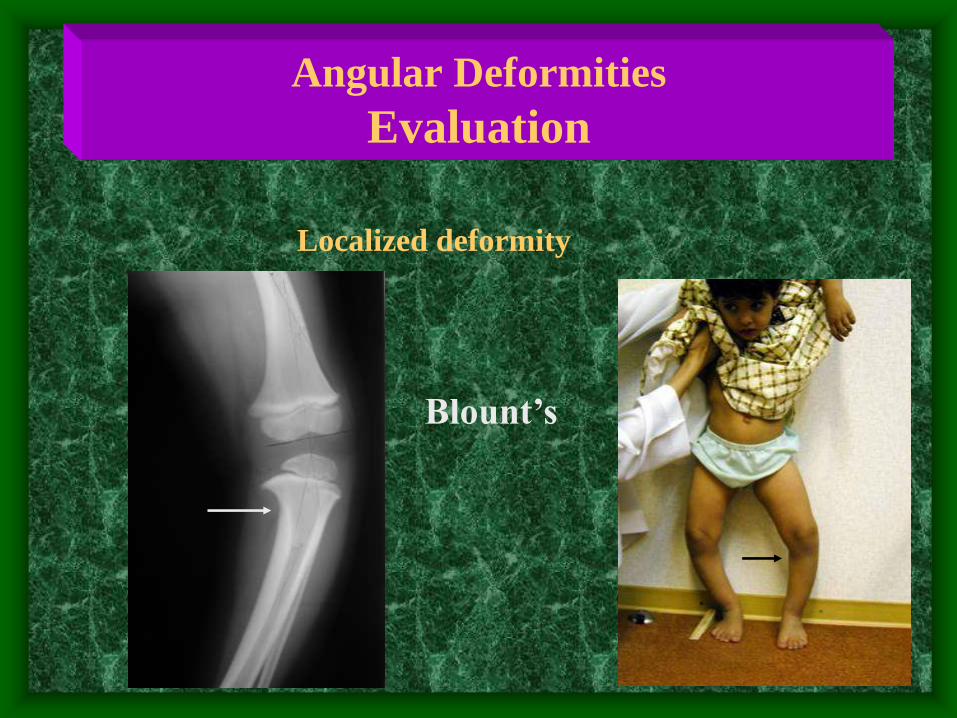
Angular Deformities
Evaluation
Localized deformity
Blount’s
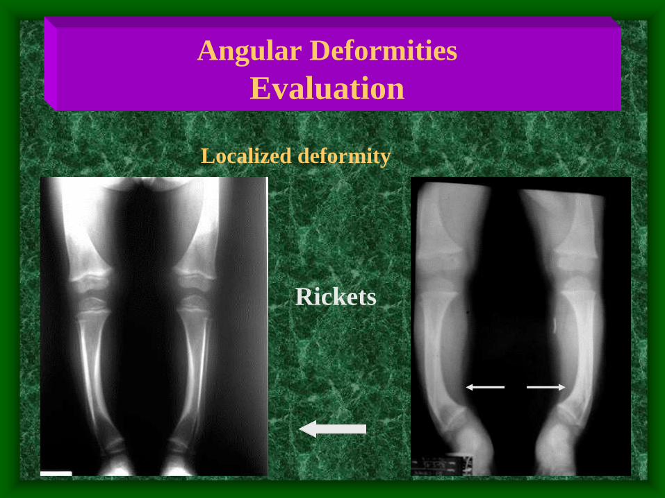
Angular Deformities
Evaluation
Rickets
Localized deformity

in bow legs / genu varum
Inter-condylar distance
Measure Angulation
( standing / supine )
Angular Deformities
Evaluation

in knock knees /genu valgum
Inter- malleolar distance
Measure Angulation
( standing / supine )
Angular Deformities
Evaluation

Measure Angulation
Angular Deformities
Evaluation
Use goneometer
measures angles directly

• Serum Calcium / Phosphorous ?
• Serum Alkaline Phosphatase
• Serum Creatinine / Urea – Renal function
Angular Deformities
Evaluation
Investigations / Laboratory

X-ray when severe or possibly pathologic
• Standing AP film
– long film ( hips to ankles ) with patellae directed forwards
• Look for diseases :
– Rickets / Tibia vara (Blount’s) / Epiphyseal injury..
– Measure angles.
Angular Deformities
Evaluation
Investigations / Radiological

Femoral-Tibial Axis Medial Physeal Slope
Angular Deformities
Evaluation
Investigations / Radiological

Angular Deformities
When To Refer ?
• Pathologic deformities:
Asymmetrical.
Localized.
Progressive.
Not expected for age.
• Exaggerated physiologic deformities:
Definition ?

Angular Deformities
Surgery
Rotational LL Deformities
• Frequently seen.
• Concerns parents.
• Frequently prompts varieties of treatment.
( often un-necessary / incorrect )
In-toeing / Ex-toeing

Rotational Deformities
• Level of affection :
Femur
Tibia
Foot

Rotational Deformities
Femur
Ante-version = more medial rotation
Retro-version = more lateral rotation

Rotational Deformities
Normal Development
• Femur : Ante-version :
– 30 degrees at birth.
– 10 degrees at maturity.
• Tibia : Lateral rotation :
– 5 degrees at birth.
– 15 degrees at maturity.

Rotational Deformities
Normal Development
Both Femur and Tibia laterally rotate with
growth in children
• Medial Tibial torsion and Femoral ante-version
improve ( reduce ) with time.
• Lateral Tibial torsion usually worsens with growth.

Rotational Deformities
Clinical Examination
Rotational Profile
• At which level is the rotational deformity?
• How severe is the rotational deformity?
• Four components:
1- Foot propagation angle.
2- Assess femoral rotational arc.
3- Assess tibial rotational arc.
4- Foot assessment.

Rotational Deformities
Clinical Examination
Rotational Profile
1- Foot propagation angle – Walking
Normal Range:
+10o _10o
? In Eastern Societies
+25o _10o

Rotational Deformities
Clinical Examination
Rotational Profile 2- Assess Femoral Rotational Arc
Supine
Extended
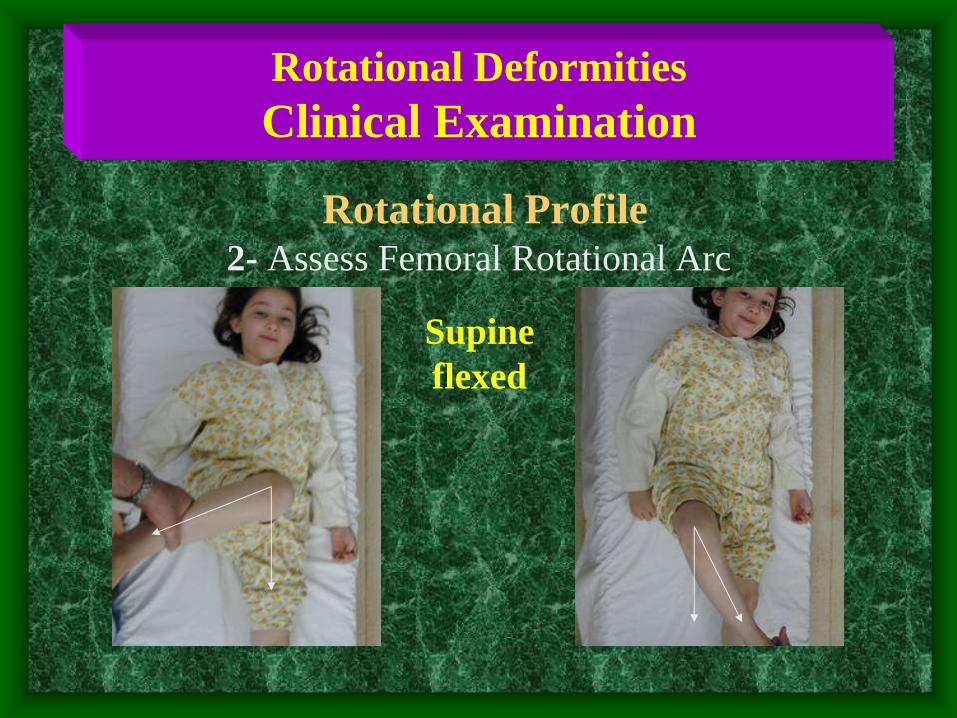
Rotational Deformities
Clinical Examination
Rotational Profile 2- Assess Femoral Rotational Arc
Supine
flexed

Rotational Deformities
Clinical Examination
Rotational Profile
3- Tibial Rotational Arc Thigh-foot angle in prone
foot position is critical
leave to fall into natural position

Rotational Deformities
Clinical Examination
Rotational Profile
4- Foot assessment
• Metatarsus adductus
• Searching big toe
• Everted foot
• Flat foot

• Out-toeing : Normal
• seen when infant positioned upright
( usually hips laterally rotate in-utero )
• Metatarsus adductus : • medial deviation of forefoot
• 90 % resolve spontaneously
• casting if rigid or persists late in 1st year
Rotational Deformities
Common Presentations
Infants

Rotational Deformities
Common Presentations
Toddlers
• In-toeing most common during second year.
( at beginning of walking )
• Causes :
– medial tibial torsion.
– metatarsus adductus.
– abducted great toe.

Rotational Deformities
Common Presentations
Toddlers - Medial Tibial Torsion
• The commonest cause of in-toeing
• Observational management is best
• Avoid special shoes / splints / braces
– unnecessary, ineffective, interferes with activity and
cause psychological and behavioral problems.
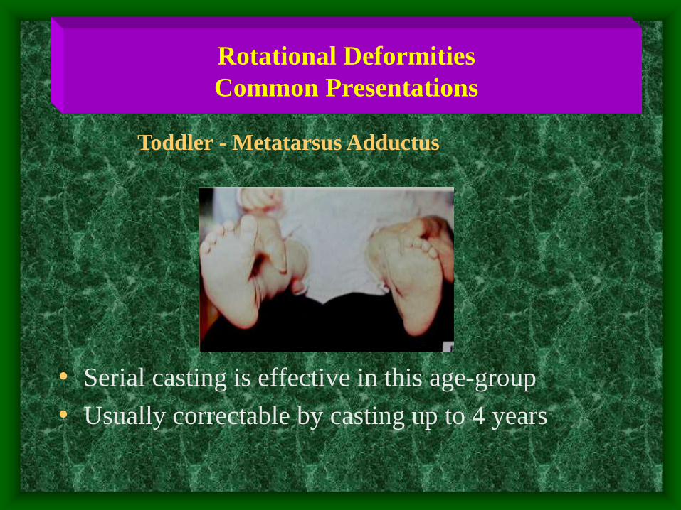
Rotational Deformities
Common Presentations
• Serial casting is effective in this age-group
• Usually correctable by casting up to 4 years
Toddler - Metatarsus Adductus

Rotational Deformities
Common Presentations
• Dynamic deformity
• Over-pull of Abductor
Hallucis Muscle during
stance phase
Toddlers - Abducted Great Toe
• Spontaneously resolve - no treatment

Rotational Deformities
Common Presentations
Child
• In-toeing : due to medial femoral torsion
• Out-toeing : in late childhood
lateral femoral / tibial torsion

Rotational Deformities
Common Presentations
Child Medial Femoral Torsion
• Usually: - starts at 3 - 5 years,
- peaks at 4 – 6 years,
- then resolves spontaneously.
• Girls > boys.
• Look at relatives - family history – normal.
• Treatment usually not recommended.
• If persists > 8 years and severe, may need surgery.

Rotational Deformities
Common Presentation
• Stands with knees medially rotated (kissing patellae).
• Sits in W position.
• Runs awkwardly (egg-beater).
Family History
Medial Femoral Torsion (Ante-version)

Rotational Deformities
Common Presentations
Child Lateral Tibial Torsion
• Usually worsens.
• May be associated with knee pain (patellar)
specially if LTT is associated with MFT.
( knee medially rotated and ankle laterally rotated )

Rotational Deformities
Common Presentations
Child Medial Tibial Torsion
• Less common than LTT in older child
• May need surgery if :
– persists > 8 year,
– and causes functional disability

Rotational Deformities
Management
• Challenge : dealing effectively with family
• In-toeing : spontaneously corrects in vast
majority of children as LL externally rotates
with growth - Best Wait !

Rotational Deformities
Management
Convince family that only observation is
appropriate
• < 1 % of femoral & tibial torsional
deformities fail to resolve and may require
surgery in late childhood.

Rotational Deformities
Management
• Attempts to control child’s walking, sitting and sleeping positions is impossible and ineffective cause frustration and conflicts.
• She wedges and inserts : ineffective.
• Bracing with twisters :ineffective - and limits activity.
• Night splints : better tolerated - ? Benefit.

Rotational Deformities
Management
Shoe wedges Ineffective Twister cables Ineffective

Rotational Deformities
When To Refer ?
• Severe & persistent deformity.
• Age > 8-10y.
• Causing a functional dysability.
• Progressive.

Rotational Deformities
Management
When Is Surgery Indicated ?
•In older child ( > 8 – 10 years ).
•Significant functional disability.
•Not prophylactic !

Leg Aches / Growing Pains

Leg Aches / Growing Pains
• Incidence : 15-30 % of children.
• More In girls / At night / In LL.
• Diagnosis is made by exclusion.

Leg Aches / Growing Pains
History
• Vague pain.
• Poorly localised.
• Bilateral.
• Nocturnal.
• Seldom alters activity.
• Long duration.

Leg Aches / Growing Pains
Examination
• General health is normal.
• No deformities.
• No joint stiffness.
• No tenderness.
• Normal gait.
• No limping.

Leg Aches / Growing Pains
Management
• When atypical history or signs present on examination:
– Imaging and lab. Studies.
• If all negative :
– Symptomatic treatment :
• Heat / Analgesics.
– Reassure family :
• Benign.
• Self-limiting.
• Advise to re-evaluate if clinical features change.

Leg Aches / Growing Pains
Feature Growing Pain Serious Problem
History :
Long duration Often Usually not
Pain localised No Often
Pain bilateral Often Unusual
Ulters activity No Often
Cause limping No Sometimes
General health Good May be ill
From Stahili : Practice of Pediatric Orthopedics 2001

Leg Aches / Growing Pains
Feature Growing Pain Serious Problem
Physical examination :
Tenderness No May show
Guarding No May show
Reduced rang of motion No May show
Laboratory :
CBC Normal ? Abnormal
ESR Normal ? Abnormal
From Stahili : Practice of Pediatric Orthopedics 2001

CDH / DDH
Congenital Dislocation of Hip.
Developmental Dysplasia of Hip.

CDH Spectrum
• Teratologic Hip : Fixed dislocation
Often with other anomalies
• Dislocated Hip : Completely out
May or may not be reducible
• Subluxated Hip : Only partially in
• Unstable Hip : Femoral head can be dislocated
• Acetabular Dysplasia : Shallow Acetabulum
Head Subluxated or in place

CDH
Etiology & Risk Factors
• Prenatal : – Positive family history (increases risk 10X)
– Primi-gravida
– Female (4-6 X > Males)
– Oligo-hydramnious
– Breech position (increases risk 5-10 X)
• Postnatal : – Swaddling / Strapping ( ? Knees extended)
– Ligament Laxity
– Torticollis (CDH in 10-20 % cases)
– Cong. Knee recurvatum / dislocation
– Metatarsus adductus / calcaneo-valgus

CDH
Risk Factors
When Risk Factors Are Present
• The infant should be examined repeatedly
• The hip should be imaged by
– U/S
– or X-ray

CDH
Clinical Examination

CDH
Neonatal Examination
LOOK :
• Asymmetric thigh
folds
– Posterior
– anterior

CDH
Clinical Examination
Look :
• Shortening ( not in neonates )
- Galeazzy sign
- in supine

CDH
Neonatal Examination
MOVE :
• Hip instability
in early infancy
• Limited hip abduction
in flexion - later
• (careful in bilateral)
if <600 on both sides:
request imaging

CDH
Neonatal Examination
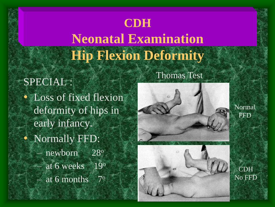
CDH
Neonatal Examination
Hip Flexion Deformity
SPECIAL :
• Loss of fixed flexion
deformity of hips in
early infancy.
• Normally FFD:
– newborn 28o
– at 6 weeks 19o
– at 6 months 7o
Normal
FFD
CDH
No FFD
Thomas Test

CDH
Neonatal Examination
Ortolani Barlow
Feel Clunk
Not hear click !

CDH
Neonatal Examination
Ortolani / Barlow
clunk
Ortolani Barlow

CDH
Neonatal Examination
Ortolani Test Barlow Test

CDH
Clinical Examination
• Hip clicks :
- fine, short duration, high pitched sounds
- common and benign – from soft tissues
• Hip clunks :
- sensation of the hip displacing over the
acetabular margin
• If in doubt : U/S in young infants
single radiograph if > 2-3 months

CDH
Clinical Examination
• Neonate (up to 2-3 months) :
– Instability/ Ortolani-Barlow
• Infant ( > 2-3 months) :
– Limited abduction
– Shortening ( Galeazzi )
• Toddler :
– Limited abduction
– Shortening ( Galeazzi )
• Walker :
– Trendelenburgh limpimg

CDH
Ultrasound Screening
• Early U/S screening not recommended
• Delayed U/S screening :
– Older than 3 weeks
– Those at risk or suspicious by:
• History
• Clinical exam

CDH
Treatment
• Birth to 6 months : – Pavlik harness or hip spica cast
• 6 months – 12 months : – closed reduction UGA and hip spica casts
• 12 months – 18 months : – possible closed / possible open reduction
• Above 18 months : – open reduction and ? Acetabuloplasty
• Above 2 years : – open reduction,acetabulplasty, and femoral osteotomy

CDH
Treatment
• Method depends on Age
• The earlier started, the easier the treatment
& the better the results
• Should be detected EARLY
• UREGENT referral once an abnormality is
detected.

Anatomy/Terminology
•3 main sections
1.Hindfoot – talus,
calcaneus
2.Midfoot – navicular,
cuboid, cuneiforms
3.Forefoot –
metatarsals and
phalanges
Anatomy/Terminology
• Important joints
1. tibiotalar (ankle) – plantar/dorsiflexion
2. talocalcaneal (subtalar) – inversion/eversion
• Important tendons
1. achilles (post calcaneus) – plantar flexion
2. post fibular (navicular/cuneiform) – inversion
3. ant fibular (med cuneiform/1st met) – dorsiflexion
4. peroneus brevis (5th met) - eversion

Anatomy/Terminology
• Varus/Valgus

Calcaneovalgus foot

Calcaneovalgus foot
• ankle joint dorsiflexed, subtalar joint everted
• classic positional deformity
• more common in 1st born, LGA, twins
• 2-10% assoc b/w foot deformity and DDH
• treatment requires stretching: plantarflex
and invert foot
• excellent prognosis
Congenital Vertical Talus
• true congenital deformity
• 60% assoc w/ some neuro impairment
• plantarflexed ankle, everted subtalar joint, stiff
• requires surgical correction (casting is
generally ineffective)

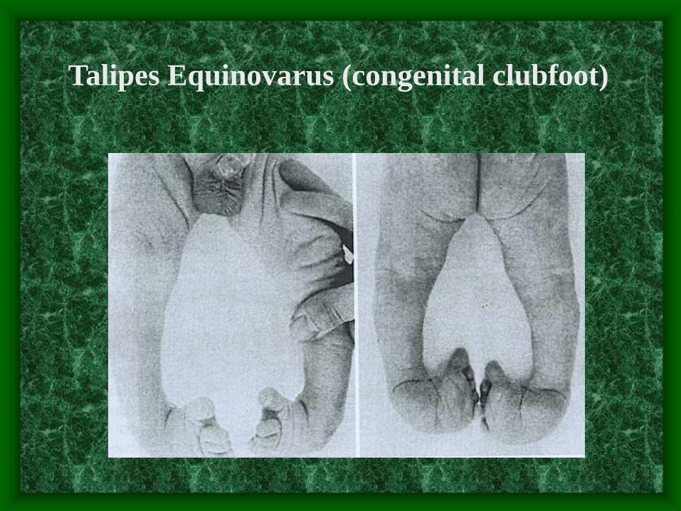
Talipes Equinovarus (congenital clubfoot)
A. General
- complicated, multifactorial deformity of
primarily genetic origin
- 3 basic components
(i) ankle joint plantarflexed/equines
(ii) subtalar joint inverted/varus
(iii) forefoot adducted
Talipes Equinovarus (congenital clubfoot)

Talipes Equinovarus (congenital clubfoot)
B. Incidence
- approx 1/1,000 live births
- usually sporadic
- bilateral deformities occur 50%
C. Etiology
- unknown
- ?defect in development of talus leads to
soft tissue changes in joints, or vice
versa

Talipes Equinovarus (congenital clubfoot)
D. Diagnosis/Evaluation
- distinguish mild/severe forms from other disease
- AP/Lat standing or AP/stress dorsiflex lat films
E. Treatment
• Non-surgical
- weekly serial manipulation and casting
- must follow certain order of correction
- success rate 15-80%
• Surgical
- majority do well; calf and foot is smaller

Talipes Equinovarus (congenital clubfoot)

Pes Planus (flatfoot)
A. General
- refers to loss of normal medial long. arch
- usually caused by subtalar joint assuming an
everted position while weight bearing
- generally common in neonates/toddlers
B. Evaluation
- painful?
- flexible? (hindfoot should invert/dorsiflex
approx 10 degrees above neutral
- arch develop with non-weight bearing pos?

Pes Planus (flatfoot)

Pes Planus (flatfoot)
C. Treatment
(i) Flexible/Asymptomatic
- no further work up/treatment is necessary!
- no studies show flex flatfoot has increased
risk for pain as an adult
(ii) rigid/painful
- must r/o tarsal coalition – congenital fusion or
failure of seg. b/w 2 or more tarsal bones
- usually assoc with peroneal muscle spasm
- need AP/lat weight bearing films of foot

In-Toeing
A. General
- common finding in newborns and children
- little evidence to show benefit from treatment

In-Toeing
B. Evaluation
- family hx of rotational deformity?
- pain?
- height/weight normal?
- limited hip abduct or leg length discrepancy?
- neuro exam
C. 3 main causes
(i) metatarsus adductus
(ii) internal tibial torsion
(iii) excessive femoral anteversion

In-Toeing
(i) metatarsus adductus
- General
• normal hindfoot,
medially deviated
midfoot
• diagnosis made if
lateral aspect of foot
has “C” shape, rather
than straight

In-Toeing
(i) metatarsus adductus
- Evaluation
• should have normal
ankle motion
• assess flexibility by
holding heel in
neutral position,
abducting forefoot

In-Toeing
(i) metatarsus adductus
• treatment
- if flexible, stretching; Q diaper change, 10 sec
- if rigid, or if no resolution by 4-8 months,
refer to ortho
- prognosis is good: 85-90% resolve by 1yr
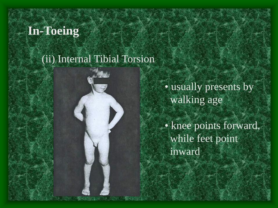
In-Toeing
(ii) Internal Tibial Torsion
• usually presents by
walking age
• knee points forward,
while feet point
inward
In-Toeing
(ii) Internal Tibial Torsion
• Treatment
- reassurance! spontaneous resolution in 95%
children, usually by 7-8yrs
- controversy with splints, casts, surgery

In-Toeing
(iii) Excessive Femoral Anteversion
• both knees and feet
point inward
• presents during early
childhood (3-7yrs)
• most common cause
of in-toeing

In-Toeing
(iii) Excessive Femoral Anteversion
• int rotation 70-80 deg
ext rotation 10-30 deg
• “W” position

In-Toeing
(iii) Excessive Femoral Anteversion
• increase in internal
rotation early with
gradual decrease
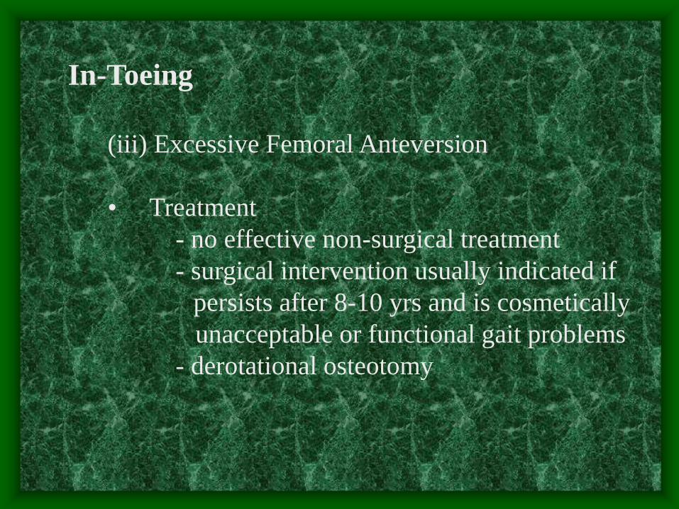
In-Toeing
(iii) Excessive Femoral Anteversion
• Treatment
- no effective non-surgical treatment
- surgical intervention usually indicated if
persists after 8-10 yrs and is cosmetically
unacceptable or functional gait problems
- derotational osteotomy
References
• Hoffinger SA. Evaluation and Management of Pediatric
Foot Deformities. Pediatric Clinics of North America. 1996.
43(5):1091-1111
• Yamamoto H. Nonsurgical treatment of congenital clubfoot
with manipulation, cast, and modified Denis Browne splint.
J Pediatric Ortho. 1998. 18(4): 538-42
• Sullivan JA. Pediatric flatfoot: evaluation and management.
J Am Acad Orthop Surg 1999. 7(1): 44-53
• Dietz FR. Intoeing-Fact, Fiction and Opinion. American
Family Physician. 1994. 50(6): 1249-1259
• Canale. Campbell’s Operative Orthopedics, 9th ed. 1998
1713-1735; 938-940

CLUB FOOT Gross deformity of the foot that is giving it the
stunted lumpy appearance

CLUB FOOT
Definitions
Talipes: Talus = ankle
Pes = foot
Equinus: (Latin = horse)
Foot that is in a position of
planter flexion at the ankle,
looks like that of the horse.
Calcaneus: Full dorsiflexion at the ankle

CLUB FOOT
Planus: flatfoot
Cavus: highly arched foot
Varus: heal going towards
the midline
Valgus: heel going away
from the midline
Adduction: forefoot going
towards the midline
Abduction: forefoot going away
From the midline
Forefoot Hind foot

CLUB FOOT
Types Postural :
Calcaneo-Valgus Equino-Varus
Look for CDH Minor and correctable

CLUB FOOT
Types
Idiopathic (Unknown Etiology) :
• Congenital Talipes Equino-Varus CTEV
Acquired, Secondary to :
• CNS Disease : Spina bifida, Poliomyelitis
• Arthrogryposis
• Absent Bone : fibula / tibia

Congenital Talipes Equino-Varus
CTEV Congenital clubfoot or CTEV occurs
typically in an otherwise normal child.

Congenital Talipes Equino-Varus
CTEV
Etiology
• Polygenic
• Multifactorial
although many of these factors are speculative

Congenital Talipes Equino-Varus
CTEV
Etiology
Some of these factors are :
• Abnormal intrauterine forces
• Arrested fetal development
• Abnormal muscle and tendon insertions
• Abnormal rotation of the talus in the mortise
• Germ plasm defects

Congenital Talipes Equino-Varus
CTEV
Incidence • Occurs approximately in one of every 1000 live
birth
• In affected families, clubfeet are about 30 times
more frequent in offspring
• Male are affected in about 65% of cases
• Bilateral cases are as high as 30 – 40 %

Congenital Talipes Equino-Varus
CTEV
Geographic Distribution
• Middle East , KSA common
• Mediterranean Coast & North Africa
• White race

Congenital Talipes Equino-Varus
CTEV
Basic Pathology
• Abnormal Tarsal Relation
Congenital Dislocation / Subluxation
Talo Calcaneo Navicular Joint
• Soft Tissue Contracture
Congenital Atresia
EGG & CHICKEN

Congenital Talipes Equino-Varus
CTEV

Congenital Talipes Equino-Varus
CTEV
Adaptive Changes Wolff’s Law “ Every change in the use of static function of bone caused a
change in the internal form or architecture as well as alteration in its external formation and function according
to mechanical law ”
Davis Law “ When ligaments and soft tissue are in loose or lax state;
they gradually shorten ”

Congenital Talipes Equino-Varus
CTEV
Adaptive Changes • Bony :
Change in the shape of tarsal and metatarsal
bones especially after walking
• Soft Tissue :
Shortening ? Contracture in the Concave Side
1- Muscles 2- Tendons
3- Ligaments 4- Joints Capsule
5- Skin 6- Nerves & Vessels

Congenital Talipes Equino-Varus
CTEV

Congenital Talipes Equino-Varus
CTEV Diagnosis
General Examination : Exclude • Neurological lesion that can cause the deformity “Spina
Bifida”
• Other abnormalities that can explain the deformity “Arthrogryposis, Myelodysplasia”
• Presence of concomitant congenital anomalies
“Proximal femoral focal deficiency”
• Syndromatic clubfoot
“Larsen’s syndrome, Amniotic band Syndrome”

Congenital Talipes Equino-Varus
CTEV Diagnosis
Spina Bifida = Paralytic TEV

Congenital Talipes Equino-Varus
CTEV
Diagnosis Characteristic Deformity :
Hind foot
• Equinus (Ankle joint)
• Varus (Subtalar joint)
Fore foot
• Adduction (Med tarsal joint)
• Supination fore foot
• Cavus

Congenital Talipes Equino-Varus
CTEV
Diagnosis

Congenital Talipes Equino-Varus
CTEV
Diagnosis
“ Hind foot “ “ Fore foot “
Equinus, Varus Adduction, Supination, Cavus

Congenital Talipes Equino-Varus
CTEV
Diagnosis

Congenital Talipes Equino-Varus
CTEV
Diagnosis • Short Achilles tendon
• High and small heel
• No creases behind Heel
• Abnormal crease in middle of the foot
• Foot is smaller in unilateral affection
• Callosities at abnormal pressure areas
• Internal torsion of the leg
• Calf muscles wasting
• Deformities don’t prevent walking

Congenital Talipes Equino-Varus
CTEV
Diagnosis

Congenital Talipes Equino-Varus
CTEV
Diagnosis
X-Ray needed to assess progress of treatment

Congenital Talipes Equino-Varus
CTEV
Treatment
The goal of treatment for clubfoot is to obtain
a plantigrade foot that is functional, painless,
and stable over time
A cosmetically pleasing appearance
is also an important goal sought by
the surgeon and the family
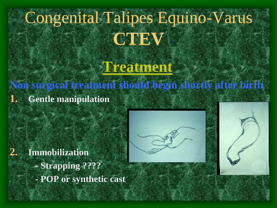
Congenital Talipes Equino-Varus
CTEV
Treatment Non surgical treatment should begin shortly after birth
1. Gentle manipulation
2. Immobilization
- Strapping ????
- POP or synthetic cast

Congenital Talipes Equino-Varus
CTEV
Treatment Non surgical treatment should begin shortly after birth
3. Splints to maintain correction
- Ankle-foot orthosis ????
- Dennis Brown splint

Congenital Talipes Equino-Varus
CTEV
Treatment
Manipulation and serial casts
• Validity, up to 6 months !
• Technique “Ponseti”
• Avoid false correction
• When to stop ?
• Maintaining the correction
• Follow up to watch and avoid recurrence

Congenital Talipes Equino-Varus
CTEV
Treatment Ponseti technique
1. Always use long leg casts, change weekly.
2. First manipulation raises the 1st metatarsal to decrease the cavus
3. All subsequent manipulations include pure abduction of forefoot with counter-pressure on neck of talus.
4. Never pronate !
5. Never put counter pressure on calcaneus or cuboid.

Congenital Talipes Equino-Varus
CTEV Treatment
Ponseti technique (cont.) 6. Cast until there is about 60 degrees of external rotation
(about 4-6 casts)
7. Percutaneous tendo Achilles tenotomy in cast room under local anesthesia, followed by final cast (3 weeks)
8. After final cast removal, apply Normal last shoes with Denis Browne bar set at 70 degrees external rotation (40 degrees on normal side)
9. Denis Browne splint full time for two months, then night time only for two-four years.
10.35% need Anterior Tibialis tendon transfer at age 2-3

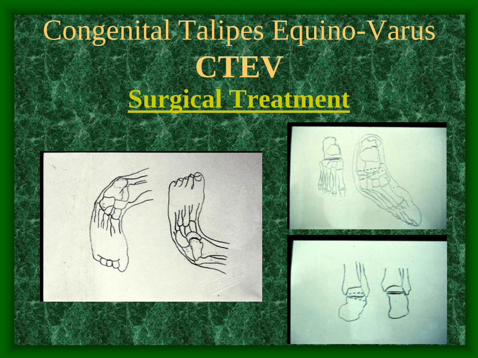
Congenital Talipes Equino-Varus
CTEV
Surgical Treatment
Indications
• Late presentation, after 6 months of age !
• Complementary to conservative treatment
• Failure of conservative treatment
• Residual deformities after conservative treatment
• Recurrence after conservative treatment

Congenital Talipes Equino-Varus
CTEV
Surgical Treatment
• Types (soft tissue and bony operations)
• Time of surgery
• Selection of the procedure and the incision
• Post operative care
• Follow up
• Complications

Congenital Talipes Equino-Varus
CTEV
Surgical Treatment
Soft tissue operations
1. Release of contractures
2. Tenotomy
3. Tendon elongation
4. Tendon transfer
5. Restoration of normal bony relationship

Congenital Talipes Equino-Varus
CTEV
Surgical Treatment

Congenital Talipes Equino-Varus
CTEV
Surgical Treatment

Congenital Talipes Equino-Varus
CTEV Surgical Treatment
Bony operations
• Indications
• Usually accompanied with soft tissue operation
• Types:
- Osteotomy, to correct foot deformity or int. tibial torsion
- Wedge excision
- Arthrodesis (usually after bone maturity)
one or several joints
- Salvage operation to restore shape
Congenital Talipes Equino-Varus
CTEV Surgical Treatment

Congenital Talipes Equino-Varus
CTEV Surgical Treatment

Congenital Talipes Equino-Varus
CTEV Surgical Treatment

Congenital Talipes Equino-Varus
CTEV Surgical Treatment