common pediatric dental problems - university · pdf file · 2013-04-27common...
TRANSCRIPT

PEDIATRIC SURGERY FOR THE PRZMARY CARE PEDIATRICIAN, PART II 0031-3955/98 $8,00 + .OO
COMMON PEDIATRIC DENTAL PROBLEMS
Paul R. Creighton, DDS
Pediatric dentistry is one of the eight specialties recognized by the American Dental Association. In the early 1900s, children were treated as ”little adults” and the focus of routine dentistry was to treat the effects of dental decay, such as pulpitis, and resultant pain from this condition. Initially, dental decay resulted in extraction and restorative treatment with emphasis on space maintenance and arch integrity. Since the early 1900s, tremendous improvements have taken place in restorative and preventative techniques. Today, pediatric dentistry is prevention oriented. The cornerstone of prevention-based pediatric dentistry is early referral to the dentist and routine follow-up visits.
For many years, the American Academy of Pediatrics has recommended that children make their first dental visit after their third birthday. The Academy of Pediatric Dentistry, on the other hand, has recommended that children be seen by the age of 1 year. The goal of a pediatric dental practice is to emphasize the importance of oral health to the child and the child’s parents. Behavior management is still very much the backbone of the specialty. A primary goal of the treatment-oriented pediatric dental profession is behavior management. A prevention-oriented pediatric dental profession concentrates on educating the parents of very young children on the dental milestones seen in the pediatric population, proper diet, and other issues that prevent dental disease. Given the etiology, pathogenesis, and the treatment of dental diseases, prevention is the only true cure, although realistically, restorative treatment will always be a significant component to the specialty. The preventive component will become a larger emphasis of the specialty if preventive strategies are initiated earlier than 3 years of age. By educating patients and families and by recognizing and appropriately referring dental pathology pediatricians have a central role to the promotion of dental health in infants and children (Fig. 1).
From the Department of Pediatric Dentistry, School of Dental Medicine of The State University of New York at Buffalo; and Pediatric Dental Services, The Children’s Hospital of New York at Buffalo, Buffalo, New York
PEDIATRIC CLINICS OF NORTH AMERICA
VOLUME 45 * NUMBER 6 DECEMBER 1998 1579

1580 CREIGHTON
Figure 1. The pediatric treatment triangle.
Pediatricians are typically the first to examine intraoral and extraoral struc- tures and soft and hard tissues postnatally. Intraoral and extraoral clinical exami- nation should focus on symmetry of the maxillary and mandibular arches, the gingiva, and mandibular and maxillary lip support. The palate, tongue, and frenal attachment should be thoroughly examined. Palatal integrity and the depth of the palate should be noted, and tongue should be observed for any restriction in range of motion. Maxillary, labial, and frenal attachments are typically prominent in children. Two epithelial layers, enclosing a loose, vascular connective tissue and, many times, muscle fibers, make up the attachment. This attachment should be noted and monitored because surgical intervention may be necessary to improve orthodontic relationships of the dentition. Any clinical appearance indicating suspicious asymmetry or abnormal soft or hard intraoral or extraoral structures should be referred to a dentist as soon as detected.
INITIAL VISIT
Pediatric dentists should evaluate all infants at the time the first tooth erupts. At this time, the dentist examines the oral cavity for natal teeth and structural abnormalities. The parents are counseled on proper diet and proper feeding techniques to prevent tooth decay. The normal chronologic development of deciduous teeth (Table 1) and permanent teeth (Table 2) are also reviewed.
If the child is born prematurely or has a craniofacial abnormality, an ad-

COMMON PEDIATRIC DENTAL PROBLEMS 1581
Table 1. NORMAL CHRONOLOGIC DEVELOPMENT OF PRIMARY TEETH
Initiation Calcification Begins Crown Completed Eruption Tooth (Wk in Utero) (Wk in Utero) (W (W
Central incisor 7 14 (13-16) 1 -3 6-9
Canine 7.5 17 (15-18) 9 16-20 Lateral incisor 7 16 (14.5-16.5) 2-3 7-1 0
First molar 8 15.5 (14.5-17.0) 6 12-16 Second molar 10 18.8 (16.0-23.5) 10-12 20-30
Modified from Gorlin RJ, Pindborg JJ, Cohen MM: Syndromes of the Head and Neck, ed 2. New York, McGraw-Hill, 1976, p 190; with permission.
justed chronologic development is reviewed with the parents. For example, delayed eruption of permanent teeth may be seen in patients diagnosed with Down’s syndrome, Cleidocranial Dysostosis, hypothyroidism, hypopituitarism, hemifacial atrophy, Apert syndrome, and chondroectodermal dysplasia. Early or precocious eruption is rare but can be seen in hyperthyroidism, hemifacial atrophy, precocious puberty, and the affected areas in Sturge-Weber syndrome. Premature loss of primary teeth is seen in juvenile periodontitis, acrodynia,
Table 2. NORMAL CHRONOLOGIC DEVELOPMENT OF PERMANENT TEETH
Crown Completed Eruptlon Tooth Initiation Calcification (Y) (Y)
Maxilla Central 5-5.5 mo in utero 3-4 mo 4-5 7-8
Incisors
Incisors Lateral 5-5.5 mo in utero 1 y 4-5 8-9
Canine 5.5-6 mo in utero 4-5 mo 6-7 11-12 First Birth 1.5-1.75 y 5-6 10-11
Second 7.5-8 mo 2-2.5 y 6-7 10-12
First molar 3.5-4 mo in utero Birth 2.5-3 6-7 Second molar 8.5-9 mo 2.5-3 y 7-8 12-1 3 Third molar 3.5-4 y 7-9 y 12-1 6 17-25
Central 5-5.5 mo in utero 3-4 mo 4-5 6-7
Lateral 5-5.5 mo in utero 3-4 mo 4-5 7-8
Canine 5.5-6.0 in utero 4-5 mo 6-7 9-1 1 First Birth 1.75-2 y 5-6 10-12
Second 7.5-8 mo 2-2.5 y 6-7 11-12
First molar 3.5-4 mo in utero Birth 2.5-3 6-7 Second molar 8.5-9 mo 2.5-3 y 7-8 11-13 Third molar 3.5-4 y 8-10 y 12-1 6 17-25
Premolar
Premolar
Mandible
Incisor
Incisor
Premolar
Premolar
Modified from Gorlin RJ, Pindborg JJ, Cohen MM: Syndromes of the Head and Neck, ed 2. New York, McGraw-Hill, 1976; with permission.

1582 CREIGHTON
Figure 2. A patient with cleft palate. Note supernumerary tooth in site of cleft (white arrow).
Hand-Schiiller-Christian disease, hypophosphatasia, .and Papillon-Lefgvre syn- drome. Missing or supernumerary teeth can be seen in patients with cleft palate (Fig. 2). Missing teeth and complete anodontia are commonly seen in patients with ectodermal dysplasia.
Premature neonates and infants with cleft palates have unique dental prob- lems. Dentists should interact with pediatricians and neonatologists early in children’s lives if patients are pematurely delivered and require intubation. Orogastric and orotracheal tubes have been known to cause maxillary anatomic deformities in deciduous teeth and supporting arches, as well as speech irregu- larities. Several investigators have shown that low-birth-weight, preterm infants who have orogastric or orotracheal tubes are likely to have palatal deformities, including grooves in the palate, high V-shaped palates, and posterior crossbites (Fig. 3).6 Individual tooth abnormalities include notched and deformed incisal
Figure 3. Study models of two patients that had an orotrachael tube. The model on the left had protective appliance placed 48 hours after birth. The model of the child on the right had no protective appliance resulting in vaulted palate.

COMMON PEDIATRIC DENTAL PROBLEMS 1583
edges, as well as missing teeth. The dental alveolar abnormalities may be indicative of pressure from a laryngoscope blade on the alveolar ridge at the time of intubationP For these patients, a pediatric dentist should be called to place a protective appliance (Fig. 4) to prevent the occurrence of these abnormalities.
Patients with cleft palate have significant structural development issues. Tooth development and orientation as related to the major and minor segments of the cleft are monitored very closely. The pediatric dentist works to coordinate the dental restorative needs with other medical and dental disciplines involved in the treatment of children with cleft deformity (see article by Denk, this issue). The pediatric dentist looks to sequence the care so that comprehensive rehabilitation of the dental arches is successful.
GROWTH AND DEVELOPMENT OF THE TEETH
The seven stages of tooth development are : (1) initiation, (2) proliferation, (3) histodifferentiation, (4) morphodifferentiation, (5) apposition, (6) calcification, and (7) eruption. At any of these stages, abnormalities may occur, resulting in a variety of dysmorphogenic features.
Initiation
The teeth are derived from ectodermal and mesodermal tissues. In the maxillary arch, the anterior teeth initiate from the dental lamina in the frontona- sal process, and the posterior maxillary teeth from the paired lateral maxillary processes. For this reason, children with cleft lip and palate frequently show malformations in the maxillary anterior teeth. The mandibular teeth form from the lamina in the mandible at the time of maxillary development at approxi- mately 37 days’ gestation.
Figure 4. Palatal protective appliance.

1584 CREIGHTON
Proliferation
Proliferation occurs when the dental laminae extend into the underlying mesenchyme at specific locations and form the primary dental organs. At the same time, another epithelial sheet develops next to the dental lamina and is responsible for the formation of the lips, cheeks, and gums. Abnormalities such as cleft lip and palate, ectodermal dysplasia, orofacial-digital syndrome, cleidocranial dysplasia, Gardner syndrome, Hallermann-Streiff syndrome, Crou- zon syndrome, Ellis-van Creveld syndrome, and Apert syndrome result in prob- lems during the initiation and proliferation stages and thus result in dental anomalies of both number and structure.
Histodifferentiation and Morphodifferentiation
Histodifferentiation and morphodifferentiation are stages at which the teeth assume their recognizable shapes. At approximately the sixth week of intrauter- ine life, the deciduous anterior tooth germs begin these processes. The deciduous posterior teeth begin about 1 week later. At the 16th week of gestation, the histodifferentiation and morphodifferentiation stage begins for the first perma- nent molar and continues after birth for the second and third permanent molars. Problems during these two stages lead to abnormalities in tooth size and shape and can result in amelogenesis and dentinogenesis imperfecta. Dentinogenesis imperfecta presents with a paucity of enamel and an opalescent appearance in both the deciduous and permanent dentition. Amelogenesis imperfecta also involves deciduous and permanent dentition and is mainly a disorder that affects the enamel.
Apposition
Ameloblasts and odontoblasts are considered formative cells that deposit the enamel and dentin matrix according to a definite rate and pattern. Any systemic disturbance or local trauma resulting in an insult to the matrix affects the appositional development of the tooth. Hypoplasia of the enamel, and more rarely the dentin, may result.
Calcification
The deciduous teeth begin to calcify in the fourth month of gestation. Enamel hypoplasia is defined as defective or incomplete formation of dental enamel and is the result of problems in the calcification stage. Enamel hypoplasia can be seen in patients with cleft lip and palate, Down’s syndrome, pseudohy- perparathyroidism, epidermal nevus, and Treacher-Collins and Turner s p - dromes.
Eruption
Eruption patterns for children provide major milestones in an infant’s life. The actual eruption times are approximate, and variability depends on genetic, systemic, and environmental factors. The eruption pattern of permanent teeth

COMMON PEDIATRIC DENTAL PROBLEMS 1585
seems to be more variable than deciduous teeth. The eruption pattern in girls seems to be earlier than for boys. Early eruption is considered premature if the teeth erupt before 3 months of age. If they are present at birth, the teeth are termed natal teeth. If they erupt between birth and 30 days, the teeth are called neonatal teeth. Natal teeth are three times as common as neonatal teeth. The majority of natal and neonatal teeth are anterior, and almost 90% are normal, primary teeth erupting early. No apparent sex predilection exists, yet familial occurrences and hereditary transmission have been most characteristic of an autosomal dominant mode. These occurrences have been associated with three syndromes: (1) chondroectodermal dysplasia, (2) Hallermann-Streiff syndrome, and (3) pachyonychia congenita. Early or precocious eruption is rare but can be seen in patients with hyperthyroidism, hemifacial atrophy, precocious puberty, and the affected areas in Sturge-Weber syndrome. Premature loss of primary teeth is seen in patients with juvenile periodontitis, acrodynia, Hand-Schuller- Christian disease, hypophosphatasia, and Papillon-Lefevre ~yndrome.~
Delayed eruption may be caused by systemic disorders and has been associ- ated with Down’s syndrome, cleidocranial synostosis, hypothyroidism, hypopi- tuitarism, hemifacial atrophy, Apert syndrome, and chondroectodermal dyspla- ~ i a . ~
DENTAL CARIES
One of the most prevalent disease processes in children is dental caries. Dental caries is a multifactorial disease process that requires the following ”ingredients”: the presence of a susceptible host, cariogenic microflora, a diet that is conducive to enamel demineralization, and time (Fig. 5 ) . Streptococcus
Figure 5. The ingredients for dental decay. All must be overlap for the decay process to occur.

1586 CREIGHTON
mutans present within the oral cavity is the organism responsible for the initia- tion of dental caries. Sugar is metabolized by anaerobic bacteria to acid end products, which lower salivary pH and the pH within the adherent bacteria-rich plaque. This bacteria-rich plaque is relatively unavailable to salivary buffering. The lowered pH near the tooth surface can cause ionic dissolution from the hydroxyapatite crystals, increase surface roughness, and enhance the ability for plaque to be more adherent and for caries to be i~~i t ia ted.~
Diet is the major factor in the development of dental caries. The quantity and frequency of ingestion of carbohydrates and the consistency of foods in- gested are the most important factors in assessing cariogenicity of the diet. Parents provide to their children many foods that can result in tooth decay. For example, parents that supply snacks such as raisins, dried fruit snacks, sweet- ened cereals, and fruit juices may be contributing to the demise of their chil- dren's dentition.
Over the past 30 years, a decrease has occurred in the incidence of dental caries, yet caries still exists at a high rate in some of our population. The earliest appearance of specific caries is nursing caries, which usually appears before the age of 20 months. The terms baby-bottle tooth decay (BBTD) and, more recently, early childhood caries are assigned to this caries pattern. Nursing caries results from nighttime feeding combined with the effects of Streptococcus mutans coloni- zation. These night feedings can either be from a bottle or the breast. Breast milk contains lactose, which, if allowed to pool and when not cleared from the mouth, results in a decay sequence similar to that of bottle feedings. The decay pattern usually involves smooth surfaces on maxillary deciduous anterior teeth, fol- lowed by the maxillary first molars (Fig. 6). Mandibular dentition is not affected because significant salivary flow clears the pooling bottle substrate. Parents of children with nursing caries report common factors. Typically, these children are placed in bed with a bottle of milk or juice. As these children fall asleep, the liquid pools around the upper teeth, providing an excellent environment for
Figure 6. Smooth surface decay.
Figure 7. Pit and fissure decay.

COMMON PEDIATRIC DENTAL PROBLEMS 1587
bacteria in the mouth. Further encouraging this environment is the fact that salivary flow and swallowing diminish as children fall asleep. Additional predis- posing factors include children’s genetic predisposition to caries, being reared in a single-parent household, a strong temperament (as perceived by the parent), sleeping difficulties, and a reward system involving s w e e t ~ . ~
Pit and fissure caries (Fig. 7) usually begins at about 3 years of age and involves grooves and fissures of the posterior teeth. Candy and retentive forms of food (e.g., raisins) contribute to the decay process seen in a large percentage of the population. Caries of the proximal surfaces of posterior teeth can begin after the molars move into adjacent contact and is a smooth surface decay process. This process is usually seen soon after the third birthday.
Severe caries may have significant impact on children’s overall health. Figure 8 depicts a patient who had not followed through with restorative treatment, and all 20 of the primary teeth had to be extracted. This child’s nutritional health was greatly affected, and he was subsequently diagnosed with failure to thrive.
Facial cellulitis (Fig. 9) and lymphadenopathy include dental decay as a possibility in the differential diagnosis. When patients present with facial celluli- tis, a panorex, periapical radiographs, and clinical examination are necessary. The antibiotic of choice for a facial cellulitis from dental decay is penicillin.
One aspect of children’s lives that has received recent attention is the effect of over-the-counter medications and liquid or chewable pharmaceutical preparations on dentition. For example, many liquid or chewable pharmaceutical preparations for children are made palatable by the addition of sucrose, glucose, or fructose as sweeteners. Medications containing these sweeteners (e.g., analge- sics, antibiotics, and vitamins) are given either before bedtime or are held in the mouth for extended periods of time and, therefore, may pose a risk of caries. A controlled study of patients taking chronic doses of medications reported a significant increase in dental caries and gingivitis in a population of children taking liquid or chewable medications continuously for a minimum of 6 months. l4
Figure 8. Severe dental decay.

1588 CREIGHTON
Figure 9. Facial cellulitis due to dental caries.
Salivary flow is critical in maintaining the health of oral soft and hard tissues.’h Children with decreased salivary flow may be more susceptible to smooth surface caries. It has been suggested that mouth breathers have modified plaque accumulations and associated soft tissue changes. Particular attention should be made of the numerous medications that mimic mouth breathing by decreasing salivary flow.17
After carious teeth have been identified, restorative treatment is planned. The pediatric dentist’s goal is to complete the necessary restorative work while respecting the potentially vulnerable psychological development of young chil- dren.
Many children are capable of completing restorative dental work in the office setting (nonpharmacotherapeutic). Such treatment plans include behavior management techniques, such as ”tell-show-do,” voice control, and various forms of restraint that range from ”hand-over-mouth” to utilization of a papoose board. These techniques are being considered more frequently because of their lesser risk for the patient than that incurred with pharmacotherapeutic measures. Pharmacotherapeutic techniques include the use of agents such as chloral hy- drate, hydroxyzine, midazolam, diazepam, or “kiddie cocktails.” These agents or combinations of agents are respiratory depressants and may cause increased medical risk to the patient and legal exposure to the dentist, who uses them in his or her office.
If a child is unable to cooperate because of age or disability, then conscious sedation may be a consideration. If numerous teeth are involved and the child is considered inappropriate for treatment in the office setting, then a general anesthetic, administered by a pediatric anesthesiologist, should be considered.

COMMON PEDIATRIC DENTAL PROBLEMS 1589
PREVENTIVE DENTISTRY
As mentioned previously, prevention is a major concern of pediatric den- tistry. The prevention of the decay processes is not difficult if the type of decay process is identified and parents are counseled and receptive to suggested preventive measures. Early detection is important, so great value is placed on seeing children soon after the first teeth erupt. Waiting until the third birthday in many cases is too late to counsel patients’ parents.
Typically, prevention programs target altering one or more of the following causes: susceptibility of the host, the presence of bacteria, or diet. Fluoride treatment is considered one of the most effective measures in preventing tooth decay in children. The intention of fluoride is to alter the susceptibility of the host. Fluoride is introduced either systemically or topically. Systemically, fluoride is introduced in the drinking water or through vitamin supplementation. As a result of this ingestion, fluoride is incorporated into the dentin and enamel of developing teeth, which makes them more resistant to demineralization by acids. Topically, fluoride is introduced in toothpaste, rinses, and professionally applied gels and foams. The result of topical application results in remineralization of demineralized enamel and increasing strength of the enamel of erupted teeth to make them more resistant to acid attack.*
Numerous studies have documented caries reduction of 40% to 50% in primary dentition and 50% to 65% in the permanent dentition of children drinking fluoridated water from birth.’* Proper dosages of fluoride should be prescribed according to the age of the child and the fluoride concentration of the drinking water. Table 3 shows the recommended supplemental fluoride dosage schedule. #en discussing fluoride with a parent, the concept that “more is better” should be discouraged. The dentist prescribing fluoride must take a thorough history to ensure that the child is not already receiving significant amounts of fluoride in his or her diet. In the most severe cases of excess fluoride ingestion, mottled enamel may result. In mild or moderate cases, the enamel presents with white or brown areas. Acute fluoride toxicity can result in nausea and vomiting. #en prescribed, the appropriate storage and administration of these products must be emphasized to the parent.
Improved oral hygiene and diet control are equally emphasized in preven- tive programs. Brushing the teeth as soon as they erupt is very important. Behavioral patterns are established early in life and consistent, effective brushing lowers the amount of sugar substrate and reduces plaque. Many have argued that diet is the most difficult aspect of a preventive program. This is important because without substrate, the chance for caries is less. Parents are informed of
Table 3. AMERICAN DENTAL ASSOCIATION RECOMMENDED SUPPLEMENTAL FLUORIDE DOSAGE SCHEDULE
Water Fluoride Concentration (ppm)
Age of Child <0.3 0.3-0.6 >0.6
Birth to 6 months 0 0 0 6 months to 3 years 0.25-mg liquid drops 0 0 3 to 6 years 0.5-rng drops or tablet 0.25 mg 0 6 to 16 years 1.00 mg 0.5 mg 0

1590 CREIGHTON
early childhood caries and suggestions on proper feeding techniques, such as the proper use of a bottle, the timing of when to discontinue the bottle, and the possibility for cross-contamination of bacteria when feeding utensils are placed first in a parent’s mouth and then in a child’s mouth.
ORTHODONTICS
Orthodontics is the specialty of dental medicine Concerned with the diagno- sis, supervision, guidance, and treatment of growing and mature dentofacial structures. This often includes moving teeth or bones to correct for tissue defi- ciencies and malformations of the craniofacial complex. The orthodontist is mainly concerned with diagnosis and treatment of mal-relationships of the teeth and the overlying soft tissues within and between the maxilla and the mandible. The correction of any abnormal relationships is accomplished by applying forces to the teeth and, if necessary, orthopedic forces, to redirect abnormal bone growth.
Referral to initiate orthodontic care is based on a sagittal relationship of the maxillary and mandibular dental units developed by Angle in 1910.3 This relationship focuses on the canines and first permanent molars. In primary dentition, the relationship is between the canines and second deciduous molars. Class I occlusion (ideal occlusion) exists when the mandibular molars and canines are mesial (forward) of maxillary first molars and canines (Fig. 10). Class I1 occlusion occurs when the mandibular molars and canines are distal (behind) the maxillary first molars and canines. The maxillary incisors are typically positioned forward of their bony base resulting in the incisors protruding for- ward. Patients with Class I1 occlusions often present with “deep bites.” De- pending on the type of malocclusion, the deep bite may be caused by the supereruption of the maxillary or mandibular central incisors (Fig. 11). Class 111 occlusion (Fig. 12) exists when the mandibular molars‘and canines are mesial of maxillary molars and canines. Typically, the maxillary incisors are retrusive and are tucked behind the mandibular molars. The most common cause of Class I11 occlusion is excessive growth of the mandible.
Treatment of the orthodontic patient is directed toward establishing correct or normal occlusion. Three dentoskeletal relationships have a direct effect on dental occlusion. These relationships in turn have an effect on the overlying facial soft tissue, which influences the facial aesthetics. These relationships in- clude (1) the relationship of the maxilla and mandible to the rest of the craniofa- cia1 structures, (2) the relationship of the upper and lower dentitions to the respective jaw bones, and (3) the relationship of the maxillary dentition to the mandibular dentition.
The development of the maxilla and mandible needs to be in concert with the rest of the craniofacial structures. For adequate development of occlusion, the maxilla and mandible must have normal position. Treatment to establish what is considered ”normal aesthetics” is a very important consideration for patients, and the proper underlying skeletal relationship of the maxilla and mandible is reflected in the overlying facial soft tissue contours. A systematic facial examination should be undertaken and should include an evaluation of the skeletal and dental relationships in three spatial planes: (1) anteroposterior, (2) vertical, and (3) transverse.
The relationships of the upper and lower dentitions to their respective jawbones are referred to as intra-arch relationships. For example, the relationship of the mandibular teeth to mandible is called the lower intra-arch relationship.

COMMON PEDIATRIC DENTAL PROBLEMS 1591
Figure 10. Class I occlusion (ideal occlusion). (From Pinkham JR: Pediatric Dentistry Infancy Through Adolescence, 2nd ed. Philadelphia, W.B. Saunders Company, 1994; with permission.)
Figure 11. Class II occlusion. (From Pinkham JR: Pediatric Dentistry Infancy Through Adolescence, 2nd ed. Philadelphia, W.B. Saunders Company, 1994; with permission.)
Figure 12. Class 111 occlusion. (From Pinkham JR: Pediatric Dentistry Infancy Through Adolescence, 2nd ed. Philadelphia, W.B. Saunders Company, 1994; with permission.)
Interarch relationships are the relationship of the mandibular and maxillary teeth. When the maxillary or mandibular, intra-arch and interarch relationships are considered to be within a normal range, then mastication, speech articulation, and temporomandibular joint function are not ~hallenged.~
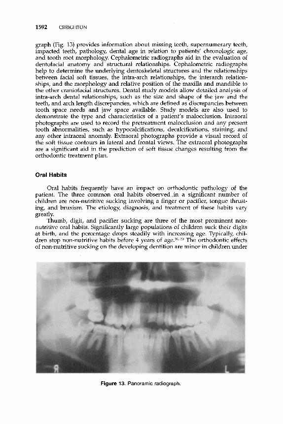
It is the orthodontist’s responsibility to evaluate the dentoskeletal anatomy and its relationship to the overall facial aesthetics and oral function. Along with the clinical examination, other diagnostic tools are necessary for an accurate treatment plan to be developed. These include a panoramic radiograph or a complete set of intraoral radiographs (if a panoramic radiograph is not avail- able), cephalometric radiographs (posteroanterior and lateral), dental study mod- els, and intraoral and extraoral photographs. The clinical examination allows for a complete caries evaluation, a thorough evaluation of the interarch and intra- arch dental relationships, an assessment of the soft tissue contours and facial aesthetics, and a comprehensive assessment of the temporomandibular joints and dental relationships during the mandibular movements. A panoramic radio-
1592 CREIGHTON
graph (Fig. 13) provides information about missing teeth, supernumerary teeth, impacted teeth, pathology, dental age in relation to patients’ chronologic age, and tooth root morphology. Cephalometric radiographs aid in the evaluation of dentofacial anatomy and structural relationships. Cephalometric radiographs help to determine the underlying dentoskeletal structures and the relationships between facial soft tissues, the intra-arch relationships, the interarch relation- ships, and the morphology and relative position of the maxilla and mandible to the other craniofacial structures. Dental study models allow detailed analysis of intra-arch dental relationships, such as the size and shape of the jaw and the teeth, and arch length discrepancies, which are defined as discrepancies between tooth space needs and jaw space available. Study models are also used to demonstrate the type and characteristics of a patient’s malocclusion. Intraoral photographs are used to record the pretreatment malocclusion and any present tooth abnormalities, such as hypocalcifications, decalcifications, staining, and any other intraoral anomaly. Extraoral photographs provide a visual record of the soft tissue contours in lateral and frontal views. The extraoral photographs are a significant aid in the prediction of soft tissue changes resulting from the orthodontic treatment plan.
Oral Habits
Oral habits frequently have an impact on orthodontic pathology of the patient. The three common oral habits observedin a significant number of children are non-nutritive sucking involving a finger or pacifier, tongue thrust- ing, and bruxism. The etiology, diagnosis, and treatment of these habits vary greatly.
Thumb, digit, and pacifier sucking are three of the most prominent non- nutritive oral habits. Significantly large populations of children suck their digits at birth, and the percentage drops steadily with increasing age. Typically, chil- dren stop non-nutritive habits before 4 years of age.1o, 2o The orthodontic effects of non-nutritive sucking on the developing dentition are minor in children under
Figure 13. Panoramic radiograph.

COMMON PEDIATRIC DENTAL PROBLEMS 1593
Figure 14. Orthodontic habit-breaking appliance that is cemented in the mouth.
3 years of age. The amount of time the child engages in the non-nutritive habit dictates the amount of displacement of the maxillary anterior teeth and premaxilla. Active intervention to discourage non-nutritive sucking is encour- aged after 4 years of age. If the habit is discouraged before the permanent central incisors erupt, then the negative effects of non-nutritive sucking tend not to be permanent. It is easier to discontinue a pacifier habit than a digit habit. Pacifiers can be made less accessible. Pacifiers should be of one-piece construc- tion, with a nipple of appropriate size. Parents should not alter the pacifier (e.g., putting a hole in the nipple to deflate it). If the habit continues through the mixed and eventual permanent dentition stages, significant displacement of teeth and supporting bone may occur. Orthodontic appliances (Fig. 14) have been successful in aiding patients who should stop the habit. The main reason for these appliances is to act as a reminder to patients. If the patient does not want to stop the non-nutritive habit, then appliances tend not to be successful.
Orthodontic therapy has limited success unless tongue-thrust habits are addressed first. Children that are tongue-thrusters move their tongues past their front teeth upon swallowing, thus earning the name of "deviate swallowers." Constant pressure from the tongue may act as an orthodontic force and cause significant relapse of an orthodontic case. Investigators have questioned the relationship of digit-sucking habits and anterior open bite (the anterior teeth are not in occlusion when the posterior teeth are in occlusion) to tongue-thrust swallowing.'*, 19, As with non-nutritive habits, tongue-thrust habits become less of an issue as children grow older. The highest incidence of tongue thrusting during swallowing appears in children under the age of 4 years. More than 50% of children under 6 years of age demonstrate a tongue-thrust swallow, compared with less than 6% at the age of 18 years.13 Tongue thrusting is much more common than anterior open bite, so a cause-and-effect relationship is question- able. Digit sucking may be instrumental in anterior open bite, which causes an adaptive tongue-thrusting swallow. Treatment is controversial, difficult, and requires a combined effort of the patient, dentist, and speech therapist. Patients must have a desire to stop the tongue-thrusting habit. Dentists may use several different orthodontic appliances, and the speech therapist uses patterning and psychological techniques to eliminate this problem.
Bruxism is not uncommon to the pediatric population. It rarely is seen to

1594 CREIGHTON
cause any long-term damage to the permanent dentition. The effect of bruxism on the temporomandibular joint is still questioned.'O As with the other oral habits, the frequency and duration of bruxism are the determining factors of the effect on the dentition. Typically, if young infants are found to brux, then there is a decrease seen until the mixed dentition stage (6 years), at which time it is not uncommon for patients to increase the frequency. From this age on, an obvious decrease in the habit occurs. The cause of bruxism is unknown; however, a correlation exists between physical and psychological stress and bruxism patterns. When bruxism results in damage to permanent teeth, the dentist may introduce intraoral splints. Psychological counseling may also be a component of the treatment. Bruxism in the special-care population is not uncommon, yet the treatments used vary as the severity of the situation dictates. Bruxism that results in damage to dentition and the surrounding hard and soft tissues is commonly seen in comatose patients. Intraoral splints are successful for short periods of treatment and must be introduced at the request and coordination of the medical personnel caring for the patients' overall conditions.
TRAUMA
Dental injuries present unique problems in children and young adults with developing dentition. Following facial trauma, after the patient is medically stable, a thorough examination and evaluatiop of any oral injuries should be completed. An examination starts with the evaluation of all the hard and soft tissues. Any tears or lacerations are noted, controlled, and addressed later. The facial bones and temporomandibular joint are palpated to rule out fractures or dislocation. The mandible is opened and closed to rule out malocclusion and any deviation or discomfort. The mouth is occluded with teeth opposed to check for any irregularities that possibly indicate bony fracture or tooth displacement. The teeth are then examined individually for fractures or displacements. Radio- graphs, essential for a complete examination, serve as a baseline for follow-up. In infants and children, the effects of trauma on primary dentition on permanent teeth are of ultimate ~oncern.~
Trauma of Primary Dentition
Prevalence and Etiology Most injuries to primary teeth occur during the toddler stage, between 1
and 3 years of age. It is at this time that children are learning to walk yet lack protective coordination. The teeth most frequently injured are the maxillary central incisors. Many times the maxillary central incisors are in a class 2 malocclusion and protrude precariously, almost inviting injury. Falls account for most injuries, yet there are a significant number of injuries that occur with motor vehicle accidents and child abuse.
The supporting alveolar bone is quite resilient. Because of the pliable, hemopoietic nature of the alveolar bone, teeth are displaced or luxated rather than fractured. Damage caused by a luxation injury not only involves the teeth but also the periodontal ligament (PDL), which is considered the physiologic "hammock that supports the tooth in its socket. Although the viability of the PDL is important, the effect of the luxation injury on the developing permanent teeth is even more important. Furthermore, maintaining the neurovascular bun-

COMMON PEDIATRIC DENTAL PROBLEMS 1595
dle, which is at the apex of the tooth and responsible for the vitality of the tooth, is the primary objective of treatment of all luxation injuries.’
Intrusion and luxation injuries are of particular importance because they present a bigger risk to the unerupted developing permanent teeth. The perma- nent anterior teeth develop in close proximity to the apices of the primary incisors. If the luxation injury occurs during the development of the permanent tooth crown (before the age of 4 years) enamel hypoplasia or hypocalcification may be seen on the facial and lingual surface of the permanent crown. These injuries can also alter the path of the developing permanent tooth crown, causing root dilaceration or ectopic eruptions. Periodic radiographic and clinical evaluation are necessary in the follow-up of these in j~r ies .~
Tooth fractures can involve the root, crown, or both. Figure 15 illustrates the more common fractures involving different regions of the tooth. At the time of the initial clinical examination, all soft and supporting hard tissues are examined. The presence of foreign matter in the lacerations of the lips or cheeks, such as tooth fragment or soil, should be identified and removed. Each tooth in the affected area should be clinically examined and transilluminated, and appropriate radiographs (periapical radiographs) should be taken. Transillumi- nation allows the clinician to visualize a through-and-through fracture versus a craze line that may exist in the enamel.
Treatment of the traumatic injuries to the primary dentition is preceded by a complete diagnostic work-up, which includes a complete medical history, history of the accident, and clearly defined short-term and long-term plans for treatment. Pulpal necrosis caused by the severance of the neuromuscular bundle at the apex is of great concern. Any effect of the trauma on the development of the permanent teeth is the ultimate concern. For this reason, a child that has sustained trauma to the anterior or posterior primary dentitions should be examined by the dentist as soon as possible.
Figure 15. Common fracture sites that involve different regions of the tooth. PDL, periodon- tal ligament.

1596 CREIGHTON
Fractures
Enamel FracturesKlass 1. Small enamel fractures can be smoothed out, and no restoration is necessary. Larger enamel fractures are routinely restored with acid-etch composite restorations.
Enamel and Dentin FracturesKlass 2. Exposed dentin is covered with an acid-resistant calcium hydroxide paste or with glass ionomer cement to “insu- late’’ the pulp and prevent insult to the pulp. Resin is used to restore the tooth.
Fractures Involving the Pulp/Class 3. If the pulpal tissue is believed to be vital, the treatment includes complete removal of the coronal pulpal tissue, zinc oxide, eugenol placement, and the placement of a resin restoration. If the pulp tissue is believed to be nonvital, then the tooth is extracted.
Posterior Crown Fractures. A blow to the underside of the chin is responsi- ble for fractures to posterior primary crowns. Treatment is consistent with the treatment of the anterior teeth as just described.
Root Fractures. Displaced root fractures have a poor prognosis, and a reduction of the segments should not be attempted. The more coronal segment should be removed, and if the apical portion is not easily accessible, it should be left to be clinically and radiographically monitored. The level of the root fracture determines the course of treatment in nondisplaced root fractures. Teeth that have fractures in the apical third of the root typically maintain their vitality and are minimally mobile. Midroot or cervical third fractures are routinely extracted, and every attempt to avoid insult to the developing permanent tooth bud should be made.
Concussion
The concussed tooth is not mobile and is not displaced. The PDL absorbs the injury and is inflamed, which leaves the tooth tender to biting pressure and percussion. Depending on the severity, the prognosis for a concussed tooth is usually good. If the child is symptomatic, then the tooth is gently taken out of occlusion.
Mobility
Mobility is a common result of trauma. The tooth is loosened but is not displaced from its socket. Diet restrictions and follow-up are important on regular intervals during the 3 months after the trauma. Splinting the teeth many times is successful. If a child presents to the pediatrician’s office and the history indicates sudden mobility, the pediatrician should consult a dentist (keeping in mind the normal chronologic development of the teeth).
Intrusion
With intrusion, the tooth is driven into its socket. This compresses the PDL and commonly causes a crushing fracture of the alveolar socket. Three types of intruding injuries exist. The first involves a force that displaces the root palatally toward the developing tooth bud. If the root ends up in direct contact with the permanent tooth bud, then the primary tooth is removed. The second intrusion injury involves a palatally displaced root and no resultant contact with the developing tooth bud. This primary tooth is allowed to re-erupt. The third type involves a force that displaces the root of the primary tooth facially away from the developing tooth bud.” This also should be allowed to re-erupt. Intrusion

COMMON PEDIATRIC DENTAL PROBLEMS 1597
injuries are considered the most threatening to the developing tooth bud. Re- eruption usually takes up to 6 months to occur. Follow-up appointments typi- cally are scheduled every 3 months. If a fistula or perapical radiolucency devel- ops, then the tooth is extracted.” Parents are reminded that the prognosis for the developing permanent teeth varies from person to person.
Extrusion and Lateral Luxation Injuries
With extrusion, the tooth is centrally dislocated from its socket. The PDL is usually torn in this injury. With lateral luxation, the tooth is displaced in a labial, lingual, or lateral direction. The PDL is torn, and contusion or fracture of the supporting alveolar bone occurs. Severe damage to the developing PDL usually occurs in these injuries. With primary teeth, the danger of disrupting the perma- nent tooth bud does not outweigh the advantage of maintaining mobile teeth. Extraction of mobile, depressible, primary extruded or laterally luxated teeth is recommended.
Avulsion
With avulsion, the tooth is completely displaced from the alveolus. The PDL is severed, and fractures of the alveolus may occur. If the patient does not present with the tooth in hand, then an effort must be made to locate the avulsed tooth. Many times, the tooth is actually driven into the socket (intrusion) and is not avulsed. If the tooth is not found, then appropriate radiographs should be taken to rule out aspirated or intranasally displaced teeth. Primary teeth that have been avulsed should‘not be reimplanted. Space loss in the maxillary anterior region is negligible if one or more of the incisors have been lost. Parents should be advised that avulsion injuries may insult the developing permanent tooth buds, and the permanent teeth may be affected. The eruption of the permanent teeth may be delayed as the gingiva may form a fibrotic scar in the path of the eruption?
As children grow, the supporting alveolar bone changes in density. The bone is considered less hemopoietic, and the marrow spaces are smaller. Similarly, as children get older, the resultant injuries seen in permanent teeth involve more crown fractures and fewer displacement injuries. The maxillary incisors are most commonly injured, and ”orthodontically” compromised teeth (protruding incisors) are at greatest risk for injury.
Trauma of Permanent Dentition
The classification of tooth fractures and luxation injuries in permanent teeth is similar to primary teeth. The principles of diagnosis and treatment are also similar; however, resultant pulpal necrosis and resorption of the root structure are of great concern in the development of permanent teeth. Luxation injuries damage the supporting structures of the teeth, which include the PDL and alveolar bone. The pulpal tissue of luxated permanent teeth with closed apices frequently becomes necrotic. If the apex of the tooth is open, the chance of the pulp becoming necrotic is not as great. The vitality of the periodontal ligament is the primary objective in the treatment of luxated permanent teeth. A tooth that has a healthy PDL has a good chance for long-term survival.
Pulpal management and stabilization of the tooth are two areas that differ in the management of trauma of primary and permanent teeth. Fractures that

1598 CREIGHTON
involve the pulp typically require root canal therapy designed to either encour- age the completion of the formation of the root structure of an immature tooth or to preserve the root structure of a mature tooth. Pulpal tissue is necessary for the development of the root structure and not necessarily for the retention of the tooth. A healthy PDL is necessary for long-term retention of traumatized permanent teeth. When the long-term health of the root is secured, prosthetic dental procedures (crown restoration) may be completed. Permanent teeth that are avulsed are reimplanted. The long-term prognosis for teeth that are avulsed decreases the longer the tooth is out of the socket.2 The primary concern is the health of the PDL fibers. The viability of these fibers is reduced the longer the tooth is out of the mouth. If the tooth cannot be replaced immediately, it should be placed in some transport medium that will maintain the PDL fibers, Milk or a professionally marketed tooth-saving transport medium are the two most ideal solutions that preserve the PDL.
Avulsions, extrusions, lateral luxations with bone damage, and root fractures are four indications for the stabilization of the permanent tooth by using a splint. Stabilization of a reimplanted avulsed tooth is designed to promote a healthy PDL. The stabilization period for an avulsed tooth is 1 or 2 weeks. Splinting the avulsed tooth is designed to restore normal physiologic movement of the tooth, which is necessary to interrupt areas of incipient resorption and ankylosis on the PDL. Splinting of an extruded tooth usually lasts for 2 or 3 weeks. The purpose is also to restore normal physiologic function. Stabilization of the luxation and intrusion injuries with bone involvement involves application of a splint for 3 to 8 weeks because of initial resorption of injured bone, which retards the periodontal healing. Stabilization-of a root fracture is usually for the longest period (3-6 mo) and is designed to encourage the formation of a callus that aids in a hard tissue union of the fracture sites7
Prevention
Orofacial protection is important in the prevention of much of the trauma previously described. Any sport that could result in trauma to the dentition should require orofacial protection. Sports that have the most documented orofacial trauma are soccer, football, baseball, and hockey. A well-fitting, custom- made mouth guard (Fig. 16) has been shown to decrease a significant amount of orofacial trauma. The proper construction of a custom mouth guard requires information concerning the patient's dentition, age, and orthodontic condition and predisposition. By separating the condyle from the base of the skull, the mouth guard significantly reduces the incidence of cerebral and dental concus- ion.^
There are many "stock or "store-bought" mouth guards on the market today. Many are considered effective if the resultant mouth guard is firmly fitting in the mouth. Custom-made mouth guards fabricated by dentists provide the ultimate orofacial protection.
SUMMARY
Physicians who provide primary care for children have a unique position to provide diagnostic, triage, educational, and preventive dental care for patients. Several papers have been published regarding primary pediatricians' participa- tion in the preventive dental health care of their patients. One publication, a survey of physicians in Alabama focusing on physicians' overall awareness of

COMMON PEDIATRIC DENTAL PROBLEMS 1599
Figure 16. Custom-made mouth guard.
dental issues, concluded that most physicians believe they have a role in the oral health of their patients, yet most were not aware of many of the American Academy of Pediatric Dentistry’s recommendation^.^^
Most physicians report that they routinely perform oral examinations during physical examinations of children and deliver preventive, oral information by the age of 6 months or earlier; however, most recommend that infants’ first visit should be at 3 years of age, not at the time of first-tooth eruption as the authors recommend. Furthermore, many primary care physicians do not talk about oral health during prenatal counseling. Many physicians understand the preventive advantages of fluoride, yet most do not prescribe vitamin combinations that contain fluoride. If an understanding of the aforementioned issues of dental care, as well as aspects of preventive care in infants and children, become more uniform among primary care physicians, the prevention-based practice of pediatric dentistry will become much more successful, and children and adults will enjoy better dental health.R
References
1. Andreasen JO, Andreasen FM (eds): Essentials of Traumatic Injuries to the Teeth.
2. Andreasen JO, Hjorting-Hansen E: Intraalveolar root fractures: Radiographic and his-
3. Angle EH (ed): Treatment of Malocclusion of the Teeth, vol 7. Philadelphia, White
4. Brodsky L, Holt L, Ritter-Schmidt (eds): Craniofacial Anomalies. St. Louis, CV
5. Camp J: Emergency: Dealing with sports related dental trauma. Journal of the Ameri-
6. Kopra D, Creighton PR: The oral effects of neonatal intubation [abstract]. J Dent Res
7. McDonald RE, Avery DR (eds): Dentistry for the Child and Adolescent, ed 6. St. Louis,
8. Nainar SM: Implications of evidence-based practice on preventive procedures in pedi-
Copenhagen, Munksgaard, 1990
torical study of 50 cases. J Oral Surg 25:414426, 1967
Dental Manufacturing Company, 1907, p 132
Mosby, 1992
can Dental Association 127812-814, 1996
6768,1988
CV Mosby 1994
atric dentistry. J Pediatr Dent 19:384-385, 1997

1600 CREIGHTON
9. Nowak A Conference Report Feeding and dentofacial development. J Dent Res
10. Nowak A, Bishara S, Lancial L, et al: Changes in nutritive and non-nutritive sucking habits: Birth to two years [abstract]. J Dent Res 651525, 1986
11. Pinkham JR (ed): Pediatric Dentistry Infancy through Adolescence, ed 2. Philadelphia, WB Saunders, 1994
12. The prevalence of dental caries in United States children: 1979-80. US Department of Health and Human Services, NIH Publication No 82-2245, Washington DC, US Government Printing Office, 1981
13. Proffit WR, Mason RM. Myofunctional therapy for tongue thrusting: Background and recommendations. Journal of the American Dental Association 9040M11, 1975
14. Roberts IF, Roberts GJ: Relation between medicines sweetened with sucrose and dental disease. Br Med J 214-16, 1979
15. Sanchez OM, Childers KC, Fox L, et a1 Physicians’ view on pediatric preventive dental care. J Pediatr Dent 19377-382, 1997
16. Ship JA, Fox PC, Baum BJ: How much saliva is enough? “Normal” function defined. Journal of the American Dental Association 12263-69, 1991
70159-160, 1991
17. Sreebny LM, Schwartz SS A reference guide to drugs and dry mouth. Geriodontology 5175-79. 1986
18. Straub WJ: Malfunction of the tongue: I. The abnormal swallowing habit: Its cause and results in relation to orthodontic treatment and speech therapy. Am J Orthodont 46:404,1960
19. Subtelny JD, Subtelny J D Oral habits: Studies in form, function and therapy. Angle Orthod 43:347, 1973
20. Traisman AS, Traisman H Thumb and finger sucking: A study of 2,650 infants and children. J Pediatr 52:566-577, 1958
21. Tulley WJ: Critical appraisal of tongue thrusting. Am) Orthod 55:640, 1969
Address reprint requests to Paul R. Creighton, DDS
Department of Pediatric Dentistry The Children’s Hospital of Buffalo
219 Bryan Street Buffalo, NY 14222
e-mail: [email protected]