common orthopedic problems
TRANSCRIPT
Common Orthopedic Problems
Carolyn M. O’Donnell, MD, PGY-3

Developmental Dysplasia of the HIp
• Abnormal development of the hip with1.) Instability (joint dislocatability) and2.) Dysplasia or abnormal shape of the acetabulum• Most hip instability resolves shortly after birth• Unresolved instability is often painless and not obvious
since a dislocated hip may function well for many years • Unresolved, it can lead to pain (often knee pain),
abnormal gait, functional disability and degenerative hip disease
• These problems are preventable if it is caught and treated early
DDH
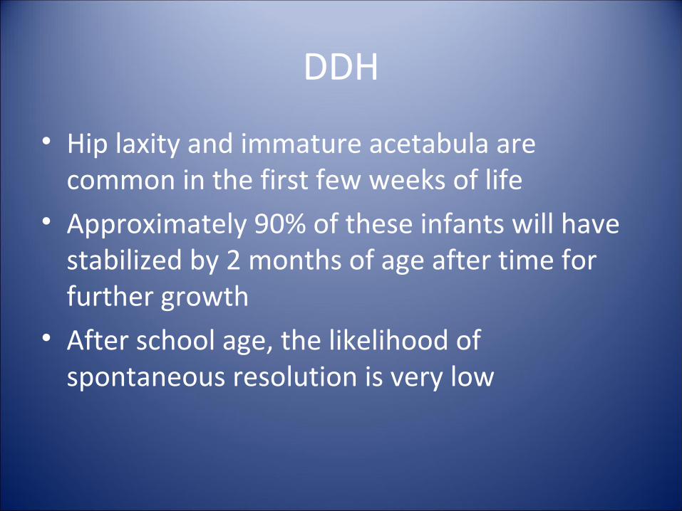
• Hip laxity and immature acetabula are common in the first few weeks of life
• Approximately 90% of these infants will have stabilized by 2 months of age after time for further growth
• After school age, the likelihood of spontaneous resolution is very low
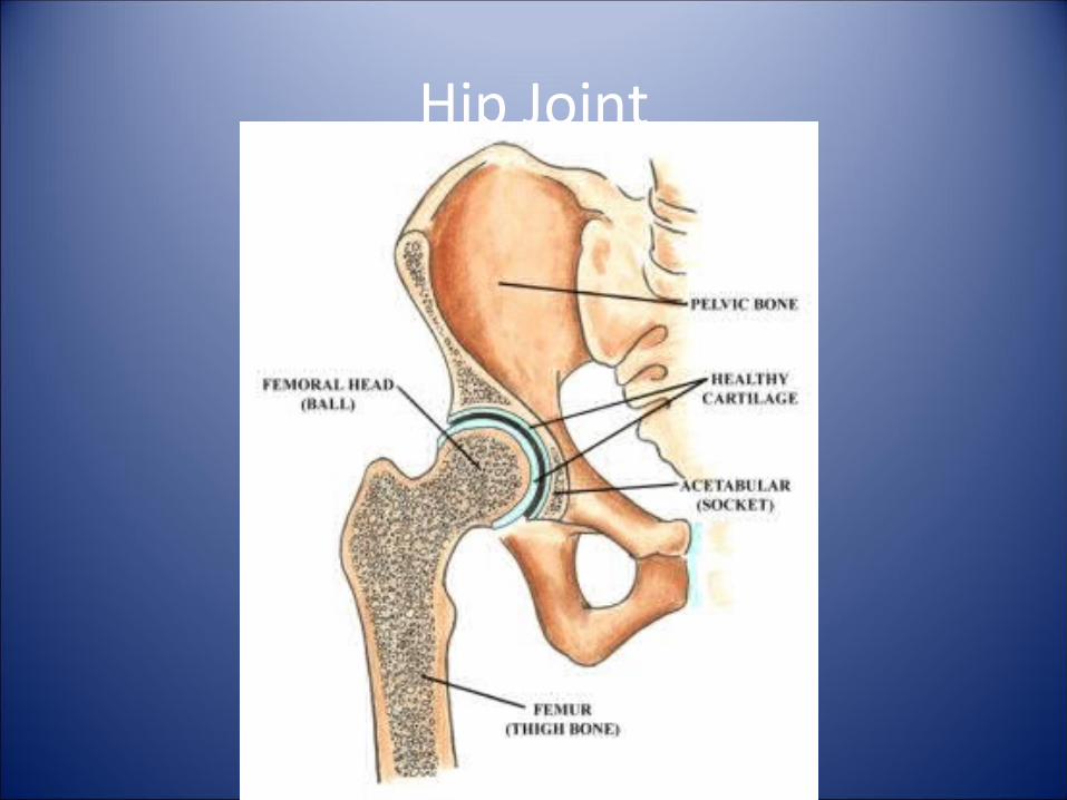
Hip Joint
Clinical Features
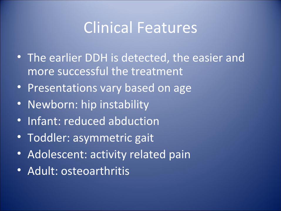
• The earlier DDH is detected, the easier and more successful the treatment
• Presentations vary based on age• Newborn: hip instability• Infant: reduced abduction• Toddler: asymmetric gait• Adolescent: activity related pain• Adult: osteoarthritis
Risk factors
• Breech presentation• Female gender• Family history• Factors related to tight positioning in utero
(can see associated torticollis and metatarsus adductus
Risk factors
• Girls with a breech presentation — 12 percent • Girls with a positive family history — 4.4
percent • Boys with a breech presentation — 2.6
percent • Girls — 1.9 percent • Boys with a positive family history — 0.9
percent
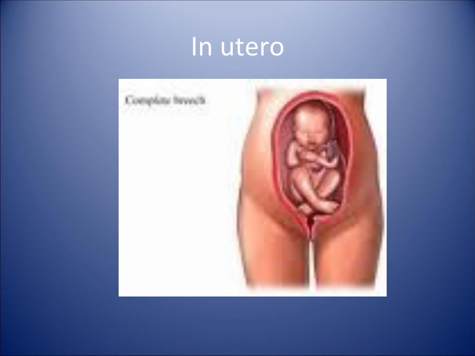
In utero
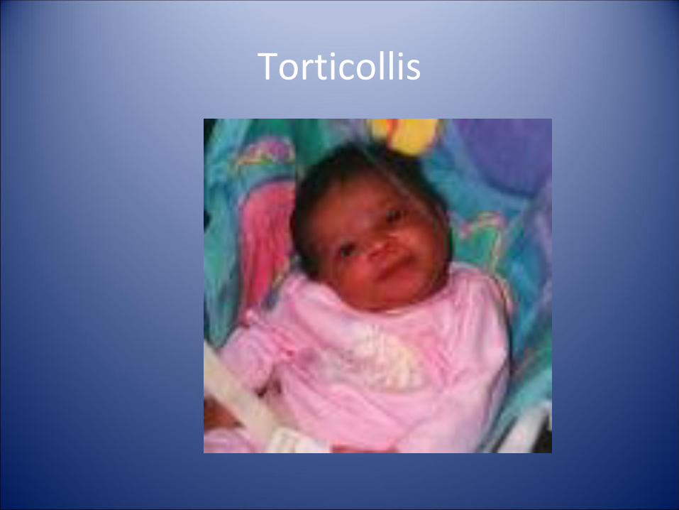
Torticollis
Physical Exam
• Hips should be checked at every visit until the child is walking normally
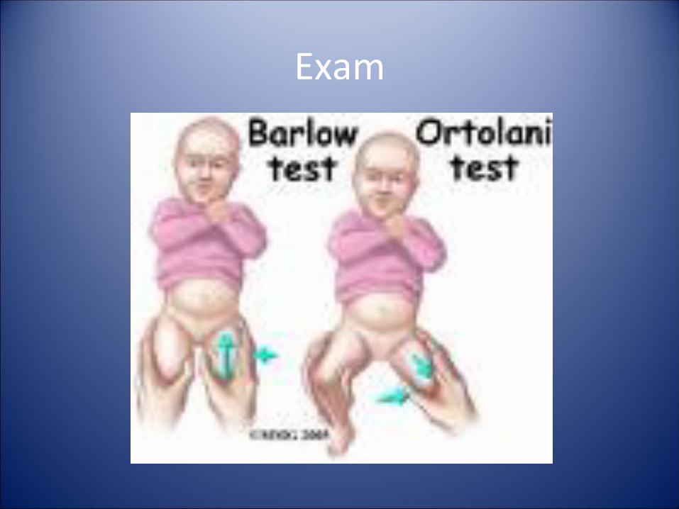
• Exam technique uses adduction and posterior pressure to feel for dislocation and abduction and elevation to feel for reduction
• Hip Instability: The sensation of dislocation or reduction is best described as a clunk or a jerk
• Clicks and pops not associated with a palpable clunk are very common and not worrisome
Exam
• Examine each hip individually while the child is calm and not crying
• Examine on a stable surface with the child supine and with hips flexed 90 degrees in neutral rotation
Exam
Galeazzi Test
Exam
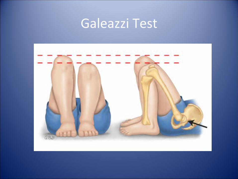
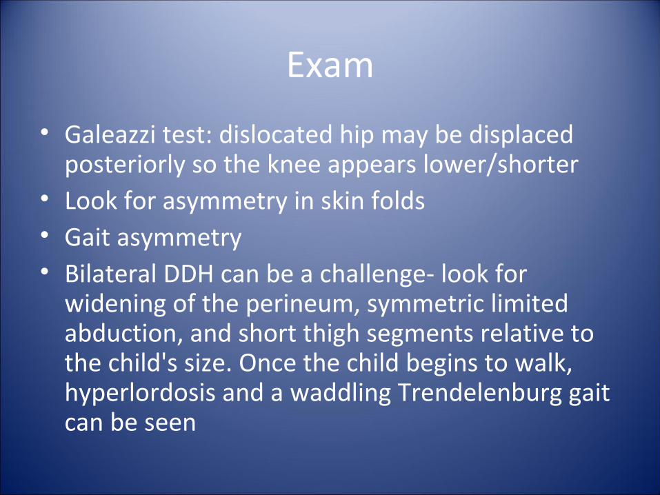
• Galeazzi test: dislocated hip may be displaced posteriorly so the knee appears lower/shorter
• Look for asymmetry in skin folds• Gait asymmetry• Bilateral DDH can be a challenge- look for
widening of the perineum, symmetric limited abduction, and short thigh segments relative to the child's size. Once the child begins to walk, hyperlordosis and a waddling Trendelenburg gait can be seen
Exam
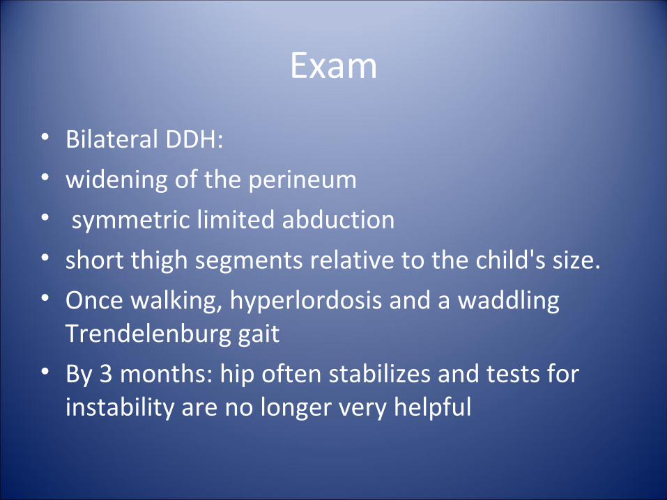
• Bilateral DDH: • widening of the perineum• symmetric limited abduction• short thigh segments relative to the child's size. • Once walking, hyperlordosis and a waddling
Trendelenburg gait• By 3 months: hip often stabilizes and tests for
instability are no longer very helpful
Imaging
• Ultrasound• Plain radiographs- limited value early on due
to femoral heads cartilaginous and not ossified
• Radiographs should be with hips flexed 20 to 30 degrees, or neutral if the child is older
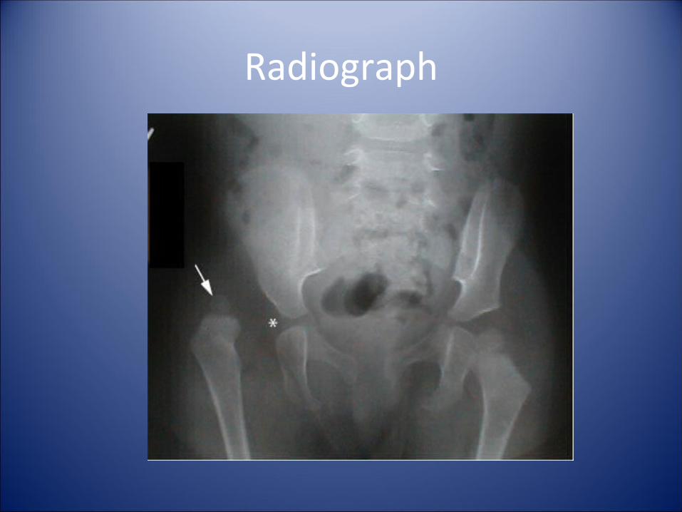
Radiograph
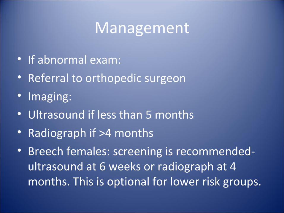
Management
• If abnormal exam:• Referral to orthopedic surgeon• Imaging:• Ultrasound if less than 5 months• Radiograph if >4 months• Breech females: screening is recommended-
ultrasound at 6 weeks or radiograph at 4 months. This is optional for lower risk groups.
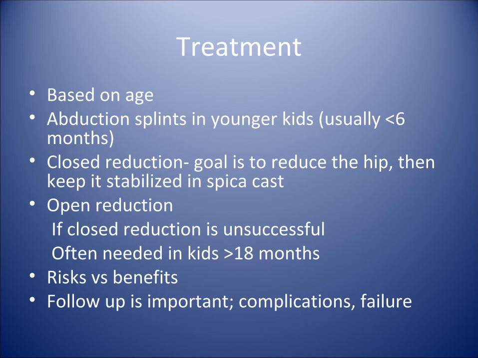
Treatment
• Based on age• Abduction splints in younger kids (usually <6
months)• Closed reduction- goal is to reduce the hip, then
keep it stabilized in spica cast• Open reduction If closed reduction is unsuccessful Often needed in kids >18 months• Risks vs benefits• Follow up is important; complications, failure
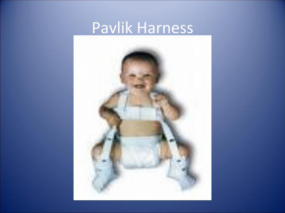
Pavlik Harness
Septic Joint
• Infection and inflammation of usually sterile joint space
• Typically affects large joints and joints of the lower extremity- knee, ankle and hip (approximately 80%)
• Up to 10% in more than 1 joint• Predominant age: 2-6 years; adolescent• Males>Females
Pathophysiology
• Bacterial entry:• Hematogenous spread• Direct inoculation• Extension from adjacent infection (bone)• Influx of inflammatory Cells• Rapid destruction of cartilaginous structures
by bacterial enzymes; may -> necrosis
Etiologies
• Viral- parvovirus, EBV, herpes, CMV, varicella, Hep B or C, mumps, rubella
• Fungal- Candida albicans• Spirochete- Lyme (B. burgdorferi)• Tuberculosis• Bacterial • <5 years: S. aureus, Group B strep, HIB, gram
negative bacteria• >5 years: S. aureus, Group A strep, N.
gonorrhoeae
Etiologies
• Also Kingella kingae, salmonella, N. meningitidis
• S. aureus most common outside of neonatal period
• Sickle cell associated with salmonella• Immunocompromised: Mycoplasma,
ureoplasma, or Aspergillus
Neonates and Infants
• Can be subtle• >1 joint• Microbiology: GBS, Gram negative bacilli such as
E. Coli, S. aureus• Often presents with septicemia or fever without
source• Subtle features: positional preference, decreased
use, pain with handling, extremity swelling• Hip arthritis

Children
• Fever and constitutional symptoms• Pain with active and passive movement• Limp/refusal to bear weight• Joint related findings can be subtle: swelling,
warmth• Hip/shoulder: often no external signs• Hip involvement may be referred (knee)• Sacroiliac may present similarly to appendicitis,
neoplasm or UTI
Symptoms
• Do not wax and wane• Can wake up at night with pain• Worsens with time• Specific joint symptoms:• Pain- often exquisite tenderness through any
range of motion• Warmth, erythema, swelling
Evaluation
• Should be PROMPT• NO DELAYS• History and Physical• Labs and imaging• Joint aspiration/joint fluid analysis ASAP• Risks vs. benefits- diagnosis vs seeding the
joint if overlying cellulitis
Evaluation
• History: • ?direct inoculation• ?rash- can implicate type of infection• ?skin/soft tissue infections- source for bacteremia• ?Recent antibiotic use- may attenuate symptoms• ?recent illnesses/URIs- consider post-infectious
synovitis• ?LMP- disseminated gonococcal in 1st 7d of menses• ?Exposures• ?Immunization Status
Exam
• Observation (use parents if young child)• Soft tissue/skin exam• Joint exam: swelling, redness, erythema, pain• Active and passive range of motion• Exam: eyes, skin, heart, lungs, abdomen, etc
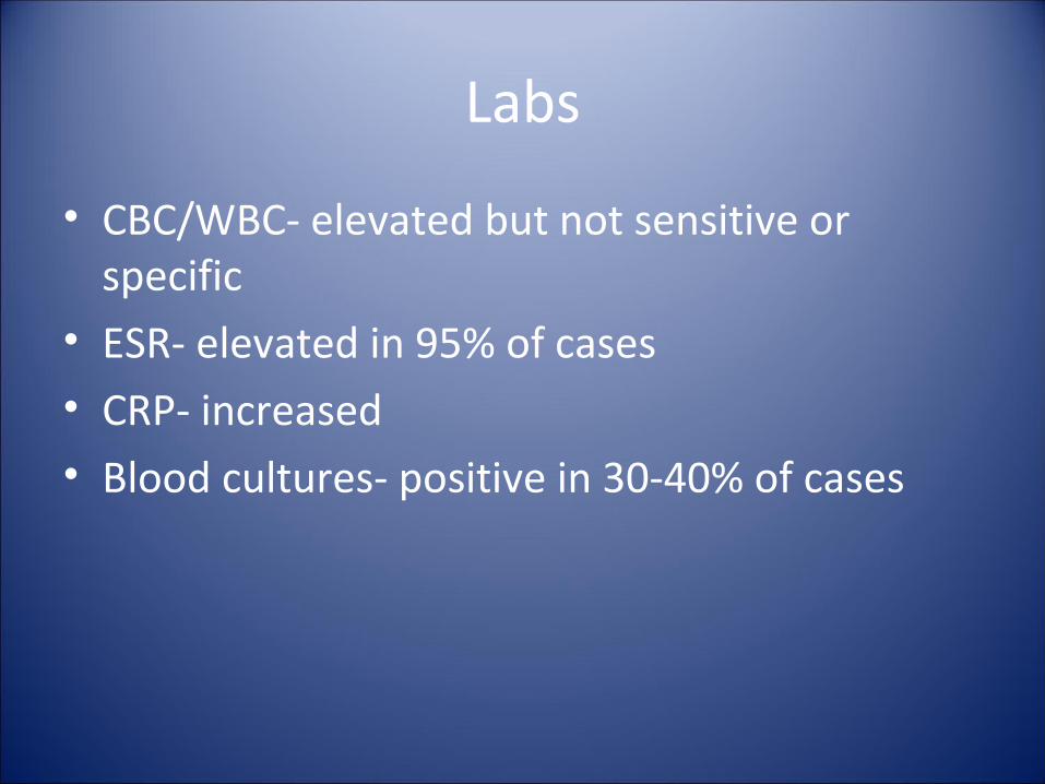
Labs
• CBC/WBC- elevated but not sensitive or specific
• ESR- elevated in 95% of cases• CRP- increased• Blood cultures- positive in 30-40% of cases
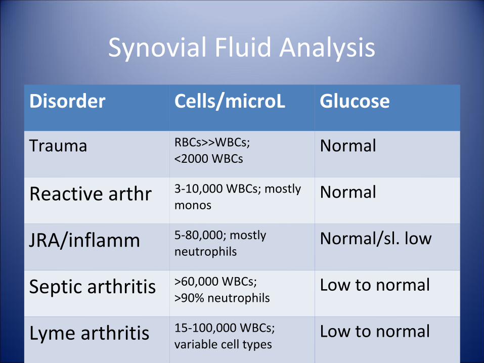
Synovial Fluid Analysis
Disorder Cells/microL Glucose
Trauma RBCs>>WBCs;<2000 WBCs
Normal
Reactive arthr 3-10,000 WBCs; mostly monos
Normal
JRA/inflamm 5-80,000; mostly neutrophils
Normal/sl. low
Septic arthritis >60,000 WBCs;>90% neutrophils
Low to normal
Lyme arthritis 15-100,000 WBCs; variable cell types
Low to normal
Imaging
• Radiography- may (or may not) show widening of the joint space +/- displacement of the normal fat pads
• Ultrasound- can identify fluid in the joint space
• CT scan• MRI• Bone scan
Algorithm
• Look for 4 or more of the following:• ESR > 20 mm/h• CRP > 1 mg/dL• WBC > 11,000 cells/mL• Joint space fluid apparent on radiograph
Differential Diagnosis• Osteomyelitits• Deep cellulitits• Abscess• Septic bursitis• Bacterial endocarditis• Inflammatory/autoimmune arthritis• Transient synovitis• Acute rheumatic fever• Trauma• Legge-Calves-Perthes• SCFE• Tumor/Malignancy
Treatment
• IV antibiotics• 1st line: Anti-staph PCN, 1st generation
cephalosporin• If MRSA in community, consider vanco or clinda• Sickle cell pts: add ceftriaxone• Duration of therapy depends on bacteria • Supportive care (sepsis, etc)• Drainage of infection- ASAP• Open surgical drainage/irrigation for hip and
usually shoulder, inability to aspirate
Complications
• Septic shock• Death• Joint destruction• Limited range of motion which may be
permanent• Growth disturbance if the epiphysis is
involved.