common complications of mechanical ventilation and
TRANSCRIPT

Common complications of mechanical ventilation andmultimodal prevention strategies
Markle, Andrew Gordon
Master's thesis / Diplomski rad
2014
Degree Grantor / Ustanova koja je dodijelila akademski / stručni stupanj: University of Zagreb, School of Medicine / Sveučilište u Zagrebu, Medicinski fakultet
Permanent link / Trajna poveznica: https://urn.nsk.hr/urn:nbn:hr:105:931105
Rights / Prava: In copyright
Download date / Datum preuzimanja: 2022-01-05
Repository / Repozitorij:
Dr Med - University of Zagreb School of Medicine Digital Repository

UNIVERSITY OF ZAGREB
SCHOOL OF MEDICINE
Andrew Markle
Common Complications of Mechanical
Ventilation and Multimodal Prevention
Strategies
GRADUATE THESIS
Zagreb, 2014
2
UNIVERSITY OF ZAGREB
SCHOOL OF MEDICINE
Andrew Markle
Common Complications of Mechanical
Ventilation and Multimodal Prevention
Strategies
GRADUATE THESIS
Zagreb, 2014

3
This graduation paper was made at the department of Anaesthesia and Intensive Care at Sveti Duh hospital under supervision of Prof. dr. sc. Dinko Tonković and it was submitted for evaluation in the academic year 2014.
Graduation paper was made at the department of Anaesthesia and Intensive
Care, University of Zagreb School of Medicine.
Mentor: Prof. dr. sc. Dinko Tonković

Table of Contents
INTRODUCTION 2 ABSTRACT 2 KEY WORDS 2 LIST OF ABBREVIATIONS 3
RESPIRATORY PHYSIOLOGY 4 NORMAL RESPIRATORY FUNCTION 4 NORMAL RESPIRATORY IMMUNE RESPONSE 8
POSITIVE PRESSURE VENTILATION 11 HISTORY AND MECHANISMS OF POSITIVE PRESSURE VENTILATION 11 ADVANTAGE AND REASONS FOR POSITIVE PRESSURE VENTILATION 13 CARDIOVASCULAR RESPONSES TO VENTILATION 14 COMMON COMPLICATIONS OF MECHANICAL VENTILATION 16 DIFFICULTY WEANING 16 VENTILATOR-ASSOCIATED PNEUMONIA 18 VENTILATOR-ASSOCIATED/INDUCED LUNG INJURY (VALI/VILI) 20 COMPLICATIONS AND EFFECTS OF GENERAL ANAESTHESIA 24
STRATEGIES FOR THE PREVENTION OF VENTILATOR-ASSOCIATED COMPLICATIONS 26 PERIOPERATIVE PREVENTION STRATEGIES 26 SMOKING CESSATION 27 ANALGESIA 29 POST-OPERATIVE RESPIRATORY FAILURE (PRF) 30 INSPIRATORY MUSCLE TRAINING 31 SEDATIVE REDUCTION AND SEDATIVE SELECTION 32 EARLY IMMUNOMODULARY NUTRITION 34 LUNG-PROTECTIVE STRATEGIES DURING GENERAL ANAESTHESIA AND MECHANICAL VENTILATION 36 LOW-VOLUME VENTILATION 36 POSITIVE END EXPIRATORY PRESSURE (PEEP) 39 RECRUITMENT MANOEUVRES 42 VENTILATOR-ASSOCIATED PNEUMONIA BUNDLES 44
CONCLUSION 46
BIBLIOGRAPHY 49

2
Introduction Abstract Mechanical ventilation has undergone tremendous change in its fifty years of
mainstream usage. In that time it has made the treatment of previously fatal
diseases possible. Importantly, research has shown that it also has the
potential to cause or exacerbate disease. This paper seeks to explore some
of the more common complications of mechanical ventilation, and methods in
which they may be prevented or ameliorated.
Key Words Mechanical ventilation, weaning, ventilator-associated pneumonia, ventilator -associated lung injury, positive end expiratory pressure, recruitment, post-operative respiratory failure.
3
List of Abbreviations Abbreviation Meaning ALI acute lung injury APACHE acute physiology and chronic health evaluation II ARDS acute respiratory distress syndrome BAL broncho-alveolar lavage CDC centres for disease prevention and control ETCO2 end-tidal carbon dioxide FiO2 fraction of inspired oxygen ICU intensive care unit LA left atrium LV left ventricle PaO2 partial pressure of arterial oxygen PBW predicted body weight PEEP positive end expiratory pressure POPC post-operative pulmonary complications PRF post-operative respiratory failure RA right atrium RASS Richmond agitation and sedation scale RV right ventricle TIVA total intravenous anaesthesia TNF tumour necrosis factor V:Q ventilation to perfusion ratio VALI ventilator-associated lung injury VAP ventilator-associated pneumonia VILI ventilator-induced lung injury Vt tidal volume WOB work of breathing
4
Respiratory Physiology Normal Respiratory Function Breathing in humans is typically facilitated through two groups of muscles (1).
The diaphragm lengthens the thorax during inspiration and shortens via
elastic recoil during expiration. The diaphragm is usually the only respiratory
muscle playing a significant role during normal, quiet breathing.
The second group of muscles are those that cause changes in thoracic
dimensions. The major muscles of thoracic excursion are the external
intercostals, the sternocleidomastoid, the anterior serati, and the scaleni (2).
During heavy, exertional breathing, the ribs are pulled caudally, expanding the
thorax some twenty per cent. Muscles that contract the thorax are the
abdominus recti and the interior intercostals. These muscles are active during
times of heavy breathing. Pathologies causing either an increase in
abdominal pressure or dysfunction of abdominal musculature may interfere
with breathing (3).
The tracheobronchial tree consists of 23 generations (trachea = 0, alveoli =
23), which are counted using the Weibel classification system (2, 3). The
number of passages roughly doubles with each generation so the number of
passages can be mathematically expressed as 𝑛𝑢𝑚𝑏𝑒𝑟 𝑜𝑓 𝑝𝑎𝑠𝑠𝑎𝑔𝑒𝑠 =
(𝑔𝑒𝑛𝑒𝑟𝑎𝑡𝑖𝑜𝑛 𝑛𝑢𝑚𝑏𝑒𝑟)!. The mean diameter of the trachea is 1.8cm and is
supported by U-shaped cartilaginous rings. Posteriorly, the trachea joins the
oesophagus via bands of smooth muscle. This is of import because, despite
the solid nature of the cartilaginous rings, it is possible to occlude the trachea
if the intrathoracic pressure exceeds 50 – 70 cmH20.
Small bronchi begin at approximately generation number 7 and have a
diameter ranging from 3.5mm – 1mm. Because smaller bronchi do not attach
directly to the pulmonary parenchyma, they rely on a discontinuous network of
cartilage to maintain patency, and on transmural pressure for airflow. By the
eleventh generation, the cartilage disappears from the airway wall (now ≤1mm
in diameter) and the pulmonary parenchyma and the elastic recoil of the lung

5
instead hold the passages open (3). The bronchioles also have a layer of
helical smooth muscle under three types of control. The sympathetic nervous
system causes dilation of the bronchioles via ß adrenergic stimulation by
nor/adrenaline. Conversely, the parasympathetic nervous system via
acetylcholine and the vagus nerve promote constriction. Additionally,
histamine exerts local control and leads to constriction as well.
The average adult lung contains 130.000 primary lobules, with each lobule
containing approximately 2000 alveoli in a diameter of 3.5mm. From a
functional standpoint, each primary lobule can be viewed as a large alveolus
(3). Ranging from 200 – 600 million, the average, healthy adult lung contains
around 300 million alveoli. The size of the alveoli is directly proportional to
lung volume, except at the point of maximal inflation. This size differential is
exhibited in a vertical, gravity-dependant gradient, with the larger alveoli found
at the apex of the lung. At functional residual capacity, the mean alveolar
diameter is 0.2mm (3).
Gravity-dependence is seen to a greater degree in the pulmonary vasculature.
The pulmonary vasculature carries the same flow as its systemic counterpart,
however is does so at about 1/6 the pressure. These lower pressure vessels
are significantly thinner than their systemic counterparts, with the media being
approximately half as thick (2, 3). Pulmonary arterioles are mostly elastic and
lack significant muscle. At rest, approximately 75% of the pulmonary vascular
bed is filled, creating a gravity-dependant V/Q ratios. Inflation of the alveoli at
higher (or lower) pressures will cause a change in vascular resistance,
leading to redistribution of blood within the pulmonary system.
The walls of the alveoli consist of a basement membrane sandwiched
between 2 layers of pulmonary epithelium. Within the walls are the pulmonary
capillaries, elastin, collagen, and various immunologically active cells such as
neutrophils and macrophages (4). Intra-alveolar septa are fenestrated by
pores of Kohn, allowing for movement of air and immunologic cells. In the
area of gas exchange, the capillary and alveolar epithelia are closely
opposed, sitting 0.4µm apart.

6
Pressure drives air movement in the respiratory system, and as such, a brief
review of pressures and volumes is warranted.
The alveolar pressure at rest, with unimpeded airways and an open glottis, is
0cmH2O (or equal to atmospheric pressure).
Plural pressure holds the alveoli open and in a normal, healthy lung sits at
about -5cmH2O at the beginning of inspiration. During inspiration, the plural
pressure drops to -7.5cmH2O.
Transpulmonary pressure is the difference between the alveolar and plural
pressures. It is also referred to as the recoil pressure.
Compliance is used as a measure by which certain changes in ventilation are
monitored, and is defined as the extent to which the lungs will expand per unit
of increase in transpulmonary pressure. There are two characteristics of
compliance. The first is the tissue-elastic forces tending towards the collapse
of the air-filled lung. The tissue-elastic forces account for approximately 1/3 of
the total lung elasticity. Alveolar fluid:air surface tension accounts for the
remaining 2/3. Importantly, if one were to measure the compliance of the
entire lung-thorax system, it would be approximately half of the lungs alone.
This means that the thorax contributes the same tendency to collapse the air-
filled lungs as do the lungs and pleura themselves (3, 4).
7
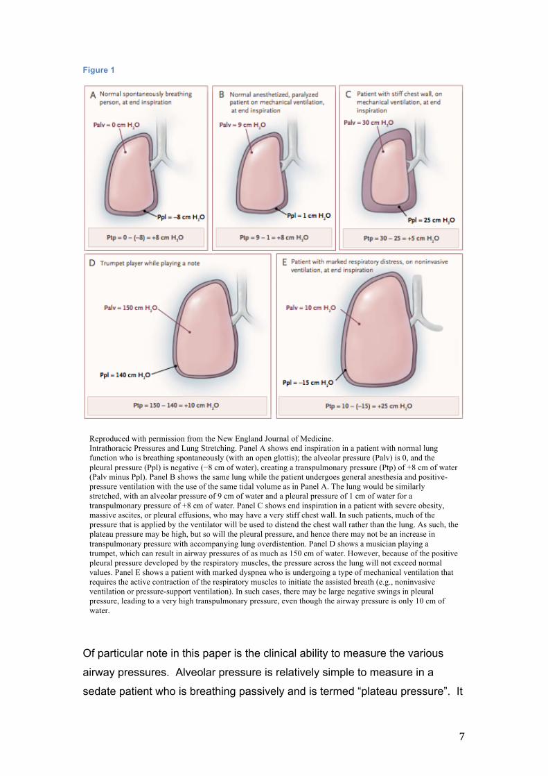
Figure 1
Of particular note in this paper is the clinical ability to measure the various
airway pressures. Alveolar pressure is relatively simple to measure in a
sedate patient who is breathing passively and is termed “plateau pressure”. It
Reproduced with permission from the New England Journal of Medicine. Intrathoracic Pressures and Lung Stretching. Panel A shows end inspiration in a patient with normal lung function who is breathing spontaneously (with an open glottis); the alveolar pressure (Palv) is 0, and the pleural pressure (Ppl) is negative (−8 cm of water), creating a transpulmonary pressure (Ptp) of +8 cm of water (Palv minus Ppl). Panel B shows the same lung while the patient undergoes general anesthesia and positive-pressure ventilation with the use of the same tidal volume as in Panel A. The lung would be similarly stretched, with an alveolar pressure of 9 cm of water and a pleural pressure of 1 cm of water for a transpulmonary pressure of +8 cm of water. Panel C shows end inspiration in a patient with severe obesity, massive ascites, or pleural effusions, who may have a very stiff chest wall. In such patients, much of the pressure that is applied by the ventilator will be used to distend the chest wall rather than the lung. As such, the plateau pressure may be high, but so will the pleural pressure, and hence there may not be an increase in transpulmonary pressure with accompanying lung overdistention. Panel D shows a musician playing a trumpet, which can result in airway pressures of as much as 150 cm of water. However, because of the positive pleural pressure developed by the respiratory muscles, the pressure across the lung will not exceed normal values. Panel E shows a patient with marked dyspnea who is undergoing a type of mechanical ventilation that requires the active contraction of the respiratory muscles to initiate the assisted breath (e.g., noninvasive ventilation or pressure-support ventilation). In such cases, there may be large negative swings in pleural pressure, leading to a very high transpulmonary pressure, even though the airway pressure is only 10 cm of water.

8
represents the pressure required to distend the chest wall and lung together.
Likewise, mean airway and peak inspiratory pressures are both easily
obtained. Unfortunately, if we are to calculate transpulmonary pressure, then
measurement of pleural pressure is also required. Currently, no method
exists to directly measure this pressure in the clinical environment. The only
readily available surrogate is oesophageal pressure, which provides only a
rough estimate as it is affected by the cardiac movement, the lungs, and by
the oesophagus itself. (5)
Normal Respiratory Immune Response Protection of the respiratory system actually begins at the nose where the air
is warmed, humidified, and filtered via turbulent airflow and gravitational
precipitation (2-4).
In its entirety the respiratory system is coated in mucous (to varying degrees)
secreted by goblet cells and sub-mucosal glands. This mucous serves
several purposes. It keeps the surfactant moist, allowing for a continued
decrease in surface tension. Respiratory mucous also traps small particles
before they reach the terminal airways.
The particles trapped in the mucous are the removed to the pharynx by
ciliated epithelium which beats approximately 10 – 20 times per second (3).
The cilial beat has two phases, fast and slow. The fast beats are always
directed caudally and are responsible for moving particulate matter out of the
airways. Normally a particle within the mucous layer will be moved a few
millimetres every minute (2). Upon reaching the pharynx, the particle-
containing mucous is either coughed or swallowed.
Though comprising only 8% of pulmonary cells, the type I pneumocyte
(epithelium) covers 95% of the alveolar surface area. Importantly, these cells
are able to take in particulate matter and then either facilitate movement of the
particle away from the alveoli or undergo oxidative apoptosis. Type I
pneumocytes are not mitotically active.

9
Type II pneumocytes are best known for supplying surfactant to the alveoli via
the release of osmophilic granules. Surfactant reduces the alveolar surface
tension and prevents collapse at lower pressures. Type II cells are mitotically
active and are able to replace damaged epithelium, eventually transforming
into type I cells as required (2, 3).
There are 3 types of macrophage commonly found within the respiratory
system: airway, interstitial, and alveolar. The three types share many
commonalities and move between the three areas to varying degrees. As a
group they are derived from bone marrow, though – in the presence of bone
marrow dysfunction, and in times of stress – they are able to undergo
functional division for several generations (3). All contain primary and
secondary lysosomes. All have surface receptors for IgG, IgM, C3, and
various other opsonins. All function as antigen-presenting cells for T cells.
Finally, pulmonary macrophages produce mediators such as fibronectin,
prostaglandins, leukotrienes, interferons,
and α1 anti-trypsin.

10
Figure 2
critical care medicine
n engl j med 369;22 nejm.org november 28, 2013 2129
Biologic alterations Physiological abnormalities
Systemic effects
C Structural consequences
A Ventilation at low lung volume
Atelectrauma Lung inhomogeneity
B Ventilation at high lung volume
Air leaks Overdistention
Increased physiological
dead space
Biologic alterations
Hydroxyproline Transforming growth factor-β Interleukin-8
Increased concentrations of:
Tumor necrosis factor α (TNF-α) β-catenin Interleukin-6 (IL-6) Interleukin-1β (IL-1β)
Release of mediators:
Decreased PaO2
Increased PaCO2
Activation of epithelium and endothelium
Capillary
Decreased compliance
Recruitment of: Pulmonary alveolar macrophages (PAMs) Neutrophils
PAM
Translocation of:
Lipopolysaccharides (LPS)
Bacteria
Various mediators
Multiple mechanisms (e.g., increased apoptosis)
Multiorgan dysfunction
Death
TNF-αβ-catenin
MediatorsBacteria
IL-6
LPS
IL-1β
Structural consequences
Alveolus
Epithelial–mesenchymal transformations
Surfactant dysfunction
Pulmonaryedema
Increased alveolar–capillarypermeability
Fibroproliferation
Hyaline membranes
Sloughing of bronchial epithelium
Atelectasis
Barotrauma
Alveolus
Alveolus
PAM
IL-6IL-1β
PMN
B Ventilation at high lung volumeVentilation at high lung volumeVentilation at high lung volume
Normal Hyperinflation
CT Image CT Image
End expiration End inspiration
2
Drazen
11/8/13
AUTHOR PLEASE NOTE:Figure has been redrawn and type has been reset
Please check carefully
AuthorFig #
Title
ME
DEArtist
Issue date
COLOR FIGURE
Draft 7Slutsky
N Koscal
11/28/13
Ventilator Induced Lung Injury
Figure 2, previous page. Reproduced with permission from the New England Journal of Medicine. Lung Injury Caused by Forces Generated by Ventilation at Low and High Lung Volumes. When ventilation occurs at low lung volumes, lung in- jury can be caused by the opening and closing of lung units (atelectrauma) as well as by other mechanisms. This injury is magnified when there is increased lung inhomogeneity, as shown on computed tomography (Panel A), especially in patients with the acute respira- tory distress syndrome (ARDS) who have surfactant dysfunction, pulmonary edema, and atelectasis.11 In addition, ventilation may be very inhomogeneous, a status that may be partially or fully reversed by the use of positive end-expiratory pressure (PEEP), as shown in a ventilated ex vivo rat lung (see video in Slutsky and Hudson12). At high lung volumes, overdistention can lead to gross barotrauma (air leaks)13 (Panel B). Over- distention can also lead to increased alveolar–capillary permeability and gross pulmonary edema. Ventilation at both high and low lung volumes has structural, physiological, biologic, and systemic effects (Panel C). Mediators that are released into the lung can cause further lung injury, recruit neutrophils to the lung, or set the stage for the development of pulmonary fibro- sis. In addition, the increased alveolar–capillary permeability associated with ventilator-induced lung injury can lead to translocation of mediators, lipopolysaccharides, and bacteria into the systemic circulation, potentially leading to multiple-organ dysfunction and death. PaCO2 denotes partial pressure of arterial carbon dioxide, PaO2 partial pressure of arterial oxygen, and PMN polymorphonuclear leukocytes.

11
The pulmonary lymphatic system has a capacity of approximately 500ml with
the vessels lying mostly within potential spaces surrounding the vasculature
and the various air passages. Lymphatic flow from the left lung is to the
thoracic duct, while the right lung drains to the lymphatic duct (2).
Positive Pressure Ventilation
History and Mechanisms of Positive Pressure Ventilation The idea of artificial ventilation is credited to Paracelsus, who used fire
bellows connected to a tube inserted into the patient’s mouth. In 1744 John
Fathergill reported the first effective use of mouth-to-mouth resuscitation. In
1775 John Hunter devised a double-bellows system, a kind of a push-pull
system designed to both introduce and draw out air. In 1911 Dräger Medical
produced what was probably the first commercially available, purpose built
artificial respirator, called the Dräger Pulmoter; it was designed to be used by
first responders. The late 1800s and early 1900s also saw the development
of negative pressure pulmonary ventilators, largely in response to polio. Two
main types reached common usage. The first and most dramatic was the iron
lung. The patient was inserted, bodily, into a metal tube or box with his or her
head left protruding. The pressure in the device was reduced cyclically
thereby drawing air in through the patient’s nose and or mouth. The second
type was known as the chest cuirass. It was designed to enclose only the
thorax and work on a similar principle to the iron lung. It had the advantage of
being significantly small and simpler, allowing many patients to remain at
home. (6)
Mechanical ventilation entered mainstream medicine as a result of its
extensive use in the 1952 polio epidemic in Copenhagen, Denmark.
Throughout the course of the epidemic, many principles were developed
which have today become standard. These include the use of cuffed
endotracheal tubes, periodic sighs, and weaning. It was also in this time
period that the first mechanical positive-pressure ventilators were designed for
long-term use. During this time, the use of mechanical ventilation to support
12
patients with paralytic polio reduced the mortality rate from approximately
80% to 40%. (7, 8)
The 1960s gave rise to the era of respiratory intensive care with the
production of the first proper volume-controlled and pressure-controlled
ventilators. While they are positively archaic by today’s standards, it is
interesting to note the designers attempted to mimic normal breathing with
sinusoidal waveforms, periodic sighs, and even PEEP to mimic pursed-lip
breathing. During the late 1960s and early 1970s, the first reports and
conferences concerning what would later be termed ARDS began to appear.
(6)
The mid 1970s though the 1980s harkened the development of low-pressure
endotracheal tubes to combat tracheal necrosis, permissive hypercapnoea,
gas mixtures, high-frequency oscillatory ventilation, and high-frequency jet
ventilation. Various modes of ventilation, some still in use today, were also
developed during this time. These include pressure-support, pressure-
control, and interestingly inverse ratio ventilation. (6) The period covering the
1980s until now has brought about not only derivative modes of ventilation,
often focused on synchronisation, but more importantly an incredible amount
of research. Some, such as the ARDSnet studies, have forever changed the
way we treat patients. While others, such as the disappointing transition of
high-frequency oscillatory ventilation to the adult population, have severed as
a scientific head-scratch. What has become clear is that mechanical
ventilation can represent a potentially life-saving intervention, but that it also
requires careful use and on-going, patient-specific tailoring. (6)
Between forty and 60 per cent of ICU patients will require some sort
mechanical ventilatory support. This makes it only less common than the use
of intravenous therapy, antibiotics, and – presumably – a bed. From a
different point-of-view, this represents about 3% to 4% of the hospitalised
population. (9, 10) Unfortunately, there is no good European data available
calculating the cost of mechanical ventilation. However, in the US in 2005,
mechanical ventilation represented approximately 12% of hospital costs at

13
around twenty-seven billion US dollars per year. (10) Whilst one must
acknowledge that US healthcare costs are the highest in the world and that
the increase in cost is likely not analogous to Europe, there is – regardless of
location – an increase in labour intensity and complications regardless of
location. For the most part the increase in labour intensity occurs at the
beginning of each patient’s mechanical ventilation.
Advantage and reasons for Positive Pressure Ventilation Mechanical ventilation has essentially two main advantages. Firstly, in the
spontaneously breathing patient ,it allows us to reduce the work of breathing.
Whilst this may sound trivial to some, work of breathing in a patient suffering
from respiratory distress can represent up to forty per cent of the body’s
energy and oxygen consumption. Work of breathing is normally expressed in
joules and is quite complex, (3) but in healthy individuals ranges from 0.3 –
0.5 joules/L. (11) Normal determinants of work of breathing are essentially a
sum of the inspiratory and expiratory forces to be overcome by the patient.
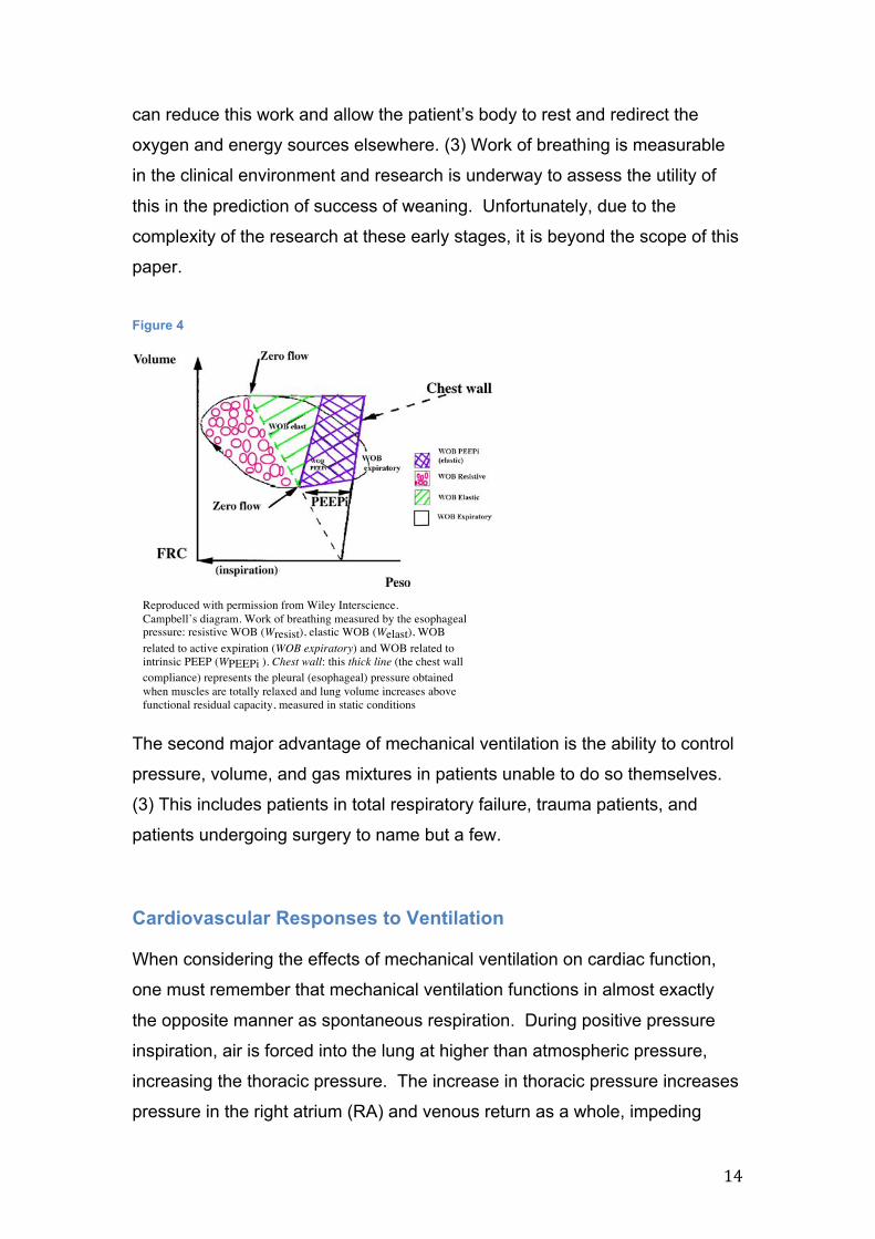
Figure 3
These include the chest wall elastance, lung elastance, transpulmonary
pressure, resistive forces, and intrinsic PEEP. This is best described using
Campbell’s diagram in figure 2 below. Obviously, when lung function is
deranged, work of breathing increases significantly, mechanical ventilation
Reproduced with permission from AnaestheisaUK
14
can reduce this work and allow the patient’s body to rest and redirect the
oxygen and energy sources elsewhere. (3) Work of breathing is measurable
in the clinical environment and research is underway to assess the utility of
this in the prediction of success of weaning. Unfortunately, due to the
complexity of the research at these early stages, it is beyond the scope of this
paper.
Figure 4
The second major advantage of mechanical ventilation is the ability to control
pressure, volume, and gas mixtures in patients unable to do so themselves.
(3) This includes patients in total respiratory failure, trauma patients, and
patients undergoing surgery to name but a few.
Cardiovascular Responses to Ventilation When considering the effects of mechanical ventilation on cardiac function,
one must remember that mechanical ventilation functions in almost exactly
the opposite manner as spontaneous respiration. During positive pressure
inspiration, air is forced into the lung at higher than atmospheric pressure,
increasing the thoracic pressure. The increase in thoracic pressure increases
pressure in the right atrium (RA) and venous return as a whole, impeding
1312
Fig. 1 Campbell’s diagram. Work of breathing measured by theesophageal pressure: resistive WOB (Wresist), elastic WOB (Welast),WOB related to active expiration (WOB expiratory) and WOB re-lated to intrinsic PEEP (WPEEPi). Chest wall: this thick line (the chestwall compliance) represents the pleural (esophageal) pressure ob-tained when muscles are totally relaxed and lung volume increasesabove functional residual capacity, measured in static conditions
The WOB is normally expressed in joules. One joule isthe energy needed to move 1 l of gas through a 10-cmH2Opressure gradient. The work per liter of ventilation (J/l) isthe work per cycle divided by the tidal volume (expressedin liters). In a healthy subject the normal value is around0.35 J/l [7]. Lastly, WOB can be expressed in work per unitof time, multiplying joules per cycle by the respiratory rate(expressed in breaths per minute) to obtain the power ofbreathing (joules/minute). In a healthy subject the normalvalue is around 2.4 J/min [7]. As illustrated by the Camp-bell diagram, two other phenomena affect the WOB: in-trinsic PEEP (positive end-expiratory pressure, or PEEPi)and active expiration.
PEEPi and active expirationThe distending pressure of the lungs is called the transpul-monary pressure and it can be estimated as the differencebetween airway and esophageal (pleural) pressure. At theend of a normal expiration, alveolar and airway pressuresare zero relative to atmosphere, and esophageal pressureis negative, reflecting the resting transpulmonary pressure(around 5 cmH2O in normal conditions). However, in thepresence of PEEPi, the alveolar pressure remains positivethroughout expiration, because of either dynamic airwaycollapse or inadequate time to exhale [8]. This implies thatsome degree of dynamic hyperinflation does exist (lungvolume at end-expiration is higher than passive functionalresidual capacity). Importantly, for lung volume to furtherincrease in a patient with PEEPi, the inspiratory musclescontract to an amount equal to PEEPi before any volumeis displaced.
PEEPi can be quite high in patients with chronicobstructive pulmonary disease (COPD) and may representa high proportion of the total WOB [9]. For example,a patient who displaces 0.5 l of tidal volume througha 7-cmH2O pressure gradient will perform an amountof work of 0.35 J/cycle. If nothing else changes exceptthat this patient develops 5 cmH2O of PEEPi, 0.25 Jwill be required to counterbalance this, meaning that thetotal WOB will be 0.60 J (0.35 + 0.25), which representsaround 40% of the total work required for the inspiration.The PEEPi value is measured as the drop in esophagealpressure occurring during expiration when the inspiratorymuscles start contraction, until the flow reaches the pointof zero (see Fig. 1).
In the case of ineffective respiratory efforts, that is,muscle contraction without volume displacement, WOBcannot be measured from the Campbell diagram, sincethis calculation is based on volume displacement. Inthis situation, measurement of the pressure–time product(PTP) may more accurately reflect the energy expenditureof these muscles. The PTP is the product of the pressuredeveloped by the respiratory muscles multiplied by thetime of muscle contraction, expressed in cmH2O persecond. The relevant pressure is again the differencebetween the measured esophageal pressure and the staticrelaxation curve of the chest wall.
Expiration normally occurs passively. However, the co-existence of PEEPi and active expiration is common, espe-cially in COPD patients [10]. Positive expiratory swingsin gastric pressure are observed during active expiration asa consequence of abdominal muscle recruitment. When thepatient starts contracting the inspiratory muscles, the expi-ratory muscles also start to relax. The drop in esophagealpressure used to estimate PEEPi is therefore also due tothe relaxation of the expiratory muscles. To avoid overesti-mating the value of PEEPi, the abdominal pressure swingresulting from the active expiration must thus be subtractedfrom the initial drop in esophageal pressure [10].
Technical aspects of WOB calculationTwo other calculations can be obtained from pressureand volume measurements: airway pressure WOB andtranspulmonary pressure WOB. The airway pressureWOB displays the energy dissipated by the ventilatorto inflate the respiratory system. The transpulmonarypressure WOB shows the energy needed to inflate the lungparenchyma and reflects the mechanical characteristicsof the pulmonary tissue. The limitation of these twomeasurements is that the amount of WOB performed bythe patient’s respiratory muscles is ignored.
The main tools used to measure the WOB are a double-lumen polyethylene gastro-esophageal catheter–balloonsystem and a pneumotachygraph. The catheter has anesophageal and a gastric balloon, usually filled with
Reproduced with permission from Wiley Interscience. Campbell’s diagram. Work of breathing measured by the esophageal pressure: resistive WOB (Wresist), elastic WOB (Welast), WOB related to active expiration (WOB expiratory) and WOB related to intrinsic PEEP (WPEEPi ). Chest wall: this thick line (the chest wall compliance) represents the pleural (esophageal) pressure obtained when muscles are totally relaxed and lung volume increases above functional residual capacity, measured in static conditions
15
filling. Increases in thoracic pressure are inversely proportional to right
ventricular (RV) preload. (1, 12, 13). In fact, one of the most common early
complications of mechanical ventilation is the so-called “post-intubation
cardiovascular collapse”. Likely this is an unmasking of the patient’s fluid
status as a result of decreased venous return, possible in combination with
increased pulmonary vascular resistance and impaired RV function. This can
be largely offset by fluid challenge or appropriately, careful monitoring and
fluid maintenance.
RV function is also affected by lung volume. At hyper-inflated lung volumes,
lung volume may raise pressure on the pulmonary vessels enough to produce
an increase in pulmonary vascular resistance. Likewise, a decrease in lung
volume can result in an increase in pulmonary vascular resistance through
two mechanisms. First, in ateletic areas of the lung, the pulmonary
vasculature becomes tortuous and tends towards collapse. Secondly, in both
ateletic and poorly ventilated areas hypoxic vasoconstriction appears. Any of
these mechanisms either alone or, more likely in combination, have the
potential to significantly increase pulmonary vascular resistance,
consequently increasing RV afterload and inhibiting RV function. (1, 12, 13)
Outside of these two extremes, the pulmonary vascular resistance tends to
remain within physiologic norms. There is, however, a small but significant
rise in pulmonary arterial pressure.
Interestingly, the left ventricle (LV) is differently affected than the right.
Because the aorta lies within the pleura it is subject to plural pressures. For
this reason, the pressure increase during mechanical ventilation decreases
LV afterload, actually increasing cardiac output in certain populations of
patients. Likewise the application of PEEP may be of cardiovascular benefit
in patients suffering the effects of ventricular interdependence, such as those
with a fluid overloaded RV or weak LV. (1, 12, 13) More often, however, the
increase in RV afterload leads to a decrease in LV preload, leading to
decrease LV output.

16
The use of PEEP, while beneficial from a pulmonary standpoint, prevents the
thoracic pressure from ever returning to atmospheric. This creates a situation
in which, outside of the normal tidal pressure changes, intra-thoracic pressure
remains elevated. This leads to a sustained decrease in venous return, and
impaired RV filling. (1, 12, 13) Further, the increase in intra-thoracic pressure
stimulates baroreceptors and has the potential to blunt adrenergic reflexes
that may be able, to some extent, to counter these effects. This is
compounded by the sedatives and analgesics that are often employed in
mechanical ventilation.
Though difficult to quantify, one can reasonably suspect that all of these
effects are present to varying degrees in the majority of patients under-going
mechanical ventilation. With this in mind, it is incumbent upon the user to
anticipate and ameliorate them. At present, it is possible to measure, or at
least estimate, all of the relevant pressures in the clinical environment.
Oesophageal pressure in this setting is an acceptable substitute for intra-
thoracic pressure, because the numeric value in this case is less important
than the trend. The pressures of all four cardiac chambers are attainable via
echocardiography, and via these one is able to estimate pulmonary
pressures. Of course, if estimation is not sufficient, pulmonary catheterisation
is available. (3) Realistically, this may trumpet the era of specifically tailored
ventilator settings in response to cardiovascular function. While this is has
been suggested, it has yet to undergo significant clinical research.
Common Complications of Mechanical Ventilation
Difficulty Weaning Weaning from mechanical ventilation is the process by which ventilatory
support is gradually removed. Typically this occurs after resolution of the
pathology requiring mechanical ventilation. Success is defined by the
absence of ventilatory support forty-eight hours after extubation. (1) Weaning
is dependant on respiratory drive, physical and functional nerve integrity,
motor end plate integrity, intact respiratory structures, sufficient muscle
17
strength, and the load against which they work. Failure of any component can
lead to weaning failure. (14) Most commonly, failure is a result of inadequate
or incomplete resolution of the original problem, but may also be a problem of
new onset related or unrelated to mechanical ventilation. In the second
category, the most common problems are ventilator-associated pneumonia
and ventilator-associated lung injury. (14) Weaning generally comprises about
half of the patient’s time on mechanical ventilation and is without complication
between 70% and 80% of the time. (15)Regardless of the reason for failure,
the result is an increase in morbidity and mortality, lengthened ICU stay,
poorer general prognosis, and an increase in cost. (16) Data show that
prolonged periods of mechanical ventilation are associated with a mortality
increase of between twenty and forty per cent. Further, in those that do
survive, many require assistance with activities of daily living. (17)
Immobility, prolonged mechanical ventilation, systemic infection and
inflammation are all associated with skeletal muscle dysfunction in critically ill
patients. (18) Ventilator-associated diaphragmatic dysfunction was first
described in 2004 (19) and is a function of altered gene expression, deranged
protein synthesis, oxidative stress, and proteolysis producing rapid atrophy of
the diaphragm. To a lesser degree, the ancillary respiratory muscles are also
accepted. Human studies have shown the onset of ventilator-induced
diaphragmatic dysfunction can occur in a little as 2 – 6 hours, however some
degree of dysfunction almost universally presents after forty-eight hours. (20)
One study measured a decrease in diaphragmatic mass of 6% per day. (21)
This is in agreement with animal studies. Additionally, a 2002 study (22) found
that the incidence of ICU-associated paralysis was ≥ 25%.
Risk factors for failure to wean include recent infection, cardiovascular
dysfunction, electrolyte imbalance, psychological dysfunction, endocrine
disorders, neuromuscular weakness, open tracheostomy and previous failure
to wean. (14) Additionally, female sex, higher acuity, pre-existing diabetes,
and hepatorenal impairment were also associated with poorer outcomes. (22)
18
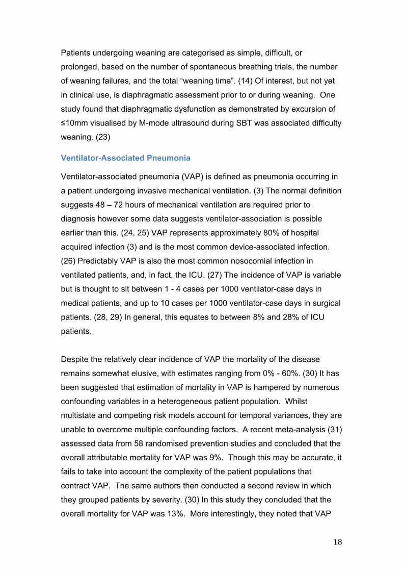
Patients undergoing weaning are categorised as simple, difficult, or
prolonged, based on the number of spontaneous breathing trials, the number
of weaning failures, and the total “weaning time”. (14) Of interest, but not yet
in clinical use, is diaphragmatic assessment prior to or during weaning. One
study found that diaphragmatic dysfunction as demonstrated by excursion of
≤10mm visualised by M-mode ultrasound during SBT was associated difficulty
weaning. (23)
Ventilator-Associated Pneumonia Ventilator-associated pneumonia (VAP) is defined as pneumonia occurring in
a patient undergoing invasive mechanical ventilation. (3) The normal definition
suggests 48 – 72 hours of mechanical ventilation are required prior to
diagnosis however some data suggests ventilator-association is possible
earlier than this. (24, 25) VAP represents approximately 80% of hospital
acquired infection (3) and is the most common device-associated infection.
(26) Predictably VAP is also the most common nosocomial infection in
ventilated patients, and, in fact, the ICU. (27) The incidence of VAP is variable
but is thought to sit between 1 - 4 cases per 1000 ventilator-case days in
medical patients, and up to 10 cases per 1000 ventilator-case days in surgical
patients. (28, 29) In general, this equates to between 8% and 28% of ICU
patients.
Despite the relatively clear incidence of VAP the mortality of the disease
remains somewhat elusive, with estimates ranging from 0% - 60%. (30) It has
been suggested that estimation of mortality in VAP is hampered by numerous
confounding variables in a heterogeneous patient population. Whilst
multistate and competing risk models account for temporal variances, they are
unable to overcome multiple confounding factors. A recent meta-analysis (31)
assessed data from 58 randomised prevention studies and concluded that the
overall attributable mortality for VAP was 9%. Though this may be accurate, it
fails to take into account the complexity of the patient populations that
contract VAP. The same authors then conducted a second review in which
they grouped patients by severity. (30) In this study they concluded that the
overall mortality for VAP was 13%. More interestingly, they noted that VAP

19
had no statistically significant impact in either trauma or medical patients with
low (APACHE score <20 or SAPS <35) or high (APACHE >30 or SAPS >58)
severity scores. Patients with medium severity of illness scores were most
affect by VAP, with medical patients faring better than surgical. The authors
feel that the increased risk of death in VAP could then actually be attributed to
risks associated with longer ICU stays, rather than to VAP itself. This
conclusion seems reasonable, especially when one considers that the finding
are consistent with Schumacher et al. (32) and Nguile-Makao et al. (33) VAP
does clearly increase the length of ICU stay, the duration of ventilation, and
cost of care. (27, 28)
As with all bacterial pneumonia, VAP occurs when bacteria enter the normally
sterile lower respiratory tract and subsequently overcome the host-defence
system, (28, 34) a system that is often weekend in ICU patients. The Centres
for Disease Control and Prevention (CDC) proposes three mechanisms by
which colonisation occurs: aspiration of secretions, colonisation of the aero-
digestive tract, and contaminated equipment. (35) Aspiration, probably more
commonly microaspiration, from the oropharynx is thought to be the dominant
route of entry. (36) Colonisation is often found in the first week of hospital
admission in critically ill patients. (3) On top of the complications often seen
with pneumonia, VAP has the added complications that it is difficult to
diagnose and often exhibits multiple drug-resistance. The most commonly
isolated causative pathogens in VAP are acinetobacter spp., P. aeruginosa,
and S. aureus, though there is no statistically significant difference in clinical
outcomes when the organism is considered.
Some factors associated with VAP (other than intubation itself) are
nasogastric tubes, re-intubation, aspiration, supine positioning, pooling of sub-
glottic secretions, coma, and enteral nutrition. (37, 38)
Considering the difficulties that VAP presents, the most logical solution is
prevention rather than treatment. The primary method of prevention is to
minimise intubation frequency and duration of ventilation. (3) To this end the
CDC released the recommendations found in table 1 for the prevention of

20
VAP. (39) Though the exact impact of VAP on patients remains somewhat
unclear, it has been shown to negatively affect mortality and morbidity,
prolong ICU stays, increase the duration of mechanical ventilation, and
increase associated costs.
Table 1 - CDC VAP prevention guidleines
i. Conduct VAP surveillance ii. Adhere to hand-hygiene procedures iii. Minimise invasive mechanical ventilation iv. Minimise the duration of ventilation v. Conduct daily assessments for readiness to wean vi. Conduct VAP education vii. Maintain head-of-bed (HOB) angle between 30° - 45° whenever
possible viii. Avoid gastric overdistension ix. Avoid unplanned extubation and re-intubation x. Use a cuffed ETT with in-line or subglottic suction xi. Maintain a cuff pressure of at least 25cm H2O xii. Employ orotracheal intubation (vs. nosotracheal) whenever possible xiii. Avoid the use of H2 receptor-blockers and PPIs when possible xiv. Perform regular oral care xv. Remove condensate from the breathing circuit whilst it remains closed xvi. Change or replace the breathing circuit only when it is malfunctioning
or visibly contaminated
Ventilator-Associated/Induced Lung Injury (VALI/VILI) The concept of lung injury as a result of mechanical ventilation is not a new
one. The term “ventilator lung” first appeared in 1967 and described the post-
mortem findings of diffuse alveolar infiltrates and hyaline membrane formation
in patients who had been mechanically ventilated. (5) Deaths often occurred
despite normalisation of arterial blood gasses, and have been attributed to
barotrauma, oxygen toxicity, and haemodynamic comprise induced by
mechanical ventilation. (40) Today ventilator-induced lung injury and the
clinical counterpart, ventilator-associated lung injury, are understood to
involve inflammatory cell infiltrates, hyaline membranes, increased vascular
permeability, and pulmonary oedema. (5) It is largely acknowledged that VILI
is as complex and can be as damaging as the diseases supported by
ventilation. In fact, the pathology exhibited in VILI is almost identical to that
21
seen in acute lung injury (ALI) and acute respiratory distress syndrome
(ARDS). So much so that there is supporting the hypothesis that VILI may
actually be capable of inducing both ALI/ARDS and its extra-pulmonary
manifestation multiple organ failure (MOF). (35, 41) VILI is a common
complication in critically ill patients, contributing to increased mortality,
morbidity, and length of stay. (5) Unfortunately, due to the variable nature of
the disease, the exact incidence are unknown. It is thought to affect
approximately one-quarter of mechanically ventilated patients, though this
number is almost certainly higher in those with pre-existing lung injury. VILI
appears to be induced via three major mechanisms: volutrauma,
atelectrauma, and biotruama.
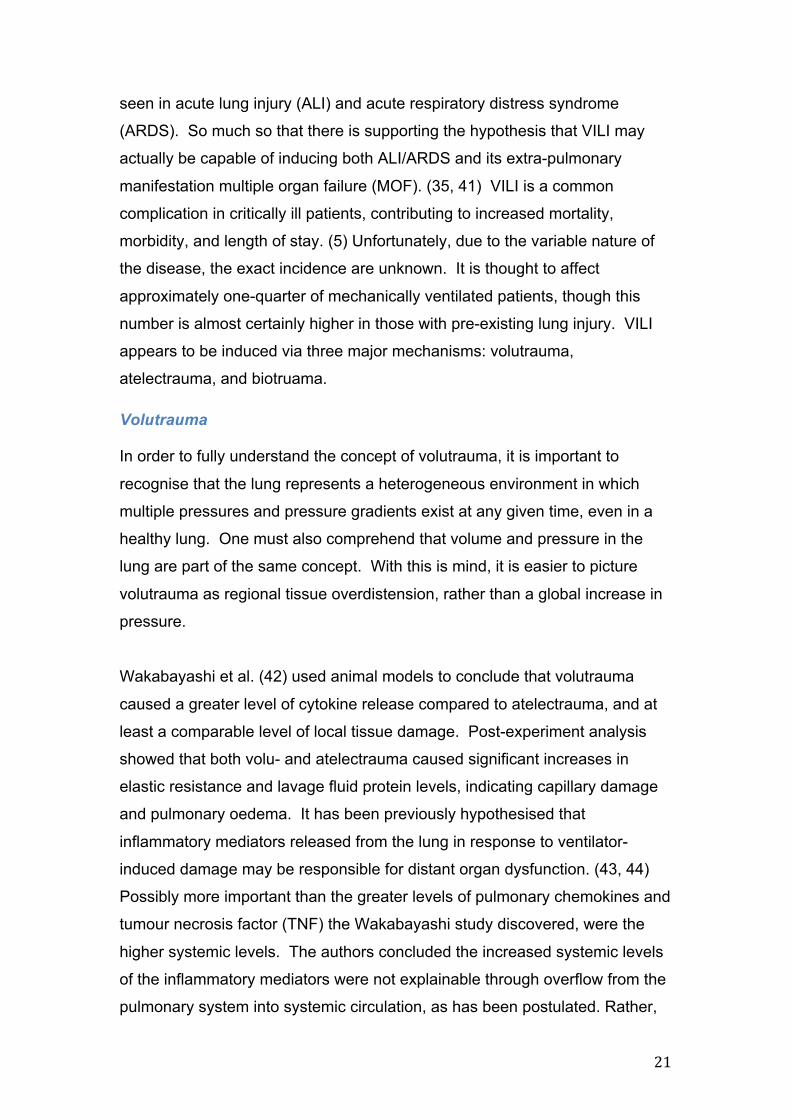
Volutrauma In order to fully understand the concept of volutrauma, it is important to
recognise that the lung represents a heterogeneous environment in which
multiple pressures and pressure gradients exist at any given time, even in a
healthy lung. One must also comprehend that volume and pressure in the
lung are part of the same concept. With this is mind, it is easier to picture
volutrauma as regional tissue overdistension, rather than a global increase in
pressure.
Wakabayashi et al. (42) used animal models to conclude that volutrauma
caused a greater level of cytokine release compared to atelectrauma, and at
least a comparable level of local tissue damage. Post-experiment analysis
showed that both volu- and atelectrauma caused significant increases in
elastic resistance and lavage fluid protein levels, indicating capillary damage
and pulmonary oedema. It has been previously hypothesised that
inflammatory mediators released from the lung in response to ventilator-
induced damage may be responsible for distant organ dysfunction. (43, 44)
Possibly more important than the greater levels of pulmonary chemokines and
tumour necrosis factor (TNF) the Wakabayashi study discovered, were the
higher systemic levels. The authors concluded the increased systemic levels
of the inflammatory mediators were not explainable through overflow from the
pulmonary system into systemic circulation, as has been postulated. Rather,
22
they believe it is representative of an extra-pulmonary source. Predictably,
high levels of lung-marginated leukocytes were also seen and may be
responsible for secreting TNF into the circulation propagating further
production. This seems possible if one accepts that the lungs have the
largest available pool of leukocytes in circulation. Also lending credence to
this theory is the finding that monocyte-depleted lungs exposed to similar
stimulus did not induce such high levels of inflammatory mediators. The
monocyte-depleted lungs also showed less pulmonary oedema. Further
supporting the concept of volutrauma, another study showed that induced
overdistension in spontaneously breathing animals produced lung injury
consistent with high-volume mechanical ventilation. (45)
Whilst atelectrauma produced elevated levels of inflammatory mediators it did
so only in the alveolar compartment. Both atelectrauma and volutrauma
instigated similar levels of pulmonary oedema. This last finding contradicts
those of another study showing that high volume ventilation caused a greater
level of pulmonary oedema than did high-pressure ventilation. (46)
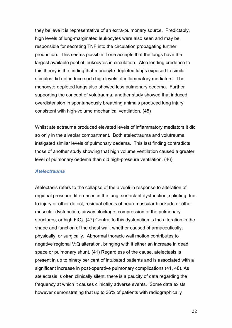
Atelectrauma
Atelectasis refers to the collapse of the alveoli in response to alteration of
regional pressure differences in the lung, surfactant dysfunction, splinting due
to injury or other defect, residual effects of neuromuscular blockade or other
muscular dysfunction, airway blockage, compression of the pulmonary
structures, or high FiO2. (47) Central to this dysfunction is the alteration in the
shape and function of the chest wall, whether caused pharmaceutically,
physically, or surgically. Abnormal thoracic wall motion contributes to
negative regional V:Q alteration, bringing with it either an increase in dead
space or pulmonary shunt. (41) Regardless of the cause, atelectasis is
present in up to ninety per cent of intubated patients and is associated with a
significant increase in post-operative pulmonary complications (41, 48). As
atelectasis is often clinically silent, there is a paucity of data regarding the
frequency at which it causes clinically adverse events. Some data exists
however demonstrating that up to 36% of patients with radiographically

23
diagnosed atelectasis proceed to develop pneumonia in the post-operative
setting. Atelectasis is also associated with an increase in alveolar bacterial
growth. (49, 50)
Figure 5
Atelectrauma is thought to be caused by repetitive shear stress and direct
inflammation caused by the repeated collapse and inflation of the alveoli in
the presence of dysfunctional surfactant or other pathologies. (51) This
concept may be thought of as tidal recruitment/derecruitment injury. The
healthy lung uses surfactant, alveolar interdependence, collateral ventilation,
extracellular matrices, and the thorax to form an incredibly stable structure
avoiding both collapse and over-inflation. In general, a healthy lung will show
only minimal changes in alveolar size or shape provided the total lung
capacity remains between 10% - 80%. (51-53) Loss of any of these
mechanisms creates instability and increases the risk of atelectrauma, and
subsequently of VILI. A disparate yet plausible theory is that small gas
bubbles in non-aerated alveoli create large pressure fronts along the alveolar
wall. (54) Such a gradient would cause large local pressure differences
Copyright © Lippincott Williams & Wilkins. Unauthorized reproduction of this article is prohibited.
macrophage recruitment and activation [77]. It waspostulated that this lung injury was independent ofthe stresses caused by mechanical ventilation as itcan be reversed when increasing FiO2 and/or remov-ing atelectasis by lung recruitment [25]. Futurestudies are needed to determine the clinical signifi-cance of any injury induced by atelectasis in patientswithout previous pulmonary diseases. Whereas thismechanism appears to be of little clinical import-ance in anesthetized patients with previouslyhealthy lungs, in the context of a ‘multiple hittheory’ (the cumulative effects of a series of lessintense insults contributing to injury), that atelec-tasis may exert a synergistic or additive effect inconcert with other mechanisms known to contri-bute to VILI [78,79].
Ventilator-induced lung injuryFully developed VILI presents as bilateral pulmonaryinfiltrates consistent with edema in the absence ofheart failure or severe, chronic lung disease [80].Postoperatively VILI may present as acute lunginjury (ALI) or acute respiratory distress syndrome(ARDS) [81,82&&,83]. The incidence of these entitiesfluctuates depending on the population and
surgeries studied. For example, ALI has an incidencebetween 2 and 6% in high-risk patients undergoingthoracic surgeries [81,83–85]. Postoperative respir-atory failure has estimated mortality that exceeds45% in some populations [86,87]. Evidence isaccumulating that alveolar instability and atelecta-sis may predispose to development of VILI [88,89].Two main mechanisms are proposed: one is tidalrecruitment, the cyclic opening and closing of lungunits at the boundary between normally aerated andcollapsed tissue [90&]. During the respiratory cyclethese units are exposed to repetitive shear stress,which ultimately destroys their cellular structures(Fig. 2) [91]. The other mechanism is tidal over-distension of nonatelectatic lung units. Atelectasiscauses the distribution of ventilation within thelungs to become more heterogeneous shifting theinspiratory flow preferentially to those parts ofthe lungs that are already ‘open’. In this way alveolibecome functionally overinflated [92]. Tissue stressis directly related to the size of tidal volume andpressures applied by the mechanical ventilator.
On the basis of the lessons learned frommechanical ventilation in patients with ALI/ARDS[88,93,94], there is increasing evidence that sup-ports the notion that the same mechanisms of
De-recruitment Overdistention
Pre-VILI BarotraumaVolutrauma
Release ofinflammatory
cytokines
BiotraumaIL-1β, IL-6, IL-8, TNFα
Atelectotrauma
Release ofinflammatory
cytokines
Low VT High VT, high PIPNo recruitmentNo PEEP
FIGURE 2. Ventilation of atelectatic areas can result in cyclic opening and closing of lung units (tidal recruitment), leading torelease of inflammatory cytokines causing atelectotrauma. High tidal volume and overexpansion of lungs can rupture alveolar-capillary membranes (inset), leading to accumulation of neutrophils and release of inflammatory cytokines. IL, interleukin; TNF,tumor necrosis factor; VILI, ventilator-induced lung injury. Adapted with permission from [91].
Atelectasis and postoperative pulmonary complication Tusman et al.
0952-7907 ! 2012 Wolters Kluwer Health | Lippincott Williams & Wilkins www.co-anesthesiology.com 5
Reproduced with permission from Wolters Kluwer Health; Lippincott, Williams & Wilkins. Ventilation of atelectatic areas can result in cyclic opening and closing of lung units (tidal recruitment), leading to release of inflammatory cytokines causing atelectotrauma. High tidal volume and overexpansion of lungs can rupture alveolar- capillary membranes (inset), leading to accumulation of neutrophils and release of inflammatory cytokines. IL, interleukin; TNF, tumor necrosis factor; VILI, ventilator-induced lung injury.

24
amplifying the interaction of tissues and causing cellular injury. (55) Indirectly,
atelectrauma also causes volutrauma via the preferential aeration and over-
inflation of the open areas lung as a derivative of the relative decrease in lung
size from the loss of the non-aerated areas. (47) Additionally, and perhaps
more simply, is the theory that the non-ventilated areas become hypoxic
leading to cell death and the release of inflammatory mediators. Given the
evidence supporting multiple mechanisms of injury, it seems more likely that
no one mechanism is responsible for the damage caused by atelectasis, and
that instead multiple small insults accrue until such time as atelectrauma is
manifest.
Biotruama Related to the two above mechanisms, biotruama refers to the “translocation
of mediators, bacteria, or lipopolysaccharide from the airspaces into the
systemic circulation” (5) in cases of increased alveolar-capillary permeability
as seen in ARDS, volutrauma, or epithelial microtears. This translocation may
then propagate further pulmonary damage or distant organ dysfunction. All
epithelial, endothelial, and of course inflammatory cells in the pulmonary
system are capable of participating in inflammatory reactions via various
signalling pathways. (5, 42) Specifically IL-6, IL-8, and TNF have been
implicated in pulmonary biotruama during clinical trials. (56) Mediator release
can occur via two methods. First, tissue trauma my cause cell death (or
rupture) provoking the release of mediators. (57) Alternatively, lesser, non-
fatal injury to either the cytoskeleton or the extracellular matrix may cause
inflammation via discrete intracellular signalling pathways. (58)
Although volu-, bio-, and atelectrauma have been discussed separately, they
must be considered, if not as different parts of the same entity, then as 3
extremely interrelated entities.
Complications and Effects of General Anaesthesia In general, one can predict several changes in patients under general
anaesthesia. Volatile agents impair mucous transport from the pulmonary
tree, one of the respiratory systems primary protective measures. Data
25
shows that sevoflurane significantly impairs ciliary beat frequency, thus
decreasing bronchial mucous transport velocity (59). The same impairment is
not seen in patients undergoing total intravenous anaesthesia. Likewise,
almost all anaesthetic agents decrease respiratory drive and blunt respiratory
reflexes.
The use of inhaled volatile agents at any point is associated with a significant
decrease in CD3+, CD4+ and, CD8+ T lymphocytes, NK cells, and B
lymphocytes. Total intravenous anaesthesia is associated with a smaller
decrease and more rapid return to normal. (60)
All volatile anaesthetics, and in fact all intravenous anaesthetics save
ketamine, cause a decrease in both systemic vascular resistance and mean
arterial blood pressure. This change in blood pressure seams almost entirely
to occur via a decrease in systemic vascular resistance, as the cardiac index
as measured by echocardiography is largely unchanged. (61) Still important
but of little impact to this paper: some inhaled anaesthetics demonstrate a
propensity towards dysrhythmogenicity and cardiac conduction disturbances,
as well as a prolongation of the QT interval.
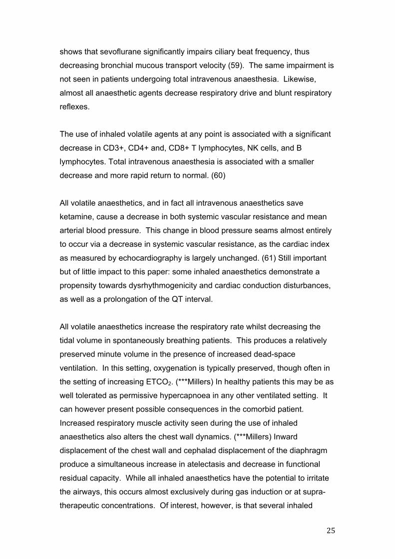
All volatile anaesthetics increase the respiratory rate whilst decreasing the
tidal volume in spontaneously breathing patients. This produces a relatively
preserved minute volume in the presence of increased dead-space
ventilation. In this setting, oxygenation is typically preserved, though often in
the setting of increasing ETCO2. (***Millers) In healthy patients this may be as
well tolerated as permissive hypercapnoea in any other ventilated setting. It
can however present possible consequences in the comorbid patient.
Increased respiratory muscle activity seen during the use of inhaled
anaesthetics also alters the chest wall dynamics. (***Millers) Inward
displacement of the chest wall and cephalad displacement of the diaphragm
produce a simultaneous increase in atelectasis and decrease in functional
residual capacity. While all inhaled anaesthetics have the potential to irritate
the airways, this occurs almost exclusively during gas induction or at supra-
therapeutic concentrations. Of interest, however, is that several inhaled
26
anaesthetics are capable of bronchodilation. This probably demonstrates a
negligible effect in most patients, however it does have significant potential to
benefit those with pulmonary comorbidities. (***Millers)
Total intravenous anaesthetic (TIVA) procedures all, to one extent or another,
produce a similar decrease in blood pressure, though most often in the
presence of a decrease in heart rate. (***Millers) The two can be used in
conjunction, termed mixed anaesthesia, in order to blunt the tachycardia seen
in gas anaesthesia. Contrary to volatile anaesthetics, TIVA generally inhibits
respiratory drive. If not supported, this leads to hypoxia and hypercarbia.
Though not a general anaesthetic, spinal and epidural anaesthesia deserve
some mention here. Because the delivery of an anaesthetic to one specific
level or dermatome is somewhat imprecise, this procedure sometimes
produces hypotension and respiratory distress. (***Millers) The most
dangerous potential side effect is termed total spinal anaesthesia. It
represents a total derangement of cardiovascular and respiratory control, and
requires immediate, careful, and aggressive management.
Strategies for the Prevention of Ventilator-Associated Complications
Perioperative Prevention Strategies Perioperative complications are those conditions related to surgery arising
within one month prior and three months after the procedure. Perioperative
complications occur in 20% - 67% of elective surgeries, and more often in
emergent surgeries. They account for 80% of surgery-related death and
reduce median survival by up to 70%. (62-64) Post-operative pulmonary
complications (POPC) occur at least as often as post-operative cardiac
complications and contribute significantly to increased mortality and ICU
admission. (65-68) POPC may be divided into acute, sub-acute, and delayed
phases.

27
Residual neuromuscular blockade is associated with increased incidence of
POPC. (66) This is explained via respiratory muscle weakness leading to
hypoxia and respiratory failure. Even in the absence of respiratory failure,
residual blockade lengthens stays in the post-operative care unit. (47) It is
advisable then to use appropriately timed neuromuscular blocking agents and
ensure complete reversal through antagonistic agents prior to extubation as
this has been shown to decrease POPC. (69)
Figure 6
Failure to wean from the ventilator after surgery may be due to retained
anaesthetic or neuromuscular agents, but is more commonly associated with
indirect lung injury resulting from the pathology at hand. (66)
Aggressive fluid administration during the perioperative and operative periods
is also associated with POPC. (66, 70) Every effort, therefore, should be
made to avoid excess fluid whilst continuing to maintain an appropriate fluid
balance.
Smoking Cessation Ample data demonstrates that patients who smoke experience both an
increased rate of surgical complications (71-75), and of perioperative death
Copyright © Lippincott Williams & Wilkins. Unauthorized reproduction of this article is prohibited.
THE ROLE OF ATELECTASIS IN THEPATHOGENESIS OF POSTOPERATIVEPULMONARY COMPLICATIONSThe variable rates of PPCs reported are due to thedifferent types of complications studied, the clinicalcriteria used in their definition, and the differentsurgical populations reported [4,6]. The rate ishigher in older patients, in those with more comor-bidities, and after complex operations. Up to 5.4%high-risk patients may develop postoperative respir-atory failure after elective operations, with the 27%2-month mortality for those who developed acutelung injury [40].
Intraoperative atelectasis is an important factorin the pathogenesis of PPCs [4,41–43]. Atelectasis isseldom visible on chest radiograms, but ratherrequires chest computed tomography for detection,so it is frequently underappreciated [4,14,36,44].However, in many instances atelectasis itself doesnot have clinical sequellae. Nonetheless, atelectasiscannot be ignored because if it persists over time itmay become the starting point of a wide range ofother PPCs, not only limited to those in lungs(Fig. 1), as further discussed.
HypoxemiaAbnormalities of gas exchange in the perioperativeperiod range from a slight decrement in PaO2 to life-threatening hypoxemia [45]. Atelectasis results inV/Q mismatching due to either pulmonary shuntor dead space [37]. Correlations exist between
pulmonary shunting and the amount of atelectasis,and between areas of low V/Q and airway closure inelderly [38,46,47]. Other causes of hypoxemiainclude hypoventilation due to pain or residualanesthetics, lung edema due to fluid overload,exacerbation of chronic obstructive pulmonarydisease, laryngospasm or bronchospasm. However,persistent postoperative hypoxemia in the absenceof other apparent causes should suggest the presenceof atelectasis [14,36,44]. The relief of hypoxemia bytreating lung collapse with noninvasive ventilationsupports a causative relationship. Use of continuouspositive airway pressure (CPAP) reduces the risk foratelectasis [48], and noninvasive ventilation can beused as a prophylactic and/or therapeutic tool toimprove gas exchange postoperatively [15]. Further-more, the use of CPAP may decrease the rate ofreintubations and other complications after electivesurgeries [49].
Deleterious effects of atelectasis-related hypo-xemia depend on the severity and duration ofhypoxemia, and on patient’s tolerance to hypoxia.Several postoperative complications directly relatedto hypoxemia have been identified:
(1) Hypoxemia-related delirium may occur in up to65% of postoperative patients [50,51] andresolves with oxygen therapy [52].
(2) Wound infection is a potential consequenceof hypoxemia because the neutrophils’ abilityto kill bacteria depends on the surroundingO2 tension [53–55]. A meta-analysis suggested
Anesthetic factors Surgical factors Patient factors* Muscle dysfunction * Thoraco-abdominal surgeries * Age* Muscle disruption * Surgical retractors * Body weight* Low phrenic nerve output * Faulty surgical techniques * Smoking* Pain * Pneumoperitoneum *Previous respiratory diseases* Mucociliary dysfunction * Body positioning *Meteorism –abdominal
compartment syndrome.* Blood displacement between thorax and abdomen
* Fluid therapy
Respiratory restrictive pattern( FRC-FVC)
Lung collapse* airway closure* atelectasis
Postoperative pulmonary complicationsHypoxemia Pneumonia Local inflammatory response Ventilator induced lung injury
* Decreased DO2 * Macrophage dysfunction * Local hypoxia or hyperoxia * Cyclic tidal recruitment* Systemic ischemia-
reperfusion injury* Loss of surfactant * Local mechanical
parenchymal stress* Tidal overdistension
* Delirium * Bacterial growth * Biotrauma *Time-prolonged ventilation* Wound infection * Bacterial translocation* Arrhythmias* Myocardial ischemia
FIGURE 1. Link between pathophysiological factors, lung collapse and postoperative pulmonary complications.
Atelectasis and postoperative pulmonary complication Tusman et al.
0952-7907 ! 2012 Wolters Kluwer Health | Lippincott Williams & Wilkins www.co-anesthesiology.com 3
Reproduced with permission from Wolters Kluwer Health; Lippincott, Williams & Wilkins. Link between pathophysiological factors, lung collapse and postoperative pulmonary complications.

28
(73, 75-78). A Cochrane review indicates that pre-operative smoking
cessation, especially ≥ 4 weeks prior to surgery may significantly improve
outcomes (79). The reduction in perioperative complications ranges from
20% to 34% (80-82). Difficulties in smoking cessation include mainly
unwillingness on the part of the patient, increased workload on the staff, and
the incurred expenses surrounding support programmes required for patient
success (73).
Smoking is a univariate risk factor for respiratory failure, unanticipated
intensive care admission, pneumonia, adverse airway events, and
unanticipated airway and breathing interventions (83).
Smoking is relevant to anaesthetists for several reasons. First, smoking
increases the level of carboxyhaemoglobin, which can reach up to 10% in
heavier smokers (83). The increased level of carboxyhaemoglobin not only
decreases the total amount of oxygen that is available, but also shifts the
oxyhaemoglobin dissociation curve leftwards, impeding the release of oxygen
to the tissues (84) This is particularly relevant in patients with cardiovascular
disease, or risk, because smoking also causes coronary vasoconstriction (85).
This occurs via both direct and indirect sympathetic outflow (86).
In addition to altered gas dynamics smoking also impairs mucociliary
clearance (87, 88). This is in addition to the impairment resulting from inhaled
volatile agents. Cigarette smoking also impairs the function of both alveolar
macrophages and neutrophils (89, 90). One can also expect goblet cell
hyperplasia accompanied by increased mucous production, as well as airway
hyperreactivity (87, 88).
The above data clearly demonstrates the negative impact of cigarette
smoking on perioperative morbidity and mortality, and the benefits of
perioperative smoking cessation. It seems therefore worthwhile to promote
smoking cessation as part of a structured programme during pre-operative
evaluations.

29
Analgesia Traditional approaches to post-operative analgesia involve opioids with or
without NSAIDS. Whilst this approach may be appropriate for some patients
in some cases it has several serious limitations. The larger doses of opioids
that are often required to manage post-operative pain following major
thoracic, abdominal, or orthopaedic surgery also tend to centrally depress
respiratory function. (3) Depression of respiratory function then increases the
odds of POPCs. (66)
Alternatively post-operative pain may be managed via neuraxial blockade. A
meta-analysis published in 2000 examined 158 trials of neuraxial blockade
and found an overall decrease in perioperative mortality of approximately one-
third when compared to general anaesthesia and traditional opioid
management. (91) Specific to post-operative pulmonary complications the
authors identified a decrease in the occurrence of pneumonia as well as a
decrease in the rate of respiratory depression. Deaths related to these two
reasons dropped in a similar fashion. Interestingly, most studies analysed
documented the continued benefit of neuraxial blockade regardless of
whether or not it was maintained after the surgery. As far as overall mortality
was concerned, there was little difference between neuraxial blockade alone
or combined with general anaesthesia. The authors attributed the better
outcomes to ease of pain-free breathing, increased blood flow, altered
coagulopathy, and a decrease in the “surgical stress response”. Ease of early
pain-free movement may also have played a role.
COPD is identified as an independent risk factor for POPC, as these patients
can be extremely sensitive to alterations in respiratory drive and function. (47)
There is some data supporting the notion that COPD patients may be
between 300% - 700% more likely to develop pulmonary complications in the
post-operative environment. (92) A study conducted in the Netherlands using
this high-risk population documented a decreased risk of pneumonia within 30
post-operative days without evidence of a decrease in pulmonary function as
a result of epidural anaesthesia. (93) Neither the pulmonary function nor the

30
pneumonia rates varied with disease severity. As a secondary end point, they
also demonstrated better pain control in the group with epidural anaesthesia
versus the traditionally managed group. Despite a clear decrease in the rates
of pneumonia, this study failed to demonstrate a statistically significant
decrease in the overall mortality rate. This is consistent with a large
population-based cohort study that identified a small but statistically significant
decrease in 30-day mortality. (94) It is possible that smaller studies will miss
this data point as the number-needed-to-treat in that study was 447. More
significantly, the study did confirm the safety of epidural anaesthesia in this
setting and its efficacy both in preventing POPC and improving pain control.
Though not without risk, neuraxial blockade appears both safe and efficacious
when used for the prevention of POPCs and the management of pain.
Though the current evidence does not support its routine use to decrease
crude mortality, it may be appropriate to employ it in patients at high risk for
post-operative pulmonary complications or difficult to manage pain.
Post-Operative Respiratory Failure (PRF) Post-operative respiratory failure represents the most common post-operative
pulmonary complication, at approximately 50%. It shows an incidence of up
to 3.4% in general surgical populations and carries a mortality rate of up to
25%. (47, 95) PRF is defined as the impairment of pulmonary gas exchange
and hypoxia with or without hypercapnoea, in the absence of cardiac failure
and as a result of surgery or anaesthesia. (47) Using PaO2:FiO2 levels, post-
operative respiratory failure is classified as mild, moderate, or severe.
Respiratory muscle dysfunction contributes heavily to PRF. (96) Muscular
dysfunction arises from residual anaesthetic agents, opiate effects on the
central respiratory centre, and splinting from either pain or trauma. (47)
Additionally, residual neuromuscular block affects not only the muscles of
respiration, but also the airway muscles. This means that, not only are the
diaphragm and thoracic muscles potentially unable to completely expand the
thorax due to weakness, but also that the muscles of the airways remain
dilated. This predisposes to lower pressures and increased risk of airway

31
collapse and atelectasis. (97) An inability to fully expand the thorax or the
predisposition to airway collapse can maintain or perpetuate atelectasis
potentially leading to a lengthened post-operative course or further lung injury.
Inspiratory Muscle Training As inspiratory muscle weakness can be seen within two hours of the initiation
of mechanical ventilation and is almost always present to some degree within
forty-eight hours, it has been thought that inspiratory muscle conditioning
could reverse this process, increasing respiratory muscle strength and
endurance, especially that of the diaphragm. Inspiratory training is delivered
using a variety of methods that increase the pressure against which patients
must draw a breath. Two common methods are increasing the trigger
pressure on the ventilator, and the use of an external impedance device. (15)
In both methods the resistance is increased incrementally for short intervals
over several days until the patient is thought to be capable of weaning.
Whilst this represents a logical intervention, data is contradictory. A 2011
review (15) concluded that although inspiratory muscle training was favoured,
it did not statistically improve the duration of weaning in a significant manner.
The same conclusion was reached for overall survival. No difference was
found when considering re-intubation rates. This is in concordance with a
randomised trialconducted more recently that demonstrated no significant
shortening of the weaning period. (98) This study however is limited in that
patients in both the control and intervention group received non-invasive
ventilatory support following extubation. The study also failed to publish re-
intubation rates.
Conversely, several studies have successfully demonstrated the benefit of
inspiratory muscle training when considering the duration and success of
weaning. (99-101) These studies showed small yet significant differences in
both duration and outcome.
All studies however reported a significant increase in peak inspiratory
pressure, suggesting a significant increase in muscle strength. Peak

32
inspiratory pressure currently represents the only clinically useful method of
measuring respiratory muscle strength. No study found any association
between inspiratory muscle training and worsened outcomes. It is possible
that the inconsistent outcomes are a reflection of the homogenous nature of
ICU patients requiring mechanical ventilation. It would therefore be plausible
to consider that there may be a subset of patients who would benefit from
inspiratory muscle training. As no danger has been demonstrated, it seems
reasonable to consider inspiratory muscle training in patients at risk of failure
to wean, such as those with a high Tomin index. Although ultrasound has
been shown capable of estimating diaphragmatic mass, no study has
combined this with inspiratory training. This may also prove of benefit in the
future, assuming a minimum required muscle mass could be determined.
Sedative Reduction and Sedative Selection Sedation in mechanically ventilated patients is employed to improve
patient:ventilator synchrony, reduce patient stress and anxiety, facilitate
medical care, and to improve patient comfort. Amelioration of this stress also
has the possible effect of reducing endogenous catecholamine release and
oxygen consumption. Significant evidence exists, however, demonstrating
that excessive and prolonged sedation promotes longer durations of
mechanical ventilation, longer ICU stays and worsened outcomes, including
long-term cognitive impairment. (102-106)
A recent multi-centre trial showed that deep sedation in the first forty-eight
hours is an independent risk factor for delayed extubation and increased
mortality. (106) The study concluded that each Richmond Agitation Sedation
Scale (RASS) finding in the deep range was associated with a delay in
extubation of 12.3 hours, a 10% increase in in-hospital death, and an 8%
increase in mortality at six months. Additionally, this study found that the use
of midazolam was associated with longer periods of mechanical ventilation.
This supports another study associating the use of benzodiazepines with
longer periods of ventilation. (107) The fact that this study addressed the first
48 hours is important, as many studies do not. One other study assessed the

33
outcomes of patients without sedation in the first 48 hours and found
significant improvements. (108)
The use of daily sedation interruption, commonly known as a “sedation
vacation” offers the theoretical benefit of avoiding the accumulation of the
sedative agents and their active or toxic metabolites, providing the opportunity
of assessment of the requirement for continued sedation, and allowing the
patient to participate in daily spontaneous breathing tests. As prolonged
ventilation and deep sedation are known to promote worse outcomes, this
potentially leads to a reduction in the duration of ventilatory support,
shortened ICU and hospital stay, and an improvement in long-term outcomes.
(102, 109)
Currently, the two most common methods for reduction in exposure to
sedation are daily sedative interruption, and protocolised targeted light
sedation. Hughes et al (104) found that, when used alone, these strategies
were equally effective in reducing exposure to sedatives and improving
outcomes when compared to deeply sedated patients. The authors further
suggested that either the combination of the two, or the combination of daily
interruption with an awake spontaneous breathing trial, might be of greater
benefit. A finding that supports this is that outcomes are unchanged when the
total dose of sedative is not decreased, even in the presence of a sedation
vacation or lightened sedation. Girard (110) actually studied the combination
of daily sedation vacations in combination with spontaneous breathing tests
and found a significant decrease in ventilator time, ICU stay, hospital stay,
and overall mortality.
To date only one trial has attempted to combine both targeted light sedation
with daily interruption. Paradoxically, the study found that there was no
difference in outcome and that the patients in the combined group required
higher doses of sedatives. (105) Several potential problems exist within this
study. First, the sedation scores published in the article are more consistent
with moderate sedation, not light. Also, the compliance with the study
protocol was only 72%. Potentially this means that the authors were not
34
actually studying lightly sedated patients, and that the data rather reflects
what is already known about more deeply sedated patients. Interestingly, the
authors did identify a sub-set of surgical/trauma patients that benefitted from
daily withdrawal of sedation alone.
A recent-meta analysis of five randomised trials covering 699 patients found
that daily interruption of sedation alone was not associated with reduced
ventilatory duration, nor earlier extubation times. (102) Likewise no decrease
in the length of ICU or total hospital stay was found. The analysis did
however identify a reduction in the likelihood of requiring tracheostomy. No
difference was noted in the re-intubation rates. Importantly, the study found
no detrimental effect associated with the use of daily sedation withdrawal.
Good evidence exists to show that over-sedation of patients results in longer
ICU and hospital stays, longer ventilation times, and poorer long-term
outcomes. It has been shown that total dose reduction of sedatives and that
the avoidance of benzodiazepines can improve outcomes. Evidence
becomes inconsistent however when examining wither daily withdrawal of
sedation or targeted light sedation. Potentially, the introduction of these two
practices has simply made clinicians more cognizant of sedation levels and
total sedative doses. Alternatively the inconsistent finding of various studies
may be the result of an as yet unidentified sub-set of patients that benefit (or
suffer) from both or either of these two practices. Regardless of the
inconsistencies in the data the adoption of either of these methods of sedation
reduction appears safe, and when compared to previous model of sedation is
certainly of benefit.
Early Immunomodulary Nutrition Early perioperative enteral nutrition with substances promoting
immunomodulation is controversial and contradictory. Endotoxinaemia is
reported in several studies and is thought to occur following abdominal
surgery
via direct contamination of more often as a result bacterial translocation.
Endotoxinaemia thought to act in a pro-inflammatory manner and is

35
associated with higher levels of markers of inflammation and poorer
outcomes. (111) Theoretically, early enteral nutrition would increase
splanchnic circulation and decrease bacterial translocation, preventing an
inflammatory cascade. TNF alpha is known to be responsive to
endotoxaemia and is probably responsible for the adverse effects, including
protein/fat redistribution, cachexia, chemotaxis and inflammatory recruitment,
and promoting anaerobic metabolism. (111) Elevated levels of TNFa are
associated with worse outcomes. The most common constituents of
immunonutrition are omega-3 fatty acids, RNA, glutamine, and arginine. Both
animal and in vitro studies have demonstrated their ability to modulate cell-
mediated immune function, acute phase proteins, insulin release, and the
post-traumatic inflammatory cascade. (112, 113) Unfortunately, despite more
than two decades of clinical trials, studies are often equivocal and reliable
data is still lacking.
One study examined early perioperative immunomodulatory nutrition
compared to a traditional approach and concluded that there was an overall
attenuation of the inflammatory response. (114) This conclusion was only
reached after the authors discounted the increased cytokine levels found in
the interventional group. No overall difference in immunoglobulin levels was
reported, nor was a difference in overall mortality.
An industry-funded trial in 2007 reported a statistically significant decrease in
the levels of endotoxin, CRP and TNFa. (115) It did not report rates of clinical
complications or outcome.
By far the best study is a 2010 meta-analysis reviewing 21 randomised
controlled trials including 2730 patients. (116) The study showed that
perioperative infections decreased in those receiving specialised early
nutrition, as did the total number of days in the hospital. Overall mortality
following surgery in the interventional group was unchanged. They further
concluded that the cost associated with the programme was probably less
than the cost associated with treating the prevented infections.

36
Current data suggests that early enteral immunonutrition may decrease the
rate of postoperative infection, as well as the number of in-hospital days. One
possible limitation is that many studies used predominantly cancer patients, a
subset of patients known to be immunocompromised and prone to infection.
Whether or not these results are transferable to the general surgical
population remains to be seen. No study reported statistically significant
improvements in outcome or mortality. As no study however reported worse
outcomes it seems reasonable that perioperative immunonutrition could be
offered to at-risk patients without fear of complication.
Lung-Protective Strategies During General Anaesthesia and Mechanical Ventilation
Low-Volume Ventilation In 2000 a landmark trial using lower tidal volumes than conventional
ventilation protocols in ARDS patients changed the way in which ALI/ARDS
patients are ventilated. (117) The trial randomised 861 patients to either a
lower Vt group (6ml/kg or predicted body weight) or to a convention ventilation
group (10ml/kg – 15 ml/kg predicted body weight). This higher tidal volume
time represented the average protocol for a patient with ALI/ARDS at the time,
as the goal was often both to ensure not only a viable FiO2 but also to
normalise PaCO2 and pH. So successful was this trial that it was suspended
prematurely, as it was no longer possible to ethically enrol patients to the
control group. This study went on to form the foundation of the low tidal
volume ARDSnet protocol in use around the world today. Overall the
reduction in mortality in the ALI/ARDS population requiring mechanical
ventilation was reduced 22 per cent. The authors further demonstrated that at
all times the peak airway pressure and mean airway pressure remained lower
in the interventional group. Moreover, the PaO2 was not statistically or
clinically altered in the interventional group, despite a significant increase in
PaCO2. In spite of this permissive hypercapnoea the patients were found to
have a lower IL-6 level at every point where the two groups were compared.
They also spent fewer days undergoing mechanical ventilation and had more
favourable FiO2:PaO2 ratios. This data has been confirmed by several
studies since and is now the standard of care in ALI/ARDS. (5)
37
While a complete discussion of ventilation in the ARDS patient is well outside
the scope of this paper, it is important to understand the genesis of low tidal
volume ventilation. So great was the improvement in mortality stemming from
these recommendations, that the concept of low volume ventilation was
rapidly transferred to non-ALI/ARDS patients, often without good clinical
evidence. (118)
In 2010 a randomised controlled trial compared traditional ventilation (10ml/kg
pbw) to low tidal volume ventilation (6ml/kg pbw) in mechanically ventilated
patients without acute lung injury. (119) The study was designed with the
primary end point of cytokine levels in bronchoalveolar lavage and plasma
and a secondary end point of the development of ALI. Similar to the 2000
ARDSnet trial, the 2010 trial was suspended early after the second interim
analysis showed significantly higher rate of conversion to ALI in the non-
intervention (traditional) group. The authors showed that, even in the
absence of diagnosable ALI, the lung injury scores (LIS) were higher in the
traditional group. Not only were the LIS lower in the interventional group, they
decreased overall from baseline. Baseline plasma and BAL cytokine levels
were comparable in both groups. In patients not developing ALI, the plasma
IL-6 levels declined more rapidly in the interventional group. Predictably,
plasma IL-6 increased in all patients developing ALI regardless of the mode of
ventilation. No significant difference was found in oxygenation, PaO2, or
PaO2:FiO2 ratio. The authors calculate an odds ratio of 5.1 for developing ALI
at a tidal volume of 10ml/kg, and an odds ratio of 1.3 for each ml/kg above
6ml/kg. Statistical analysis of this data showed traditional ventilation to be an
independent risk factor for the development or ALI in mechanically ventilated
patients. This study may be somewhat underpowered as ARDSnet guidelines
had already been adopted at the time of study, requiring a switch to the
ARDSnet low tidal volume protocol if ALI was identified.
A 2012 meta-analysis examined 20 studies investigating the use of low Vt in
patients without ALI/ARDS. (118) It was found that not only was mortality
lowered in the low Vt group, but so was the development of lung injury.
Though they were unable to demonstrate a significant difference in either

38
number of ventilator days or number of ICU days, they did show that low Vt
decreased the total number in-hospital days. The analysis also points to
significantly lower numbers when considering VAP, atelectasis, and plateau
pressure. The authors conclude that higher (previously traditional) tidal
volumes promote VILI and that the use of low Vt is associated with better
outcomes.
A recently published translational review found that low Vt ventilation in
patients with identifiable pulmonary insult resulted in both decreased airway
pressures and strain. (120) The study was also able to identify several studied
specific types of damage to the lungs associated with mechanical ventilation.
It was found that in addition to lowering the incidence of ALI, the overall rates
of inflammation, pulmonary oedema, and capillary leakage were all
decreased. In contrast to the above meta-analysis from 2012, this study
found a shorter duration of ventilation was associated with low Vt. Lower
rates of ALI/ARDS, lower mortality, and lower rates of VAP and other
pulmonary infections were in concordance with previous studies.
Though the 2012 meta-analysis included studies examining surgical patients,
a 2006 study is worth mentioning specifically. This study concluded that the
use of more traditional tidal volumes was associated with a significantly higher
risk of post-operative pulmonary failure after only a few hours of ventilation.
(121) Fifty per cent of the patients developing post-operative respiratory
failure met the diagnostic criteria for ALI. The authors calculated an odds
ratio of 1.56 per ml/kg above 6ml/kg Vt. The study also associated
aggressive fluid management with post-operative respiratory failure.
Low tidal volume ventilation is the current standard of care for ALI/ARDS
patients requiring ventilator support. In many institutions, this mode of care is
extended to those without ALI/ARDS and the currently available data supports
this. No study reviewed during the writing of this paper demonstrated a
negative outcome as a result of low Vt ventilation. All studies demonstrated a
decrease in the number of ALI cases associated with mechanical ventilation
regardless of the clinical setting. Surgical studies included in the meta-

39
analysis showed a decrease in the rate of post-operative pulmonary failure.
In all studies in which it was considered, the PaO2 was maintained with Low
Vt, even in the face of permissive hypercapnoea, which had no statistically
significant impact. Considering the available data, it appears that low tidal
volume ventilation should now reflect the standard of care in patients requiring
mechanical ventilation.
Positive End Expiratory Pressure (PEEP) As previously discussed, atelectasis occurs in the majority of intubated
patients with or without prior lung injury. Atelectrauma occurs through a
variety of mechanisms when the alveoli are allowed to repeatedly inflate and
collapse resulting in localised inflammation and the release of inflammatory
mediators. Such a scenario may subsequently promote systemic
inflammation and increases the risk of VILI.
Positive end expiratory pressure prevents the alveolar pressure from reaching
zero (atmospheric pressure), and is used as an instrument of stabilisation,
preventing recruitment/derecruitment. (1, 122) This is explained by the fact
that alveolar derecruitment seems a function of lower plateau pressures rather
than low Vt. (123) Halter et al. (122) used diverse ventilation strategies in
animal models with alveolar dysfunction and established that higher levels of
PEEP effectively prevented alveolar collapse even in the presence of low tidal
volumes. During evaluation they found that lower levels of PEEP were
associated with an increase in neutrophil infiltration, intra-alveolar oedema,
and septal thickening. The same authors also maintain that they were able to
maintain a constant number of open alveoli throughout the high PEEP
protocol, whilst lower PEEP resulted in progressive atelectasis. This was
seen during the experiment as increasing mean airway pressures and was
confirmed histologically afterwards. In humans, the use of PEEP with low
tidal volumes is associated with a decrease in the frequency of post-operative
pulmonary complications. (124, 125)

40
Figure 7
The use of PEEP, especially higher levels of PEEP in the setting of lung
injury, appears to have more impact in the prevention of VILI than the use of
low tidal volume ventilation. (122) Predictably it appears that the effect of
PEEP on mortality is not uniform. A recent Cochrane review (125) failed to
demonstrate a decrease in the overall mortality of patients after the use of
PEEP despite an obvious decrease in the rate of post-operative pulmonary
complications. In contrast to this, another group of researchers concluded
that PEEP brought about a 5% decrease in mortality in patients with poor
paO2:FiO2 ratios. (126)
Prior to employing PEEP, one must be aware that it does have potential
haemodynamic consequences. PEEP can, depending on cardiac function,
either increase or decrease cardiac output through elevation of intrathoracic
pressures. (41) An increase in intrathoracic pressure typically reduces venous
Furthermore, mechanical ventilation can infl uence both local and systemic immune responses [1] because mechanical forces within the lungs trigger diff erent biological responses that spread into the systemic circu-lation. Although much knowledge about this issue has been generated in recent years, the specifi c contribution of ventilator settings to the infl ammatory response (that is, the biological activity brought about increasing volu-trauma or atelectrauma) is unknown, and this complex response is likely to be infl uenced by patients’ baseline conditions.
ConclusionsKnowledge of the mechanisms involved in VILI is important to develop ventilatory strategies that could result in clinical benefi ts. Recent advances suggest that isolated mechanical forces (plateau pressure, PEEP) cannot adequately explain VILI; rather, the amount of damage depends on the simultaneous interaction of these forces and the previous status of the lung parenchyma. Ultimately, dynamic changes in alveolar structure could be responsible for lung injury. Th e translation of these concepts to the bedside requires complex physiological reasoning and directed research.
Competing interestsThe authors declare that they have no competing interests.
AbbreviationsALI, acute lung injury; ARDS, acute respiratory distress syndrome; EELV, end-expiratory lung volume; PEEP, positive end-expiratory pressure; VILI, ventilator-induced lung injury; Vt, tidal volume.
Author details1Intensive Care Unit, Hospital Universitario Central de Asturias, Departamento de Biología Funcional, Universidad de Oviedo, Instituto Universitario de Oncología del Principado de Asturias (IUOPA), Celestino Villamil s/n, 33006 Oviedo, Spain. 2CIBER de Enfermedades Respiratorias, Instituto de Salud Carlos III, Spain. 3Critical Care Center, Hospital de Sabadell, Corporació Parc Taulí, Insitut Universitari Fundació Parc Tauli, Universitat Autònoma de Barcelona, Parc Taulí s/n, 08208 Sabadell, Spain.
Published: 7 April 2011
References1. dos Santos CC, Slutsky AS: The contribution of biophysical lung injury to
the development of biotrauma. Annu Rev Physiol 2006, 68:585-618.2. International consensus conferences in intensive care medicine.
Ventilator-associated lung injury in ARDS. American Thoracic Society, European Society of Intensive Care Medicine, Societe de Reanimation Langue Francaise. Intensive Care Med 1999, 25:1444-1452.
3. Brower RG, Shanholtz CB, Fessler HE, Shade DM, White P, Jr, Wiener CM, Teeter JG, Dodd-o JM, Almog Y, Piantadosi S: Prospective, randomized, controlled clinical trial comparing traditional versus reduced tidal volume ventilation in acute respiratory distress syndrome patients. Crit Care Med 1999, 27:1492-1498.
Figure 2. Eff ects of recruitment in alveolar stability. Alveolar stability can be achieved by the equilibrium between end-inspiratory and end-expiratory volumes. This stability allows defi nition of a hypothetical safe zone (blue area). Because strain can be viewed as the ratio between these volumes, this zone corresponds to normal or lower strain values. On the contrary, excessive end-inspiratory deformation in lungs with a low resting volume leads to alveolar instability (red area). The arrows represent how the tidal volume (Vt) and positive end-expiratory pressure (PEEP) modify the end-inspiratory/end-expiratory ratio. In recruitable lungs, PEEP induces an increase in end-expiratory lung volume that allows ventilation to stay in the safe zone, and strain decreases. In the absence of recruitment, however, both the Vt and PEEP lead to a predominant increase in end-inspiratory volume, therefore increasing strain. FRC, functional residual capacity.
� "Effective" FRC(FRC+Recruitment)
� E
nd-in
spira
tory
vol
ume
Vt
PEEP
"Safe" zone
� "Effective" FRC(FRC+Recruitment)
� E
nd-in
spira
tory
vol
ume
PEEP
Vt
Recruiter Non-Recruiter
"Unstable" zone
"Safe" zone
"Unstable" zoneStrainAlveolar instability
StrainAlveolar instability
Albaiceta and Blanch Critical Care 2011, 15:304 http://ccforum.com/content/15/2/304
Page 5 of 7
Reproduced with permission from BioMed Central. Effects of recruitment in alveolar stability. Alveolar stability can be achieved by the equilibrium between end-inspiratory and end- expiratory volumes. This stability allows definition of a hypothetical safe zone (blue area). Because strain can be viewed as the ratio between these volumes, this zone corresponds to normal or lower strain values. On the contrary, excessive end-inspiratory deformation in lungs with a low resting volume leads to alveolar instability (red area). The arrows represent how the tidal volume (Vt) and positive end-expiratory pressure (PEEP) modify the end-inspiratory/end-expiratory ratio. In recruitable lungs, PEEP induces an increase in end-expiratory lung volume that allows ventilation to stay in the safe zone, and strain decreases. In the absence of recruitment, however, both the Vt and PEEP lead to a predominant increase in end- inspiratory volume, therefore increasing strain. FRC, functional residual capacity.

41
return and consequently right ventricular preload. A second potential
consequence of PEEP is the formation of micro ruptures or blebs, though this
is usually only seen at high levels of PEEP. (5) Notwithstanding these two
caveats, this author is unaware of any study concluding that appropriate
levels of PEEP, even in routine use, contributes to poorer outcomes.
Considering the potential benefits of decreased atelectasis, improved V:Q
ratios, better oxygenation, and decreased rates of VAPthe routine use of
PEEP seems appropriate. More so in the absence of significant contradictory
evidence. Outside of routine patient care, there is excellent evidence to
support its use in at-risk patients as well as those already presenting
difficulties with ventilation.
Figure 8
Considering the potential benefits of decreased atelectasis, improved V:Q
ratios, better oxygenation, and decreased rates of VAP in the absence of
significant contradictory evidence its routine use of PEEP seems appropriate.
Outside of routine patient care there is excellent evidence to support its use in
at-risk patients, as well as those already presenting difficulties with ventilation.
alveolar expansion in normal lungs. Whereas some zones undergo only minimal changes in each breath, other zones undergo deformations of up to 20% of their length. Th is fi nding could have important consequences for VILI in healthy lungs.
Failure of any of the above-mentioned mechanisms leads to alveolar instability, understood as an excessive tissue deformation during each breath. Although this may be a vague term, and in fact the thresholds of stress and strain that lead to tissue injury are unknown, we will use it as a concept beyond the classic opening and closing hypothesis. In this sense, alveolar instability refers not only to tidal changes in aeration, but also to those circumstances of excessive lung tissue deformation irrespective of the alveolar initial status. Th is common mechanism could help to override the diff erent hypo the-sis on lung injury induced by cyclic changes in lung aeration (that is, opening and closing of alveoli versus fl uid and foam in airways and alveoli [37]). Most circumstances that increase alveolar instability also increase the risk of VILI. Whereas the current paradigm considers alveolar insta bility and VILI to be concomitant consequences of an unknown cause, we hypothesize that alveolar instability is the main mechanism in VILI. Lung injury increases alveolar instability, and alveolar damage starts in the unstable zones [19]; VILI could spread from these zones where tidal changes are more relevant. Th ese zones are also present in healthy lungs, so this model could explain the induction of VILI in patients with and without previous lung injury.
Some studies have explored how ventilatory parameters infl uence lung deformation (Figure 1). Halter and colleagues demonstrated that changes in subpleural alveoli correlate directly to Vt and inversely to PEEP [38]. Interestingly, even with high Vt (up to 15 ml/kg), increasing PEEP levels from 5 to 20 cmH2O decreased alveolar instability from 108% to 15%. Experiments performed in cells corroborate these fi ndings. Using cell cultures submitted to cyclic stretch, Tschumperlin and colleagues showed that the magnitude of deformation was more important than the peak stretch in inducing cell death [39].
Excessive tissue deformation can also explain why healthy lungs ventilated with high Vt and moderate pressures develop lung injury. Vt promotes a time-dependent increase in alveolar instability that could lead to lung damage [40]. Retrospective clinical studies have found that high Vt increases the risk of lung injury [28,29], and a recent randomized clinical trial found a higher risk of ARDS with 10 ml/kg Vt than with 6 ml/kg Vt [30].
Considering alveolar instability as the main mechanism of VILI explains how PEEP may reduce VILI [38]. Even during ventilation with high Vt, PEEP can signifi cantly reduce tidal changes in alveolar size. Valenza and
colleagues found that VILI was delayed in intact rats ventilated with high airway pressures and PEEP [14]. One could hypothesize that PEEP avoids the progressive impairment in alveolar stability, as seen during in vivo microscopy studies.
Measurements of lung stability at the bedsideExplained in terms of stress and strain, ventilation with moderate/high PEEP and low Vt will reduce both stress (Vt) and strain (higher EELV induced by higher PEEP), thus reducing the risk of superimposed ventilator-asso-ciated lung injury. Terragni and colleagues found that even when Vt and plateau pressure are limited, patients with larger nonaerated lung compartments measured with computed tomography are exposed to tidal hyper-infl ation, with excessive lung deformation of the aerated compartment at each tidal breath [41]. Similarly, in patients with focal ARDS ventilated with the ARDSnet protocol, using a physiologic approach to PEEP setting based on the shape of the airway pressure curve as a function of time during constant fl ow attenuated alveolar hyperinfl ation [42]. Lack of lung recruitment with PEEP during protective lung ventilation is again associated with excessive lung deformation.
Chiumello and colleagues found that the plateau pressure and Vt were inadequate surrogates for lung stress and strain due to considerable overlap at both diff erent Vt and PEEP values, and strain was clearly over-estimated when recruitment was computed [34]. Th ese results accounted for the high variability in functional
Figure 1. Infl uence of ventilatory settings in alveolar stability. Diff erences between end-expiratory and end-inspiratory alveolar size increase with tidal volume and decrease with positive end-expiratory pressure (PEEP) (measured in an experimental model of lung injury). Note the synergistic relationship between these two parameters, and that PEEP may decrease the change in size in spite of high tidal volumes. Data extracted from [38].
Albaiceta and Blanch Critical Care 2011, 15:304 http://ccforum.com/content/15/2/304
Page 3 of 7
Reproduced with permission from BioMed Central. Influence of ventilatory settings in alveolar stability. Differences between end-expiratory and end-inspiratory alveolar size increase with tidal volume and decrease with positive end-expiratory pressure (PEEP) (measured in an experimental model of lung injury). Note the synergistic relationship between these two parameters, and that PEEP may decrease the change in size in spite of high tidal volumes.

42
Recruitment Manoeuvres As discussed elsewhere in this paper, lung recruitment manoeuvres form part
of the open-lung concept and are used in an attempt to ventilate atelectic
areas of the lung so they become available for gas-exchange. Generally, this
involves temporary increases in peak inspiratory pressure, an increase in
PEEP, inspiratory hold, or a combination of the above, with a subsequent
return to open-lung ventilation. (3)
As explained above, PEEP plays a central role in open lung ventilation.
Unfortunately, little data exists on exactly how much PEEP should be
employed in the clinical setting. Using data available from experimental trials,
most authors advocate a starting PEEP of 5cmH20. This likely represents
either an under- or over-pressure in many patients. Compounding this
problem is the fact that few, if any, authors explain what to do after “starting”.
Recently explored is the concept of individualising PEEP, however this
requires more information that is routinely attained at the bedside. Optimal
PEEP would be a post-recruitment PEEP level that prevents alveolar collapse
while minimising overdistension and promoting maximal arterial oxygen
tension and compliance with minimal dead space. (127)
In order to accomplish this, it is necessary that one know that opening and
closing pressures of the lungs, or the upper and lower inflection points.
Evidence has shown that when the pressure in the lungs stays between these
two points the amount of biotruama is reduced. (3) A recent study
demonstrated that volumetric capnography and pulse oximetry after titration of
Fi02 are able to serve as accurate real time indicators of opening and closing
pressures for the purpose of PEEP individualisation. (128) The authors found
a lower prevalence of atelectasis following recruitment in patients whose
PEEP had been titrated versus those with standardised PEEP.
In patients undergoing surgery, individualised PEEP was shown to
significantly better lung mechanics, oxygenation, and atelectasis. (129) The
study demonstrated that alveolar recruitment manoeuvres effectively

43
increased static compliance, but that this was only maintained using tailored
PEEP. This suggests that standardised PEEP may often be set too low to
maintain an open lung and prevent recruitment/derecruitment injury. These
results are in concordance with Slinger et al who showed that the open lung
benefits gained via recruitment were better maintained using titrated PEEP.
(130) Likewise, in an animal model, Silva et al demonstrated that the stepwise
application of recruitment manoeuvres produced similar results with less
biotruama. (131) Alternatively, forced expiratory technique (FOT) has been
shown to accurately determine lung tissue elastance. Though requiring
significantly more study Zinnan et al demonstrated that FOT could be used to
guide ventilator settings in order to minimise recruitment/derecruitment in
animals. (132) A direct measure of atelectasis, or derecruitment, is
ultrasound. Ultrasound has been successfully used to visualise derecruitment
of alveoli during SBTs. (133) These same authors postulate that it could be
used to guide ventilator and PEEP settings.
A fairly well documented, but not often used, recruitment method is prone
positioning. Prone positioning is thought to be effective through several
mechanisms including: an increase in end-expiratory lung volume, better
ventilation perfusion matching via removal of lower lobe gravity dependence,
lessening the mass-effect of the heart, and improving regional ventilation. (5)
These effects combine to improve lung homogeneity. One study showed that
70% of hypoxic patients with ARDS had improved oxygenation once prone.
(134) The PROSEVA trial established a sixteen per cent decrease in
absolute, and a fifty per cent relative reduction in mortality in ARDS patients
when positioned prone as compared to standard. (135) A recent review of two
meta-analyses found that prone positioning was associated with improved
oxygenation and survival in ALI/ARDS patients. (136) It went on to suggest
that prone positioning in hypoxic non-ALI/ARDS patients may be of benefit. It
is important to note that prone positioning in the absence of PEEP only
improves the ratio of aerated tissue to non-aerated tissue. (137) When
combined with PEEP, however, it acts synergistically, significantly improving
compliance and gas exchange while decreasing recruitment/derecruitment
44
injury. This suggests that while prone positioning is an effective recruitment
strategy, it requires recruitment maintenance in a manner similar to others.
Good data exists demonstrating the usefulness of prone positioning as a
recruitment manoeuvre in both animal models and ALI/ARDS patients.
Similar evidence gained from clinical trials harkens improved outcomes when
used in combination with PEEP. Little data exists however outside of the
ALI/ARDS setting, though a review did suggest that it may be useful. It is
important to note that no study demonstrated negative outcomes or
complications associated with prone positioning. As such prone positioning
may be appropriate in the hypoxic, difficult to ventilate patient with or without
ALI/ARDS. Certainly this requires further study.
Ventilator-Associated Pneumonia Bundles Table 1 shows the CDC recommended guidelines for the prevention of VAP.
Several studies have attempted to validate or implement these
recommendations – alone or in combination with other strategies – in real-
world ICUs with mixed success.
Staff education regarding the pathogenesis, impact, and prevention of VAP is
an important first step towards reduction of incidence. Salahuddin et al. (27)
established a VAP education programme to reinforce the CDC guidelines and
the rational behind them. In doing so they reduced the incidence of VAP in
the study ICU by 51%, reaching 6.5 ± 1.5 cases per 1000 ventilator-case
days. Jansson et al. (26) conducted a systematic review of 8 previous studies
and found that on-going educational programmes directed towards the
reinforcement of staff knowledge had a significant impact on VAP rates. Most
studies were able to demonstrate a reduction in VAP rates of approximately
fifty per cent. There was also an overall decrease in ICU stay and cost.
Some, though not all, were further able to express an improvement in clinical
outcomes.
45
As the most probable route of entry for causative pathogens is oropharyngeal
microaspiration, the recommendation for regular oral care seems logical. Not
all studies have been able to demonstrate a clear benefit however, perhaps
because oral care is multifactorial. One conundrum is the decision to use
antiseptic or antibiotic agents. Antibiotics may be effective but could also
promote increased antibiotic resistance; antiseptics act via multiple receptor
sites theoretically reducing the risk of resistance. (138) A systematic review of
sixteen trials (139) concluded that both antiseptic and non-iseganan topical
antibiotics significantly reduced the risk of VAP. Iseganan-containing
antibiotics demonstrated a slight, but not statistically significant increase in
VAP rates. Paradoxically, they were unable to demonstrate a significant
reduction in all-cause mortality, duration of ventilation, or duration in the ICU
even in the presence of reduced VAP numbers. The study did not
unfortunately exam the difference in VAP-associated mortality.
Enteral support in critically ill patients plays several roles and is associated
with better outcomes in critically ill patients. (140, 141) In this context,
however, it can both increase and decrease the incidence of VAP, depending
mostly upon the time of implementation. (142) In the critical care environment
enteral nutrition typically results in a caloric and nitrogen deficit. This deficit is
often overcome using parenteral nutrition, however this does not improve
clinical outcomes. (143, 144) One study (142) noted that patients receiving
aggressive enteral nutritional support during the first week of ICU admission
had higher rates of VAP than those receiving less aggressive enteral nutrition.
The authors suggest that one, or a combination of nasogastric tubes, impaired
gastric motility, inhibited lower oesophageal sphincter function may contribute
to this phenomena. In spite this, and in accordance with the studies
examining VAP mortality above, they found no increase in death due to VAP,
nor prolongation of ICU stays. They did identify increased rates of
septicaemia, VAP and death in patients receiving parenteral nutrition. Finally,
no negative outcomes were attributed to lower intake in the patients receiving
fewer calories than indicated during the first week of admission. This would
seem to suggest that enteral nutrition, even at less than ideal levels required

46
to prevent the above identified complications, is superior to parenteral
nutrition in terms of VAP rates and outcomes in general.
Lastly, a three phase study examines the feasibility and efficacy of the CDC
“VAP bundle” alone, combined with regular oral chlorhexadine
decontamination, and then in combination with chlorhexadine and continuous
aspiration of subglottic secretions (CASS). (145) The authors noted a
significant increase in VAP rates when the CDC recommended VAP bundle
was implemented alone. Phase 2 added regular oral decontamination with
chlorhexadine 0.12% to the VAP bundle and showed a decrease in VAP rates
below the starting point. The phase 2 VAP rate ranged from 1.3 – 2.0 case
per 1000 ventilator-case days, a statistical rate of almost zero. Not
surprisingly then, the addition of CASS in phase three did not significantly
affect the VAP rate. CASS has been shown however to positively affect VAP
rates in non-“zero VAP” departments (146) and may be beneficial in certain
circumstances and institutions.
VAP represents a significant mortality, resource, and cost burden in ICU
patients. Prevention is paramount and good evidence exists to supporting
strategies to that end. In addition to the CDC VAP bundle data supports
ongoing staff education, oral decontamination with or without CASS, and early
non-aggressive enteral nutritional support in the ICU.
Conclusion Though mechanical ventilation only attained wide acceptance in the 1960s, it
has rapidly become one of the most widely employed interventions in critical
care units the world over. It is now employed in the treatment of almost half of
ICU patients. Crucially, since its inception, it has undergone almost constant
revision and research. Mechanical ventilation is not in-and-of itself a
treatment, rather it allows the patient to be maintained in a state that allows
healing and treatment to take place. By far the most common pathologies in
which it is employed are sepsis, ALI/ARDS, trauma, and surgical patients.
Additionally, it is often employed in specialty settings such as cardiac

47
intensive care units, and in the out-of-hospital environment. The advent and
development of mechanical respiratory support means that these patients can
often remain stable enough to undergo treatment of the underlying pathology.
Recently however a growing body of research shows that mechanical
ventilation has the ability to cause pathologies as well. In some cases, such
as VILI/VALI, the produced disease is as bad, or worse, than the one being
treated. Additionally, mechanical ventilation’s required adjuvants, such as
sedatives, analgesics, positioning, and alternative forms of nutrition all have
the potential to contribute in some way to new or worsening disease.
It should be clear that mechanical ventilation has many potential and diverse
pitfalls, especially when considered in light of the fact that it is often the
sickest and most complex patients who require it. As such, it seems
unreasonable to approach all ventilated patients in the same fashion, which
until recently is essentially what clinical practice did. Likewise, when one
considers the complexity of mechanical ventilation and its complications it
should be obvious that anything but multimodal prevention strategies are
doomed to failure. It is useless for instance to have a “zero-VAP” ICU,
something that is possible, but experience high rates of lung injury because
PEEP is not used properly.
Unfortunately it is almost certain that no one multi-modal strategy will be
effective, however. What should be universal are risk-stratification and the
anticipation of complications. All patients should undergo thorough
preoperative assessment, and should have their fluid balance carefully
monitored. All ventilated ICU patients should be subject to locally tailored
VAP prevention bundles. Low-volume ventilation and PEEP now both have
enough evidence behind them that they should be considered standard
practice. In order for these to be truly effective, however, they require
individual tailoring, something not currently done in most places. Additionally,
alternative interventions must be sought for the “difficult to ventilate” patient.
This may be simply a repositioning of the patient, or a more in-depth re-
evaluation of the current treatment using alternative means.

48
Mechanical ventilation is a powerful tool, with both negative and positive
effects. It is clear that further research is required to clarify how a ventilator
programme can be tailored to each individual patient.
49
Bibliography 1. Cairo JM, Pilbeam SP. Mechanical ventilation : physiological and clinical applications. 5th ed. St. Louis, Mo.: Elsevier; 2012. xvi, 591 p. p. 2. Hall JE, Guyton AC. Guyton and Hall textbook of medical physiology. 12th ed. Philadelphia, Pa.: Saunders/Elsevier; 2011. xix, 1091 p. p. 3. Gabrielli A, Layon AJ, Yu M, Civetta JM, Taylor RW, Kirby RR. Civetta, Taylor, & Kirby's critical care. 4th ed. Philadelphia: Lippincott Williams & Wilkins; 2009. xli, 2765 p. p. 4. Bongard FS, Sue DY, Vintch JRE. Current diagnosis & treatment : critical care. 3rd ed. New York: McGraw-Hill Medical; 2008. xi, 878 p. p. 5. Slutsky AS, Ranieri VM. Ventilator-induced lung injury. The New England journal of medicine. 2013 Nov 28;369(22):2126-36. PubMed PMID: 24283226. 6. Marini JJ. Mechanical ventilation: past lessons and the near future. Critical care. 2013;17 Suppl 1:S1. PubMed PMID: 23514222. Pubmed Central PMCID: 3603465. 7. Lassen HC. A preliminary report on the 1952 epidemic of poliomyelitis in Copenhagen with special reference to the treatment of acute respiratory insufficiency. Lancet. 1953 Jan 3;1(6749):37-41. PubMed PMID: 13011944. 8. Lassen HC. [The poliomyelitis epidemic in Copenhagen in the fall of 1952, with special consideration of the treatment of acute respiratory insufficiency; preliminary report]. Nordisk medicin. 1953 Jan 2;49(1):2-9. PubMed PMID: 13054931. Poliomyelitisepidemien i K|obenhavn efteraret 1952 med saerligt henblik pa behandlingen af acut respirationsinsufficiens; en forel|obig meddelelse. 9. Carson SS, Cox CE, Holmes GM, Howard A, Carey TS. The changing epidemiology of mechanical ventilation: a population-based study. Journal of intensive care medicine. 2006 May-Jun;21(3):173-82. PubMed PMID: 16672639. 10. Wunsch H, Linde-Zwirble WT, Angus DC, Hartman ME, Milbrandt EB, Kahn JM. The epidemiology of mechanical ventilation use in the United States. Critical care medicine. 2010 Oct;38(10):1947-53. PubMed PMID: 20639743. 11. Levy MM, Miyasaki A, Langston D. Work of breathing as a weaning parameter in mechanically ventilated patients. Chest. 1995 Oct;108(4):1018-20. PubMed PMID: 7555112. 12. Duke GJ. Cardiovascular effects of mechanical ventilation. Critical care and resuscitation : journal of the Australasian Academy of Critical Care Medicine. 1999 Dec;1(4):388-99. PubMed PMID: 16599883. 13. Shekerdemian L, Bohn D. Cardiovascular effects of mechanical ventilation. Archives of disease in childhood. 1999 May;80(5):475-80. PubMed PMID: 10208959. Pubmed Central PMCID: 1717913. 14. Yehia K, Emad I, Ahmed S, Mohamed I, Amany ELB. Assessment of risk factors responsible for difficult weaning from mechanical ventilation in adults. Egyptian Journal of Chest Diseases and Tuberculosis. 2012;61. 15. Moodie L, Reeve J, Elkins M. Inspiratory muscle training increases inspiratory muscle strength in patients weaning from mechanical ventilation: a systematic review. Journal of physiotherapy. 2011;57(4):213-21.
50
16. Vassilakopoulos T, Roussos C, Zakynthinos S. Weaning from mechanical ventilation. Journal of critical care. 1999. 17. Unroe M, Kahn JM, Carson SS, Govert JA, Martinu T, Sathy SJ, et al. One-year trajectories of care and resource utilization for recipients of prolonged mechanical ventilation: a cohort study. Annals of internal medicine. 2010 Aug 3;153(3):167-75. PubMed PMID: 20679561. Pubmed Central PMCID: 2941154. 18. Prentice CE, Paratz JD, Bersten AD. Differences in the degree of respiratory and peripheral muscle impairment are evident on clinical, electrophysiological and biopsy testing in critically ill adults: a qualitative systematic review. Critical care and resuscitation : journal of the Australasian Academy of Critical Care Medicine. 2010 Jun;12(2):111-20. PubMed PMID: 20513220. 19. Vassilakopoulos T, Petrof BJ. Ventilator-induced diaphragmatic dysfunction. American journal of respiratory and critical care medicine. 2004 Feb 1;169(3):336-41. PubMed PMID: 14739134. 20. Huang TT, Deoghare HV, Smith BK, Beaver TM, Baker HV, Mehinto AC, et al. Gene expression changes in the human diaphragm after cardiothoracic surgery. The Journal of thoracic and cardiovascular surgery. 2011 Nov;142(5):1214-22, 22 e1-20. PubMed PMID: 21463877. 21. Grosu HB, Lee YI, Lee J, Eden E, Eikermann M, Rose KM. Diaphragm muscle thinning in patients who are mechanically ventilated. Chest. 2012 Dec;142(6):1455-60. PubMed PMID: 23364680. 22. De Jonghe B, Sharshar T, Lefaucheur J-P, Authier F-J, Durand-Zaleski I, Boussarsar M, et al. Paresis acquired in the intensive care unit: a prospective multicenter study. JAMA : the journal of the American Medical Association. 2002;288(22):2859-67. 23. Kim WY, Suh HJ, Hong SB, Koh Y, Lim CM. Diaphragm dysfunction assessed by ultrasonography: influence on weaning from mechanical ventilation. Critical care medicine. 2011 Dec;39(12):2627-30. PubMed PMID: 21705883. 24. Ewig S, Bauer T, Torres A. The pulmonary physician in critical care * 4: Nosocomial pneumonia. Thorax. 2002 Apr;57(4):366-71. PubMed PMID: 11923560. Pubmed Central PMCID: 1746297. 25. Rello J, Ollendorf DA, Oster G, Vera-Llonch M, Bellm L, Redman R, et al. Epidemiology and outcomes of ventilator-associated pneumonia in a large US database. Chest. 2002 Dec;122(6):2115-21. PubMed PMID: 12475855. 26. Jansson M, Kaariainen M, Kyngas H. Effectiveness of educational programmes in preventing ventilator-associated pneumonia: a systematic review. The Journal of hospital infection. 2013 Jul;84(3):206-14. PubMed PMID: 23769315. 27. Salahuddin N, Zafar A, Sukhyani L, Rahim S, Noor MF, Hussain K, et al. Reducing ventilator-associated pneumonia rates through a staff education programme. The Journal of hospital infection. 2004 Jul;57(3):223-7. PubMed PMID: 15236851. 28. American Thoracic S, Infectious Diseases Society of A. Guidelines for the management of adults with hospital-acquired, ventilator-associated, and healthcare-associated pneumonia. American journal of respiratory and critical care medicine. 2005 Feb 15;171(4):388-416. PubMed PMID: 15699079.
51
29. Richards MJ, Edwards JR, Culver DH, Gaynes RP. Nosocomial infections in combined medical-surgical intensive care units in the United States. Infection control and hospital epidemiology : the official journal of the Society of Hospital Epidemiologists of America. 2000 Aug;21(8):510-5. PubMed PMID: 10968716. 30. Melsen WG, Rovers MM, Groenwold RH, Bergmans DC, Camus C, Bauer TT, et al. Attributable mortality of ventilator-associated pneumonia: a meta-analysis of individual patient data from randomised prevention studies. The Lancet infectious diseases. 2013 Aug;13(8):665-71. PubMed PMID: 23622939. 31. Melsen WG, Rovers MM, Koeman M, Bonten MJ. Estimating the attributable mortality of ventilator-associated pneumonia from randomized prevention studies. Critical care medicine. 2011 Dec;39(12):2736-42. PubMed PMID: 21765351. 32. Schumacher M, Wangler M, Wolkewitz M, Beyersmann J. Attributable mortality due to nosocomial infections. A simple and useful application of multistate models. Methods of information in medicine. 2007;46(5):595-600. PubMed PMID: 17938785. 33. Nguile-Makao M, Zahar JR, Francais A, Tabah A, Garrouste-Orgeas M, Allaouchiche B, et al. Attributable mortality of ventilator-associated pneumonia: respective impact of main characteristics at ICU admission and VAP onset using conditional logistic regression and multi-state models. Intensive care medicine. 2010 May;36(5):781-9. PubMed PMID: 20232046. 34. Chastre J, Fagon JY. Ventilator-associated pneumonia. American journal of respiratory and critical care medicine. 2002 Apr 1;165(7):867-903. PubMed PMID: 11934711. 35. Zubairi AB, Zafar A, Salahuddin N, Haque AS, Waheed S, Khan JA. Atypical pathogens causing community-acquired pneumonia in adults. JPMA The Journal of the Pakistan Medical Association. 2012 Jul;62(7):653-6. PubMed PMID: 23866508. 36. Rello J, Diaz E, Rodriguez A. Advances in the management of pneumonia in the intensive care unit: review of current thinking. Clinical microbiology and infection : the official publication of the European Society of Clinical Microbiology and Infectious Diseases. 2005 Oct;11 Suppl 5:30-8. PubMed PMID: 16138817. 37. Estes RJ, Meduri GU. The pathogenesis of ventilator-associated pneumonia: I. Mechanisms of bacterial transcolonization and airway inoculation. Intensive care medicine. 1995 Apr;21(4):365-83. PubMed PMID: 7650262. 38. Torres A, el-Ebiary M, Gonzalez J, Ferrer M, Puig de la Bellacasa J, Gene A, et al. Gastric and pharyngeal flora in nosocomial pneumonia acquired during mechanical ventilation. The American review of respiratory disease. 1993 Aug;148(2):352-7. PubMed PMID: 8342898. 39. Coffin SE, Klompas M, Classen D, Arias KM, Podgorny K, Anderson DJ, et al. Strategies to prevent ventilator-associated pneumonia in acute care hospitals. Infection control and hospital epidemiology : the official journal of the Society of Hospital Epidemiologists of America. 2008 Oct;29 Suppl 1:S31-40. PubMed PMID: 18840087.
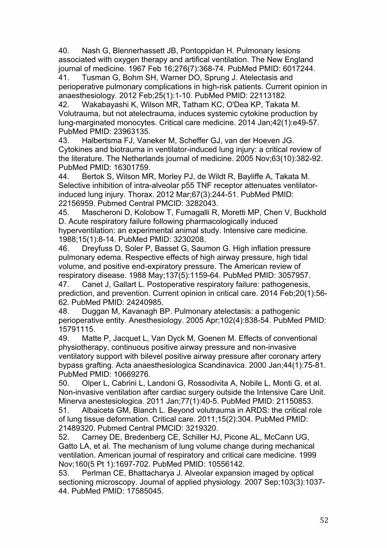
52
40. Nash G, Blennerhassett JB, Pontoppidan H. Pulmonary lesions associated with oxygen therapy and artifical ventilation. The New England journal of medicine. 1967 Feb 16;276(7):368-74. PubMed PMID: 6017244. 41. Tusman G, Bohm SH, Warner DO, Sprung J. Atelectasis and perioperative pulmonary complications in high-risk patients. Current opinion in anaesthesiology. 2012 Feb;25(1):1-10. PubMed PMID: 22113182. 42. Wakabayashi K, Wilson MR, Tatham KC, O'Dea KP, Takata M. Volutrauma, but not atelectrauma, induces systemic cytokine production by lung-marginated monocytes. Critical care medicine. 2014 Jan;42(1):e49-57. PubMed PMID: 23963135. 43. Halbertsma FJ, Vaneker M, Scheffer GJ, van der Hoeven JG. Cytokines and biotrauma in ventilator-induced lung injury: a critical review of the literature. The Netherlands journal of medicine. 2005 Nov;63(10):382-92. PubMed PMID: 16301759. 44. Bertok S, Wilson MR, Morley PJ, de Wildt R, Bayliffe A, Takata M. Selective inhibition of intra-alveolar p55 TNF receptor attenuates ventilator-induced lung injury. Thorax. 2012 Mar;67(3):244-51. PubMed PMID: 22156959. Pubmed Central PMCID: 3282043. 45. Mascheroni D, Kolobow T, Fumagalli R, Moretti MP, Chen V, Buckhold D. Acute respiratory failure following pharmacologically induced hyperventilation: an experimental animal study. Intensive care medicine. 1988;15(1):8-14. PubMed PMID: 3230208. 46. Dreyfuss D, Soler P, Basset G, Saumon G. High inflation pressure pulmonary edema. Respective effects of high airway pressure, high tidal volume, and positive end-expiratory pressure. The American review of respiratory disease. 1988 May;137(5):1159-64. PubMed PMID: 3057957. 47. Canet J, Gallart L. Postoperative respiratory failure: pathogenesis, prediction, and prevention. Current opinion in critical care. 2014 Feb;20(1):56-62. PubMed PMID: 24240985. 48. Duggan M, Kavanagh BP. Pulmonary atelectasis: a pathogenic perioperative entity. Anesthesiology. 2005 Apr;102(4):838-54. PubMed PMID: 15791115. 49. Matte P, Jacquet L, Van Dyck M, Goenen M. Effects of conventional physiotherapy, continuous positive airway pressure and non-invasive ventilatory support with bilevel positive airway pressure after coronary artery bypass grafting. Acta anaesthesiologica Scandinavica. 2000 Jan;44(1):75-81. PubMed PMID: 10669276. 50. Olper L, Cabrini L, Landoni G, Rossodivita A, Nobile L, Monti G, et al. Non-invasive ventilation after cardiac surgery outside the Intensive Care Unit. Minerva anestesiologica. 2011 Jan;77(1):40-5. PubMed PMID: 21150853. 51. Albaiceta GM, Blanch L. Beyond volutrauma in ARDS: the critical role of lung tissue deformation. Critical care. 2011;15(2):304. PubMed PMID: 21489320. Pubmed Central PMCID: 3219320. 52. Carney DE, Bredenberg CE, Schiller HJ, Picone AL, McCann UG, Gatto LA, et al. The mechanism of lung volume change during mechanical ventilation. American journal of respiratory and critical care medicine. 1999 Nov;160(5 Pt 1):1697-702. PubMed PMID: 10556142. 53. Perlman CE, Bhattacharya J. Alveolar expansion imaged by optical sectioning microscopy. Journal of applied physiology. 2007 Sep;103(3):1037-44. PubMed PMID: 17585045.

53
54. Bilek AM, Dee KC, Gaver DP, 3rd. Mechanisms of surface-tension-induced epithelial cell damage in a model of pulmonary airway reopening. Journal of applied physiology. 2003 Feb;94(2):770-83. PubMed PMID: 12433851. 55. Mead J, Takishima T, Leith D. Stress distribution in lungs: a model of pulmonary elasticity. J Appl Physiol. 1970 May;28(5):596-608. PubMed PMID: 5442255. 56. Ranieri VM, Suter PM, Tortorella C, De Tullio R, Dayer JM, Brienza A, et al. Effect of mechanical ventilation on inflammatory mediators in patients with acute respiratory distress syndrome: a randomized controlled trial. JAMA : the journal of the American Medical Association. 1999 Jul 7;282(1):54-61. PubMed PMID: 10404912. 57. Vlahakis NE, Hubmayr RD. Cellular stress failure in ventilator-injured lungs. American journal of respiratory and critical care medicine. 2005 Jun 15;171(12):1328-42. PubMed PMID: 15695492. Pubmed Central PMCID: 2718477. 58. Ridge KM, Linz L, Flitney FW, Kuczmarski ER, Chou YH, Omary MB, et al. Keratin 8 phosphorylation by protein kinase C delta regulates shear stress-mediated disassembly of keratin intermediate filaments in alveolar epithelial cells. The Journal of biological chemistry. 2005 Aug 26;280(34):30400-5. PubMed PMID: 15972820. 59. Ledowski T, Paech MJ, Patel B, Schug SA. Bronchial mucus transport velocity in patients receiving propofol and remifentanil versus sevoflurane and remifentanil anesthesia. Anesthesia and analgesia. 2006 May;102(5):1427-30. PubMed PMID: 16632821. 60. Zhang T, Fan Y, Liu K, Wang Y. Effects of different general anaesthetic techniques on immune responses in patients undergoing surgery for tongue cancer. Anaesthesia and intensive care. 2014 Mar;42(2):220-7. PubMed PMID: 24580388. 61. Cahalan MK, Weiskopf RB, Eger EI, 2nd, Yasuda N, Ionescu P, Rampil IJ, et al. Hemodynamic effects of desflurane/nitrous oxide anesthesia in volunteers. Anesthesia and analgesia. 1991 Aug;73(2):157-64. PubMed PMID: 1854030. 62. Browner WS, Li J, Mangano DT. In-hospital and long-term mortality in male veterans following noncardiac surgery. The Study of Perioperative Ischemia Research Group. JAMA : the journal of the American Medical Association. 1992 Jul 8;268(2):228-32. PubMed PMID: 1608142. 63. Moje C, Jackson TJ, McNair P. Adverse events in Victorian admissions for elective surgery. Australian health review : a publication of the Australian Hospital Association. 2006 Aug;30(3):333-43. PubMed PMID: 16879092. 64. Scott IA, Lodge RS, Russell DM. Evidence-based guide to perioperative medicine. Internal medicine journal. 2007 Jun;37(6):389-401. PubMed PMID: 17535383. 65. Haeck PC, Swanson JA, Iverson RE, Lynch DJ, Committee APS. Evidence-based patient safety advisory: patient assessment and prevention of pulmonary side effects in surgery. Part 2. Patient and procedural risk factors. Plastic and reconstructive surgery. 2009 Oct;124(4 Suppl):57S-67S. PubMed PMID: 20827240.

54
66. Johnson DC, Kaplan LJ. Perioperative pulmonary complications. Current opinion in critical care. 2011 Aug;17(4):362-9. PubMed PMID: 21734490. 67. Warner DO, Warner MA, Barnes RD, Offord KP, Schroeder DR, Gray DT, et al. Perioperative respiratory complications in patients with asthma. Anesthesiology. 1996 Sep;85(3):460-7. PubMed PMID: 8853074. 68. Garcia-Delgado M, Navarrete-Sanchez I, Colmenero M. Preventing and managing perioperative pulmonary complications following cardiac surgery. Current opinion in anaesthesiology. 2014 Apr;27(2):146-52. PubMed PMID: 24514031. 69. Plaud B, Debaene B, Donati F, Marty J. Residual paralysis after emergence from anesthesia. Anesthesiology. 2010 Apr;112(4):1013-22. PubMed PMID: 20234315. 70. Brandt S, Regueira T, Bracht H, Porta F, Djafarzadeh S, Takala J, et al. Effect of fluid resuscitation on mortality and organ function in experimental sepsis models. Critical care. 2009;13(6):R186. PubMed PMID: 19930656. Pubmed Central PMCID: 2811934. 71. Babayan RK. Wound healing and infection in surgery: the pathophysiological impact of smoking, smoking cessation, and nicotine replacement therapy: a systematic review. The Journal of urology. 2012 Dec;188(6):2243-4. PubMed PMID: 23141233. 72. Hawn MT, Houston TK, Campagna EJ, Graham LA, Singh J, Bishop M, et al. The attributable risk of smoking on surgical complications. Annals of surgery. 2011 Dec;254(6):914-20. PubMed PMID: 21869677. 73. Lee SM, Landry J, Jones PM, Buhrmann O, Morley-Forster P. The effectiveness of a perioperative smoking cessation program: a randomized clinical trial. Anesthesia and analgesia. 2013 Sep;117(3):605-13. PubMed PMID: 23868890. 74. Sorensen LT. Wound healing and infection in surgery: the pathophysiological impact of smoking, smoking cessation, and nicotine replacement therapy: a systematic review. Annals of surgery. 2012 Jun;255(6):1069-79. PubMed PMID: 22566015. 75. Turan A, Mascha EJ, Roberman D, Turner PL, You J, Kurz A, et al. Smoking and perioperative outcomes. Anesthesiology. 2011 Apr;114(4):837-46. PubMed PMID: 21372682. 76. Myles PS, Iacono GA, Hunt JO, Fletcher H, Morris J, McIlroy D, et al. Risk of respiratory complications and wound infection in patients undergoing ambulatory surgery: smokers versus nonsmokers. Anesthesiology. 2002 Oct;97(4):842-7. PubMed PMID: 12357149. 77. Sorensen LT. Wound healing and infection in surgery. The clinical impact of smoking and smoking cessation: a systematic review and meta-analysis. Archives of surgery. 2012 Apr;147(4):373-83. PubMed PMID: 22508785. 78. Sorensen LT, Karlsmark T, Gottrup F. Abstinence from smoking reduces incisional wound infection: a randomized controlled trial. Annals of surgery. 2003 Jul;238(1):1-5. PubMed PMID: 12832959. Pubmed Central PMCID: 1422652. 79. Thomsen T, Villebro N, Moller AM. Interventions for preoperative smoking cessation. The Cochrane database of systematic reviews. 2010 (7):CD002294. PubMed PMID: 20614429.

55
80. Sorensen LT, Jorgensen T. Short-term pre-operative smoking cessation intervention does not affect postoperative complications in colorectal surgery: a randomized clinical trial. Colorectal disease : the official journal of the Association of Coloproctology of Great Britain and Ireland. 2003 Jul;5(4):347-52. PubMed PMID: 12814414. 81. Moller AM, Villebro N, Pedersen T, Tonnesen H. Effect of preoperative smoking intervention on postoperative complications: a randomised clinical trial. Lancet. 2002 Jan 12;359(9301):114-7. PubMed PMID: 11809253. 82. Myles PS, Leslie K, Angliss M, Mezzavia P, Lee L. Effectiveness of bupropion as an aid to stopping smoking before elective surgery: a randomised controlled trial. Anaesthesia. 2004 Nov;59(11):1053-8. PubMed PMID: 15479310. 83. Warner DO. Perioperative abstinence from cigarettes: physiologic and clinical consequences. Anesthesiology. 2006 Feb;104(2):356-67. PubMed PMID: 16436857. 84. Rietbrock N, Kunkel S, Worner W, Eyer P. Oxygen-dissociation kinetics in the blood of smokers and non-smokers: interaction between oxygen and carbon monoxide at the hemoglobin molecule. Naunyn-Schmiedeberg's archives of pharmacology. 1992 Jan;345(1):123-8. PubMed PMID: 1538790. 85. Klein LW, Ambrose J, Pichard A, Holt J, Gorlin R, Teichholz LE. Acute coronary hemodynamic response to cigarette smoking in patients with coronary artery disease. Journal of the American College of Cardiology. 1984 Apr;3(4):879-86. PubMed PMID: 6707354. 86. Narkiewicz K, van de Borne PJ, Hausberg M, Cooley RL, Winniford MD, Davison DE, et al. Cigarette smoking increases sympathetic outflow in humans. Circulation. 1998 Aug 11;98(6):528-34. PubMed PMID: 9714109. 87. Saetta M, Turato G, Baraldo S, Zanin A, Braccioni F, Mapp CE, et al. Goblet cell hyperplasia and epithelial inflammation in peripheral airways of smokers with both symptoms of chronic bronchitis and chronic airflow limitation. American journal of respiratory and critical care medicine. 2000 Mar;161(3 Pt 1):1016-21. PubMed PMID: 10712357. 88. Maestrelli P, Saetta M, Mapp CE, Fabbri LM. Remodeling in response to infection and injury. Airway inflammation and hypersecretion of mucus in smoking subjects with chronic obstructive pulmonary disease. American journal of respiratory and critical care medicine. 2001 Nov 15;164(10 Pt 2):S76-80. PubMed PMID: 11734472. 89. Di Stefano A, Capelli A, Lusuardi M, Balbo P, Vecchio C, Maestrelli P, et al. Severity of airflow limitation is associated with severity of airway inflammation in smokers. American journal of respiratory and critical care medicine. 1998 Oct;158(4):1277-85. PubMed PMID: 9769292. 90. Lee LY, Hong JL. Involvement of prostanoids in cigarette smoking-induced pathophysiological effects in the lung. Prostaglandins, leukotrienes, and essential fatty acids. 1999 Sep;61(3):145-55. PubMed PMID: 10582654. 91. Rodgers A, Walker N, Schug S, McKee A, Kehlet H, van Zundert A, et al. Reduction of postoperative mortality and morbidity with epidural or spinal anaesthesia: results from overview of randomised trials. Bmj. 2000 Dec 16;321(7275):1493. PubMed PMID: 11118174. Pubmed Central PMCID: 27550.
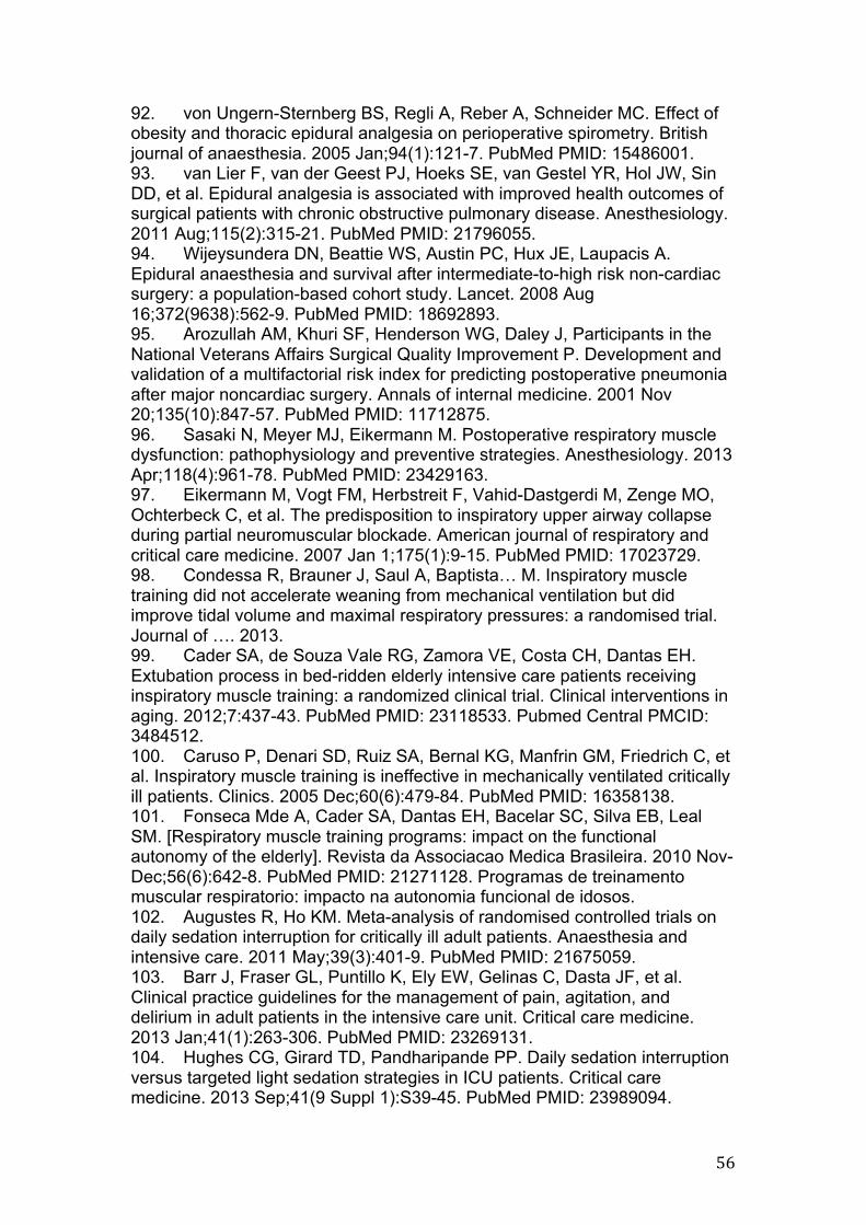
56
92. von Ungern-Sternberg BS, Regli A, Reber A, Schneider MC. Effect of obesity and thoracic epidural analgesia on perioperative spirometry. British journal of anaesthesia. 2005 Jan;94(1):121-7. PubMed PMID: 15486001. 93. van Lier F, van der Geest PJ, Hoeks SE, van Gestel YR, Hol JW, Sin DD, et al. Epidural analgesia is associated with improved health outcomes of surgical patients with chronic obstructive pulmonary disease. Anesthesiology. 2011 Aug;115(2):315-21. PubMed PMID: 21796055. 94. Wijeysundera DN, Beattie WS, Austin PC, Hux JE, Laupacis A. Epidural anaesthesia and survival after intermediate-to-high risk non-cardiac surgery: a population-based cohort study. Lancet. 2008 Aug 16;372(9638):562-9. PubMed PMID: 18692893. 95. Arozullah AM, Khuri SF, Henderson WG, Daley J, Participants in the National Veterans Affairs Surgical Quality Improvement P. Development and validation of a multifactorial risk index for predicting postoperative pneumonia after major noncardiac surgery. Annals of internal medicine. 2001 Nov 20;135(10):847-57. PubMed PMID: 11712875. 96. Sasaki N, Meyer MJ, Eikermann M. Postoperative respiratory muscle dysfunction: pathophysiology and preventive strategies. Anesthesiology. 2013 Apr;118(4):961-78. PubMed PMID: 23429163. 97. Eikermann M, Vogt FM, Herbstreit F, Vahid-Dastgerdi M, Zenge MO, Ochterbeck C, et al. The predisposition to inspiratory upper airway collapse during partial neuromuscular blockade. American journal of respiratory and critical care medicine. 2007 Jan 1;175(1):9-15. PubMed PMID: 17023729. 98. Condessa R, Brauner J, Saul A, Baptista… M. Inspiratory muscle training did not accelerate weaning from mechanical ventilation but did improve tidal volume and maximal respiratory pressures: a randomised trial. Journal of …. 2013. 99. Cader SA, de Souza Vale RG, Zamora VE, Costa CH, Dantas EH. Extubation process in bed-ridden elderly intensive care patients receiving inspiratory muscle training: a randomized clinical trial. Clinical interventions in aging. 2012;7:437-43. PubMed PMID: 23118533. Pubmed Central PMCID: 3484512. 100. Caruso P, Denari SD, Ruiz SA, Bernal KG, Manfrin GM, Friedrich C, et al. Inspiratory muscle training is ineffective in mechanically ventilated critically ill patients. Clinics. 2005 Dec;60(6):479-84. PubMed PMID: 16358138. 101. Fonseca Mde A, Cader SA, Dantas EH, Bacelar SC, Silva EB, Leal SM. [Respiratory muscle training programs: impact on the functional autonomy of the elderly]. Revista da Associacao Medica Brasileira. 2010 Nov-Dec;56(6):642-8. PubMed PMID: 21271128. Programas de treinamento muscular respiratorio: impacto na autonomia funcional de idosos. 102. Augustes R, Ho KM. Meta-analysis of randomised controlled trials on daily sedation interruption for critically ill adult patients. Anaesthesia and intensive care. 2011 May;39(3):401-9. PubMed PMID: 21675059. 103. Barr J, Fraser GL, Puntillo K, Ely EW, Gelinas C, Dasta JF, et al. Clinical practice guidelines for the management of pain, agitation, and delirium in adult patients in the intensive care unit. Critical care medicine. 2013 Jan;41(1):263-306. PubMed PMID: 23269131. 104. Hughes CG, Girard TD, Pandharipande PP. Daily sedation interruption versus targeted light sedation strategies in ICU patients. Critical care medicine. 2013 Sep;41(9 Suppl 1):S39-45. PubMed PMID: 23989094.

57
105. Mehta S, Burry L, Martinez-Motta JC, Stewart TE, Hallett D, McDonald E, et al. A randomized trial of daily awakening in critically ill patients managed with a sedation protocol: a pilot trial. Critical care medicine. 2008 Jul;36(7):2092-9. PubMed PMID: 18552687. 106. Shehabi Y, Bellomo R, Reade MC, Bailey M, Bass F, Howe B, et al. Early intensive care sedation predicts long-term mortality in ventilated critically ill patients. American journal of respiratory and critical care medicine. 2012 Oct 15;186(8):724-31. PubMed PMID: 22859526. 107. Fraser GL, Devlin JW, Worby CP, Alhazzani W, Barr J, Dasta JF, et al. Benzodiazepine versus nonbenzodiazepine-based sedation for mechanically ventilated, critically ill adults: a systematic review and meta-analysis of randomized trials. Critical care medicine. 2013 Sep;41(9 Suppl 1):S30-8. PubMed PMID: 23989093. 108. Strom T, Martinussen T, Toft P. A protocol of no sedation for critically ill patients receiving mechanical ventilation: a randomised trial. Lancet. 2010 Feb 6;375(9713):475-80. PubMed PMID: 20116842. 109. Payen JF, Chanques G, Mantz J, Hercule C, Auriant I, Leguillou JL, et al. Current practices in sedation and analgesia for mechanically ventilated critically ill patients: a prospective multicenter patient-based study. Anesthesiology. 2007 Apr;106(4):687-95; quiz 891-2. PubMed PMID: 17413906. 110. Girard TD, Kress JP, Fuchs BD, Thomason JW, Schweickert WD, Pun BT, et al. Efficacy and safety of a paired sedation and ventilator weaning protocol for mechanically ventilated patients in intensive care (Awakening and Breathing Controlled trial): a randomised controlled trial. Lancet. 2008 Jan 12;371(9607):126-34. PubMed PMID: 18191684. 111. Bennett-Guerrero E, Panah MH, Barclay GR, Bodian CA, Winfree WJ, Andres LA, et al. Decreased endotoxin immunity is associated with greater mortality and/or prolonged hospitalization after surgery. Anesthesiology. 2001 Jun;94(6):992-8. PubMed PMID: 11465625. 112. Braga M, Gianotti L, Vignali A, Carlo V. Preoperative oral arginine and n-3 fatty acid supplementation improves the immunometabolic host response and outcome after colorectal resection for cancer. Surgery. 2002;132(5):805-14. 113. Zheng Y, Li F, Qi B, Luo B, Sun H, Liu S, et al. Application of perioperative immunonutrition for gastrointestinal surgery: a meta-analysis of randomized controlled trials. Asia Pacific journal of clinical nutrition. 2007;16 Suppl 1:253-7. 114. Gianotti L, Braga M, Fortis C, Soldini L, Vignali A, Colombo S, et al. A prospective, randomized clinical trial on perioperative feeding with an arginine-, omega-3 fatty acid-, and RNA-enriched enteral diet: effect on host response and nutritional status. JPEN Journal of parenteral and enteral nutrition. 1999 Nov-Dec;23(6):314-20. PubMed PMID: 10574478. 115. Giger U, Buchler M, Farhadi J, Berger D, Husler J, Schneider H, et al. Preoperative immunonutrition suppresses perioperative inflammatory response in patients with major abdominal surgery-a randomized controlled pilot study. Annals of surgical oncology. 2007 Oct;14(10):2798-806. PubMed PMID: 17632760.
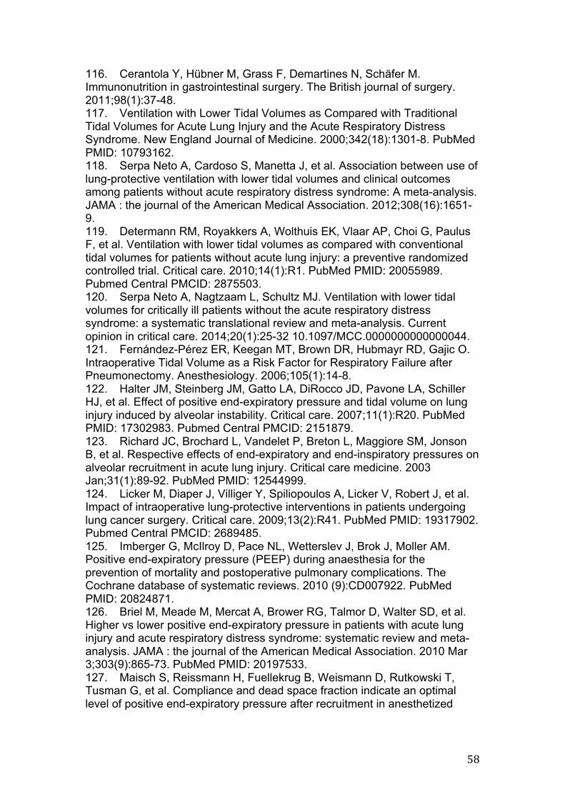
58
116. Cerantola Y, Hübner M, Grass F, Demartines N, Schäfer M. Immunonutrition in gastrointestinal surgery. The British journal of surgery. 2011;98(1):37-48. 117. Ventilation with Lower Tidal Volumes as Compared with Traditional Tidal Volumes for Acute Lung Injury and the Acute Respiratory Distress Syndrome. New England Journal of Medicine. 2000;342(18):1301-8. PubMed PMID: 10793162. 118. Serpa Neto A, Cardoso S, Manetta J, et al. Association between use of lung-protective ventilation with lower tidal volumes and clinical outcomes among patients without acute respiratory distress syndrome: A meta-analysis. JAMA : the journal of the American Medical Association. 2012;308(16):1651-9. 119. Determann RM, Royakkers A, Wolthuis EK, Vlaar AP, Choi G, Paulus F, et al. Ventilation with lower tidal volumes as compared with conventional tidal volumes for patients without acute lung injury: a preventive randomized controlled trial. Critical care. 2010;14(1):R1. PubMed PMID: 20055989. Pubmed Central PMCID: 2875503. 120. Serpa Neto A, Nagtzaam L, Schultz MJ. Ventilation with lower tidal volumes for critically ill patients without the acute respiratory distress syndrome: a systematic translational review and meta-analysis. Current opinion in critical care. 2014;20(1):25-32 10.1097/MCC.0000000000000044. 121. Fernández-Pérez ER, Keegan MT, Brown DR, Hubmayr RD, Gajic O. Intraoperative Tidal Volume as a Risk Factor for Respiratory Failure after Pneumonectomy. Anesthesiology. 2006;105(1):14-8. 122. Halter JM, Steinberg JM, Gatto LA, DiRocco JD, Pavone LA, Schiller HJ, et al. Effect of positive end-expiratory pressure and tidal volume on lung injury induced by alveolar instability. Critical care. 2007;11(1):R20. PubMed PMID: 17302983. Pubmed Central PMCID: 2151879. 123. Richard JC, Brochard L, Vandelet P, Breton L, Maggiore SM, Jonson B, et al. Respective effects of end-expiratory and end-inspiratory pressures on alveolar recruitment in acute lung injury. Critical care medicine. 2003 Jan;31(1):89-92. PubMed PMID: 12544999. 124. Licker M, Diaper J, Villiger Y, Spiliopoulos A, Licker V, Robert J, et al. Impact of intraoperative lung-protective interventions in patients undergoing lung cancer surgery. Critical care. 2009;13(2):R41. PubMed PMID: 19317902. Pubmed Central PMCID: 2689485. 125. Imberger G, McIlroy D, Pace NL, Wetterslev J, Brok J, Moller AM. Positive end-expiratory pressure (PEEP) during anaesthesia for the prevention of mortality and postoperative pulmonary complications. The Cochrane database of systematic reviews. 2010 (9):CD007922. PubMed PMID: 20824871. 126. Briel M, Meade M, Mercat A, Brower RG, Talmor D, Walter SD, et al. Higher vs lower positive end-expiratory pressure in patients with acute lung injury and acute respiratory distress syndrome: systematic review and meta-analysis. JAMA : the journal of the American Medical Association. 2010 Mar 3;303(9):865-73. PubMed PMID: 20197533. 127. Maisch S, Reissmann H, Fuellekrug B, Weismann D, Rutkowski T, Tusman G, et al. Compliance and dead space fraction indicate an optimal level of positive end-expiratory pressure after recruitment in anesthetized

59
patients. Anesthesia and analgesia. 2008 Jan;106(1):175-81, table of contents. PubMed PMID: 18165575. 128. Tusman G, Groisman I, Fiolo FE, Scandurra A, Arca JM, Krumrick G, et al. Noninvasive monitoring of lung recruitment maneuvers in morbidly obese patients: the role of pulse oximetry and volumetric capnography. Anesthesia and analgesia. 2014 Jan;118(1):137-44. PubMed PMID: 24356163. 129. Ferrando C, Mugarra A, Gutierrez A, Carbonell JA, Garcia M, Soro M, et al. Setting individualized positive end-expiratory pressure level with a positive end-expiratory pressure decrement trial after a recruitment maneuver improves oxygenation and lung mechanics during one-lung ventilation. Anesthesia and analgesia. 2014 Mar;118(3):657-65. PubMed PMID: 24557111. 130. Slinger PD, Kruger M, McRae K, Winton T. Relation of the static compliance curve and positive end-expiratory pressure to oxygenation during one-lung ventilation. Anesthesiology. 2001 Nov;95(5):1096-102. PubMed PMID: 11684977. 131. Silva PL, Moraes L, Santos RS, Samary C, Ramos MB, Santos CL, et al. Recruitment maneuvers modulate epithelial and endothelial cell response according to acute lung injury etiology. Critical care medicine. 2013 Oct;41(10):e256-65. PubMed PMID: 23887231. 132. Zannin E, Dellaca RL, Kostic P, Pompilio PP, Larsson A, Pedotti A, et al. Optimizing positive end-expiratory pressure by oscillatory mechanics minimizes tidal recruitment and distension: an experimental study in a lavage model of lung injury. Critical care. 2012 Nov 7;16(6):R217. PubMed PMID: 23134702. Pubmed Central PMCID: 3672594. 133. Soummer A, Perbet S, Brisson H, Arbelot C, Constantin JM, Lu Q, et al. Ultrasound assessment of lung aeration loss during a successful weaning trial predicts postextubation distress*. Critical care medicine. 2012 Jul;40(7):2064-72. PubMed PMID: 22584759. 134. Piedalue F, Albert RK. Prone positioning in acute respiratory distress syndrome. Respiratory care clinics of North America. 2003 Dec;9(4):495-509. PubMed PMID: 14984068. 135. Guerin C, Reignier J, Richard JC, Beuret P, Gacouin A, Boulain T, et al. Prone positioning in severe acute respiratory distress syndrome. The New England journal of medicine. 2013 Jun 6;368(23):2159-68. PubMed PMID: 23688302. 136. Guerin C. Prone position. Current opinion in critical care. 2014 Feb;20(1):92-7. PubMed PMID: 24366167. 137. Cornejo RA, Diaz JC, Tobar EA, Bruhn AR, Ramos CA, Gonzalez RA, et al. Effects of prone positioning on lung protection in patients with acute respiratory distress syndrome. American journal of respiratory and critical care medicine. 2013 Aug 15;188(4):440-8. PubMed PMID: 23348974. 138. Pittet D. Improving compliance with hand hygiene in hospitals. Infection control and hospital epidemiology : the official journal of the Society of Hospital Epidemiologists of America. 2000 Jun;21(6):381-6. PubMed PMID: 10879568. 139. Li J, Xie D, Li A, Yue J. Oral topical decontamination for preventing ventilator-associated pneumonia: a systematic review and meta-analysis of

60
randomized controlled trials. The Journal of hospital infection. 2013 Aug;84(4):283-93. PubMed PMID: 23846238. 140. Gramlich L, Kichian K, Pinilla J, Rodych NJ, Dhaliwal R, Heyland DK. Does enteral nutrition compared to parenteral nutrition result in better outcomes in critically ill adult patients? A systematic review of the literature. Nutrition. 2004 Oct;20(10):843-8. PubMed PMID: 15474870. 141. Braunschweig CL, Levy P, Sheean PM, Wang X. Enteral compared with parenteral nutrition: a meta-analysis. The American journal of clinical nutrition. 2001 Oct;74(4):534-42. PubMed PMID: 11566654. 142. Chung CK, Whitney R, Thompson CM, Pham TN, Maier RV, O'Keefe GE. Experience with an enteral-based nutritional support regimen in critically ill trauma patients. Journal of the American College of Surgeons. 2013 Dec;217(6):1108-17. PubMed PMID: 24051065. Pubmed Central PMCID: 3845006. 143. Casaer MP, Mesotten D, Hermans G, Wouters PJ, Schetz M, Meyfroidt G, et al. Early versus late parenteral nutrition in critically ill adults. The New England journal of medicine. 2011 Aug 11;365(6):506-17. PubMed PMID: 21714640. 144. Sena MJ, Utter GH, Cuschieri J, Maier RV, Tompkins RG, Harbrecht BG, et al. Early supplemental parenteral nutrition is associated with increased infectious complications in critically ill trauma patients. Journal of the American College of Surgeons. 2008 Oct;207(4):459-67. PubMed PMID: 18926446. 145. Caserta RA, Marra AR, Durao MS, Silva CV, Pavao dos Santos OF, Neves HS, et al. A program for sustained improvement in preventing ventilator associated pneumonia in an intensive care setting. BMC infectious diseases. 2012;12:234. PubMed PMID: 23020101. Pubmed Central PMCID: 3521195. 146. Muscedere J, Rewa O, McKechnie K, Jiang X, Laporta D, Heyland DK. Subglottic secretion drainage for the prevention of ventilator-associated pneumonia: a systematic review and meta-analysis. Critical care medicine. 2011 Aug;39(8):1985-91. PubMed PMID: 21478738.