common childhood infections -...
TRANSCRIPT
Common Childhood InfectionsT Avenant

Outline
� Definitions
� Septicaemia and shock
� Rash and fever
◦ Infectious
◦ Non-infectious
◦ Rash – no fever (Lecture 3 today))
� Other childhood infections

Definitions
� Hemorrhage◦ Rupture of blood vessel
� Hematoma◦ Blood trapped in tissue
� Petechiae◦ Minute hemorrhages into the skin (1-3mm)
� Purpura◦ Slightly larger, groups of adjoining petechiae
� Ecchymosis◦ Large(>1-2cm) subcutanuoushematoma� e.g. common bruise
online.epocrates.com

Vasculitis
� Vascular inflammatory injury often with necrosis of blood vessels
� Most common mechanisms are
◦ injury by infectious pathogens
◦ immune –mediated inflammation
� Other
◦ Physical
◦ Chemical
◦ Toxins

Bleeding disorders
� Vessel wall◦ Infections
� Meningococcemia, septicaemia, measles, rickettsiosis (damage to microvasculature/DIC)
◦ Drug reactions
� Deposition of immune complexes
◦ Abnormal vessel walls
� Thrombocytopenia
� Defective platelets
� Clotting factors
� Combinations

Urticaria and Angio-oedema
� Pathophysiology incompletely understood◦ Release of inflammatory mediators◦ Local vasodilatation◦ Exudation from post-capillary venules◦ Variable accumulation of mononuclear cells◦ Stimulation of local nerve endings cause itching or burning

Septicaemia

Introduction
� Bacteremia◦ recovery of bacteria in blood culture
� transient, no disease� serious extension of infection elsewhere
� Local infections usually concomitant or follow bacteremia◦ meningitis, osteomyelitis, endocarditis, epiglottitis etc.
� Instrumentation� No or very few symptoms� If bacteremia not cleared – systemic inflammatory response◦ can progress independently of original disease

Sepsis
� Systemic response to infection with bacteria, viruses, fungi, protozoa and rickettsiae
� One of the causes of systemic inflammatory response syndrome (SIRS)
� If not recognized and treated, may progress to ◦ severe sepsis
◦ septic shock
◦ multiple organ dysfunction
syndrome
◦ death
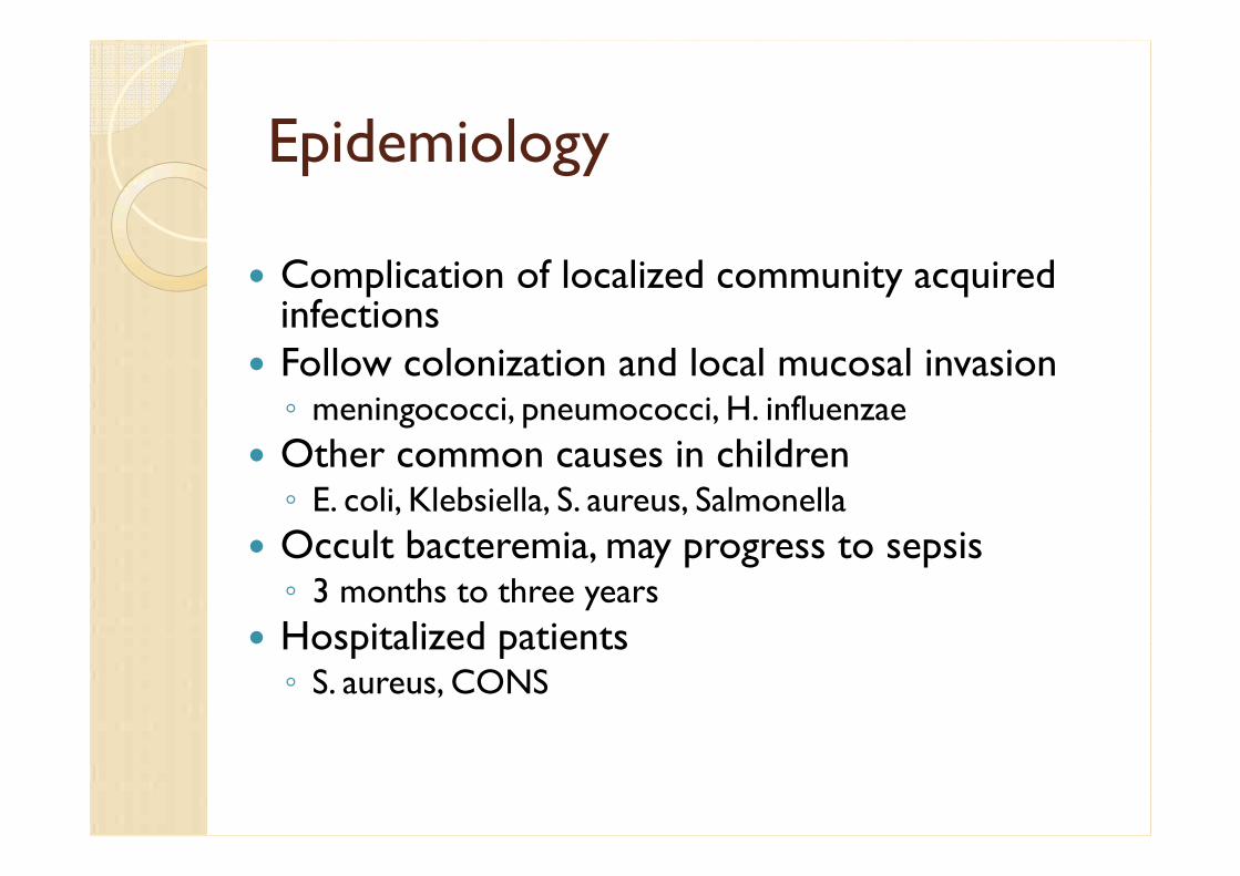
Epidemiology
� Complication of localized community acquired infections
� Follow colonization and local mucosal invasion◦ meningococci, pneumococci, H. influenzae
� Other common causes in children◦ E. coli, Klebsiella, S. aureus, Salmonella
� Occult bacteremia, may progress to sepsis ◦ 3 months to three years
� Hospitalized patients◦ S. aureus, CONS

Epidemiology
� Immunocompromised patients
◦ nosocomial infections
� gram negative, fungemia
� polymicrobial sepsis
� Unusual pathogens
◦ immunocompromised, travel
� Pseudobacteremia

Sepsis
MODS
Death

Pathogenesis
� Systemic inflammatory response syndrome results from
◦ tissue damage due to host response to bacterial products
◦ cardiopulmonary manifestations of gram negative sepsis mimicked by injection of
� TNF
� Endotoxin

Pathogenesis
� Shock
◦ disruption in circulatory function leading to poor perfusion and inadequate delivery of oxygen nutrients to tissues
� Not diagnosed by low blood pressure
◦ compensatory mechanisms maintain BP
� Low BP ominous sign

Pathogenesis
� Early phase◦ decrease systemic vascular resistance, decline in preload –tachycardia, increased cardiac output
� Endothelial damage, third space losses◦ warm, bounding pulses
◦ later cool extremities, poor perfusion
◦ lactic acidosis
� Pulmonary function impaired◦ development of ARDS poor prognosis
� Renal failure, hepatic failure, CNS dysfunction, DIC◦ alone
◦ part of MODS

Clinical Manifestations
� Primary signs and symptoms
◦ fever, chills, hyperventilation, tachycardia, hypothermia, cutaneous lesions, changes in mental status
� Secondary manifestations
◦ hypotension, cyanosis, gangrene, oliguria or anuria, jaundice, signs of heart failure
� Evidence of local infection
◦ meningitis, pneumonia, arthritis, cellulitis, pyelonephritis
� Immunocompromised status
◦ splenectomy, malignancy, HIV

Laboratory findings
� Blood cultures
� Stains
◦ blood
◦ skin lesions
� Metabolic acidosis
� Thrombocytopenia
� Abnormal clotting
� Fibrinogen
� Anemia
� Decreased PaO2 and PaCO2
� Neutrophils
◦ number and morphology
� CSF

Management
� Cultures and stains
◦ blood, urine, csf, exudates, abscesses, cutaneous lesions
� Blood count and platelets, PT and PTT, fibrinogen, ABG, CXR
� ICU
� Broad spectrum antibiotics
◦ community acquired
◦ nosocomial
◦ immunocompromised
◦ resistant S. pneumoniae

Management
� Oxygen
� Intubation and ventilation
� Circulation◦ Saline or Ringer solution 20ml/kg
◦ 5% albumin
� Sodium bicarbonate?
� Calcium and Potassium monitored
� Inotropics
� DIC◦ FFP

Management
� Modification host responses◦ IVIG, monoclonal antibodies against endotoxin, anti TNF-alpha, IL-1 receptor antagonists, granulocyte transfusions
� Corticosteroids◦ Not beneficial in adults with septic shock
◦ Useful� ARDS
� H. influenzae type b
� Adrenal hemorrhage
◦ SIRS in children, further research required

Prognosis
� Mortality for septic shock depends
◦ initial site of infection
◦ bacterial pathogen
◦ presence of MODS
◦ host immune response
� 40-60% mortality in gram negative enteric sepsis
� Meningococcal sepsis, poor prognostic signs◦ Hypotension
◦ Coma
◦ Leukopenia
◦ Thrombocytopenia
◦ Low fibrinogen level
◦ Absence of CSF pleocytosis with bacteria on gram stain
◦ Rapid appearance of petechiae
◦ Hypothermia

Prevention
� Immunization ◦ H. influenza type b◦ Streptococcus pneumoniae
� High risk patients◦ pneumococcal vaccine◦ meningococcal vaccine
� Penicillin prophylaxis ◦ splenic dysfunction, splenectomy
� Rifampicin prophylaxis for contacts◦ H. influenzae, meningococcal disease
� Immunocompromised◦ antibiotics, interferon, antivirals, isolation, etc.

Conclusion
� Septicaemia should be considered in any child with an acute, severe illness and pyrexia in whom no cause for the fever can be found
� If untreated, sepsis can lead to shock, multiple organ failure and death

Infectious Causes of Rash and Fever

Erythematous Rashes

Case study � NN, 10 months old
� Admitted
◦ Severe respiratory distress, fever and cough
� Previously healthy
� Clinical picture
◦ One week ago: URTI� Conjunctivitis, runny nose cough and fever
◦ Photophobic, red sore mouth, maculopapular rash� Started behind ears� Spread to trunk and limbs� Red becoming brown, scaling

Case study
◦ Cough
� Gradually worse
� Fever hasn’t subsided
� Progressively worsening respiratory distress and indrawing
◦ Very ill and not feeding
◦ Immunisations
� Last appointment forgotten
foxnews.com


Measles� Clinical features
� Prodrome (catarrhal phase)
� Fever
� Cough
� Coryza
� Conjunctivitis
� Kopliks
� Rash
� Erythematous, maculopapular
� Face – trunk – limbs
� Staining
� Desquamation
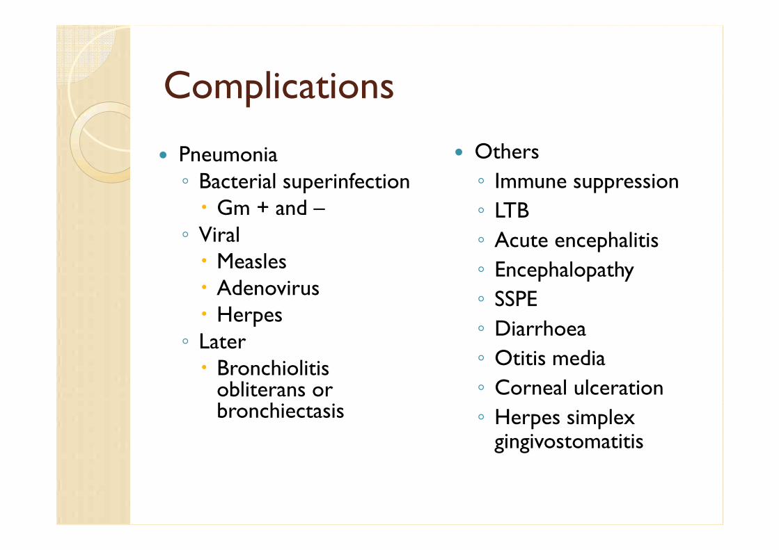
Complications
� Pneumonia◦ Bacterial superinfection� Gm + and –◦ Viral� Measles� Adenovirus� Herpes◦ Later� Bronchiolitisobliterans or bronchiectasis
� Others
◦ Immune suppression
◦ LTB
◦ Acute encephalitis
◦ Encephalopathy
◦ SSPE
◦ Diarrhoea
◦ Otitis media
◦ Corneal ulceration
◦ Herpes simplex gingivostomatitis

Rash and Fever*Viral
� Erythema infectiosum
◦ Parvovirus B19

Rash and Fever*Viral
� Roseola infantum
◦ Human Herpesvirus 6, 7

Rash and Fever*Viral
� Infectious Mononucleosis

Rash and Fever*Viral
� Infectious Mononucleosis

Rash and Fever*Viral
� German Measles

Rash and Fever*Viral
� Enterovirus
pigmcable.edu.ms

Rash and Fever*Bacterial
� Scarlet fever
◦ Group A Streptococcus

Rash and Fever*Bacterial
� Scarlet fever

Rash and Fever*Bacterial
� Scarlet fever
Rash and Fever*Bacterial
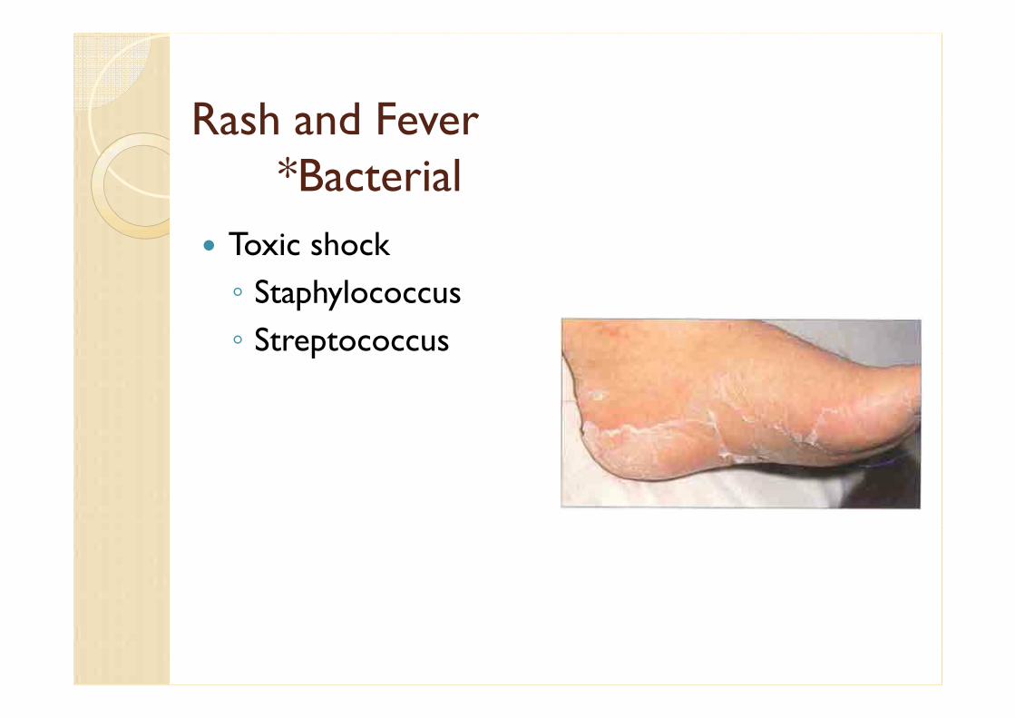
� Toxic shock
◦ Staphylococcus
◦ Streptococcus

Vesicular and Blistering Rashes

Rash and Fever*Viral
� Chicken-pox

Herpes simplex
� Primary infection between I and 5 years of age
� Infection by contaminated saliva
� Dissemination in immunosuppressed
� Clinical◦ Gingivostomatitis
� Fever salivation and refusal to eat
� Vesicles-rupture-shallow ulcers with red margin
� 4-9days� Local analgesia, tube feeds

Herpes simplex
◦ Meningo-encephalitis � High mortality and morbididty
◦ Conjunctivitis
◦ Recurrent disease� “fever blisters”
◦ Disseminated disease� Immunosuppressed
Dermatlas

Eczema herpeticum
� Infection of eczematous skin
� May have systemic reaction with fever
Dermatlas

Rash and Fever*Viral� Hand, foot and mouth disease
shanghaiist.com

Impetigo
� Staphylococci and streptococci
� Round confluent blisters
� Rupture and forms crusts
� Topical antibiotics
� If severe, systemic antibiotics

Petechial or Purpuric Rashes

Rash and Fever*Bacterial
� Meningococcal disease
� Ranges◦ Asymptomatic transient bacteraemia � Clears spontaneously
To
◦ Fulminant sepsis� Death in few hours

Meningococcaemia/Meningitis� Variable
� Early ◦ Signs of upper respiratory infection
◦ Fever, headache, lethargy, vomiting, myalgia, joint pain
� Typical◦ URTI, fever, haemorrhagic rash
◦ Circulatory collapse, purpura, shock

Meningococcaemia/Meningitis
� Skin
◦ Diffuse mottling to extensive purpuric lesions
◦ Petechiae in 50 – 60%
◦ Less than 7 petechiae in 12%
◦ No rash in 1-2
◦ Maculopapular rash in 13%in one study
◦ Purpura – not from petechiae but from thrombosis and haemorrhage

Meningitis
� Typical meningitis signs
� Complications
◦ Hydrocephalus, cranial nerve palsies, subdural effusion or empyema, cerebral oedema, cortical vein thrombosis, cerebral infarction
◦ Hearing loss in 5 – 10%

Laboratory Diagnosis
� Leukopaenia
� Thombocytopaenia
� Inappropriate ADH secretion
� Abnormal coagulation (DIC)
� Abnormal LFT

Laboratory Diagnosis
� Gold standard
◦ Culture� Blood-, CSF- or petechiae culture
� Rapid diagnosis
◦ Gram stain
� Antigen detection
◦ CSF, urine, serum� Cross reaction esp E coli
� PCR
◦ Sensitivity and specificity 91%
◦ Useful in partially treated meningitis
◦ Not available yet

Treatment
� PROMPT INITIATION OF ANTIBIOTIC THERAPY MAY BE LIFESAVING
� Empiric ◦ May need to take into account other causes of meningitis e.g. S. pneumoniae, H. influenzae
� Ceftriaxone/ Cefotaxime/ Penicillin� In penicillin allergy◦ Choramphenicol
� No conclusive advantage – use of steroids◦ Exception (WF)
� Eradicate carrier state if treated with Penicillin

Prevention
� Primary prevention◦ Vaccination
� Secondary prevention◦ Notify
◦ Chemoprophylaxis
◦ Vaccination

Chemoprophylaxis
Chance of infection
◦ House hold contacts and roommates
� 1000X rest of population
◦ Pre school contacts
� 50X
◦ Medical personnel not in close contact with oral secretions
� Similar to general population

Chemoprophylaxis
� Close Contacts◦ Household contacts◦ Other contacts� Week before onset of symptoms until 24 hours after appropriate antimicrobial therapy
� Within 3 feet of patient� At least 8 hours contact◦ Day care centre contacts◦ Significant contact with oral secretions� Kissing, sharing toothbrush◦ Medical personnel� Intensive contact with oral secretions

Antibiotics Used
� Rifampicin
◦ Suitable for all ages
◦ Easy to administer
◦ Efficacy of 90 – 95% eradication of nasopharyngeal carriage
◦ Disadvantages
� Teratogenic
� Decreases reliability of contraceptives
� Colours secretions and contact lenses

Antibiotics Used
� Ciprofloxacin◦ Single oral dose◦ Not for use in pregnancy or lactation
� Ceftriaxione◦ Single dose ◦ Only intramuscular route
� Azythromycin◦ Only studied in adults◦ 93% effective

Rash and Fever*Bacterial
� Tick bite fever

Non-infectious causes

Case study - Juvenile idiopathic arthritis� 10 year boy
� Intermittent fever and skin rash 6 months
◦ Evening or early morning, up to 39o
◦ Feeling unwell
◦ Skin rash
� Pale pink macules
� Trunk and proximal extremities
� In between attacks well
� Intermittent joint pains
� Hepatosplenomegalyotherwise well

Rash and Fever*Non infectious
� Kawasaki Disease

Rash and Fever*Non infectious
� Erythema nodosum

Rash and Fever*Non infectious
� Erythema multiforme

Case study
� 5 year old girl
� Rash on legs
� URTI and fever
◦ 1 week ago
� Bad abdominal pain
� Rash on legs
◦ Buttocks to ankles
◦ Prominent on back of legs
◦ Not painful/itchy
◦ Raised, do not blanch on pressure
� Diffuse abdominal pain
� Urine dipstick: blood

Henoch Schönlein Purpura
� Clinical features � Rash on legs
◦ Distribution
◦ Not painful or itching
◦ Raised
� URTI
� Fever
� Abdominal pain
� Nephritis

Henoch Schönlein Purpura
� Clinical features
◦ Arthritis
� Large joint
� Hepatosplenomegaly
� Lymphadenopathy
� Abdominal pain◦ Edema and damage to the vasculature of the GIT
◦ Intermittent
◦ Colicky
◦ Occult heme-positive stools in half of the patients
◦ Diarrhea
◦ Intussusception may occur

Rash – No fever
� Molluscumcontagiosum

Selected other childhood infections

Diphtheria
� Corynebacterium diphtheriae
� Rare� Clinical◦ Sore throat, fever, toxaemia◦ White to grey membrane in nose or oropharynx� Attempts to remove results in � bleeding
◦ Cervical lymphadenopathy and periadenitis (“bull neck”)◦ Myocarditis◦ Neuritis
� Palatal and pharyngeal� Ocular muscles� Intercostal� Peripheral nerves
CDC
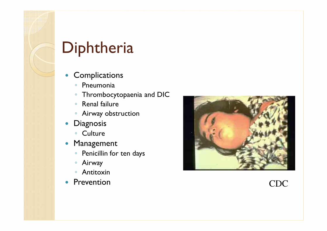
Diphtheria
� Complications◦ Pneumonia
◦ Thrombocytopaenia and DIC
◦ Renal failure
◦ Airway obstruction
� Diagnosis◦ Culture
� Management◦ Penicillin for ten days
◦ Airway
◦ Antitoxin
� Prevention CDC

Tetanus
� Clostridium tetani (Toxin)◦ Neonatal tetanus
◦ Wound contamination
� Clinical features◦ Muscle rigidity
◦ Muscle spasms
◦ Trismus (lock jaw)
◦ Facial muscle rigidity (risus sardonicus)
◦ Pharyngeal and laryngeal spasms
◦ Opisthotonus
◦ Alert and conscious
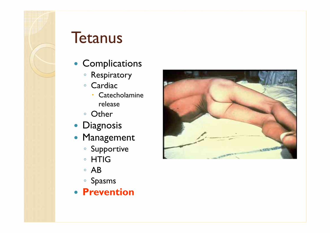
Tetanus
� Complications◦ Respiratory
◦ Cardiac� Catecholamine release
◦ Other
� Diagnosis� Management◦ Supportive
◦ HTIG
◦ AB
◦ Spasms
� Prevention
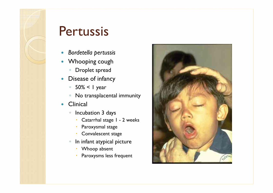
Pertussis
� Bordetella pertussis
� Whooping cough
◦ Droplet spread
� Disease of infancy
◦ 50% < 1 year
◦ No transplacental immunity
� Clinical
◦ Incubation 3 days� Catarrhal stage 1 - 2 weeks
� Paroxysmal stage
� Convalescent stage
◦ In infant atypical picture� Whoop absent
� Paroxysms less frequent

Pertussis� Diagnosis◦ Clinical
◦ Leucocytosis
◦ Culture and serology
◦ PCR
� Management◦ Hospitalize, Oxygen during spells
◦ Minimize stimuli
◦ Salbutamol
◦ Erythromycin� Eradicate organism, Prevent relapse
� Complications◦ Pneumonia, atelectasis, encephalopathy, subconjunctival
haemorrhage, epistaxis
� Prevention

Mumps
� Droplet infection� Infectivity◦ 6 days before symptoms to subsidence of swelling
� Clinical features◦ Incubation 14 – 21 days
◦ 30% sub-clinical
◦ Enlargement of parotid and other salivary glands
◦ Headache, malaise, anorexia
� Complications� Diagnosis, Treatment, Prevention
medscape.com

Poliomyelitis
� Eradication 2005
� Justification◦ There is no non-human reservoir
◦ There is no long-term carrier state
◦ The highly effective oral vaccine is cheap, available, and easy to administer
◦ Immunity is life-long, following either vaccination or natural infection
� No country can be certified free of wild poliomyelitis before it has met the minimum surveillance indicators.

Poliomyelitis
� AFP Case Definition
� Professional
◦ Any case of Acute Flaccid Paralysis including Guillian Barré syndrome, that is not caused by injury
◦ In a child less than 15 yrs of age
� Lay◦ Sudden weakness in the leg(s) and or arm(s), not caused by injury

Poliomyelitis
� Role of clinicians
◦ To notify all cases with sudden paralysis in children <15 years
◦ Investigate the case thoroughly
� By completing a case investigation form.
� By recording accurate address information, to facilitate tracing and follow-up.
� By ensuring that 2 stool specimens are collected and shipped frozen to NICD in JHB

The End