common abdominal emergencies in pediatrics … ‐schulmanetalradiology1994;193:771...
TRANSCRIPT

7/13/2015
1
Marta Hernanz-Schulman MD, FAAP, FACRProfessor, Radiology and Pediatrics
No Conflicts of Interest to DiscloseInfantile Hypertrophic Pyloric Stenosis (IHPS)
Malrotation
Intussusception
Appendicitis
+
Be able to accurately diagnose IHPS vs normal pylorus
Understand the role of US vs. UGI in the diagnosis of malrotation and volvulus
Understand the ultrasound diagnosis of intussusception
Differentiation of small vs. large bowel, unusual presentations
Understand the role of US in the evaluation and diagnosis of appendicitis in the
pediatric patient
At the conclusion of this discussion, attendees will:
2‐5 : 1000 births
Familiar to every pediatric radiologist
IHPS
500 visits per year
40 visits per month
4 visits per week
Glands are loosely packed and occupy about ½ the mucosal thickness
Histology for Pathologists: Stephen S. Sternberg
Glands are loosely packed and occupy about ½ the mucosal thickness
Histology for Pathologists: Stephen S. Sternberg
Glands are hyperplastic with multiple branches; deepening of crypts, inflammatory cellular infiltrate, edematous lamina propria
Kissane J. Pathology of infancy and childhood. Mosby 1975 Hernanz‐Schulman M. AJR 2001;177:843
IHPS: anatomy & definition
Thickened, UNRELAXING pyloric muscle
Thickening and edema of mucosaVirtually unknown prior to 1627
1627 Fabricius HildanusFirst reported case with survival
1788 Hezikiah Beardsley First reported case in North America
1799 Michael Underwood‐ postmortem description
1841 Thomas Williamson – postmortem description
1842 Siemon‐Dawosky – postmortem descriptionincludes “hypertrophy of submucous cellular tissue”
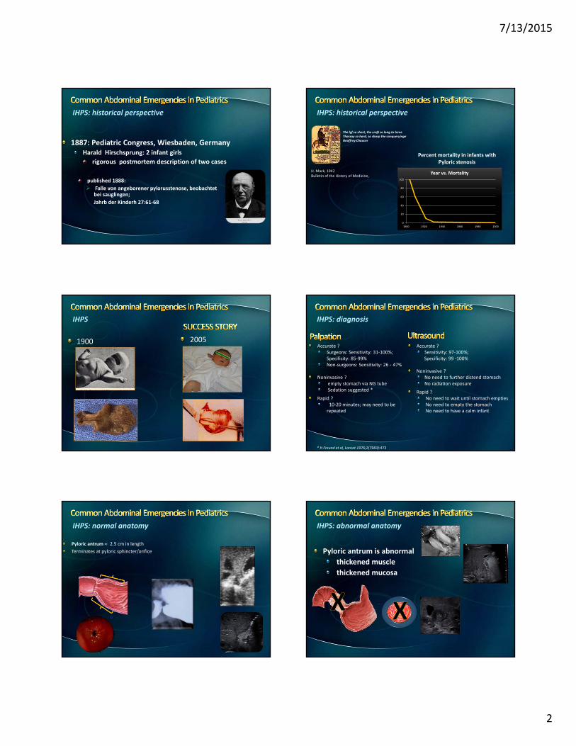
IHPS: historical perspective
7/13/2015
2
published 1888:
Falle von angeborener pylorusstenose, beobachtetbei sauglingen;
Jahrb der Kinderh 27:61‐68
1887: Pediatric Congress, Wiesbaden, GermanyHarald Hirschsprung: 2 infant girls
rigorous postmortem description of two cases
IHPS: historical perspective
The lyf so short, the craft so long to lerneThassay so hard, so sharp the conqueryingeGeoffrey Chaucer
H. Mack, 1942Bulletin of the History of Medicine,
Percent mortality in infants with Pyloric stenosis
0
20
40
60
80
100
1900 1920 1940 1960 1980 2000
Year vs. Mortality
IHPS: historical perspective
1900 2005
IHPS
Accurate ?Surgeons: Sensitivity: 31‐100%; Specificity: 85‐99%Non‐surgeons: Sensitivity: 26 - 47%
Noninvasive ?empty stomach via NG tubeSedation suggested *
Rapid ?10‐20 minutes; may need to be repeated
IHPS: diagnosis
Accurate ?Sensitivity: 97‐100%; Specificity: 99 ‐100%
Noninvasive ? No need to further distend stomachNo radiation exposure
Rapid ?No need to wait until stomach emptiesNo need to empty the stomachNo need to have a calm infant
* H Freund et al, Lancet 1976;2(7983):473
Pyloric antrum 2.5 cm in length
Terminates at pyloric sphincter/orifice
IHPS: normal anatomy
Pyloric antrum is abnormal
thickened muscle
thickened mucosa
IHPS: abnormal anatomy

7/13/2015
3
Warm room
Warm gel
Scan infant under blankets
Pacifier soaked in D5W
IHPS
6 ‐7 MHz long footprint linear transducer
Patient positioned to bring pylorus into viewbegin with patient supine
turn to right slowly if need to bring fluid to antrum
turn to left slowly if pylorus tucked behind distended stomach
IHPS
IHPS
Identify pylorusSweep caudally from GE junction anterior to aortic crus.
Identify arrowhead‐shaped duodenal cap
Landmarks: head of pancreas gallbladder
IHPS: ultrasound characteristics
Thickened muscle ≥ 3mm
Thickened mucosa
Usually hyperemic
Dimensions may change during study
IHPS is NOT a complete obstruction
UltrasoundMucosa fills pyloric channel
protrudes into antrum: “nipple sign”
IHPS: ultrasound characteristics
Mucosal hypertrophynipple sign
double track sign

7/13/2015
4
Pyloric stenosis: thickened muscle, mucosa
3.6mm; 10.7mm
Endoscopy
Mucosa protrudes through pyloric channel into antral lumen
“Typical endoscopic image of HPS” J pediatrgastroenterol nutr 18:1994“Typical endoscopic image of HPS” J pediatrgastroenterol nutr 18:1994
Failure to visualize the pylorusOverdistended stomach
Pylorus tucked behind stomach
Turn infant to the left, allowing pylorus to rise anteriorly
IHPS: pitfalls Normal pylorus: ultrasound characteristics
Muscle at rest < 2mm
Dimensions may change during study
May need observation
Turn patient, Add fluid
The stomach may or may not empty during
study
Turn infant to the right
Give fluid (e.g., Pedialyte D5W)
Normal pylorus: pitfalls
Borderline measurementsempty stomach
collapsed antrum
Watch ………… !
Normal pylorus: pitfalls
Borderline measurementsperistalsis

7/13/2015
5
Normal pylorus: pitfalls
Beware the GE junction
Unknown rate of evolution of pyloric stenosisUnknown whether pylorospasm (failure of antropyloric portion of the stomach to relax, with muscle thickness < 3mm)
is self‐resolving in some
develops into pyloric stenosis in others
IHPS – Questions
6 / 145 consecutive patients had borderline muscle thickness ≥ 2 ‐ < 3mm
2/6 developed pyloric stenosis two weeks later
7 / 152 with borderline muscle thickness
none developed pyloric stenosis
1 / 75 patients developed pyloric stenosis between 2 weeks of age (intermittent opening, 2.8mm) and 7 weeks (no opening, 3.5mm)
O’Keefe et al, Radiology 1991;178: 827 Hernanz‐Schulman et al Radiology 1994;193:771Hernanz‐Schulman et al Radiology 2003;229:389
IHPS ‐ Questions
2 wks; 1.7mm 4 wks; 2.8mm 6 wks; 2.8 – 3.5mm
Intermittent opening No opening
IHPS: Questions
What happens if US is negative?Reflux
Duodenal stenosis
Document Treat
IHPS: Questions
MALROTATION
7/13/2015
6
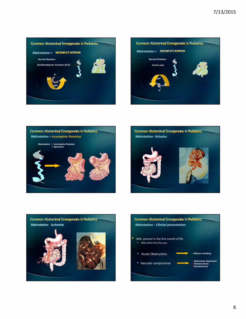
Duodenojejunal Junction (DJJ)
Normal Rotation
0o
180o
90o 270o
Malrotation =
Cecal Loop
Normal Rotation
0o
90o
180o
270o
Malrotation =
Malrotation = Incomplete Rotation= Spectrum
Malrotation = Incomplete Rotation Malrotation‐ Volvulus
Malrotation ‐ Ischemia
80% present in the first month of life90% within the first year
Acute Obstruction
Vascular compromise
Bilious vomiting
Abdominal distension Hematochezia Hematemesis
Malrotation – Clinical presentation

7/13/2015
7
Malrotation – Clinical presentation Malrotation – Clinical presentation
Malrotation – Clinical presentation
Older patients
Recurrent vomiting
Recurrent abdominal pain
Failure to thrive
Malabsorption
Malrotation – Clinical presentation
First pass
Gastric emptying is variable
Duodenal emptying is variable
Gastric overdistension with contrast
Gold Standard (?)
Malrotation – Diagnosis
Detection of Malrotation
Sensitivity 93 – 100%
Lateral view
Sensitivity 96%
Detection of volvulus
Sensitivity 54%
Specificity 88%
Reversal of SMA / SMV relationship
Sensitivity: 66 ‐ 71%
Specificity: 89 – 92%
Whirlpool sign: volvulus
Twist of duodenum and SMV around SMA
Sensitivity: 83 – 92%
Specificity: 92 – 100%
Dao, Beydoun, Youssfi
245 studies; 100% Sensitivity & Specificity
SPR 58th annual meeting, April 2015
Malrotation – Diagnosis

7/13/2015
8
US: Whirlpool Sign
Malrotation – Diagnosis
8 year old boylong history of failure to thriverecent 14 pound weight loss
recent diagnosis of “sprue”
on gluten‐free diet
Case history
Malrotation ‐ Diagnosis
Malrotation
INTUSSUSCEPTION
Intussusception
DEL POZO ET AL. RADIOGRAPHICS 1999 19:299‐13DEL POZO ET AL. RADIOGRAPHICS 1999 19:299‐13
Intussusception
Intussusceptum Intussuscipiens

7/13/2015
9
Diagnosis
Tailoring management
Sensitive – 100%
Specific – “89%”
BOWEL ‐WITHIN ‐ BOWEL
Ultrasound in the diagnosis and exclusion of intussusception Ir Med J 1997;90:64Intussusception in children: reliability of US in diagnosis Radiology 1992;191:741
Intussusception
TECHNIQUE
Curvilinear transducer for bird’s eye view
Linear transducer for focused evaluation of
bowel‐within‐bowel
trapped fluid
lead points
Intussusception
ENTRANCE END
M
Intussusception
Increased reduction failure
Age < 3 months
Duration > 48 hours
small bowel obstruction
Hematochezia Fluid within the intussusceptum complex
Diminished or absent flow to complex
Intussusception
6 months – 2 years
Ileocolic, idiopathic
Lead points
< 2‐3 months ‐ duplication cyst, Meckels
> 5 years – lymphoma ‐ Burkitt
Intussusception Intussusception‐ lead points
Duplication cyst
Burkitt lymphoma

7/13/2015
10
Intussusception – small bowel
Asymptomatic
< 3 cm wide
< 3 cm long
No obstruction
Active peristalsis
Intussusception – small bowel
Intussusception – small bowelAPPENDICITIS
Appendicitis
Most common childhood surgical condition80% of pediatric surgical emergencies
Greatest in second decade
Rare in young children
Neonates‐ appendiceal perforation may be a presentation of long‐segment Hirschsprung disease*
*J Ped Surg 1997;32(1):123
Appendicitis – Pediatric Challenges
Young children unable to verbalize symptoms
Presentation atypical in 30‐45%
Perforation rates
adults 16‐39%
children 23‐73%
infants 62‐ 88%
as high as 100% in infants < 1 year

7/13/2015
11
Appendicitis – Diagnosis
Physical examination
Typical findings
Atypical findings
SURGERY
IMAGING
ULTRASOUND
Appendicitis – Ultrasound
Sensitivity
Approximately 40‐100%
Specificity
Approximately 40‐95%
UnderscoresOperatorDependence
Appendicitis – Ultrasound
High frequency LINEAR transducer
graded compression
Gentle and STEADY pressure
Upward direction
empty bladder
visualize psoas
CALM INFANT
Crying prevents successful compression
Toys, videos
Appendicitis – Ultrasound
Find liver and right kidney
Identify ascending colon
noncompressible bowel gas ?
Follow ascending colon
identify terminal ileum
Search for appendix
cecal tip, retrocecal area, iliac fossa
Appendicitis – Ultrasound
Visualization is variable
5 – 50%
ENTIRE appendix MUST be visible
Base and length may be normal
Tip may be hidden by overlying gas
Appendicitis – Ultrasound

7/13/2015
12
Appendicitis – Ultrasound
>6mm outer‐wall‐to‐outer‐wall
during compression
Periappendiceal echogenicity
Increased flow
may not be present if necrosis has supervened
Appendicitis – Ultrasound
APPENDICITIS
RLQ PAIN
CONTROLS
6 mm Rettenbacher et al radiology 2001;218:757‐62
Appendicitis – Ultrasound
Borderline appendix size
Loop of bowel mistaken for appendix
Need to confirm blind‐ending structure
origin from cecal tip if possible
identify terminal ileum separately
Appendicitis – Ultrasound
No appendix identified
Hidden by bowel gas or bone
Normal appendix identified at cecal pole
Inflamed appendiceal tip elsewhere
Loop of bowel mistaken for “normal” appendix
+