combining microenvironment normalization strategies to ... · stroma normalization can further...
TRANSCRIPT

Combining microenvironment normalization strategiesto improve cancer immunotherapyFotios Mpekrisa, Chrysovalantis Voutouria, James W. Baishb, Dan G. Dudac, Lance L. Munnc,Triantafyllos Stylianopoulosa,1, and Rakesh K. Jainc,1
aCancer Biophysics Laboratory, Department of Mechanical and Manufacturing Engineering, University of Cyprus, 1678 Nicosia, Cyprus; bDepartment ofBiomedical Engineering, Bucknell University, Lewisburg, PA 17837; and cEdwin L. Steele Laboratories, Department of Radiation Oncology, MassachusettsGeneral Hospital and Harvard Medical School, Boston, MA 02114
Contributed by Rakesh K. Jain, December 17, 2019 (sent for review November 12, 2019; reviewed by Aleksander S. Popel and Hermann Frieboes)
Advances in immunotherapy have revolutionized the treatment ofmultiple cancers. Unfortunately, tumors usually have impaired bloodperfusion, which limits the delivery of therapeutics and cytotoxicimmune cells to tumors and also results in hypoxia—a hallmark ofthe abnormal tumor microenvironment (TME)—that causes immu-nosuppression. We proposed that normalization of TME using anti-angiogenic drugs and/or mechanotherapeutics can overcome thesechallenges. Recently, immunotherapy with checkpoint blockers wasshown to effectively induce vascular normalization in some types ofcancer. Although these therapeutic approaches have been used incombination in preclinical and clinical studies, their combined effectson TME are not fully understood. To identify strategies for improvedimmunotherapy, we have developed a mathematical frameworkthat incorporates complex interactions among various types of can-cer cells, immune cells, stroma, angiogenic molecules, and the vas-culature. Model predictions were compared with the data from fivepreviously reported experimental studies. We found that low dosesof antiangiogenic treatment improve immunotherapy when the twotreatments are administered sequentially, but that high doses areless efficacious because of excessive vessel pruning and hypoxia.Stroma normalization can further increase the efficacy of immuno-therapy, and the benefit is additive when combined with vascularnormalization. We conclude that vessel functionality dictates theefficacy of immunotherapy, and thus increased tumor perfusionshould be investigated as a predictive biomarker of response toimmunotherapy.
immunotherapy | vascular function | normalization | anti-angiogenictherapy | mechanotherapeutics
Immunotherapy is now a standard of care for multiple types ofcancer (1, 2). Cancer cells are able to evade immune responses
by activating negative regulatory pathways, also known as im-mune checkpoints, that block T-cell priming and activation. Theinhibition is mediated in large part by the binding of ProgrammedCell Death Protein 1 (PD-1) receptor of T cells to the PD-L1 li-gand or the binding of cytotoxic T lymphocyte antigen 4 (CTLA-4)receptor of T cells to the B7 molecules in response to variouscytokines, such as interferon-γ (IFNγ) (3). The recent develop-ment and Food and Drug Administration approval of anti–PD-1/anti–PD-L1 or anti–CTLA-4 antibodies, known as immunecheckpoint blockers (ICBs), enables the inhibition of immunecheckpoint pathways, thereby eliciting antitumor clinical responsesin a variety of solid cancers (4). ICBs have revolutionized thetreatment of cancer and have been approved as monotherapies orin combination with other treatments for more than a dozen ma-lignancies (4). While treatment responses are often durable, only asmall fraction of patients receiving anti–PD-1/PD-L1 therapy are“cured,” while an estimated 87% of US cancer patients currentlydo not benefit from ICB monotherapies (5). However, with morethan 3,500 ongoing clinical trials for cancer immunotherapy(ClinicalTrials.gov), immunotherapy is expected to change thestandard of care in many more cancer types.
The absence of a therapeutic benefit in patients has been at-tributed to a variety of factors, including the abnormal tumormicroenvironment (TME), characterized by dysfunctional bloodvessels that hinder the delivery of immunotherapeutic agents andcause immunosuppression (1, 6, 7). Indeed, a spatiotemporallack of sufficient tumor blood perfusion can result in hypoxia,low pH, and inadequate delivery of medicines, which in turncompromises the efficacy of cancer therapies, including immu-notherapy (1, 8, 9). One important consequence is that a hypoxicTME helps cancer cells evade the immune system (1). In partic-ular, hypoperfusion hinders the delivery of immune cells to thetumor site through the vascular system, while hypoxia renders theTME immunosuppressive and attenuates the killing potential ofimmune effector cells (10, 11). Specifically, hypoxia up-regulatesimmune checkpoints (11, 12), reprograms tumor-associated mac-rophages (TAMs) from an immunosupportive M1-like phenotypetoward an immunosuppressive M2-like state, and may hinder the
Significance
Immunotherapy has changed the standard of care in cancertreatment, but an estimated 87% of patients currently do notderive long-term benefit from immune checkpoint blockermonotherapy. Therefore, new therapeutic strategies are neededto improve the response rates in patients who are resistant toimmune checkpoint inhibition. We have developed a mathe-matical framework to determine how tumor microenvironmentnormalization strategies—specifically, vascular and stroma nor-malization—might improve immunotherapy efficacy. By in-corporating complex interactions among various types of cancercells, immune cells, stromal cells, and the vasculature, as well asphysical mechanisms, we provide guidelines for designing ef-fective combinatorial therapeutic strategies and point out areasfor future investigation.
Author contributions: J.W.B., D.G.D., L.L.M., T.S., and R.K.J. designed research; F.M. andC.V. performed research; T.S. contributed new reagents/analytic tools; F.M., C.V., J.W.B.,D.G.D., L.L.M., T.S., and R.K.J. analyzed data; and F.M., C.V., D.G.D., L.L.M., T.S., and R.K.J.wrote the paper.
Reviewers: A.S.P., Johns Hopkins University School of Medicine; and H.F., University ofLouisville.
Competing interest statement: R.K.J. has received honoraria from Amgen and consultantfees from Chugai, Merck, Ophthotech, Pfizer, SPARC, SynDevRx, and XTuit; owns equity inEnlight, Ophthotech, and SynDevRx; and serves on the Boards of Trustees of Tekla Health-care Investors, Tekla Life Sciences Investors, Tekla Healthcare Opportunities Fund, andTekla World Healthcare Fund. D.G.D. received consultant fees from Bayer, Simcere, andBMS and research grants from Bayer, Exelixis, and BMS. Neither any reagent nor anyfunding from these organizations was used in this study.
This open access article is distributed under Creative Commons Attribution-NonCommercial-NoDerivatives License 4.0 (CC BY-NC-ND).
See online for related content such as Commentaries.1To whom correspondence may be addressed. Email: [email protected] or [email protected].
This article contains supporting information online at https://www.pnas.org/lookup/suppl/doi:10.1073/pnas.1919764117/-/DCSupplemental.
First published February 3, 2020.
3728–3737 | PNAS | February 18, 2020 | vol. 117 | no. 7 www.pnas.org/cgi/doi/10.1073/pnas.1919764117
maturity, and thus the efficacy, of antigen presentation by den-dritic cells (10, 13). In addition, hypoxia and acidity affect thefunction of T lymphocytes and other immune cells (14–16), whereashypoperfusion and the dense/stiff TME is a physical barrier to T cellinfiltration into the tumor (17, 18). Indeed, increased perfusion hasbeen related to improved ICB response (19).Tumor perfusion is compromised in part by the compression
as well as the tortuosity and hyperpermeability of tumor vessels(20). Vessel compression is a result of mechanical forces accu-mulated within stroma components of tumors (21–23). Indeed, ourprevious studies (24–26) showed that inhibiting CXCL12/CXCR4or angiotensin signaling can target cancer-associated fibroblasts(CAFs) and extracellular collagen and hyaluronan, which in turnalleviates intratumoral forces, decompresses tumor vessels, andimproves perfusion as well as the outcome of ICBs. We have re-ferred to this approach as stroma normalization (27–30).Vessel hyperpermeability and tortuosity can be lowered using
judicious doses of antiangiogenic (e.g., anti-vascular endothelialgrowth factor [anti-VEGF]) agents, which increase pericyte cov-erage and fortify the immature vessels. This strategy, known asvascular normalization, can improve tumor perfusion and therebyincrease oxygen and drug delivery, as well as improve the efficacyof various treatments, including immunotherapy (31–36). As aresult, perfusion can be increased within the tumor (37). However,vessel normalization is dose-dependent, and high doses of theanti-angiogenic drug can prune the vessels and reduce perfusionand drug delivery (36, 38). In addition, vascular normalization hasa transient effect; prolonged anti-angiogenic treatment can resultin excessive vessel pruning. This dose- and time-dependency ofvascular normalization results in a “normalization window” withinwhich perfusion is improved (33, 38).Stroma and vascular normalization improve tumor perfusion
and oxygenation and can enhance immune response by skewingTAM polarization toward the M1 phenotype, which is tumor-icidal and immunostimulatory (10, 13, 24, 39, 40). This can alsoresult in the activation of dendritic cells, cytotoxic T lympho-cytes, and natural killer (NK) cells (39). Furthermore, increasedtumor perfusion via vessel normalization improves the functionof immune cells, contributing to increased killing of cancer cells,which results in the killing of the more resistant stem cell-likecancer cells (41). It has also been proposed that ICB-inducedincreases in the ratio of CD4+/CD8+ T cells may decrease thelevels of VEGF and thereby decrease tumor vessel permeability,indirectly inducing vascular normalization (10, 42).Given these complex interactions among cancer cells, immune
cells, stroma, and the tumor vasculature, the prediction of theoutcome of combined anti-angiogenic, stroma-normalizing, andimmune therapies is not intuitive. We hypothesize that a mathe-matical model that incorporates the known mechanisms of im-mune response as well as vascular and stroma normalization canexplain the experimental observations reported in the literatureand provide guidelines for its optimal use and for future experi-ments. Systems-level mathematical models for the prediction ofanti–CTLA-4 and anti–PD-1/anti-PD-L1 response have been de-veloped previously (43–45); however, they do not account forspatial variation in the TME. Here, based on our previous study(41) and the work of others (46, 47), we developed a continuummathematical modeling framework to account for interactionsamong different types of cancer cells, immune cells, tumor bloodvessels, oxygen supply, and drug delivery. We build on our previousmodels (41, 48) by incorporating M1-like and M2-like TAMs;CD4+, CD8+, and regulatory (Tregs) T cells, and vascular andperivascular cells and their reported involvement in immunother-apy response. Furthermore, we take into account the dependenceof the functional vasculature on stroma components; the concen-tration gradients of various proangiogenic molecules, includingangiopoietins (Ang1 and Ang2), platelet-derived growth factor(PDGF)-B, VEGF, and CXCL12; and the vascular normalization
effect of CD4+ and CD8+ T cells (Fig. 1). We simulate how anti-angiogenic, stroma normalization, and immunotherapy treatmentsaffect model components and compare model predictions withprevious experimental studies (19, 24, 26, 36, 49). Furthermore, weuse model predictions to provide guidelines for the optimal use ofthese three strategies and point out the gaps in our understanding.
ResultsValidation of Model Predictions with Experimental Studies. To assessthe validity of our mathematical model and justify the values of themodel parameters (SI Appendix, Table S1), we compared modelpredictions with experimental data from five studies, summarizedin Table 1. To compare model predictions with experimental data,all model parameters were kept the same as derived separately frompertinent studies. The sole model parameter that was adjusted to fitthe experimental data (19, 24, 26, 36, 49) was k1, which describes thedependence of cancer cell proliferation on oxygen concentration (SIAppendix, Table S2). The value of k1 was determined so that thepredicted final tumor volume matched the experimental value ofone of the treatment groups, and it was kept the same for com-parison of model predictions against all experimental data from allgroups of the same study. Therefore, despite the large number ofparameters in the model—which is justified by its degree of com-prehensiveness—only one parameter was varied for each study.In our previous studies (e.g., refs. 24 and 26), stroma nor-
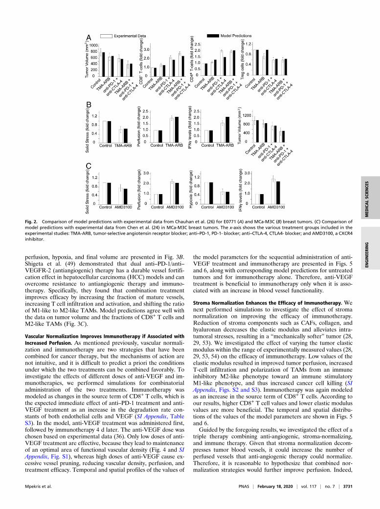
malization was applied to improve perfusion and enhance theefficacy of ICBs (anti–PD-1 and anti–CTLA-4) in murine breasttumors. We incorporated the experimental protocols in our modeland compared model predictions with experimental results. Wemodeled stroma normalization as a decrease in the tumor elasticmodulus because a primary effect of the strategy is the softening ofthe tumor. In addition, anti–PD-1 treatment was modeled as anincrease in CD8+ T cells, and anti–CTLA-4 treatment was mod-eled as a decrease in regulatory T cells (Tregs), in line with thereported effects of these two treatments (50). SI Appendix, TableS3 summarizes the model parameters associated with each treat-ment strategy. To demonstrate the comparison between the di-mensionless model parameters and the experimental measurements,the values of the different measured parameters are presentedrelative to the values of the control group. For the first study(26), model predictions agree well with the data on CD4+ andCD8+ T cells, Tregs, and tumor volume in E0771 breast tumors(Fig. 2A) and solid stress, tumor perfusion, IFNγ level, and tumorvolume in MCa-M3C breast tumors (Fig. 2B). In our model, hypoxiawas calculated as the percentage of oxygen concentration below theoxygen level in the peritumoral normal tissue, and tumor perfusionwas calculated as the ratio of the tumor functional vascular density tothat of the normal tissue. In the second breast cancer study (24),model predictions show good agreement with the data on solid stress,tumor perfusion, hypoxia, and IFNγ levels (Fig. 2C).In another breast cancer study from our laboratory, Huang
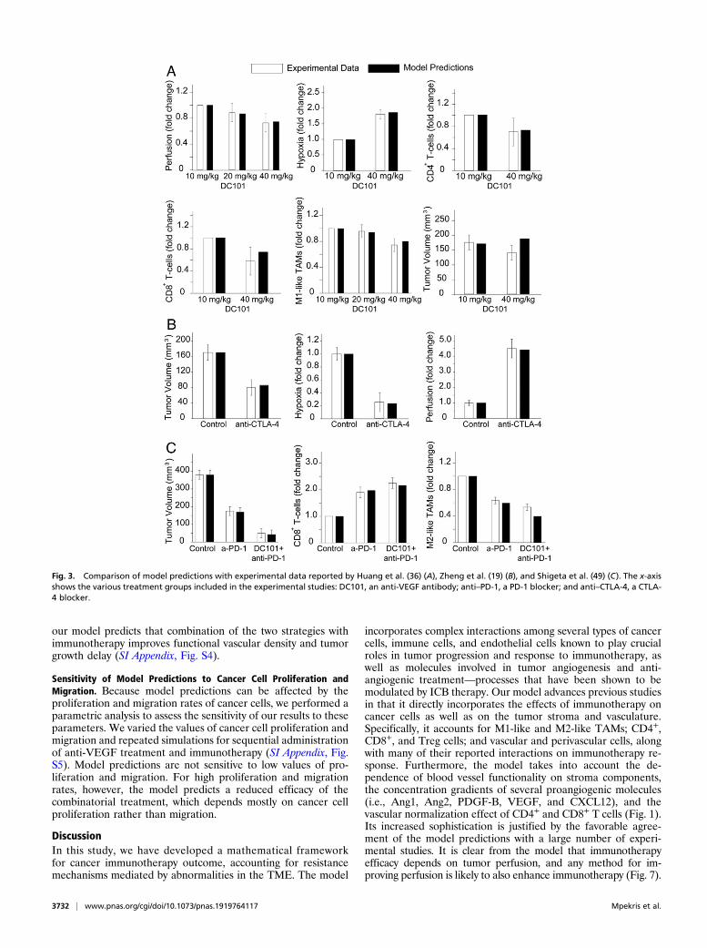
et al. (36) demonstrated that targeting the tumor vasculaturewith low vascular-normalizing doses, but not high antivasculardoses, results in a more homogeneous distribution of functionaltumor vessels. Furthermore, low doses were superior to thehigh doses in polarizing TAMs from an immune inhibitory M2-like phenotype toward an immune stimulatory M1-like phe-notype and in facilitating CD4+ and CD8+ T cell tumor in-filtration. We simulated the same experimental protocol tocompare model predictions to the results of low- and high-doseanti-VEGF treatments. In our simulations, the effects of dif-ferent doses of anti-VEGF on endothelial cells and VEGFlevels were modeled by changes in the endothelial cell andVEGF degradation rate constants, keca−vegf and kvegfa−vegf , respec-tively (SI Appendix, Eqs. S19 and S22), according to experi-mental studies (51, 52). To facilitate comparisons betweenthe dimensionless model parameters and the experimental
Mpekris et al. PNAS | February 18, 2020 | vol. 117 | no. 7 | 3729
MED
ICALSC
IENCE
SEN
GINEE
RING
measurements, the values of the different measured parametersare presented relative to the values of the lowest-dose treatment.Model predictions agree well with our data on tumor perfusion,hypoxia, tumor volume, and the numbers of CD4+ and CD8+ Tcells and M1-like TAMs (Fig. 3A).
Zheng et al. (19), found that ICBs increased tumor vesselperfusion in the immunotherapy-sensitive E0771 murine breasttumor model, and that the ability of anti–CTLA-4 therapy toincrease vessel perfusion was associated with treatment efficacy.Comparisons of model predictions with the data on tumor
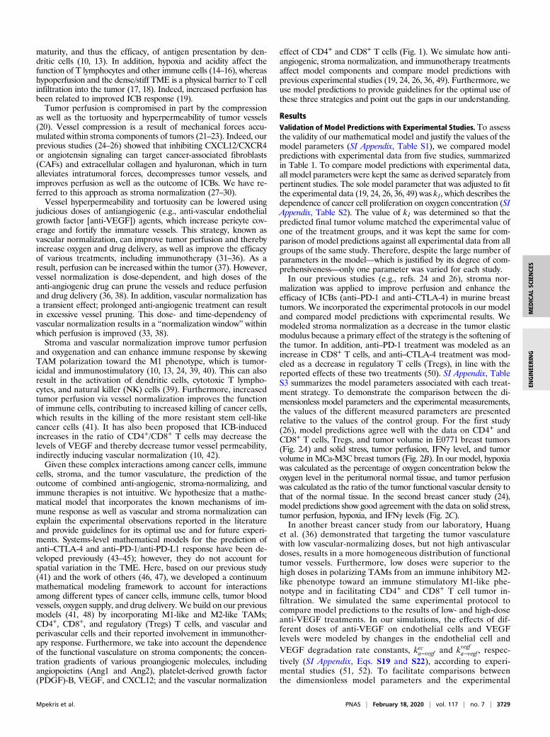
Fig. 1. Schematic of the interactions among model components. The model accounts for various cell populations (orange boxes) and tumor angiogenicfactors (blue boxes). TME component: Increases in functional vascular density and tumor perfusion enhance tumor oxygenation. Higher oxygen levels ac-celerate the proliferation rates of CCs and CD4+ T cells and the activity of immune cells and polarize TAMs from an immune inhibitory M2-like phenotypetoward an immune stimulatory M1-like phenotype. Along with the immunostimulatory action of M1-like TAMs, the model accounts for their tumoricidaleffect on CCs. According to previous studies, CD4+ T cells stimulate CD8+ T cells, and an increased immune response leads to more efficient killing of all typesof cancer cells. Increased proliferation of all cancer cell types results in increased oxygen consumption, inactivation of immune cells, and decreased vesseldiameters due to compression-induced hypoxia. Hypoxia favors proliferation of CSCs and ICCs and increases VEGF and CXCL12 levels. In addition, targeting ofstroma components through CXCL12/CXCR4 signaling alleviates solid stresses, which are associated with vascular dysfunction. Tumor vasculature component:This initiates angiogenesis through the proliferation and migration of endothelial cells that form the vessels. Angiogenesis is enhanced by high levels of Ang2,which destabilizes vessels, and inhibited by Ang1 and PDGF-B, which recruit pericytes and stabilize vessels. In addition, knockout of CD4+ T cells results inoverexpression of VEGF, which is correlated with higher numbers of M2-like TAMs. On the other hand, a decrease in M2-like TAMs results in higher numbersof effector immune cells (CD8+ T cells and NK cells). Increased numbers of CD4+ and CD8+ T cells enhances production rates of IFNγ, which is associated withdecreased vessel wall pore size and permeability, leading to vascular normalization. Vascular normalization improves the functionality of the vascular net-work, leading to an increase in functional vascular density, which enhances cancer cell proliferation.
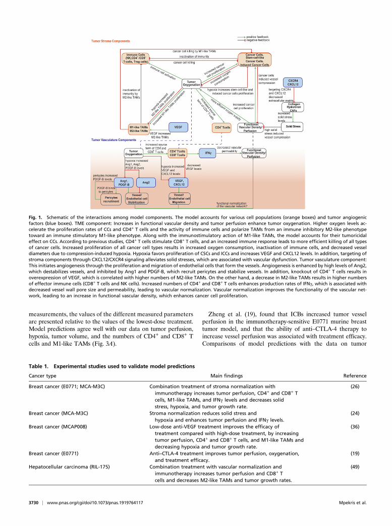
Table 1. Experimental studies used to validate model predictions
Cancer type Main findings Reference
Breast cancer (E0771; MCA-M3C) Combination treatment of stroma normalization withimmunotherapy increases tumor perfusion, CD4+ and CD8+ Tcells, M1-like TAMs, and IFNγ levels and decreases solidstress, hypoxia, and tumor growth rate.
(26)
Breast cancer (MCA-M3C) Stroma normalization reduces solid stress andhypoxia and enhances tumor perfusion and IFNγ levels.
(24)
Breast cancer (MCAP008) Low-dose anti-VEGF treatment improves the efficacy oftreatment compared with high-dose treatment, by increasingtumor perfusion, CD4+ and CD8+ T cells, and M1-like TAMs anddecreasing hypoxia and tumor growth rate.
(36)
Breast cancer (E0771) Anti–CTLA-4 treatment improves tumor perfusion, oxygenation,and treatment efficacy.
(19)
Hepatocellular carcinoma (RIL-175) Combination treatment with vascular normalization andimmunotherapy increases tumor perfusion and CD8+ Tcells and decreases M2-like TAMs and tumor growth rates.
(49)
3730 | www.pnas.org/cgi/doi/10.1073/pnas.1919764117 Mpekris et al.
perfusion, hypoxia, and final volume are presented in Fig. 3B.Shigeta et al. (49) demonstrated that dual anti–PD-1/anti–VEGFR-2 (antiangiogenic) therapy has a durable vessel fortifi-cation effect in hepatocellular carcinoma (HCC) models and canovercome resistance to antiangiogenic therapy and immuno-therapy. Specifically, they found that combination treatmentimproves efficacy by increasing the fraction of mature vessels,increasing T cell infiltration and activation, and shifting the ratioof M1-like to M2-like TAMs. Model predictions agree well withthe data on tumor volume and the fractions of CD8+ T cells andM2-like TAMs (Fig. 3C).
Vascular Normalization Improves Immunotherapy if Associated withIncreased Perfusion. As mentioned previously, vascular normali-zation and immunotherapy are two strategies that have beencombined for cancer therapy, but the mechanisms of action arenot intuitive, and it is difficult to predict a priori the conditionsunder which the two treatments can be combined favorably. Toinvestigate the effects of different doses of anti-VEGF and im-munotherapies, we performed simulations for combinatorialadministration of the two treatments. Immunotherapy wasmodeled as changes in the source term of CD8+ T cells, which isthe expected immediate effect of anti–PD-1 treatment and anti-VEGF treatment as an increase in the degradation rate con-stants of both endothelial cells and VEGF (SI Appendix, TableS3). In the model, anti-VEGF treatment was administered first,followed by immunotherapy 4 d later. The anti-VEGF dose waschosen based on experimental data (36). Only low doses of anti-VEGF treatment are effective, because they lead to maintenanceof an optimal area of functional vascular density (Fig. 4 and SIAppendix, Fig. S1), whereas high doses of anti-VEGF cause ex-cessive vessel pruning, reducing vascular density, perfusion, andtreatment efficacy. Temporal and spatial profiles of the values of
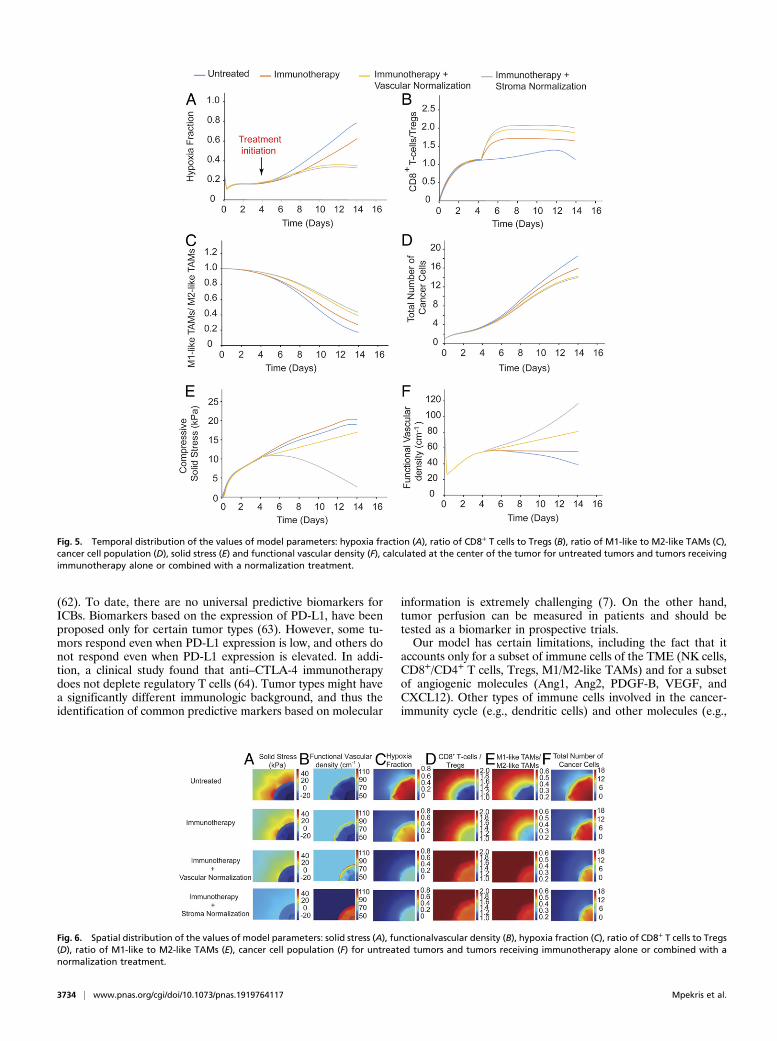
the model parameters for the sequential administration of anti-VEGF treatment and immunotherapy are presented in Figs. 5and 6, along with corresponding model predictions for untreatedtumors and for immunotherapy alone. Therefore, anti-VEGFtreatment is beneficial to immunotherapy only when it is asso-ciated with an increase in blood vessel functionality.
Stroma Normalization Enhances the Efficacy of Immunotherapy. Wenext performed simulations to investigate the effect of stromanormalization on improving the efficacy of immunotherapy.Reduction of stroma components such as CAFs, collagen, andhyaluronan decreases the elastic modulus and alleviates intra-tumoral stresses, resulting in a “mechanically softer” tumor (28,29, 53). We investigated the effect of varying the tumor elasticmodulus within the range of experimentally measured values (28,29, 53, 54) on the efficacy of immunotherapy. Low values of theelastic modulus resulted in improved tumor perfusion, increasedT-cell infiltration and polarization of TAMs from an immuneinhibitory M2-like phenotype toward an immune stimulatoryM1-like phenotype, and thus increased cancer cell killing (SIAppendix, Figs. S2 and S3). Immunotherapy was again modeledas an increase in the source term of CD8+ T cells. According toour results, higher CD8+ T cell values and lower elastic modulusvalues are more beneficial. The temporal and spatial distribu-tions of the values of the model parameters are shown in Figs. 5and 6.Guided by the foregoing results, we investigated the effect of a
triple therapy combining anti-angiogenic, stroma-normalizing,and immune therapy. Given that stroma normalization decom-presses tumor blood vessels, it could increase the number ofperfused vessels that anti-angiogenic therapy could normalize.Therefore, it is reasonable to hypothesize that combined nor-malization strategies would further improve perfusion. Indeed,
Fig. 2. Comparison of model predictions with experimental data from Chauhan et al. (26) for E0771 (A) and MCa-M3C (B) breast tumors. (C) Comparison ofmodel predictions with experimental data from Chen et al. (24) in MCa-M3C breast tumors. The x-axis shows the various treatment groups included in theexperimental studies: TMA-ARB, tumor-selective angiotensin receptor blocker; anti–PD-1, PD-1- blocker; anti–CTLA-4, CTLA4- blocker; and AMD3100, a CXCR4inhibitor.
Mpekris et al. PNAS | February 18, 2020 | vol. 117 | no. 7 | 3731
MED
ICALSC
IENCE
SEN
GINEE
RING
our model predicts that combination of the two strategies withimmunotherapy improves functional vascular density and tumorgrowth delay (SI Appendix, Fig. S4).
Sensitivity of Model Predictions to Cancer Cell Proliferation andMigration. Because model predictions can be affected by theproliferation and migration rates of cancer cells, we performed aparametric analysis to assess the sensitivity of our results to theseparameters. We varied the values of cancer cell proliferation andmigration and repeated simulations for sequential administrationof anti-VEGF treatment and immunotherapy (SI Appendix, Fig.S5). Model predictions are not sensitive to low values of pro-liferation and migration. For high proliferation and migrationrates, however, the model predicts a reduced efficacy of thecombinatorial treatment, which depends mostly on cancer cellproliferation rather than migration.
DiscussionIn this study, we have developed a mathematical frameworkfor cancer immunotherapy outcome, accounting for resistancemechanisms mediated by abnormalities in the TME. The model
incorporates complex interactions among several types of cancercells, immune cells, and endothelial cells known to play crucialroles in tumor progression and response to immunotherapy, aswell as molecules involved in tumor angiogenesis and anti-angiogenic treatment—processes that have been shown to bemodulated by ICB therapy. Our model advances previous studiesin that it directly incorporates the effects of immunotherapy oncancer cells as well as on the tumor stroma and vasculature.Specifically, it accounts for M1-like and M2-like TAMs; CD4+,CD8+, and Treg cells; and vascular and perivascular cells, alongwith many of their reported interactions on immunotherapy re-sponse. Furthermore, the model takes into account the de-pendence of blood vessel functionality on stroma components,the concentration gradients of several proangiogenic molecules(i.e., Ang1, Ang2, PDGF-B, VEGF, and CXCL12), and thevascular normalization effect of CD4+ and CD8+ T cells (Fig. 1).Its increased sophistication is justified by the favorable agree-ment of the model predictions with a large number of experi-mental studies. It is clear from the model that immunotherapyefficacy depends on tumor perfusion, and any method for im-proving perfusion is likely to also enhance immunotherapy (Fig. 7).
Fig. 3. Comparison of model predictions with experimental data reported by Huang et al. (36) (A), Zheng et al. (19) (B), and Shigeta et al. (49) (C). The x-axisshows the various treatment groups included in the experimental studies: DC101, an anti-VEGF antibody; anti–PD-1, a PD-1 blocker; and anti–CTLA-4, a CTLA-4 blocker.
3732 | www.pnas.org/cgi/doi/10.1073/pnas.1919764117 Mpekris et al.

Our study is of particular importance because it proposes newconsiderations for the use of treatments that normalize theTME. Stroma normalization is beneficial in desmoplastic tumorswith abundant compressed vessels, whereas vascular normaliza-tion should improve perfusion in tumors with hyperpermeablevessels with open lumens (20). In tumors with both compressedand leaky vessels, the two normalization strategies could becombined to further enhance perfusion. For our theoreticalpredictions to be implemented in the clinic, we would need toidentify the cause of hypoperfusion in each tumor, a challengingtask. Although we can make some broad statements (e.g., pan-creatic ductal adenocarcinomas are desmoplastic), in many tu-mors, such as breast cancers, the degree of desmoplasia is highlyvariable from one tumor subtype to another and potentially fromthe primary site to the metastatic site. In such cases, the state ofan individual tumor should be defined before the selection of anappropriate strategy.The recent finding that ICB treatment can normalize vessels
further encourages its combined use with anti-angiogenic drugs.In fact, the successful recent phase III clinical study for thecombined use of the anti–PD-L1 antibody atezolizumab with the anti-VEGFA antibody bevacizumab is consistent with this therapeuticstrategy in hepatocellular carcinoma patients (ClinicalTrials.govIdentifier: NCT03434379) (55). The addition of atezolizumab tobevacizumab plus chemotherapy significantly improved overall sur-vival among patients with metastatic nonsquamous non–small-celllung cancer (56). Furthermore, combinatorial treatment of axitinibwith pembrolizumab (57) or with avelumab (58) in patients with
advanced renal cell carcinoma increased progression free-survival. Also, the effect of lenvatinib plus pembrolizumab isbeing investigated in a randomized phase III clinical study, as itwas found that this treatment had antitumor activity in patientswith advanced recurrent endometrial cancer (59). However,the benefit of anti-angiogenic drugs is time- and dose-dependent,and identifying the normalization window of a tumor is chal-lenging. Our model predictions are further validated by recentclinical trials showing that combination treatment with a lowdose of an anti-angiogenic agent (regorafenib) with nivolumabis superior to high doses in advanced gastric or colorectalcancer (60). Our model suggests that administration of im-munotherapy with anti-angiogenic treatment could protectblood vessels from excessive pruning (49). However, the extentof ICB-induced normalization is likely to vary with tumor type,stage of disease, and location, and it might not even occur insome tumor types. We should also emphasize that for theresults presented in Figs. 4–6, we modeled vascular normali-zation considering only inhibition of VEGF. Normalization oftumor blood vessels can be also achieved with the use of ty-rosine kinase inhibitors (TKIs), which target other proangio-genic pathways besides VEGF, such as PDGF receptors onpericytes. Inhibiting pericytes, and thus vessel maturation,would lessen the impact of any anti-VEGF effect and thusinterfere with ICB-induced normalization (61).The identification of tumor perfusion as a key parameter for
the efficacy of immunotherapy also highlights the potential useof perfusion measures as markers for immunotherapy prediction
Fig. 4. Effect of different doses of anti-VEGF treatment combined with different values of the source term of CD8+ T cells to model immunotherapy forsequential administration. Shown are phase diagrams for the effect of combinatorial treatment on functional vascular density (A), tumor oxygenation (B),VEGF level (C), CD4+ T cells (D), effector immune cells (NK and CD8+ T cells) (E) , M1-like (F) and M2-like (G) TAMs, cancer cell population (H), and tumorvolume (I). Values of model parameters presented in the figure were calculated at the location equidistant from the tumor center and periphery. On thex-axis, a value of 1 corresponds to the baseline value of source term of CD8+ T cells (SI Appendix, Table S1).
Mpekris et al. PNAS | February 18, 2020 | vol. 117 | no. 7 | 3733
MED
ICALSC
IENCE
SEN
GINEE
RING
(62). To date, there are no universal predictive biomarkers forICBs. Biomarkers based on the expression of PD-L1, have beenproposed only for certain tumor types (63). However, some tu-mors respond even when PD-L1 expression is low, and others donot respond even when PD-L1 expression is elevated. In addi-tion, a clinical study found that anti–CTLA-4 immunotherapydoes not deplete regulatory T cells (64). Tumor types might havea significantly different immunologic background, and thus theidentification of common predictive markers based on molecular
information is extremely challenging (7). On the other hand,tumor perfusion can be measured in patients and should betested as a biomarker in prospective trials.Our model has certain limitations, including the fact that it
accounts only for a subset of immune cells of the TME (NK cells,CD8+/CD4+ T cells, Tregs, M1/M2-like TAMs) and for a subsetof angiogenic molecules (Ang1, Ang2, PDGF-B, VEGF, andCXCL12). Other types of immune cells involved in the cancer-immunity cycle (e.g., dendritic cells) and other molecules (e.g.,
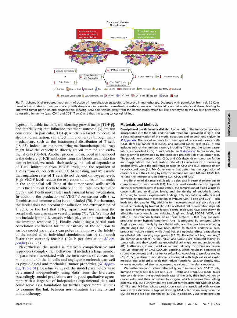
Fig. 6. Spatial distribution of the values of model parameters: solid stress (A), functionalvascular density (B), hypoxia fraction (C), ratio of CD8+ T cells to Tregs(D), ratio of M1-like to M2-like TAMs (E), cancer cell population (F) for untreated tumors and tumors receiving immunotherapy alone or combined with anormalization treatment.
Fig. 5. Temporal distribution of the values of model parameters: hypoxia fraction (A), ratio of CD8+ T cells to Tregs (B), ratio of M1-like to M2-like TAMs (C),cancer cell population (D), solid stress (E) and functional vascular density (F), calculated at the center of the tumor for untreated tumors and tumors receivingimmunotherapy alone or combined with a normalization treatment.
3734 | www.pnas.org/cgi/doi/10.1073/pnas.1919764117 Mpekris et al.
hypoxia-inducible factor 1, transforming growth factor [TGF-β],and interleukins) that influence treatment outcome (3) are notconsidered. In particular, TGF-β, which is a target molecule ofstroma normalization, can affect immunotherapy through manymechanisms, such as the intratumoral distribution of T cells(18, 65). Indeed, stroma-normalizing mechanotherapeutic drugsmight have the capacity to directly act on immune and endo-thelial cells (66–68). Another process not included in the modelis the delivery of ICB antibodies from the bloodstream into thetumor; instead, we model their activity, the lack of dependenceof T-cell infiltration from VEGF levels, and the repulsion ofT cells from cancer cells via CXCR4 signaling, and we assumethat migration rates of T cells do not depend on oxygen levels.High VEGF levels reduce the expression of adhesion moleculesin the endothelial cell lining of the tumor vessel walls, whichlimits the ability of T cells to adhere and infiltrate into the tumor(1, 69), and T cells move faster under normal tissue oxygenation.In addition, the production of VEGF from stroma cells (i.e.,fibroblasts and immune cells) is not included (70). Furthermore,the model does not account for adhesion and extravasation ofT cells, or the fact that IFNγ, apart from normalizing thevessel wall, can also cause vessel pruning (71, 72). We also didnot include lymphatic vessels, which play an important role inthe immune response (1). Finally, calculating a partial rankcorrelation coefficient for the sensitivity of the solution tovarious model parameters can potentially improve the fidelityof the model when individual simulations can be run muchfaster than currently feasible (∼24 h per simulation; SI Ap-pendix) (44, 73).Nevertheless, the model is relatively comprehensive and
reproduces complex, rich behaviors. It includes a large numberof parameters associated with the interactions of cancer, im-mune, and endothelial cells and angiogenic molecules, as wellas physiological and mechanical tumor properties (SI Appen-dix, Table S1). Baseline values of the model parameters weredetermined independently using data from the literature.Accordingly, model predictions are in good qualitative agree-ment with a large set of independent experimental data andcould serve as a foundation for further experimental studiesto examine the link between normalization treatments andimmunotherapy.
Materials and MethodsDescription of the Mathematical Model. A schematic of the tumor componentsincorporated into the model and their interrelations is provided in Fig. 1, anda detailed presentation of the model equations and assumptions is given inSI Appendix. The model accounts for three types of cancer cells: cancer cells(CCs), stem-like cancer cells (CSCs), and induced cancer cells (ICCs). It alsoincludes cells of the immune system, including TAMs and the tumor vascu-lature, as described in Fig. 1 and detailed in SI Appendix. In our model, tu-mor growth is determined by the combined proliferation of all cancer cells.The population balance of CCs, CSCs, and ICCs depends on tumor perfusionand oxygenation. The proliferation rate of CCs increases with increasingoxygen level, while the proliferation rates of CSCs and ICCs increase underhypoxic conditions (41, 74). Other events that determine the population ofcancer cells are their killing by effector immune cells and M1-like TAMs (47,75) and the interconversion among CCs, CSCs, and ICSs.
Proliferation of all cancer cells leads to a decrease in vessel diameter due tocompression of tumor vessels (21). The functional vascular density dependson the hyperpermeability of blood vessels, the compression of blood vessels bycancer cells and solid stress levels, and the density of endothelial cells.According to previous experimental findings, IFNγ concentration affects vesselpermeability; specifically, elimination of immune CD4+ T cells and CD8+ T cellsleads to a decrease in IFNγ, which in turn increases vessel wall pore size andvessel permeability by fivefold (42, 76). Endothelial cell concentration dependson levels of tumor angiogenic factors. Several molecules have been shown toaffect the tumor vasculature, including Ang1 and Ang2, PDGF-B, VEGF, andCXCL12. The common feature of all these proteins is that they are over-produced under hypoxic conditions. Ang1 is produced by pericytes, whileAng2 is produced mainly by endothelial cells, and the two have competingeffects: Ang1 and PDGF-β have been shown to stabilize endothelial cells,producing mature vessels, while Ang2 has the opposite effect, destabilizingendothelial cells, favoring angiogenesis (77, 78). The effects of Ang1 and Ang2are context-dependent (79, 80). VEGF and CXCL12 are produced mainly bytumor cells, and they coordinate endothelial cell migration and angiogenesis(81). Furthermore, in our model we account indirectly for stroma normaliza-tion via targeting of CXCL12/CXCR4 signaling, which results in decreases ofstroma components and thus tumor softening. According to previous studies(28, 29, 53), a dense tumor stroma is associated with high values of elasticmodulus and solid stress levels that reduce functional vascular density (82),whereas reduction of stroma decreases the values of the elastic modulus.
We further account for four different types of immune cells: CD4+ T cells,immune effector cells (i.e., NK cells, CD8+ T cells), and Tregs. Our model takesinto consideration the growth/death rate of the cells, their inactivation bycancer cells, and their activation by oxygen, which increases their killingpotential (41, 75). Furthermore, we account for two different types of TAMs,M1-like and M2-like, whose production rates are associated with oxygenlevels, with a decrease in hypoxia skewing TAM polarization away from theM2-like to the M1-like phenotype (33–35). In addition, VEGF overexpression
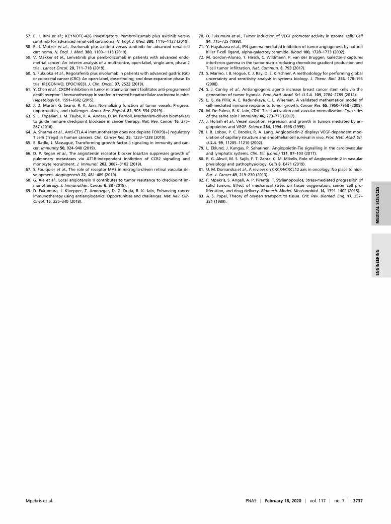
Fig. 7. Schematic of proposed mechanism of action of normalization strategies to improve immunotherapy. (Adapted with permission from ref. 7.) Com-bined administration of immunotherapy with stroma and/or vascular normalization restores vascular functionality and alleviates solid stress, leading toimproved tumor perfusion and oxygenation, skewing TAM polarization away from the immunosuppressive M2-like phenotype to the M1-like phenotype,stimulating immunity (e.g., CD4+ and CD8+ T cells) and thus increasing cancer cell killing.
Mpekris et al. PNAS | February 18, 2020 | vol. 117 | no. 7 | 3735
MED
ICALSC
IENCE
SEN
GINEE
RING

is correlated with higher numbers of M2-like TAMs (33). M2-like TAMs havebeen reported to inhibit the number of immune effector cells (NK cells andCD8+ T cells) (39). Transport of oxygen is described by the convection-diffusion reaction equation, and diffusion is the dominant mode of trans-port for oxygen (82, 83).
Additional details about the model are provided in SI Appendix.Data availability. All data supporting the findings of this study are available inthe paper and SI Appendix.Code availability. The COMSOL code is available in SI Appendix.
ACKNOWLEDGMENTS. We thank Drs. Zohreh Amoozgar, Meenal Datta,Yuhui Huang and John Martin for their insightful comments on this work.This study was supported by grants from the National Foundation for CancerResearch, the Ludwig Center at Harvard, the Jane’s Trust Foundation, theAdvanced Medical Research Foundation, the National Cancer Institute (GrantsR35 CA197743, R01 CA208205, and U01 CA224173, to R.K.J.), the ResearchPromotion Foundation of Cyprus (POST-DOC/0718/0084, INFRASTRUCTURE/1216/0052, to T.S.), and a postdoctoral fellowship from the University of Cyprus(to F.M.).
1. L. L. Munn, R. K. Jain, Vascular regulation of antitumor immunity. Science 365, 544–545 (2019).
2. J. Xin Yu, V. M. Hubbard-Lucey, J. Tang, Immuno-oncology drug development goesglobal. Nat. Rev. Drug Discov. 18, 899–900 (2019).
3. D. S. Chen, I. Mellman, Elements of cancer immunity and the cancer-immune setpoint. Nature 541, 321–330 (2017).
4. X. Li, W. Song, C. Shao, Y. Shi, W. Han, Emerging predictors of the response to theblockade of immune checkpoints in cancer therapy. Cell. Mol. Immunol. 16, 28–39(2019).
5. A. Haslam, V. Prasad, Estimation of the percentage of US patients with cancer whoare eligible for and respond to checkpoint inhibitor immunotherapy drugs. JAMANetw. Open 2, e192535 (2019).
6. M. Datta, L. M. Coussens, H. Nishikawa, F. S. Hodi, R. K. Jain, Reprogramming thetumor microenvironment to improve immunotherapy: Emerging strategies andcombination therapies. Am. Soc. Clin. Oncol. Educ. Book 39, 165–174 (2019).
7. J. D. Martin, H. Cabral, T. Stylianopoulos, R. K. Jain, Improving cancer immunotherapyusing nanomedicines: Progress, opportunities and challenges. Nat. Rev. Clin. Oncol.,10.1038/s41571-019-0308-z (2019).
8. R. K. Jain, T. Stylianopoulos, Delivering nanomedicine to solid tumors. Nat. Rev. Clin.Oncol. 7, 653–664 (2010).
9. T. Stylianopoulos, L. L. Munn, R. K. Jain, Reengineering the physical microenviron-ment of tumors to improve drug delivery and efficacy: From mathematical modelingto bench to bedside. Trends Cancer 4, 292–319 (2018).
10. Y. Huang, S. Goel, D. G. Duda, D. Fukumura, R. K. Jain, Vascular normalization as anemerging strategy to enhance cancer immunotherapy. Cancer Res. 73, 2943–2948(2013).
11. M. Z. Noman et al., Hypoxia: A key player in antitumor immune response. A review inthe theme: Cellular responses to hypoxia. Am. J. Physiol. Cell Physiol. 309, C569–C579(2015).
12. M. Z. Noman et al., PD-L1 is a novel direct target of HIF-1α, and its blockade underhypoxia enhanced MDSC-mediated T cell activation. J. Exp. Med. 211, 781–790 (2014).
13. T. E. Peterson et al., Dual inhibition of Ang-2 and VEGF receptors normalizes tumorvasculature and prolongs survival in glioblastoma by altering macrophages. Proc.Natl. Acad. Sci. U.S.A. 113, 4470–4475 (2016).
14. A. Facciabene et al., Tumour hypoxia promotes tolerance and angiogenesis via CCL28and T(reg) cells. Nature 475, 226–230 (2011).
15. A. Calcinotto et al., Modulation of microenvironment acidity reverses anergy in hu-man and murine tumor-infiltrating T lymphocytes. Cancer Res. 72, 2746–2756 (2012).
16. A. Palazón, J. Aragonés, A. Morales-Kastresana, M. O. de Landázuri, I. Melero, Mo-lecular pathways: Hypoxia response in immune cells fighting or promoting cancer.Clin. Cancer Res. 18, 1207–1213 (2012).
17. H. Salmon et al., Matrix architecture defines the preferential localization and mi-gration of T cells into the stroma of human lung tumors. J. Clin. Invest. 122, 899–910(2012).
18. S. Mariathasan et al., TGFβ attenuates tumour response to PD-L1 blockade by con-tributing to exclusion of T cells. Nature 554, 544–548 (2018).
19. X. Zheng et al., Increased vessel perfusion predicts the efficacy of immune checkpointblockade. J. Clin. Invest. 128, 2104–2115 (2018).
20. T. Stylianopoulos, R. K. Jain, Combining two strategies to improve perfusion and drugdelivery in solid tumors. Proc. Natl. Acad. Sci. U.S.A. 110, 18632–18637 (2013).
21. G. Griffon-Etienne, Y. Boucher, C. Brekken, H. D. Suit, R. K. Jain, Taxane-inducedapoptosis decompresses blood vessels and lowers interstitial fluid pressure in solidtumors: Clinical implications. Cancer Res. 59, 3776–3782 (1999).
22. T. P. Padera et al., Pathology: Cancer cells compress intratumour vessels. Nature 427,695 (2004).
23. T. Stylianopoulos et al., Causes, consequences, and remedies for growth-induced solidstress in murine and human tumors. Proc. Natl. Acad. Sci. U.S.A. 109, 15101–15108(2012).
24. I. X. Chen et al., Blocking CXCR4 alleviates desmoplasia, increases T-lymphocyte in-filtration, and improves immunotherapy in metastatic breast cancer. Proc. Natl. Acad.Sci. U.S.A. 116, 4558–4566 (2019).
25. M. Pinter, R. K. Jain, Targeting the renin-angiotensin system to improve cancertreatment: Implications for immunotherapy. Sci. Transl. Med. 9, eaan5616 (2017).
26. V. P. Chauhan et al., Reprogramming the microenvironment with tumor-selectiveangiotensin blockers enhances cancer immunotherapy. Proc. Natl. Acad. Sci. U.S.A.116, 10674–10680 (2019).
27. V. P. Chauhan et al., Angiotensin inhibition enhances drug delivery and potentiateschemotherapy by decompressing tumour blood vessels. Nat. Commun. 4, 2516 (2013).
28. P. Papageorgis et al., Tranilast-induced stress alleviation in solid tumors improves theefficacy of chemo- and nanotherapeutics in a size-independent manner. Sci. Rep. 7,46140 (2017).
29. C. Polydorou, F. Mpekris, P. Papageorgis, C. Voutouri, T. Stylianopoulos, Pirfenidonenormalizes the tumor microenvironment to improve chemotherapy. Oncotarget 8,24506–24517 (2017).
30. Y. Zhao et al., Losartan treatment enhances chemotherapy efficacy and reduces as-cites in ovarian cancer models by normalizing the tumor stroma. Proc. Natl. Acad. Sci.U.S.A. 116, 2210–2219 (2019).
31. R. K. Jain, Determinants of tumor blood flow: A review. Cancer Res. 48, 2641–2658(1988).
32. R. K. Jain, Normalizing tumor vasculature with anti-angiogenic therapy: A new par-adigm for combination therapy. Nat. Med. 7, 987–989 (2001).
33. R. K. Jain, Normalization of tumor vasculature: An emerging concept in anti-angiogenic therapy. Science 307, 58–62 (2005).
34. S. Goel et al., Normalization of the vasculature for treatment of cancer and otherdiseases. Physiol. Rev. 91, 1071–1121 (2011).
35. S. Goel, D. Fukumura, R. K. Jain, Normalization of the tumor vasculature throughoncogenic inhibition: An emerging paradigm in tumor biology. Proc. Natl. Acad. Sci.U.S.A. 109, E1214 (2012).
36. Y. Huang et al., Vascular normalizing doses of antiangiogenic treatment reprogramthe immunosuppressive tumor microenvironment and enhance immunotherapy.Proc. Natl. Acad. Sci. U.S.A. 109, 17561–17566 (2012).
37. R. K. Jain, Antiangiogenesis strategies revisited: From starving tumors to alleviatinghypoxia. Cancer Cell 26, 605–622 (2014).
38. R. K. Jain, Normalizing tumor microenvironment to treat cancer: Bench to bedside tobiomarkers. J. Clin. Oncol. 31, 2205–2218 (2013).
39. C. Rolny et al., HRG inhibits tumor growth and metastasis by inducing macrophagepolarization and vessel normalization through downregulation of PlGF. Cancer Cell19, 31–44 (2011).
40. J. Kloepper et al., Ang-2/VEGF bispecific antibody reprograms macrophages andresident microglia to anti-tumor phenotype and prolongs glioblastoma survival. Proc.Natl. Acad. Sci. U.S.A. 113, 4476–4481 (2016).
41. F. Mpekris, J. W. Baish, T. Stylianopoulos, R. K. Jain, Role of vascular normalization inbenefit from metronomic chemotherapy. Proc. Natl. Acad. Sci. U.S.A. 114, 1994–1999(2017).
42. L. Tian et al., Mutual regulation of tumour vessel normalization and immunostimulatoryreprogramming. Nature 544, 250–254 (2017).
43. O. Milberg et al., A QSP model for predicting clinical responses to monotherapy,combination and sequential therapy following CTLA-4, PD-1, and PD-L1 checkpointblockade. Sci. Rep. 9, 11286 (2019).
44. M. Jafarnejad et al., A computational model of neoadjuvant PD-1 inhibition in non-small cell lung cancer. AAPS J. 21, 79 (2019).
45. H. Wang et al., In silico simulation of a clinical trial with anti-CTLA-4 and anti-PD-L1immunotherapies in metastatic breast cancer using a systems pharmacology model.R. Soc. Open Sci. 6, 190366 (2019).
46. G. E. Mahlbacher, K. C. Reihmer, H. B. Frieboes, Mathematical modeling of tumor-immune cell interactions. J. Theor. Biol. 469, 47–60 (2019).
47. G. Mahlbacher, L. T. Curtis, J. Lowengrub, H. B. Frieboes, Mathematical modeling oftumor-associated macrophage interactions with the cancer microenvironment.J. Immunother. Cancer 6, 10 (2018).
48. C. Voutouri et al., Experimental and computational analyses reveal dynamics of tumorvessel cooption and optimal treatment strategies. Proc. Natl. Acad. Sci. U.S.A. 116,2662–2671 (2019).
49. K. Shigeta et al., Dual PD-1 and VEGFR-2 blockade promotes vascular normalizationand enhances anti-tumor immune responses in HCC. Hepatology, 10.1002/hep.30889.(2019).
50. S. A. Quezada, K. S. Peggs, M. A. Curran, J. P. Allison, CTLA4 blockade and GM-CSFcombination immunotherapy alters the intratumor balance of effector and regula-tory T cells. J. Clin. Invest. 116, 1935–1945 (2006).
51. V. Boige et al., Efficacy, safety, and biomarkers of single-agent bevacizumab therapyin patients with advanced hepatocellular carcinoma.Oncologist 17, 1063–1072 (2012).
52. C. G. Willett et al., Direct evidence that the VEGF-specific antibody bevacizumab hasantivascular effects in human rectal cancer. Nat. Med. 10, 145–147 (2004).
53. F. Mpekris et al., Sonic-hedgehog pathway inhibition normalizes desmoplastic tumormicroenvironment to improve chemo- and nanotherapy. J. Control. Release 261, 105–112 (2017).
54. J. D. Martin et al., Dexamethasone increases cisplatin-loaded nanocarrier delivery andefficacy in metastatic breast cancer by normalizing the tumor microenvironment. ACSNano 13, 6396–6408 (2019).
55. R. K. Jain, J. Wei, P. M. Gullino, Pharmacokinetics of methotrexate in solid tumors.J. Pharmacokinet. Biopharm. 7, 181–194 (1979).
56. M. A. Socinski et al.; IMpower150 Study Group, Atezolizumab for first-line treatmentof metastatic nonsquamous NSCLC. N. Engl. J. Med. 378, 2288–2301 (2018).
3736 | www.pnas.org/cgi/doi/10.1073/pnas.1919764117 Mpekris et al.
57. B. I. Rini et al.; KEYNOTE-426 Investigators, Pembrolizumab plus axitinib versussunitinib for advanced renal-cell carcinoma. N. Engl. J. Med. 380, 1116–1127 (2019).
58. R. J. Motzer et al., Avelumab plus axitinib versus sunitinib for advanced renal-cellcarcinoma. N. Engl. J. Med. 380, 1103–1115 (2019).
59. V. Makker et al., Lenvatinib plus pembrolizumab in patients with advanced endo-metrial cancer: An interim analysis of a multicentre, open-label, single-arm, phase 2trial. Lancet Oncol. 20, 711–718 (2019).
60. S. Fukuoka et al., Regorafenib plus nivolumab in patients with advanced gastric (GC)or colorectal cancer (CRC): An open-label, dose-finding, and dose-expansion phase 1btrial (REGONIVO, EPOC1603). J. Clin. Oncol. 37, 2522 (2019).
61. Y. Chen et al., CXCR4 inhibition in tumor microenvironment facilitates anti-programmeddeath receptor-1 immunotherapy in sorafenib-treated hepatocellular carcinoma inmice.Hepatology 61, 1591–1602 (2015).
62. J. D. Martin, G. Seano, R. K. Jain, Normalizing function of tumor vessels: Progress,opportunities, and challenges. Annu. Rev. Physiol. 81, 505–534 (2019).
63. S. L. Topalian, J. M. Taube, R. A. Anders, D. M. Pardoll, Mechanism-driven biomarkersto guide immune checkpoint blockade in cancer therapy. Nat. Rev. Cancer 16, 275–287 (2016).
64. A. Sharma et al., Anti-CTLA-4 immunotherapy does not deplete FOXP3(+) regulatoryT cells (Tregs) in human cancers. Clin. Cancer Res. 25, 1233–1238 (2019).
65. E. Batlle, J. Massagué, Transforming growth factor-β signaling in immunity and can-cer. Immunity 50, 924–940 (2019).
66. D. P. Regan et al., The angiotensin receptor blocker losartan suppresses growth ofpulmonary metastases via AT1R-independent inhibition of CCR2 signaling andmonocyte recruitment. J. Immunol. 202, 3087–3102 (2019).
67. S. Foulquier et al., The role of receptor MAS in microglia-driven retinal vascular de-velopment. Angiogenesis 22, 481–489 (2019).
68. G. Xie et al., Local angiotensin II contributes to tumor resistance to checkpoint im-munotherapy. J. Immunother. Cancer 6, 88 (2018).
69. D. Fukumura, J. Kloepper, Z. Amoozgar, D. G. Duda, R. K. Jain, Enhancing cancerimmunotherapy using antiangiogenics: Opportunities and challenges. Nat. Rev. Clin.Oncol. 15, 325–340 (2018).
70. D. Fukumura et al., Tumor induction of VEGF promoter activity in stromal cells. Cell94, 715–725 (1998).
71. Y. Hayakawa et al., IFN-gamma-mediated inhibition of tumor angiogenesis by naturalkiller T-cell ligand, alpha-galactosylceramide. Blood 100, 1728–1733 (2002).
72. M. Gordon-Alonso, T. Hirsch, C. Wildmann, P. van der Bruggen, Galectin-3 capturesinterferon-gamma in the tumor matrix reducing chemokine gradient production andT-cell tumor infiltration. Nat. Commun. 8, 793 (2017).
73. S. Marino, I. B. Hogue, C. J. Ray, D. E. Kirschner, A methodology for performing globaluncertainty and sensitivity analysis in systems biology. J. Theor. Biol. 254, 178–196(2008).
74. S. J. Conley et al., Antiangiogenic agents increase breast cancer stem cells via thegeneration of tumor hypoxia. Proc. Natl. Acad. Sci. U.S.A. 109, 2784–2789 (2012).
75. L. G. de Pillis, A. E. Radunskaya, C. L. Wiseman, A validated mathematical model ofcell-mediated immune response to tumor growth. Cancer Res. 65, 7950–7958 (2005).
76. M. De Palma, R. K. Jain, CD4+ T cell activation and vascular normalization: Two sidesof the same coin? Immunity 46, 773–775 (2017).
77. J. Holash et al., Vessel cooption, regression, and growth in tumors mediated by an-giopoietins and VEGF. Science 284, 1994–1998 (1999).
78. I. B. Lobov, P. C. Brooks, R. A. Lang, Angiopoietin-2 displays VEGF-dependent mod-ulation of capillary structure and endothelial cell survival in vivo. Proc. Natl. Acad. Sci.U.S.A. 99, 11205–11210 (2002).
79. L. Eklund, J. Kangas, P. Saharinen, Angiopoietin-Tie signalling in the cardiovascularand lymphatic systems. Clin. Sci. (Lond.) 131, 87–103 (2017).
80. R. G. Akwii, M. S. Sajib, F. T. Zahra, C. M. Mikelis, Role of Angiopoietin-2 in vascularphysiology and pathophysiology. Cells 8, E471 (2019).
81. U. M. Domanska et al., A review on CXCR4/CXCL12 axis in oncology: No place to hide.Eur. J. Cancer 49, 219–230 (2013).
82. F. Mpekris, S. Angeli, A. P. Pirentis, T. Stylianopoulos, Stress-mediated progression ofsolid tumors: Effect of mechanical stress on tissue oxygenation, cancer cell pro-liferation, and drug delivery. Biomech. Model. Mechanobiol. 14, 1391–1402 (2015).
83. A. S. Popel, Theory of oxygen transport to tissue. Crit. Rev. Biomed. Eng. 17, 257–321 (1989).
Mpekris et al. PNAS | February 18, 2020 | vol. 117 | no. 7 | 3737
MED
ICALSC
IENCE
SEN
GINEE
RING