color and power doppler sonography of liver hemangiomas: a dream unfulfilled?
TRANSCRIPT
Color and Power Doppler Sonography ofLiver Hemangiomas: A Dream Unfulfilled?
Alexandra B. Perkins, MD, Khursheed Imam, MD, Wendy J. Smith, RTR, John J. Cronan, MD
Department of Diagnostic Imaging, Brown University School of Medicine, Rhode Island Hospital, 593 EddyStreet, Providence, Rhode Island 02903
Received 17 June 1999; accepted 23 November 1999
ABSTRACT: Purpose. The aim of this study was todetermine whether color Doppler or power Dopplersonography can aid in the diagnosis of hepatic cav-ernous hemangiomas.
Methods. We imaged 25 hepatic cavernous hem-angiomas in 17 patients with gray-scale, color Dopp-ler, and power Doppler sonography. Five malignantlesions were also imaged in the same manner for ref-erence. Hemangiomas had been previously diag-nosed by biopsy in 8 patients (15 lesions) and by CT,MRI, and/or tagged red blood cell scanning in 9 pa-tients (10 lesions).
Results. Of the 25 hemangiomas, color or powerDoppler imaging showed no internal blood flow in 23.Of these 23 lesions, 11 showed a peripheral flow pat-tern believed to represent flow in displaced blood ves-sels. This pattern was better visualized with powerDoppler imaging in 3 lesions and equally well visual-ized with color and power Doppler imaging in 8 le-sions. Two hemangiomas that had unusual central fi-brosis with large vessels in 1 patient showed diffuselyincreased blood flow on power Doppler study. All 5malignant lesions showed flow in peripheral vessels,and 1 showed internal vascularity as well.
Conclusions. Neither color nor power Doppler im-aging improved the capability of sonography for mak-ing a specific diagnosis of benign hepatic cavernoushemangioma. © 2000 John Wiley & Sons, Inc. J ClinUltrasound 28:159–165, 2000.
Keywords: liver neoplasms; ultrasonography; Dopplerultrasonography
Distinguishing cavernous hemangioma, themost common benign hepatic lesion, from
malignant neoplasms represents a diagnostic
challenge. Because lesions are frequently de-tected incidentally at sonography, a reliablemeans of differentiating benign cavernous hem-angioma from malignant disease at the initialsonographic examination would obviate furtherimaging studies or biopsy.
Although hemangiomas are vascular lesionsconsisting of blood-filled channels, their evalua-tion with color Doppler imaging has yielded vary-ing results. One group reported that color Dopplerimaging revealed a characteristic spot pattern ofinternal vascularity in up to 50% of hemangio-mas,1 but others found that this techniqueshowed no internal vascularity in most lesions.2,3
Presumably, the lack of apparent flow is due tothe flow’s being multidirectional and its velocitybeing below the sensitivity limits of the colorDoppler technique.
Power Doppler imaging has been demon-strated to improve the visualization of vascularityover that of color Doppler imaging.3,4 Whereascolor Doppler imaging is based on the mean fre-quency shift, power Doppler imaging is based onthe integral of the total power Doppler spectrum,which is related in turn to the number of reflec-tors. These characteristics give power Dopplerimaging several advantages over conventionalcolor Doppler imaging, including improved sensi-tivity over a larger dynamic range and lack ofaliasing or angle dependence.5 Power Doppler im-aging also depends less on flow velocity than doescolor Doppler imaging and therefore theoreticallycan demonstrate the internal flow characteristicsof hemangiomas to better advantage. In 1 series,12 of 12 imaged hemangiomas showed internalpower Doppler signals, and 7 had a characteristicpattern of marked internal power Doppler signals
Correspondence to: J. J. Cronan
© 2000 John Wiley & Sons, Inc.
VOL. 28, NO. 4, MAY 2000 159

and no color Doppler signals.3 Because this hasnot been our experience, we prospectively evalu-ated whether color or power Doppler sonographycould demonstrate a characteristic appearance ofinternal vascularity in proven cavernous heman-giomas of the liver.
PATIENTS AND METHODS
We attempted to contact all patients with patho-logically proven or imaging-confirmed diagnosesof cavernous hemangioma of the liver made be-tween January 1985 and June 1997 at Rhode Is-
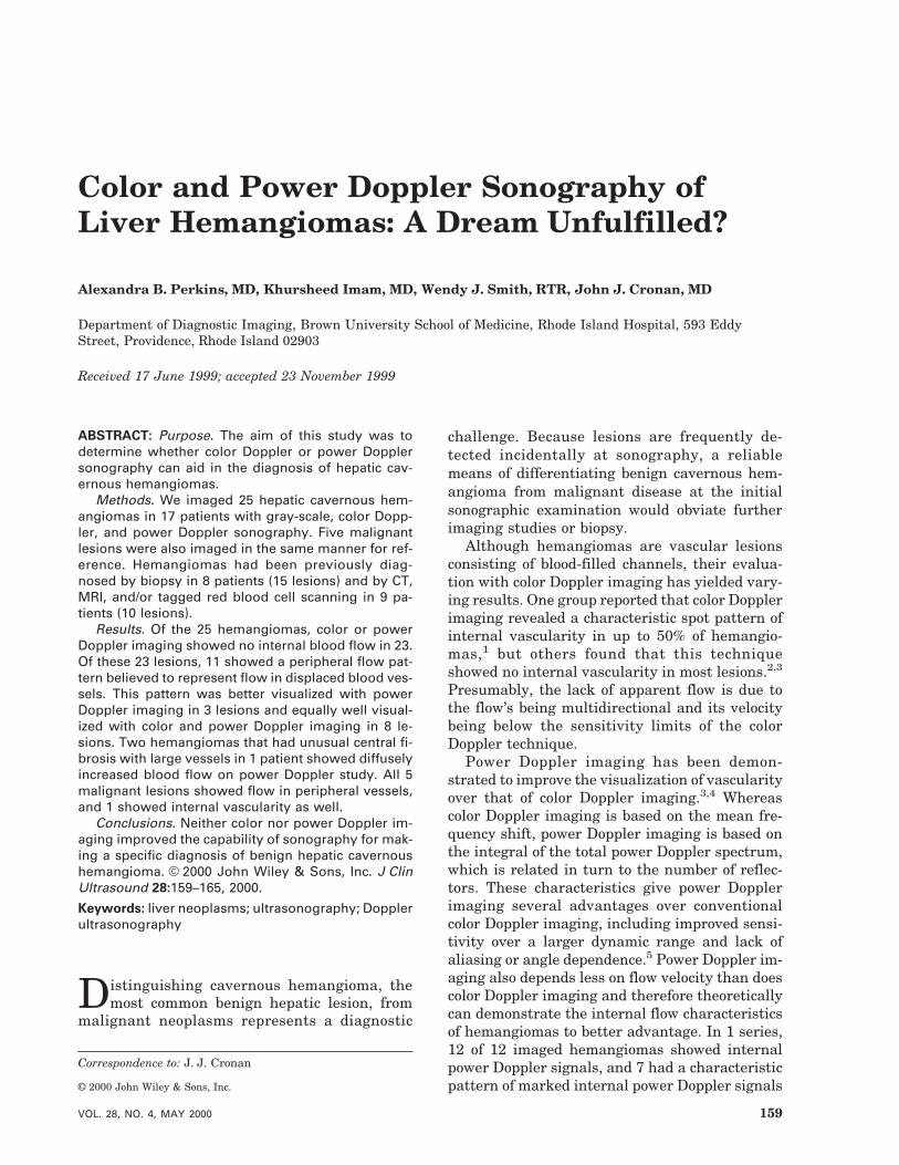
FIGURE 1. Color (A) and power (B) Doppler sonograms of a cavernous hemangioma (large arrows) in the right hepatic lobe demonstrate no flowwithin the lesion but peripheral flow in adjacent displaced vessels (small arrows). The diagnosis was confirmed by tagged red blood cell scanning(C). Homogeneous high uptake within the lesion (arrows) is apparent on this delayed image but not on earlier images (not shown).
PERKINS ET AL
160 JOURNAL OF CLINICAL ULTRASOUND

land Hospital. The 17 patients (8 men and 9women) located ranged in age from 30 to 78 years(mean, 60 years). The study was approved by ourinstitutional review board, and written informedconsent to participate was obtained from all pa-tients.
The color and power Doppler characteristics of25 focal liver lesions in these 17 patients wereanalyzed. Lesions were already known to repre-sent cavernous hemangiomas by open surgical bi-opsy (4 lesions in 2 patients), by percutaneousbiopsy (11 lesions in 6 patients6), or by a combi-nation of nuclear medicine–tagged red blood cellscanning, MRI, or contrast-enhanced CT (10 le-sions in 9 patients). In all cases of histologicallyproven hemangioma, the biopsies had been per-formed several years before the sonographic ex-aminations.
We also included 5 malignant lesions detectedand proven by biopsy for comparison; 4 weremetastatic lesions (2 from colon cancer and 2 fromlung cancer), and 1 was a primary hepatocellularcarcinoma. These 3–5-cm lesions were selectedbecause of their similar appearance to the cavern-ous hemangiomas. The 5 patients (3 men and 2women) who had malignant lesions ranged in agefrom 45 to 72 years.
All patients were examined with either an HDI3000 or an HDI 9 ultrasound scanner (AdvancedTechnology Laboratories, Bothell, WA) or an MR
700 ultrasound scanner (General Electric, Mil-waukee, WI). Either 3–5-MHz phased-array or4–7-MHz linear-array transducers were used forimaging, depending on the depth of the lesion.The color and power Doppler parameters were op-timized for each patient at the level of the lesionby using the lowest scale and filter settings andthe highest gain that did not produce backgroundnoise that obscured the signal within the lesion.Color gain was optimized by manipulating thegain until color noise first became apparent.Power gain was optimized by manipulating thegain until a few random speckles were visibleabove the homogeneous monochromatic back-ground or electronic noise. All patients were ableto suspend respiration adequately to obtain colorand power Doppler images. All images were ana-lyzed for the presence or absence of vascularitywithin the lesion by 3 of the authors (A. B. P.,K. I., and J. J. C., an experienced sonologist).
RESULTS
The 25 hemangiomas imaged ranged in size from1.0 to 8.4 cm (mean, 3.0 cm).
Twenty-three hemangiomas (92%) showed noevidence of internal flow with either power orcolor Doppler imaging. Of these lesions, 11 (48%)demonstrated foci of color flow that were periph-eral and distinctly separate from the lesion (Fig-
FIGURE 1. Continued.
DOPPLER SONOGRAPHY OF LIVER HEMANGIOMAS
VOL. 28, NO. 4, MAY 2000 161

ure 1). Although these areas were not examinedwith pulsed Doppler imaging, they were believedto represent displaced blood vessels adjacent tothe lesion and could not be shown to be related tothe hemangioma itself. The remaining 12 lesions(52%) showed no flow within or adjacent to thehemangioma.
Among the 11 hemangiomas that showed adja-cent vascular flow, power Doppler visualizedthose peripheral vessels better than did color
Doppler in 3 lesions (27%) (Figure 2); peripheralvessels in the remaining 8 lesions (73%) wereseen equally well with power or color Doppler im-aging (Figure 3).
Two hemangiomas in 1 patient showed markedinternal signals on power Doppler imaging thatwere absent on color Doppler study. These lesionshad the atypical histologic characteristics of largeamounts of connective tissue with sclerosis of vas-cular channels; they were also unusual in that
FIGURE 2. Color (A) and power (B) Doppler sonograms of a histologically proven hepatic cavernous hemangioma (solid arrows) demonstrate noflow within the lesion but some flow in adjacent displaced vessels (open arrow). Peripheral flow was better seen with power Doppler imaging.
PERKINS ET AL
162 JOURNAL OF CLINICAL ULTRASOUND

they contained 2 zones, a central fibrotic and scle-rotic zone and a peripheral zone with large, tor-tuous vessels.
Of the 5 malignant lesions evaluated, the 4
metastatic lesions showed flow in peripheral ves-sels, and the single case of primary hepatocellularcarcinoma showed flow in peripheral vessels aswell as internal branching vascularity.
FIGURE 3. Color (A) and power (B) Doppler sonograms of histologically proven hepatic hemangioma (solid arrows) both demonstrate equally wellthe absence of flow within the lesion and the presence of flow in adjacent displaced vessels (open arrows). An early contrast-enhanced CT scan(C) and a high-signal-intensity proton-density MRI scan (D) showed typical peripheral puddling of the contrast agent; the lesion (arrows) becamecompletely filled with contrast material on later images (not shown).
DOPPLER SONOGRAPHY OF LIVER HEMANGIOMAS
VOL. 28, NO. 4, MAY 2000 163

DISCUSSION
Cavernous hemangioma is a common benign he-patic lesion with a reported prevalence at autopsyof 0.4–7.5%.7,8 Distinguishing between benignhemangioma and malignant neoplasm is a diag-nostic challenge, particularly for patients with aknown primary malignancy, and generally in-volves percutaneous biopsy or imaging modalitiessuch as nuclear medicine, contrast-enhanced CT,or MRI. These additional tests can substantiallyincrease the cost of the workup. For these rea-sons, the ability to characterize hemangiomas bysonography is desirable.
The addition of power Doppler imaging to ab-dominal sonography has been shown to improvevisualization of vascular flow, particularly in therenal cortex.4 The application of power Dopplersonography to hepatic imaging is still being de-veloped. Unfortunately, we found no characteris-tic power Doppler appearance in 25 cavernoushemangiomas, most of which had been histologi-cally verified. In our series, 23 cavernous heman-giomas (92%) showed no internal vascularity oneither power or color Doppler imaging, an appear-ance that overlaps with that of metastases to theliver. Moreover, the incidence of that appearancein our series correlates well with the findings inanother series in which only color Doppler imag-ing was used.
Only 2 of the hemangiomas in our seriesshowed internal signals on power Doppler imag-ing. The unusual histopathologic characteristicsof these lesions, ie, central fibrosis and large ves-sels, may account for their different appearanceon power Doppler imaging. Cavernous hemangi-omas are histologically heterogeneous, and theirimaging appearance can vary depending on thesize of the vascular channels and the intervening
connective tissue septa.9 In our 2 unusual lesions,a slight elevation in gain in combination with thefibrotic central internal architecture could havecreated an illusion of flow.
Our results contrast with those of Choi et al,3
who described internal signals with power Dopp-ler imaging in 12 of 12 hemangiomas studied, aswell as those of Young et al,10 who described a“diffuse blush” on power Doppler imaging in 27 of27 hemangiomas studied. We suggest that the in-ternal architecture of the lesion determines theapparent “flow” visualized by power Doppler im-aging. The “blush” noted by Young et al is likelysimilar to the flow we observed in the 2 fibroticcavernous hemangiomas. Even if we postulatethat our population of cavernous hemangiomaswas biased by the inclusion of 15 lesions previ-ously verified by histopathology, this does not ex-plain the lack of flow seen on power Doppler im-aging in the remaining 10 lesions. Perhaps anincrease in the gain could create the appearanceof flow on power Doppler imaging, but so couldthe presence of fibrosis.
A potential limitation of our study is the sub-jectivity inherent in our optimizing the Dopplerparameters for each patient. However, given thevariability in habitus and lesion location amongpatients, we believe that an individualized opti-mization would provide the best possible assess-ment for each patient.
In summary, the great majority of cavernoushemangiomas in our series showed no inter-nal blood flow on power or color Doppler imaging,findings that overlap with the appearanceof metastatic lesions. Further studies of otherhepatic lesions may yet yield characteristicsonographic appearances. The addition ofsonographic contrast media to power and color
FIGURE 3. Continued.
PERKINS ET AL
164 JOURNAL OF CLINICAL ULTRASOUND

Doppler imaging may also improve the abil-ity to distinguish benign from malignant liverlesions11 and may provide areas for futurestudy.
REFERENCES
1. Tanaka S, Kitamura T, Fujita M, et al. Color Dopp-ler flow imaging of liver tumors. AJR Am J Roent-genol 1990;154:509.
2. Nino-Murcia M, Ralls PW, Jeffrey RB Jr, et al.Color flow Doppler characterization of focal hepaticlesions. AJR Am J Roentgenol 1992;159:1195.
3. Choi BI, Kim TK, Han JK, et al. Power versus con-ventional color Doppler sonography: comparison inthe depiction of vasculature in liver tumors. Radi-ology 1996;200:55.
4. Bude RO, Rubin JM, Adler RS. Power versus con-ventional color Doppler sonography: comparison inthe depiction of normal vasculature. Radiology1994;192:777.
5. Bude RO, Rubin JM. Power Doppler sonography.Radiology 1996;200:21.
6. Cronan JJ, Esparza AR, Dorfman GS, et al. Cav-ernous hemangioma of the liver: role of percutane-ous biopsy. Radiology 1988;166:135.
7. Mungovan JA, Cronan JJ, Vaccaro JP. Hepaticcavernous hemangiomas: lack of enlargement overtime. Radiology 1994;191:111.
8. Anthony PP. Tumours and tumour-like lesions ofthe liver and biliary tract. In: MacSween RN, An-thony PP, Scheuer PJ, editors. Pathology of theliver. 2nd ed. New York: Churchill Livingstone;1987. p 622.
9. Tung GA, Vaccaro JP, Cronan JJ, et al. Cavernoushemangioma of the liver: pathologic correlationwith high-field MR imaging. AJR Am J Roentgenol1994;162:1113.
10. Young LK, Yang WT, Chan KW, et al. Hepatichemangioma: quantitative color power US angiog-raphy—facts and fallacies. Radiology 1998;207:51.
11. Ernst H, Hahn EG, Balzer T, et al. Color Dopplerultrasound of liver lesions: signal enhance-ment after intravenous injection of the ultrasoundcontrast agent Levovist. J Clin Ultrasound 1996;24:31.
DOPPLER SONOGRAPHY OF LIVER HEMANGIOMAS
VOL. 28, NO. 4, MAY 2000 165