colon capsule endoscopy: a new method of investigating the ... · colon capsule endoscopy seems to...
TRANSCRIPT
Received:20.06.2008 Accepted: 02.07.2008J Gastrointestin Liver DisSeptember 2008 Vol.17 No 3, 347-352Address for correspondence: Silviu Iobagiu 3rd Medical Clinic, 19-21, Croitorilor Street, 400162, Cluj-Napoca, Romania E-mail: [email protected]
Colon Capsule Endoscopy: a New Method of Investigating the Large BowelSilviu Iobagiu, Lidia Ciobanu, Oliviu Pascu
3rd Medical Clinic, University “Iuliu Haţieganu” Cluj-Napoca, Romania
AbstractUsing the same principles as small bowel capsule
endoscopy, a new wireless device has been developed to explore the colon. Colon capsule endoscopy (CCE) is a safe and non-invasive method in visualizing the entire large bowel mucosa. A special preparation of the digestive tract is necessary to ensure the cleanliness of the colon and the progression of the capsule. CCE acquires images with a very good quality and accuracy. The actual experience confirmsthe possibility of using CCE as a method for screening colorectal polyps and cancer.
Key wordsColon – colon capsule – colonoscopy – screening
IntroductionWireless capsule endoscopy is a safe, non-invasive
procedure especially developed for exploring visually the small bowel. This very modern method is useful and cost-effective in probing bleeding of obscure origin, inflammatorybowel disease lesions, tumors or lesions produced by non-steroidal anti-inflammatory drugs. Capsule endoscopy hasas primary indication the visualization of small bowel areas that are inaccessible at upper and lower endoscopy.
The new technology has been recently improved so that it can be used to image the whole length of the colon as well. Capsule colon endoscopy (CCE) can serve as a complementary procedure to conventional colonoscopy when colonoscopy is incomplete, when colonoscopy is contraindicated or in patients who are unwilling to undergo it [1].
Conventional colonoscopy offers excellent means to explore colonic mucosa and has as major advantages the possibilities to obtain biopsies and to perform a polypectomy when necessary. At the same time, a traditional colonoscopy is a relatively invasive technique and involves high costs when performed under sedation or general anaesthesia. The potential complications decrease its applicability in screening and surveillance of colonic polyps and cancer or colonic inflammatory bowel disease. The quality of dataobtained through colonoscopy is highly operator-dependent [2]. Useful tests in detecting colon cancer in the general population are noninvasive screening methods (such as fecal occult blood test), but they have not a very high sensitivity [3]. The ideal screening modality in colorectal cancer should be noninvasive, safe, well accepted by the target population, cost-effective and with a high diagnostic accuracy [4]. The cost-effectiveness of two screening strategies using colonoscopy or CCE was evaluated through a computer model based on a Markov process [5]. It depends mainly on the ability to improve compliance to colorectal screening.
Colon capsule endoscopy seems to be a safe non-invasive procedure, with higher sensitivity than the fecal occult blood test in colon cancer screening in the general population. Other advantages are the direct visualization of colon mucosa with no need of sedation, intubation, insufflationor radiation [6].
Technical backgroundColon capsule endoscopy uses an ingestible wireless
miniature device especially designed for propulsion into the colon. The colon capsule has two cameras ensuring the capture of 4 images per second and 11 x 31 mm in dimension. This increased capsule length is generated by the need to achieve a specific weight-to-volume ratio allowing the deviceto progress into the large bowel within the pre-programmed time frame [7]. The device shuts off 3 minutes after the activation to save the battery energy and automatically turns back on after 1h 45min, before entering the colon. The special pre-programmed delay ensures a total operating time of about 10 hours. A 2-hour delay was observed to be

348 Iobagiu et al
the most appropriate for capsule reactivation in the terminal ileum in human healthy volunteers [7]. The colon capsule has a wide visual optic field and automatic light control.During the battery life, the colon capsule captures about 144,000 images. The whole progression of the capsule in the colon can be visualized using a real-time viewer and the captured data are downloaded to a computer workstation. After one booster, 70% patients eliminate the capsule through the anus during 10 hours after its ingestion. The second booster increases the elimination rate at 78%. The patients who eliminate the capsule have a colon residence time of 170+132 min (with sodium phosphate) and 178+128 min (without it) [8]. Patients who do not eliminate the capsule naturally may have to ingest an additional dose of laxatives or may need supplementary suppositories or colonoscopic capsule retrieval [9].
The mean time taken to read the CCE images is 62 minutes (range 45-90 minutes), with a speed range of 7-12 frames per second [4]. The capsule video review involves a “2 steps-both sides” technique. Physicians need training to ensure an accurate interpretation of the CCE images [9]. Reading the CCE images is time-consuming, similar to the reading of the small bowel images obtained with the earlier version of the procedure. The interobserver agreement in
reading CCE results is 80% [7], suggesting the need for improvement and the fact that a learning curve will be mandatory.
Based upon an expert-reading in detecting colon lesions, CCE has a sensitivity of 76%, a specificity of 100%, apositive predictive value of 100% and a negative predictive value of 78% [10].
PreparationBefore CCE, the colon has to be properly prepared.
The preparation methods should accomplish several aims: to clean the colon, to promote the capsule progression (‘booster”) and to allow a good visualization of the mucosa (“submarine view”).
One day before CCE, the patient has to ingest a clear liquid diet. In the evening before the procedure, 3 liters of polyethyleneglycol (PEG) will be ingested. Another liter of PEG is ingested in the next morning, then the capsule is swallowed with a glass of water. Prokinetic agents (domperidone), one or two “boosters” (sodium phosphate) – pending the capsule passage out of the stomach, as visualized through the real-time viewer - and laxatives (bisacodyl suppositories) ensure the capsule propulsion. During the CCE, an optional low-fiber snack is allowed.
Fig 1. The assessment of colon cleanliness using CCE. a. excellent preparation (only small fragments of adherent feces); b. good preparation (small quantities of dark fluid or feces, withno interference with the examination); c. fair preparation (dark fluid or feces precluding acompletely reliable examination); d. poor preparation (large amount of fecal residue).
Colon capsule endoscopy: a new method to investigate the large bowel 349
Placing patients in the right lateral position after capsule ingestion and before the device enters the pylorus is a simple method to increase the complete examination rate of the bowel by reducing the gastric transit time [11].
During the reading of the CCE images, the physician has to assess the colon cleanliness (Fig. 1). It is considered poor (a large amount of fecal residues), fair (enough feces or dark fluid precluding a completely reliable visualization of themucosa), good (only small quantities of feces or dark fluid,not interfering with visualization of colonic wall) or excellent (presence of small bits of adherent feces). In a Belgian study [7], the preparation was evaluated as excellent in 30% of
cases, good in 58%, fair in 6% and poor in 3%.
ContraindicationsColon capsule endoscopy should not be performed in
pregnant female patients and in patients with high risk for capsule retention: patients with Crohn’s disease with known or suspected stenosis, patients with large small bowel tumors, patients with surgical anastomosis. The known allergy to sodium phosphate, polyethyleneglycol solution, domperidone or bisacodyl is also a contraindication for the procedure.
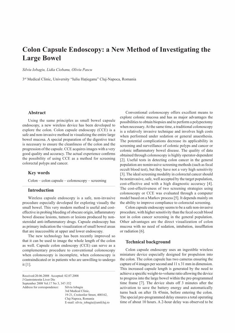
Fig 2. Representative normal CCE images at different sites in the colon. a. ileocecal valve; b. ascending colon; c. transverse colon; d. descending colon; e. sigmoid colon; f. rectum.
350 Iobagiu et al
Acquired imagesUsing CCE in visualizing the colon mucosa, very good
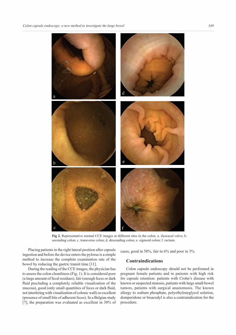
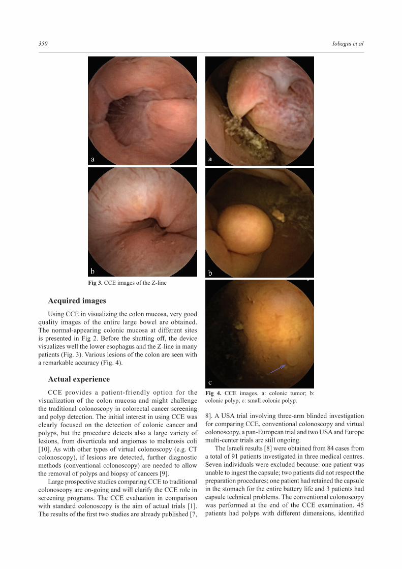
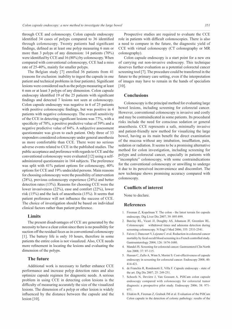
quality images of the entire large bowel are obtained. The normal-appearing colonic mucosa at different sites is presented in Fig 2. Before the shutting off, the device visualizes well the lower esophagus and the Z-line in many patients (Fig. 3). Various lesions of the colon are seen with a remarkable accuracy (Fig. 4).
Actual experienceCCE provides a patient-friendly option for the
visualization of the colon mucosa and might challenge the traditional colonoscopy in colorectal cancer screening and polyp detection. The initial interest in using CCE was clearly focused on the detection of colonic cancer and polyps, but the procedure detects also a large variety of lesions, from diverticula and angiomas to melanosis coli [10]. As with other types of virtual colonoscopy (e.g. CT colonoscopy), if lesions are detected, further diagnostic methods (conventional colonoscopy) are needed to allow the removal of polyps and biopsy of cancers [9].
Large prospective studies comparing CCE to traditional colonoscopy are on-going and will clarify the CCE role in screening programs. The CCE evaluation in comparison with standard colonoscopy is the aim of actual trials [1]. The results of the first two studies are already published [7,
8]. A USA trial involving three-arm blinded investigation for comparing CCE, conventional colonoscopy and virtual colonoscopy, a pan-European trial and two USA and Europe multi-center trials are still ongoing.
The Israeli results [8] were obtained from 84 cases from a total of 91 patients investigated in three medical centres. Seven individuals were excluded because: one patient was unable to ingest the capsule; two patients did not respect the preparation procedures; one patient had retained the capsule in the stomach for the entire battery life and 3 patients had capsule technical problems. The conventional colonoscopy was performed at the end of the CCE examination. 45 patients had polyps with different dimensions, identified
Fig 3. CCE images of the Z-line
Fig 4. CCE images. a: colonic tumor; b: colonic polyp; c: small colonic polyp.
Colon capsule endoscopy: a new method to investigate the large bowel 351
through CCE and colonoscopy. Colon capsule endoscopy identified 34 cases of polyps compared to 36 identifiedthrough colonoscopy. Twenty patients had significantfindings, defined as at least one polyp measuring 6 mm ormore than 3 polyps of any dimension: 14 patients (70%) were identified by CCE and 16 (80%) by colonoscopy. Whencompared with conventional colonoscopy, CCE had a miss rate of 25-40%, mainly for smaller polyps.
The Belgian study [7] enrolled 36 patients from 41 (reasons for exclusion: inability to ingest the capsule in one patient and technical problems in four patients). Significantlesions were considered such as the polyps measuring at least 6 mm or at least 3 polyps of any dimension. Colon capsule endoscopy identified 19 of the 25 patients with significantfindings and detected 7 lesions not seen at colonoscopy.Colon capsule endoscopy was negative in 6 of 25 patients with positive colonoscopic findings, but was positive in 4patients with negative colonoscopy. The overall sensitivity of the CCE in detecting significant lesions was 77%, with aspecificity of 70%, a positive predictive value of 59% and anegative predictive value of 84%. A subjective assessment questionnaire was given to each patient. Only three of 33 responders considered colonoscopy under general anesthesia as more comfortable than CCE. There were no serious adverse events related to CCE in the published studies. The public acceptance and preference with regards to CCE and the conventional colonoscopy were evaluated [12] using a self-administered questionnaire in 164 subjects. The preference was split with 41% patient options for colonoscopy, 40% options for CCE and 19% undecided persons. Main reasons for choosing colonoscopy were the possibility of intervention (28%), previous colonoscopy experience (24%) and better detection rates (13%). Reasons for choosing CCE were the lower invasiveness (22%), ease and comfort (22%), lower risk (15%) and the lack of anaesthesia (15%). It seems that patient preference will not influence the success of CCE.The choice of investigation should be based on individual clinical factors rather than patient preference.
Limits The present disadvantages of CCE are generated by the
necessity to have a clear colon since there is no possibility for suction off the residual feces as in conventional colonoscopy [1]. The battery life is only 10 hours, therefore in some patients the entire colon is not visualized. Also, CCE needs more refinement in locating the lesions and evaluating thedimension of the polyps.
The futureAdditional work is necessary to further enhance CCE
performance and increase polyp detection rates and also optimize capsule regimen for diagnostic needs. A serious problem in using CCE in detecting colon lesions is the difficulty of measuring accurately the size of the visualizedlesions. The dimension of a polyp or other lesion is widely influenced by the distance between the capsule and thelesion [10].
Prospective studies are required to evaluate the CCE role in patients with difficult colonoscopies. There is alsoa need to compare in the future, the diagnostic yield of CCE with virtual colonoscopy (CT colonography or MR colonography).
Colon capsule endoscopy is a start point for a new era of carrying out non-invasive endoscopy. This technique deserves further evaluation as a potential colorectal cancer screening tool [7]. The procedure could be transferred in the future to the primary care setting, even if the interpretation of images may have to remain in the hands of specialists [10].
ConclusionsColonoscopy is the principal method for evaluating large
bowel lesions, including screening for colorectal cancer. However, conventional colonoscopy is invasive and costly and may be contraindicated in some patients. Its procedural risks include the need for conscious sedation or general anaesthesia. CCE represents a safe, minimally invasive and patient-friendly new method for visualizing the large bowel, having as its main benefit the direct examinationof the mucosa without any intubation, insufflation, pain,sedation or radiation. It seems to be a promising alternative method for colon investigation, including screening for polyps and colorectal cancer, especially in patients with “incomplete” colonoscopy, with some contraindications for the conventional colonoscopy or unwilling to undergo it due to its perceived inconvenience and discomfort. The new technique shows promising accuracy compared with colonoscopy.
Conflicts of interestNone to declare.
References 1. Fireman Z, Kopelman Y. The colon - the latest terrain for capsule
endoscopy. Dig Liver Dis 2007; 39: 895-899. 2. Barclay RL, Vicari JJ, Doughty AS, Johanson JF, Greenlaw RL.
Colonoscopic withdrawal times and adenoma detection during screening colonoscopy. N Engl J Med 2006; 355: 2533-2541.
3. Faivre J, Dancourt V, Lejeune C et al. Reduction in colorectal cancer mortality by fecal occult blood screening in a French controlled study. Gastroenterology 2004; 126: 1674-1680.
4. Mandel JS. Screening for colorectal cancer. Gastroenterol Clin North Am 2008; 37: 97-115.
5. Hassan C, Zullo A, Winn S, Morini S. Cost-effectiveness of capsule endoscopy in screening for colorectal cancer. Endoscopy 2008; 40: 414-421.
6. de Franchis R, Rondonotti E, Villa F. Capsule endoscopy - state of the art. Dig Dis 2007; 25: 249-251.
7. Schoofs N, Devière J, Van Gossum A. PillCam colon capsule endoscopy compared with colonoscopy for colorectal tumor diagnosis: a prospective pilot study. Endoscopy 2006; 38: 971-977.
8. Eliakim R, Fireman Z, Gralnek IM et al. Evaluation of the PillCam Colon capsule in the detection of colonic pathology: results of the

352 Iobagiu et al
first multicenter, prospective, comparative study. Endoscopy 2006;38: 963-970.
9. Tran K. Capsule colonoscopy: PillCam Colon. Issues Emerg Health Technol 2007; 106: 1-4.
10. Galmiche JP, Coron E, Sacher-Huvelin S. Recent developments in capsule endoscopy. Gut 2008; 57: 695-703.
11. Liao Z, Li F, Li ZS. Right lateral position improves complete examination rate of capsule endoscope: a prospective randomized, controlled trial. Endoscopy 2008; 40: 483-487.
12. Teoh WC, Abey S, Ooi M, Mcdonald J. Public Perception of the PillCam Colon Versus Colonoscopy. Gastrointestinal Endoscopy 2008; 67: AB323.