colloidal micro- and nano-particles as templates for polyelectrolyte multilayer capsules
TRANSCRIPT

Advances in Colloid and Interface Science xxx (2014) xxx–xxx
CIS-01401; No of Pages 12
Contents lists available at ScienceDirect
Advances in Colloid and Interface Science
j ourna l homepage: www.e lsev ie r .com/ locate /c i s
Colloidal micro- and nano-particles as templates for polyelectrolytemultilayer capsules
Bogdan V. Parakhonskiy a,b, Alexey M. Yashchenok c, Manfred Konrad d, Andre G. Skirtach c,e,⁎a BIOtech Center, Department of Physics, University of Trento, via delle Regole 101, 38123 Mattarello, Italyb Shubnikov Institute of Crystallography, Russian Academy of Science, Leninskii prospekt 59, Moscow 119333, Russiac Department of Interfaces, Max-Planck Institute of Colloids and Interfaces, Golm, Potsdam D-14476, Germanyd Enzyme Biochemistry Laboratory, Max Planck Institute for Biophysical Chemistry, D-37077 Gottingen, Germanye Department of Molecular Biotechnology & Nano-Bio (NB)-Photonics, Ghent University, 9000 Ghent, Belgium
⁎ Corresponding author at: University of Ghent, BelgiumE-mail address: [email protected] (A.G. Skirta
0001-8686/$ – see front matter © 2014 Elsevier B.V. All rihttp://dx.doi.org/10.1016/j.cis.2014.01.022
Please cite this article as: Parakhonskiy BV,Colloid Interface Sci (2014), http://dx.doi.org
a b s t r a c t
a r t i c l e i n f oAvailable online xxxx
Keywords:MicrocapsulesColloidal particlesTemplatesPolyelectrolytesMultilayers
Colloidal particles play an important role in various areas ofmaterial and pharmaceutical sciences, biotechnology,and biomedicine. In this overview we describe micro- and nano-particles used for the preparation of poly-electrolyte multilayer capsules and as drug delivery vehicles. An essential feature of polyelectrolyte multilayercapsule preparations is the ability to adsorb polymeric layers onto colloidal particles or templates followed bydissolution of these templates. The choice of the template is determined by various physico-chemical conditions:solvent needed for dissolution, porosity, aggregation tendency, as well as release of materials from capsules.Historically, the first templates were based on melamine formaldehyde, later evolving towards more elaboratematerials such as silica and calcium carbonate. Their advantages and disadvantages are discussed here incomparison to non-particulate templates such as red blood cells. Further steps in this area include developmentof anisotropic particles, which themselves can serve as delivery carriers. We provide insights into application ofparticles as drug delivery carriers in comparison to microcapsules templated on them.
© 2014 Elsevier B.V. All rights reserved.
Contents
1. Introduction . . . . . . . . . . . . . . . . . . . . . . . . . . . . . . . . . . . . . . . . . . . . . . . . . . . . . . . . . . . . . . . 02. Particles and templates . . . . . . . . . . . . . . . . . . . . . . . . . . . . . . . . . . . . . . . . . . . . . . . . . . . . . . . . . . 0
2.1. Synthesis of particles . . . . . . . . . . . . . . . . . . . . . . . . . . . . . . . . . . . . . . . . . . . . . . . . . . . . . . . 02.1.1. Calcium phosphate . . . . . . . . . . . . . . . . . . . . . . . . . . . . . . . . . . . . . . . . . . . . . . . . . . . . 02.1.2. Calcium carbonate (CaCO3) . . . . . . . . . . . . . . . . . . . . . . . . . . . . . . . . . . . . . . . . . . . . . . . . 02.1.3. Manganese carbonate (MnCO3) . . . . . . . . . . . . . . . . . . . . . . . . . . . . . . . . . . . . . . . . . . . . . . 02.1.4. Cadmium carbonate (CdCO3) . . . . . . . . . . . . . . . . . . . . . . . . . . . . . . . . . . . . . . . . . . . . . . . 02.1.5. Synthesis of dextran-hydroxyethyl methacrylate (dex-HEMA) microgels . . . . . . . . . . . . . . . . . . . . . . . . . . . . 02.1.6. Fabrication of alginate hydrogel microspheres . . . . . . . . . . . . . . . . . . . . . . . . . . . . . . . . . . . . . . . . 02.1.7. Formation of mesoporous silica particles . . . . . . . . . . . . . . . . . . . . . . . . . . . . . . . . . . . . . . . . . . 0
2.2. Stability and decomposition of particles . . . . . . . . . . . . . . . . . . . . . . . . . . . . . . . . . . . . . . . . . . . . . . . 02.3. Release from particles . . . . . . . . . . . . . . . . . . . . . . . . . . . . . . . . . . . . . . . . . . . . . . . . . . . . . . . 0
2.3.1. Release by recrystallization at normal pH . . . . . . . . . . . . . . . . . . . . . . . . . . . . . . . . . . . . . . . . . . 02.3.2. Release at low pH . . . . . . . . . . . . . . . . . . . . . . . . . . . . . . . . . . . . . . . . . . . . . . . . . . . . 0
2.4. Microparticles as carriers of enzymes . . . . . . . . . . . . . . . . . . . . . . . . . . . . . . . . . . . . . . . . . . . . . . . . 02.5. Biomolecule-based particles . . . . . . . . . . . . . . . . . . . . . . . . . . . . . . . . . . . . . . . . . . . . . . . . . . . . 0
3. Capsules . . . . . . . . . . . . . . . . . . . . . . . . . . . . . . . . . . . . . . . . . . . . . . . . . . . . . . . . . . . . . . . . 03.1. From particles to capsules: dissolution of templates . . . . . . . . . . . . . . . . . . . . . . . . . . . . . . . . . . . . . . . . . 03.2. Release of cargo from capsules . . . . . . . . . . . . . . . . . . . . . . . . . . . . . . . . . . . . . . . . . . . . . . . . . . . 0
4. Complex morphologies of particles and capsules . . . . . . . . . . . . . . . . . . . . . . . . . . . . . . . . . . . . . . . . . . . . . . 04.1. Multi-compartment particles and capsules . . . . . . . . . . . . . . . . . . . . . . . . . . . . . . . . . . . . . . . . . . . . . 04.2. Anisotropic capsules and anisotropic carriers . . . . . . . . . . . . . . . . . . . . . . . . . . . . . . . . . . . . . . . . . . . . 0
.ch).
ghts reserved.
et al, Colloidal micro- and nano-particles as templates for polyelectrolyte multilayer capsules, Adv/10.1016/j.cis.2014.01.022

2 B.V. Parakhonskiy et al. / Advances in Colloid and Interface Science xxx (2014) xxx–xxx
5. Summary . . . . . . . . . . . . . . . . . . . . . . . . . . . . . . . . . . . . . . . . . . . . . . . . . . . . . . . . . . . . . . . . 0References . . . . . . . . . . . . . . . . . . . . . . . . . . . . . . . . . . . . . . . . . . . . . . . . . . . . . . . . . . . . . . . . . . 0
1. Introduction
The significance of micro- and nano-particles ranges from earlydevelopments in the area of colloidal particles to a broad range of indus-trial and nanotechnology applications in physical, biological, and medi-cal sciences. In this overview, we focus on the class of organic andinorganic particles which are used as templates for the preparation ofpolyelectrolyte multilayer capsules. The choice of the template for thepreparation of these capsules is particularly important in different fieldsof application, with advantages and disadvantages pertaining to thetemplate dissolution, the stability of the polymer shell, and tendencyto aggregation. In addition, these particles can serve as drug deliverycarriers themselves.
There are two important classes of templates, possessing eithersmooth or porous surface. The surface porosity of templates determinesa number of parameters of polyelectrolyte multilayer capsules, includ-ing the easiness of preparation, polymer shell thickness, and efficiencyof loading of molecules. For example, smooth particles usually needthe addition of more layers of the polyelectrolytes, and the shell wallis quite thin, on the order of one to two nanometers. The interior ofsuch capsules is typically clean and transparent, and after dissolutionof the core particle, such capsules look like empty balloons. On the con-trary, during the layer-by-layer (LbL) adsorption on porous cores, thepolyelectrolytes penetrate into the matrix, resulting in a substantiallythicker polymer shell. In this case, the capsules look like sponges afterthe core dissolution. This differential property is very important forloading of molecules and for protection of encapsulated materials. Forthe capsules templated on porous particles, pores are utilized for load-ing producing relatively large capacity. Polyelectrolyte capsules basedon smooth particles have a bigger inner volume, can easily encapsulatesubstances by heating and thereby shrinking of surface, and release ofcargo from such particles can be more easily manipulated by externalstimuli.
Colloidal particles used directly as carriers represent an importantclass of materials and stand on their own in various areas of research.
Table 1Types and sizes of particles, and chemicals used for their decomposition in the construction of
Type of the particles Size of the particles used as templates for polyelectrol
Smooth particlesSilica 0.5 [2] 1 [2], 1.9 [3], 4.5–5 [4–7]Melamine–formaldehyde 1 [8], 2 [9,10] 3.2–3.5 [8,11,12], 4.2 [13], 5–5.7 [14–16
Polystyrene 0.64 [8] 4.4–4.6 [5,17,18], 9.6–10.6 [19,20] 20 [20]CdCO3 cubic shape 2.5, 4–6 [21]CaCO3 cubic shape 4–5 [22]Calcium phosphate modified by PAH 158–300 nm [23]Gold nanoparticles 13.5 nm [24]Micro gel dex-HEMA 150 [25]Alginate hydrogel microspheres 4.25 [26]poly-DL-lactic acid particles (PLA) 4–8 [10]
Halloysite Cylinder 50 nm × 300 nm [27]
Porous particlesMesoporous silica 0.4 [28], 2–4 [1,29]Calcium carbonate spherical 4–5 [18,22,30,31], 9 [32], 11.5 [33]MnCO3 1.9.[9] 2.5–2.8 [9,34], 3.6 [32], 5–5.5 [35,36]Calcium carbonate elliptic 2–4 [22]
Hybrid templatesHuman erythrocytes 5–6 [37–42]5–6 [37–42]Virus-like particles ~4 [43]Liposomes 0.1–0.5 [44]
Please cite this article as: Parakhonskiy BV, et al, Colloidal micro- and naColloid Interface Sci (2014), http://dx.doi.org/10.1016/j.cis.2014.01.022
We elaborate here on the applicability and potential role of particlesas templates for the preparation of polyelectrolyte multilayer capsuleson one hand, and as stand-alone carriers on the other hand.
2. Particles and templates
2.1. Synthesis of particles
A number of different particles are used as templates for the prepa-ration of polyelectrolyte multilayer capsules. Information about parti-cles most frequently used for the fabrication of polymeric capsules issummarized in Table 1. Templates can be divided into two major clas-ses, namely smooth and porous particles. The smooth ones encompass,for example, melamine–formaldehyde, silica, and polystyrene. Most ofthem are commercially available in a large range of sizes: from approx-imately 100 nm to millimeters. Advantages of these particles includegood stability (zeta-potential of ~35mV) and excellentmonodispersity.Specific modifications of the surface of these particles with amino-,carboxy-, mercapto-, maleimido-, hydroxy-, epoxy-, sulfo-, and otherfunctional groups are available. The tailored properties of particlesallow extending their potential application to medical diagnostics,pharmaceutical industry, biotechnology, molecular biology, analytics,etc. For drug delivery purposes, these types of particles are of limiteduse due to low loading efficacy on the functionalized surface. Themost advanced approach consists in the loading of the hollowpolymericshell which is obtained when the solid core is decomposed, Fig. 1.
Porous particles constitute another type of solid particles. They havea number of advantages over their smooth counterparts, including facilesynthesis, low cost of processing, and high porositywhich leads to an in-creased loading capacity. The relatively big variation in size (polydisper-sity) and strong aggregative behavior are some disadvantages of thistype of templates. Production of porous particles (calcium carbonate)is relatively easy, including the mixing of two salt solutions either inthe absence or in the presence of special additiveswhich are used to ob-tain certain surface modifications or a particular morphology.
polymeric capsules.
yte capsules, microns Size range, microns Solvent
2–5 HF, HF/NH4F] 3.2–5.7 HCl, N,N-dimethylformamide (DMF) or
dimethyl sulfoxide (DMSO).4–20 THF2.5–6 HCl, pH = 14–5 HCl, EDTA0.16–0.3 HCl0.14 Potassium cyanide150 NaOH solution4.254–8 1:1 mixture of 1-methyl-2-pyrrolindinone
acid and organic solvent (acetone)0.05 × 0.3
0.4–4 HF, HF/NH4F4–11.5 EDTA pH 6; acid solution pH b 62.8–5.52–4 EDTA pH 6; acid solution pH b 6
5–6 140 mM NaCl and 1.2% NaOCl~40.1–0.5
no-particles as templates for polyelectrolyte multilayer capsules, Adv

Fig. 1. Transmission electron microcopy (TEM) image of a) catalase-loaded mesoporous silica (MS) spheres coated with (PLL/PGA)3 multilayers, and b) following removal of the MStemplate; c) fluorescence images of (PLL/PGA)3 microcapsules loaded with FITC-labeled catalase. The scale bar in the inset corresponds to 800 nm. PLL/PGA layers were assembledfrom a 0.05 M MES, pH 5.5 buffer. The MS spheres were dissolved using HF/NH4F at pH= 5. Figure reprinted from [1]. Copyright Wiley-VCH Verlag GmbH & Co. KGaA.
3B.V. Parakhonskiy et al. / Advances in Colloid and Interface Science xxx (2014) xxx–xxx
2.1.1. Calcium phosphateCalcium phosphate synthesis usually involves mixing of two salt
solutions to initiate crystal growth. Calcium phosphate precipitatesform as a result of colloidal aggregation during mixing of CaCl2 andNa2HPO4 in a buffer solution (TRIS pH 7.4) [45]. The PAH-modifiednanoparticles of calcium phosphate were prepared at room tem-perature by rapidly pumping aqueous solutions of calcium lactate,(NH4)2HPO4, and poly(allylamine hydrochloride)(PAH) in a volumeratio of 1:1:1 into 4 volume parts of water, forming particles withdiameter of about 158 nm [23]. Another approach for the synthesis
Fig. 2. Bimodal microscopic images of CaCO3 particles: Scanning electron microscope (SEM) imcrystals after recrystallization (c). The 2-photon excited fluorescence images show a single varesidual amount of dye attached to the calcite edges could be observed (d). The insets in (b) a[50]. Copyright Wiley-VCH Verlag GmbH & Co. KGaA.
Please cite this article as: Parakhonskiy BV, et al, Colloidal micro- and nanColloid Interface Sci (2014), http://dx.doi.org/10.1016/j.cis.2014.01.022
of smaller calcium phosphates nanoparticles (20 nm) was proposedby Y. Cai [46]: A CaCl2 solution was dropwise added to a Na2HPO4
solution containing different concentrations of hexadecyl(cetyl)trimethyl ammonium bromide (CTAB) in magnetically stirred vesselsat 20 °C.
2.1.2. Calcium carbonate (CaCO3)Synthesis of calcium carbonate templates is based on crystal growth
of polycrystalline spherical vaterite particles precipitated by mixing ofCaCl2 and Na2CO3 solutions [47]. The nucleation and growth rate of
ages show the 430 nm vaterite containers after their synthesis (a), and the calcite singleterite container loaded with the Rhodamine Rh6G dye (b); after recrystallization, only and (d) show fluorescence intensity profiles along the marked axes. Figure reprinted from
o-particles as templates for polyelectrolyte multilayer capsules, Adv

4 B.V. Parakhonskiy et al. / Advances in Colloid and Interface Science xxx (2014) xxx–xxx
the vaterite particles is determined by the supersaturation level of thedissolved amorphous CaCO3 [48]. The final size of the vaterite particlesdepends strongly on the concentration of the reagents, the solubilityof the salts, the reaction time, and the rotation speed during mixing. Itwas shown that at high salt concentrations (1 M), at stirring rates ofup to 1500 rpm, and reaction time of about 2 min, the size of CaCO3
particle was reduced to 3 μm [49]. Recently, size control of sphericalvaterite particles in the range from 10 down to 0.4 μm (Fig. 2a) hasbeen achieved in a mixture of water and ethylene glycol (EG) [50].The presence of EG during synthesis of CaCO3 particles diminishes themolecular diffusion, reduces the rate and the probability of nucleation,which leads to stabilization of the particles. Depending on the size ofthe particles, the pore size can be varied from 10 nm to 60 nm. Thus,with pore sizes well above 10 nm, the CaCO3 particles are not nano-porous, but can be considered mesoporous [51]. In 430 nm vateriteparticles, the average pore size is ~30 nm [52], in 4 μm-particles it is~35 nm, and 18 μm-particles have a pore size distribution rangingfrom 20 to 60 nm [51]. Thus, it appears that upon decreasing the sizeof the particles to less than 4 μm, the pore size reaches an averagevalue of 30–35 nm. Biocompatibility tests for vaterite particles indicatedno cytotoxicity and no influence on viability or metabolic activity [52].
2.1.3. Manganese carbonate (MnCO3)The procedure for manganese carbonate particles synthesis is simi-
lar to calcium carbonate synthesis described before, using salt solutionswhich contain appropriate ions [21] The acidic manganese sulfate solu-tion is added to NH4HCO3 in a volume ratio of 1:1, the stirred mixture isthen aged at 50 °C for 16 h. The resulting MnCO3 particles have roundshapes, and their diameters range from 1.85 μm to 0.3 mm [9].
2.1.4. Cadmium carbonate (CdCO3)This type of particles is often prepared according to the method de-
scribedby Janekovic andMatijevic [53]. A 10Murea solution aged for 24h at 80 °C is quickly added to a preheated 20 mM CdSO4 solution. Thisprocedure leads to monodisperse cubic CdCO3 crystals with a linearsize of about 2.5 μm. Temperature has been found to be critical for theshape of the CdCO3 crystals. Thus, in the range of temperatures used(70–75 °C), large polydisperse spherical CdCO3 particles (4–6 μm) areformed [21].
2.1.5. Synthesis of dextran-hydroxyethyl methacrylate (dex-HEMA)microgels
Dex-HEMAwas slowly introducedwith a pipette into a PEG solutionunder slow continuous stirring in a glass beaker. Upon formation of ahomogeneous dispersion, 35 ml dimethyl aminoethyl methacrylate(DMAEMA) was added, and the mixture was slowly stirred for 1 min.In the next step, radical polymerization was started by addition of N,N,N′,N′-tetramethylethylenediamine (TEMED) (100 ml; pH neutralizedby 4 M NaCl) and potassium persulfate (KPS). The mixture was stirredfor 15 min, and then allowed to stand for 30 min. The resultingmicrogels were centrifuged/washed with 20 ml water (twice) andstored in 5 ml water at 20 °C [25].
2.1.6. Fabrication of alginate hydrogel microspheresAn emulsification method was used for the preparation of alginate
hydrogel microspheres. Briefly, 50 g of 3 wt.% sodium alginate aqueoussolution was dispersed in 75 g of isooctane containing 1.7 g of thesurfactant SPAN 85 (sorbitan trioleate) using an ultrasonicator at 60%power for 5 min. A solution containing 0.9 g of TWEEN 85(polyoxyethylene sorbitan trioleate) in 5 g of isooctane was thenadded to the emulsion and ultra-sonicated at the same power foranother 5 min to achieve stable water/oil emulsion droplets. Followingthis step, 20 mL of aqueous solution containing 10 wt.% of calciumchloride was added. The microspheres were then rinsed three timeswith deionized water by centrifugation.
Please cite this article as: Parakhonskiy BV, et al, Colloidal micro- and naColloid Interface Sci (2014), http://dx.doi.org/10.1016/j.cis.2014.01.022
2.1.7. Formation of mesoporous silica particlesThe reaction mixture for the synthesis of mesoporous silica particles
was prepared directly in an autoclavable polypropylene bottle at~30 °C. A total of 19.6 g of CTAB followed by 10 g of solid Na2SiO3
were dissolved in 350 ml of distilled water, resulting in a clear solution.Then, 25 ml of ethyl acetate were quickly added under stirring. After30 s, the stirring was stopped, and the mixture was allowed to standat ambient temperature for 5 h; after this period of aging, the pHreached a constant value. During the aging process, organic solventswere allowed to evaporate through leaks in the cap of the bottle. Theresulting solids were recovered by filtration of the warm reaction mix-ture, extensively washed with distilled water and ethanol, and driedat ambient temperature. The template was removed by calcination at600 °C for 20 h in flowing air [54].
2.2. Stability and decomposition of particles
The most important parameter of colloidal systems is the stability ofparticles which depends on the degree of ion adsorption, and, therefore,on the zeta potential. The smooth particles melamine–formaldehyde,silica, and polystyrene have relative high zeta-potentials (~50 mV),and, therefore, high stability. The high surface charge prevents these par-ticles from aggregation; furthermore, their morphology is unchangeable.
In contrast to smooth particles, porous ones, for example CaCO3, arenot stable in aqueous solution or during extended steps of synthesis,and they transform to the more stable calcite structure [50]. Early stud-ies have demonstrated the influence of aqueous and PBS buffer onCaCO3 morphology (Fig. 2c). In order to prevent recrystallization, parti-cles can be either transferred to pure ethanol or dried at 70 °Cduring 1 hand then kept as a powder.
In contrast to the stability of the aforementioned particles, the de-composable ones can be treated to obtain polymeric hollow carriersby immersion into appropriate solvents. These chemicals are listed inTable 1. Depending on thematerial of the particles, the dissolution pro-cess lasts from several minutes to several hours, and this is followed bythorough washing with water. When using chemicals for dissolution ofparticles, caution should be taken into account, since in certain casesthese may include acidic media or might have toxic effects. Specifically,silica particles usually are decomposed in 0.3 M HF consisting of a mix-ture of HF and NH4F. N,N- dimethylformamide (DMF)/dimethyl sulfox-ide (DMSO) and tetrahydrofuran (THF) have been used for melamine-formaldehyde and polystyrene particles, respectively. Calcium carbon-ate particles have been found to dissolve by immersing them into acidicmedia, such as NaCl/HCl buffer (pH 2.0), as well as by complexationwith the chelating agent EDTA in appropriate solution (0.2 M, pH 7.5)[31,55].
The gold core was removed and transformed in a gold cyanidecomplex according to Eq. (1).
2Auþ 1=2O2 þ H2Oþ 4KCN→2K Au CNð Þ2� �þ 2KOH ð1Þ
Red blood cells can be removed by exposure to amixture of 140mMNaCl and 1.2% NaClO.
2.3. Release from particles
The release of biomolecules, such as small drug molecules fromparticles [56], is governed by an interplay of drug desorption and carrierdissolution [57]. This process usually is very slow, especially in theabsence of a payload-specific solvent, but could be increased when thecarrier size is reduced. During the desorption/adsorption process,molecules get detached and reattached until a dynamic equilibrium isreached, causing small oscillatory modulations in the release curve[57–59]. Furthermore, the particles themselves can be degraded ordissolved by the surrounding medium, causing the payload to diffusefrom the carrier.
no-particles as templates for polyelectrolyte multilayer capsules, Adv

5B.V. Parakhonskiy et al. / Advances in Colloid and Interface Science xxx (2014) xxx–xxx
The typical time-dependence of release from porous silica particleshas been discussed earlier. The release is observed to be very fast duringthe first few hours (releasing up to 75%), and then the release curvereaches saturation, with around 10% of encapsulated material remain-ing bound for longer time. This has been demonstrated for the releaseof doxorubicin from silica particles [60]; Brilliant Blue F from porous sil-ica particles [61]; Orange 2, Rhodamine 6G, and doxorubicin encapsu-lated in millimeter-sized silicagel particles [62], and for gentamicinreleased over a period of 20 days from mesoporous silica [63].
When the particles start to dissolve, the release is enhanced andreaches 100%when all carriers are dissolved. [64]. This is commonly ob-served for biodegradable carriers such as the well-known gelatin,polyalginate capsules. One of the typical examples is poly(D,L-lactic-co-glycolic) acid PLGA particles: Continuous release of serum albuminfrom biodegradable PLGA particles during 60 days has been demon-strated [65]. Also, a number of particles based on carbonate and gelatincan dissolve at low pH in the stomach, and cargo is rapidly released.
2.3.1. Release by recrystallization at normal pHThe combination of dissolution and desorption processes is a unique
property of slowly dissolving crystals, or it happens during recrystalliza-tion of the containers. This effect can be seen on the calcium carbonateparticles in the vaterite phase. The vaterite particles (Fig. 3a) can dis-solve or can get transferred into a crystal phase (to calcite, Fig. 3b),where the external layer of vaterite starts to ionize [55]. The simulta-neous occurrence of the two processes (desorption and dissolution) of-fers the opportunity to realize the delayed burst release.
In previous work [52], the authors demonstrated the release based onthis process and studied it for encapsulated cargo of different sizes: forhigh molecular weight tetramethyl-rhodamine isothiocyanate TRITC-dextran [52], for the low molecular weight model substance Rhodamine6G [52,66], and for the drug photosensitizer “Photosens” (a mixture ofsulfonated aluminum phthalocyanines AlPcSn, with n = 2, 3 or 4) [55].The size of particles can vary between 400 nm and 4 μm. Typically, the re-crystallization process starts when vaterite particles are immersed in anaqueous solution: This formation of particles can be explained by the dis-solution and ionization of the external layer of vaterite [67].
Particles can be loadedwithmolecules of lowmolecular weight (e.g.Rhodamine Rh6G). The dye is slowly released until equilibrium isreached. An interesting feature is that after three to four days, the re-crystallization process accelerates, raising the amount of dye moleculesin the aqueous solution to 40%. The complete recrystallization of allvaterite spheres takes place within less than one week (Fig. 3c).
Immersion of the vaterite particles in ethanol induces slow releasevia adsorption–desorption. The total amount of released dye was
Fig. 3. Particle recrystallization in different media: (a) and (b) represent a scheme of the releascontainers, with SEM images as insets. In (a), the carriers are in a pure vaterite phase loadedwitresiduals attached to the crystal edges; (c) shows dye release curves for the two payloads Rhimmersion in water and in ethanol. Reproduced from [52] with permission from The Royal Soc
Please cite this article as: Parakhonskiy BV, et al, Colloidal micro- and nanColloid Interface Sci (2014), http://dx.doi.org/10.1016/j.cis.2014.01.022
reported to be around 22% after one day, and did not increase signifi-cantly during one week of monitoring. This points to the formation ofthe desorption–adsorption equilibrium without crystal phase transi-tion. Colloidal stability of containers results from their high negativeZ-potential. For particles loadedwith high molecular weight molecules,no recrystallization was observed regardless of the media used (exper-iments lasted for oneweek). In this time-frame, only slow release is pos-sible, while recrystallization is expected to end after one week, possiblydue to strong attachment of TRITC-dextran to the surface of vaterite.This view is supported by the observed strong negative Z-potential,which is thought to prevent aggregation; in addition, TRITC-dextranslows down the rate of carrier dissolution. [68,69]. Thorough under-standing of thesemechanisms can be used to tune the release dynamics.
2.3.2. Release at low pHDissolution of calcium carbonate containers at pH b 5 was used for
creating the matrix of encapsulated drug [70], or for simple release ofthe compound [66]. This pH-dependence of the dissolution of particlesavails to adjust conditions for controlled and targeted cellular delivery.This is due to the fact that the microenvironment in tumors is typicallymore acidic than that in normal tissues [71]. Thus, during endocytosis,the pH level decreases in the endocytotic vesicles to around 5.0, in com-parison to 7.4 in the cytoplasm. This can trigger the release of loaded orattachedmolecules because the acidic pH in these vesicles ismaintainedby proton pumps; therefore, pH-dependent release would be ideallysuited for targeting viable cancer cells [72].
This pH-sensitivity could be functionally exploited for delaying drugrelease from particles, which is particularly relevant in the bloodstream(pH= 7.4).More detailed data on release of lowmolecularweightmol-ecules (“Photosens”) from vaterite microparticles was recently pub-lished [73]. The time course of release was determined starting from 5min after immersion and was continued up to almost one week. In dif-ferent experiments, the pH value of the ambient medium was varied,while Photosens was confined inside the carrier particles and was re-leased during the transformation from vaterite to calcite particles, oramorphous calcium carbonate, except for residual amounts of thedrug which reattached to the external surfaces. An interesting resulthere is the overall tendency of particles to dissolve quickly with de-creasing pH, thus leading to formation of calcite crystals, and/or amor-phous CaCO3. This points to increasing solubility of calcium carbonatewith decreasing pH, in good agreement with the observed 3.7-fold rel-ative difference in solubility between vaterite and calcite [74].
The time interval until complete dissolution of the vaterite crystalsdecreases significantly upon decrease of pH. As such, the drug releaseis governed to a larger extent by the dissolution of the particles. At
e mechanism, and the corresponding 2-photon microscopy fluorescence images from theh Rh6G; (b) shows the calcite phasewhere the dyewas released to themedium apart from6G (Rhodamine) and TD4 (TRITC-dextran) measured by spectrofluorometry during theiety of Chemistry.
o-particles as templates for polyelectrolyte multilayer capsules, Adv

6 B.V. Parakhonskiy et al. / Advances in Colloid and Interface Science xxx (2014) xxx–xxx
intermediate acidity in the range of pH 6.5 to 5, the dissolution times areshorter, and the vaterite particles dissolve completely already after oneday. The shortest timeof the phase transitionwas observed to take placeat quite a low pH range (5 to 4.5), where both sizes of vaterite particlesdissolved within the first five minutes. This led to immediate (burst)release of cargo from particles.
The above-described processes can be interpreted as follows: Drugrelease is first controlled by desorption. Complete release can thentake place during thephase transition. In contrast to drugdelivery appli-cations performed in anopen environment, a complete dispersion of theloaded compound can be expected after the dissolution of the vateriteparticles. The differences between the time-dependent release curvesof large and small particles are twofold: a) the time for reaching satura-tion increases from ~1 to ~4 days, and b) the saturation level is lowerfor larger particles because of their potential for an enhanced re-adsorption due to more efficient re-attachment to newly formed calcitestructures [55,66].
2.4. Microparticles as carriers of enzymes
Primarily, the role of particles has been considered here as templatesfor building polyelectrolyte multilayer capsules. However, the particleson which capsules are templated can also be used as carriers ofenzymes. The polyelectrolyte multilayer shell can be constructed
Fig. 4. Schematic representation of enzyme-catalyzed reaction in porous microparticles. Confotaken at the indicated time intervals. Figure reprinted from [75]. Copyright Wiley-VCH Verlag
Please cite this article as: Parakhonskiy BV, et al, Colloidal micro- and naColloid Interface Sci (2014), http://dx.doi.org/10.1016/j.cis.2014.01.022
over such particles loaded with enzymes, providing an importantfunctionality — protection of enzymes, just as in the case of capsules[75].
Capsules carrying enzymes in their pores have been prepared basedon calcium carbonatematrix. Enzyme for the substrate can be added di-rectly into a solution, although co-encapsulation of the substrate in lipo-somes, which are capable of carrying smaller molecules, has beenshown applicable for initiating enzyme-catalyzed reactions. Peroxidasehas been used in the above studies as themodel system,while release ofsubstrate from liposomes attached to the particles has been performedby ultrasound (Fig. 4).
Besides calcium carbonate particles serving as a reservoir for bio-molecules, calcium phosphate cores are promising candidates for intra-cellular delivery of geneticmaterial [76]. A number of studies have beendevoted to the packaging of DNA, RNA, and oligonucleotides moleculesin calcium phosphate nanoparticles [77]. The advantage of using calci-um phosphate particles is that the size of these carriers can be adjustedon the ten-to-hundred nanometer scalewhich is important in cell trans-fection [78]. Furthermore, these carriers exert a protective role on theembeddedmaterial against the cellular environment, and allow for spe-cific targeting functionalization of the calcium phosphate surface [79].Designing artificial cells, initiating an enzyme-catalyzed reaction withprotection of the enzyme fromdegradation in the surroundingmedium,or drug delivery are some of the application areas of these approaches.
cal transmission (middle row) and fluorescence microscopy images (bottom row) wereGmbH & Co. KGaA.
no-particles as templates for polyelectrolyte multilayer capsules, Adv

7B.V. Parakhonskiy et al. / Advances in Colloid and Interface Science xxx (2014) xxx–xxx
2.5. Biomolecule-based particles
Particles are also ideally suited to the delivery of some of the mostrelevant biomolecules. For example, insulin [80] or hemoglobin can bedirectly loaded into particles by calcium carbonate [81,82] or manga-nese carbonate templating [83]. Such particles could also be suitablefor biocompatible polymer coverage using the layer-by-layer (LbL) as-sembly approach. The advantage ofmicroparticles of high deformabilityin combination with high loading capacity of hemoglobin, mimickingred blood cells [84], is the simplicity of manufacture as well as the factthat the particles themselves determine size and surface properties ofthe protein carriers.
3. Capsules
3.1. From particles to capsules: dissolution of templates
This overview covers the basics of capsule preparation andthe role of particles in the properties inherited by capsules. En-capsulation efficiencies for capsules which are templated on ei-ther porous or smooth particles differ strongly. There are three
Fig. 5. Schematic of loading based on smooth and porous particles, using the layer-by-layer (Lb
Please cite this article as: Parakhonskiy BV, et al, Colloidal micro- and nanColloid Interface Sci (2014), http://dx.doi.org/10.1016/j.cis.2014.01.022
basic methods of capsule formation (Fig. 5): i) co-precipitationwhereby cargo molecules are loaded on the particles duringtheir synthesis; ii) adsorption of cargo on pre-synthesized parti-cles which are then used as template for the assembly of thepolyelectrolyte shell; and iii) the loading of pre-formed hollowpolyelectrolyte capsules.
Encapsulation during synthesis is widely used for molecules of highmolecular weight, such as proteins. This method is based on the addi-tion of cargo material to the salt solution reaction mixture with subse-quent incorporation of the target molecules into the particle matrix.Porous particles like CaCO3 have been widely used for loading of mole-cules by this technique and exhibited a relatively high efficiency as com-pared to the adsorption method [85]. However, co-precipitation mightdecrease, or increase, the size of particles, change the surface morphol-ogy and the shape of the particles which could then be significantly dif-ferent from particles produced in the absence of loaded molecules.Work of She et al., comparing encapsulation of molecules by bothmethods, i.e. co-precipitation and adsorption, demonstrated that load-ing efficiency of TRITC-BSA was higher in the co-precipitation approach[58]; we have recently obtained similar results for encapsulation of thetherapeutic enzyme L-asparaginase [85]. The efficiency of adsorption of
L) technique for capsule shell formation, followed by decomposition of the core material.
o-particles as templates for polyelectrolyte multilayer capsules, Adv

8 B.V. Parakhonskiy et al. / Advances in Colloid and Interface Science xxx (2014) xxx–xxx
molecules on a porous matrix depends on the surface area and the sizeof pores: The higher the surface area and the larger the pore size, thehigher the number ofmolecules containedwithin the particles. The sur-face charge is another parameter which determines the amount of mol-ecules to be loaded into the particles. Two fluorescent markers wereloaded into the vaterite containers: Rhodamine 6G (480 Da) andTRITC dextran, whereby the attachment of the dextran group increasesthe molecular weight by one order of magnitude to ~4.4 kDa. In thisway, the uptake of payload of different molecular properties could bequantitated: For the highmolecular weight cargo, the loading efficiencywas 157 ± 24 μg/mg, whereas for the low molecular weight substanceRhodamine 6G the loading efficiency depended on the number ofwash-ings. One to four repetitions resulted in efficiencies from 8±2 μg/mg to(7 ± 2) · 10−2 μg/mg. These efficiencies are of the same order as thoseof other substances loaded via adsorption to porous carriers [86–88].Loading in pre-assembled capsules strongly depends on the type ofcores used for the assembly of capsules. In the case of porous templates,the first polyelectrolyte layer determines the structure of the polymericshell which is usually thick and appears as a polyelectrolyte network[31]. This polymeric matrix can be used for the loading of moleculesby the adsorption technique, similar to the use of simple porous parti-cles. In contrast to porous cores, the smooth ones display very differentfeatures because the polymers assembled onto the smooth surface forma thin polyelectrolyte shell that is only a few nanometers thick. Theloading into polyelectrolyte capsules formed on smooth particles canbe performed by changing physical parameters of environmental condi-tions. However, encapsulation methods based on variation of pH, ionicstrength, and solvent are not easy to implement, since for all of them acertain compound (acid/base, salt, organic solvent) has to be added tothe capsule suspension, which can induce deformations through a tran-sient osmotic shock.
The applicability of the threemethods described in the previous sec-tion is restricted to certain compounds and/or capsule systems. Further-more, the distribution of the large (above 4 kDa) entrapped moleculeswould be higher in the capsule wall than in their interior [89]. Of highimportance for biomedical applications (e.g. laser-induced release) isthe response to thermal shrinking of microcapsules functionalizedwith gold nanoparticles (AuNPs). Heating of (PDADMAC/PSS)n(where n is the number of layers) microcapsules has been shown to in-duce the reorganization of loosely arranged polyelectrolyte layers into adenser structure [90]. Thermally increasing the entropy incites more ef-ficient, stronger electrostatic interactions between the oppositelycharged layers (enthalpy increase), accompanied by densification ofthe polymeric shell and subsequent reduction of its inner volume. Theshrinking behavior can be adjusted by the total number of bilayers,while its direction is predetermined by an odd (swelling) or even(shrinking) layer number due to the interplay of hydrophobic and elec-trostatic forces [91]. While denser microshells tend to be mechanicallymore stable, their permeability decreases with increasing bilayer num-ber [92].
Modifications of the particle surface, or surface functionalization, aredifferent for smooth and porous particles. The difference between poly-electrolyte capsules based on porous particles and non-porous ones re-garding their optical properties was demonstrated in several works[33,93,94]. Polyelectrolyte capsules modified by gold [33,93] or silvernanoparticles possessing plasmon resonance effects have been reported[94], revealing that the amount and distribution of metal nanoparticlesin shells of polyelectrolyte capsules depend on the nature of the tem-plate. Capsules based on porous particles have better adsorption prop-erties and exhibit higher surface-enhanced Raman spectroscopy(SERS) sensor effects [93]. If the number of nanoparticles used for ad-sorption also was controlled, capsules built on porous particles demon-strated that nanoparticles form larger elaborate aggregates [50,94,95]versus smaller aggregates on capsules with smooth surface [5,96],whichprovide other interesting optical properties, such as control of ab-sorption in the near-IR range [33,94].
Please cite this article as: Parakhonskiy BV, et al, Colloidal micro- and naColloid Interface Sci (2014), http://dx.doi.org/10.1016/j.cis.2014.01.022
3.2. Release of cargo from capsules
Release of payload molecules frommicrocapsules, which can be ini-tiated by various stimuli (pH, ionic strength, solvent, and temperature),substantially depends on the morphology of the core. These stimulihave been shown to alter the permeability of capsule shells in a control-lable and reversible way by creating tiny pores in the polymeric struc-ture and thus facilitating diffusion of molecules. Such methods arebroadly used for small-molecule drug delivery. However, applicationof chemical approachesmight be limited in cellular systemswhere dras-tic changes in the chemical composition of the solution are not a viableoption.
Fig. 6 presents data of a comparative study on release from micro-capsules templated on either smooth silica particles (Fig. 6a,b), or onporous calcium carbonate particles (Fig. 6c,d). In the first case(Fig. 6c), thermally shrunk microcapsules have been used. These cap-sules can be functionalized with nanoplasmonic gold nanoparticleswhich serve as centers of localized temperature rise and permeabilitychange. A distinctive feature of such capsules is the possibility of com-plete control over spatial and temporal release profiles. Alternatively,capsules templated on porous calcium carbonate are often used for re-lease which is achieved through dissolution of the core material upondecomposition of the surrounding biodegradable polymers (Fig. 6d).We note that multifunctionality of release is yet another aspect whichreceives increasing attention [97]. In this regard, multi-functionalityand multi-compartmentalization are essential properties [98,99].
4. Complex morphologies of particles and capsules
4.1. Multi-compartment particles and capsules
The multi-compartmentalization, which was mentioned inSection 2.4 andwhich represents a higher level of complexity, is a prom-ising way to increase functionality of capsules destined for cargo trans-port into cells. Both particles and capsules can constitute multi-compartmentalized carriers,whosemain advantage is simultaneous de-livery of different molecules, targeting [100,101] using one carrier enti-ty. The aforementioned encapsulation methods can also be adapted tothe construction of a certain compartment of multi-compartmentcapsules: a) assembly from pre-formed different sub-compartments;b) synthesis of particles onwhich capsules are assembled; c) a combina-tion of synthesis and assembly from pre-formed sub-compartments[102]. Of all multi-compartment capsules structures, the concentricand pericentric ones are most frequently used. Strategies for the con-struction of concentric capsules were demonstrated by Kreft and col-leagues [103]. They demonstrated fabrication of the shell-in-shellcapsules with two concentric calcium carbonate shells. These shellswere independently loaded with biomolecules separated by a semiper-meable polyelectrolyte membrane. Furthermore, this system has beentested as a reactorwhere the coupling of an enzymatic reaction througha semipermeable membrane was achieved via encapsulation of a sub-strate in one ball and an enzyme in the other one. Triggered release ofthe inner compartment of two-compartment capsules has been demon-strated [104]. The inner capsules weremodified bymetal nanoparticles,and upon near-infrared laser illumination the inner part of such dual-capsules was released. Three coupled enzymes were encapsulated intoconcentric multi-compartment CaCO3 particles via co-precipitation[105]. These enzymes were incorporated in separated compartmentstogether with two spacing compartments made of bovine serum albu-min (BSA). Strong influence of the spacing between compartments onthe reaction kinetics was monitored through confocal laser scanningmicroscopy (CLSM). The cascade reaction catalyzed by enzymes intro-duced in separate polymersomes, without artificial transport mediatorlocated in themembrane, has been demonstrated by Kuiper et al. [106].
Pericentricmicrocapsules are another example ofmulti-compartmentcarriers. This type of complex capsules can be constructed by
no-particles as templates for polyelectrolyte multilayer capsules, Adv

Fig. 6. Upper row: Microcapsulesmade of synthetic polymers: a) TEM, empty capsule, b) CSLM image ofmicrocapsules after TRITC-dextran encapsulation. Bottom row: Microcapsuleswithbiodegradable shell thatwere constructed byusing CaCO3 templates: c) AFMof a driedmicrocapsule, d) CSLM image after FITC-dextran encapsulation. Figure reprinted from [7]. CopyrightWiley-VCH Verlag GmbH & Co. KGaA.
9B.V. Parakhonskiy et al. / Advances in Colloid and Interface Science xxx (2014) xxx–xxx
surrounding a large container with small ones. One approach to manu-facture pericentric capsules is to use polyelectrolyte-coated particlesmodified via electrostatic attraction between small and large counter-parts [86]. Porous colloids, such as calcium carbonate cores, can alsobe applied for assembly of pericentric compartments due, in a largepart, to their large surface-to-volume ratio and plenty of cavities[74,80]. By using pericentric structures made of calcium carbonate asinner core, filled with an enzyme surrounded by liposomes containinga substrate, an enzyme-catalyzed reaction was performed [74]. Bydisrupting liposomes via ultrasonication, the course of the reaction inlarge particle volumes was observed by CLSM. Detachment of smallcontainers from the inner particles has been demonstrated to permitcontrol of the release of small carriers via biodegradation of polyelectro-lytes upon enzymatic disruption [80]. An overview of various morphol-ogies of multi-compartment particles and capsules has been recentlypresented [107], while different ways of inwards build-up of concentricpolymeric (polyamines and polyacids layers) capsules templated onagarose or alginate hydrogels in an organic phase [108] have beenshown. Generation of hybrid liposome–polymersome multi-compartment assemblies [109] has been also demonstrated.
4.2. Anisotropic capsules and anisotropic carriers
Of all parameters pertaining to particles as drug delivery vehicles,the shape has been shown to be a valuable characteristic which influ-ences the internalization and further fate of the particles in the cell. Re-cent findings suggest that the geometry of particles could change their
Please cite this article as: Parakhonskiy BV, et al, Colloidal micro- and nanColloid Interface Sci (2014), http://dx.doi.org/10.1016/j.cis.2014.01.022
efficiency of uptake into cells in the phagocytotic process [110]. The car-rier geometry of polymer particles was found to influence endothelialtargeting in the vasculature, and the rate of endocytosis and lysosomaltransport within endothelial cells [111]. Specifically, non-spherical(elongated, rod-like) particles revealed higher intracellular transportas compared to that of spherical carriers [112,113]. Attachment of parti-cles to specific sites of cells can be tuned not only via surface chemistryof vehicles, but also by changing the geometry of particles even at thenanoscale [114–116]. Thus, future trends in particle-directed drug de-livery are toward novel methods of synthesis and elaboration of aniso-tropic particles which in specific cases resemble natural vehicles likebacteria [117]. Not only particles of certain shapes can be used as deliv-ery system, but also capsules templated on specifically shaped particleswould be beneficial to certain applications [118,119]. The flexibility ofpolyelectrolytes molecules allows reproducing a number of capsuleshapes after decomposition of the initial solid cores [119–121].Shchepelina et al. compared the morphology, mechanical properties,and permeability of hydrogen-bonded layer-by-layer (LbL) microcap-sule shells assembled on squared cadmium carbonate particles againstthe same shells assembled on spherical silica. [122] The patternedtemplate-assisted assembly of the cubic micro-particles driven by thecompeting capillary, Coulomb, and van der Waals forces was comparedto the traditional spherical colloidal microparticles that had been stud-ied by Lisunova et al. [123]. A set of shaped (spherical, elliptical, andsquared) CaCO3 particleswas synthesized through changing the stirringspeed, time, pH value, and salt ratio (Fig. 7). The particles were furtherused for building polyelectrolyte capsules loaded with labeled dextran
o-particles as templates for polyelectrolyte multilayer capsules, Adv
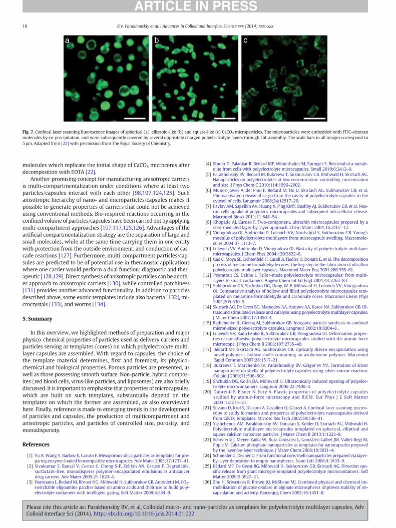
Fig. 7. Confocal laser scanning fluorescence images of spherical (a), ellipsoid-like (b) and square-like (c) CaCO3 microparticles. The microparticles were embedded with FITC–dextranmolecules by co-precipitation, and were subsequently covered by several oppositely charged polyelectrolyte layers through LbL assembly. The scale bars in all images correspond to5 μm. Adapted from [22] with permission from The Royal Society of Chemistry.
10 B.V. Parakhonskiy et al. / Advances in Colloid and Interface Science xxx (2014) xxx–xxx
molecules which replicate the initial shape of CaCO3 microcores afterdecomposition with EDTA [22].
Another promising concept for manufacturing anisotropic carriersis multi-compartmentalization under conditions where at least twoparticles/capsules interact with each other [98,107,124,125]. Suchanisotropic hierarchy of nano- and microparticles/capsules makes itpossible to generate properties of carriers that could not be achievedusing conventional methods. Bio-inspired reactions occurring in theconfined volumeof particles/capsules have been carried out by applyingmulti-compartment approaches [107,117,125,126]. Advantages of theartificial compartmentalization strategy are the separation of large andsmall molecules, while at the same time carrying them in one entitywith protection from the outside environment, and conduction of cas-cade reactions [127]. Furthermore, multi-compartment particles/cap-sules are predicted to be of potential use in theranostic applicationswhere one carrier would perform a dual function: diagnostic and ther-apeutic [128,129]. Direct synthesis of anisotropic particles can be anoth-er approach to anisotropic carriers [130], while controlled patchiness[131] provides another advanced functionality. In addition to particlesdescribed above, some exotic templates include also bacteria [132], mi-crocrystals [133], and worms [134].
5. Summary
In this overview, we highlighted methods of preparation and majorphysico-chemical properties of particles used as delivery carriers andparticles serving as templates (cores) on which polyelectrolyte multi-layer capsules are assembled. With regard to capsules, the choice ofthe template material determines, first and foremost, its physico-chemical and biological properties. Porous particles are presented, aswell as those possessing smooth surface. Non-particle, hybrid compos-ites (red blood cells, virus-like particles, and liposomes) are also brieflydiscussed. It is important to emphasize that properties ofmicrocapsules,which are built on such templates, substantially depend on thetemplates on which the former are assembled, as also overviewedhere. Finally, reference is made to emerging trends in the developmentof particles and capsules, the production of multicompartment andanisotropic particles, and particles of controlled size, porosity, andmonodispersity.
References
[1] Yu A, Wang Y, Barlow E, Caruso F. Mesoporous silica particles as templates for pre-paring enzyme-loaded biocompatible microcapsules. Adv Mater 2005;17:1737–41.
[2] Sivakumar S, Bansal V, Cortez C, Chong S-F, Zelikin AN, Caruso F. Degradable,surfactant-free, monodisperse polymer-encapsulated emulsions as anticancerdrug carriers. Adv Mater 2009;21:1820–4.
[3] Hartmann L, BedardM, Börner HG,Möhwald H, Sukhorukov GB, Antonietti M. CO2-switchable oligoamine patches based on amino acids and their use to build poly-electrolyte containers with intelligent gating. Soft Matter 2008;4:534–9.
Please cite this article as: Parakhonskiy BV, et al, Colloidal micro- and naColloid Interface Sci (2014), http://dx.doi.org/10.1016/j.cis.2014.01.022
[4] Studer D, Palankar R, Bédard MF, Winterhalter M, Springer S. Retrieval of a metab-olite from cells with polyelectrolyte microcapsules. Small 2010;6:2412–9.
[5] Parakhonskiy BV, Bedard M, Bukreeva T, Sukhorukov GB, Möhwald H, Skirtach AG.Nanoparticles on polyelectrolytes at low concentration: controlling concentrationand size. J Phys Chem C 2010;114:1996–2002.
[6] Muñoz-Javier A, del Pino P, Bedard M, Ho D, Skirtach AG, Sukhorukov GB, et al.Photoactivated release of cargo from the cavity of polyelectrolyte capsules to thecytosol of cells. Langmuir 2008;24:12517–20.
[7] Pavlov AM, Sapelkin AV, Huang X, P'ng KMY, Bushby AJ, Sukhorukov GB, et al. Neu-ron cells uptake of polymeric microcapsules and subsequent intracellular release.Macromol Biosci 2011;11:848–54.
[8] Khopade AJ, Caruso F. Two-component, ultrathin microcapsules prepared by acore-mediated layer-by-layer approach. Chem Mater 2004;16:2107–12.
[9] Vinogradova OI, Andrienko D, Lulevich VV, Nordschild S, Sukhorukov GB. Young'smodulus of polyelectrolyte multilayers from microcapsule swelling. Macromole-cules 2004;37:1113–7.
[10] Lulevich VV, Andrienko D, Vinogradova OI. Elasticity of polyelectrolyte multilayermicrocapsules. J Chem Phys 2004;120:3822–6.
[11] Gao C, Moya SE, Lichtenfeld H, Casoli A, Fiedler H, Donath E, et al. The decompositionprocess of melamine formaldehyde cores: the key step in the fabrication of ultrathinpolyelectrolyte multilayer capsules. Macromol Mater Eng 2001;286:355–61.
[12] Peyratout CS, Dähne L. Tailor-made polyelectrolyte microcapsules: from multi-layers to smart containers. Angew Chem Int Ed Engl 2004;43:3762–83.
[13] Sukhorukov GB, Shchukin DG, Dong W-F, Möhwald H, Lulevich VV, VinogradovaOI. Comparative analysis of hollow and filled polyelectrolyte microcapsules tem-plated on melamine formaldehyde and carbonate cores. Macromol Chem Phys2004;205:530–5.
[14] Skirtach AG, De Geest BG, Mamedov AA, Antipov AA, Kotov NA, Sukhorukov GB. Ul-trasound stimulated release and catalysis using polyelectrolyte multilayer capsules.J Mater Chem 2007;17:1050–4.
[15] Radtchenko IL, Giersig M, Sukhorukov GB. Inorganic particle synthesis in confinedmicron-sized polyelectrolyte capsules. Langmuir 2002;18:8204–8.
[16] Lulevich VV, Radtchenko IL, Sukhorukov GB, Vinogradova OI. Deformation proper-ties of nonadhesive polyelectrolyte microcapsules studied with the atomic forcemicroscope. J Phys Chem B 2003;107:2735–40.
[17] Bédard MF, Skirtach AG, Sukhorukov GB. Optically driven encapsulation usingnovel polymeric hollow shells containing an azobenzene polymer. MacromolRapid Commun 2007;28:1517–21.
[18] Bukreeva T, Marchenko IV, Parakhonskiy BV, Grigor'ev YV. Formation of silvernanoparticles on shells of polyelectrolyte capsules using silver-mirror reaction.Colloid J 2009;71:596–602.
[19] Shchukin DG, Gorin DA, Möhwald H. Ultrasonically induced opening of polyelec-trolyte microcontainers. Langmuir 2006;22:7400–4.
[20] Dubreuil F, Elsner N, Fery A. Elastic properties of polyelectrolyte capsulesstudied by atomic-force microscopy and RICM. Eur Phys J E Soft Matter2003;12:215–21.
[21] Silvano D, Krol S, Diaspro A, Cavalleri O, Gliozzi A. Confocal laser scanning micros-copy to study formation and properties of polyelectrolyte nanocapsules derivedfrom CdCO3 templates. Microsc Res Tech 2002;59:536–41.
[22] Yashchenok AM, Parakhonskiy BV, Donatan S, Kohler D, Skirtach AG, Möhwald H.Polyelectrolyte multilayer microcapsules templated on spherical, elliptical andsquare calcium carbonate particles. J Mater Chem B 2013;1:1223–8.
[23] Schwiertz J, Meyer-Zaika W, Ruiz-Gonzalez L, González-Calbet JM, Vallet-Regí M,Epple M. Calcium phosphate nanoparticles as templates for nanocapsules preparedby the layer-by-layer technique. J Mater Chem 2008;18:3831–4.
[24] Schneider G, Decher G. From functional core/shell nanoparticles prepared via layer-by-layer deposition to empty nanospheres. Nano Lett 2004;4:1833–9.
[25] Bédard MF, De Geest BG, Möhwald H, Sukhorukov GB, Skirtach AG. Direction spe-cific release from giant microgel-templated polyelectrolyte microcontainers. SoftMatter 2009;5:3927–31.
[26] Zhu H, Srivastava R, Brown JQ, McShane MJ. Combined physical and chemical im-mobilization of glucose oxidase in alginate microspheres improves stability of en-capsulation and activity. Bioconjug Chem 2005;16:1451–8.
no-particles as templates for polyelectrolyte multilayer capsules, Adv

11B.V. Parakhonskiy et al. / Advances in Colloid and Interface Science xxx (2014) xxx–xxx
[27] Abdullaev E, Shchukin D, Lvov Y. Halloysite Clay Nanotubes as a Reservoir for Cor-rosion Inhibitors and Template for Layer-by-Layer Encapsulation. Polymer MaterSci Eng 2008;99:121–2.
[28] Wang Y, Yan Y, Cui J, Hosta-Rigau L, Heath JK, Nice EC, et al. Encapsulation of water-insoluble drugs in polymer capsules prepared using mesoporous silica templatesfor intracellular drug delivery. Adv Mater 2010;22:4293–7.
[29] Wang Y, Yu A, Caruso F. Nanoporous polyelectrolyte spheres prepared by sequen-tially coating sacrificial mesoporous silica spheres. Angew Chem Int Ed Engl2005;44:2888–92.
[30] Borodina TN, Rumsh LD, Kunizhev SM, Sukhorukov GB, Vorozhtsov GN, Feldman BM,et al. Polyelectrolyte microcapsules as the systems. Biochemistry 2008;2:88–93.
[31] Volodkin DV, Petrov AI, Prevot M, Sukhorukov GB. Matrix polyelectrolytemicrocapsules: new system for macromolecule encapsulation. Langmuir2004;20:3398–406.
[32] Déjugnat C, Sukhorukov GB. pH-responsive properties of hollow polyelectrolytemicrocapsules templated on various cores. Langmuir 2004;20:7265–9.
[33] Bukreeva T, Parakhonskiy BV, Skirtach AG, Susha AS, Sukhorukov GB. Preparationof polyelectrolyte microcapsules with silver and gold nanoparticles in a shell andthe remote destruction of microcapsules under laser irradiation. Crystallogr Rep2006;51:863–9.
[34] Shchukin DG, Sukhorukov GB. Selective YF3 nanoparticle formation in polyelectro-lyte capsules as microcontainers for yttrium recovery from aqueous solutions.Langmuir 2003;19:4427–31.
[35] De Geest BG, Déjugnat C, PrevotM, Sukhorukov GB, Demeester J, De Smedt SC. Self-rupturing and hollow microcapsules prepared from bio-polyelectrolyte-coatedmicrogels. Adv Funct Mater 2007;17:531–7.
[36] Lu Z, Prouty MD, Guo Z, Golub VO, Kumar CSSR, Lvov YM. Magnetic switch of per-meability for polyelectrolyte microcapsules embedded with Co@Au nanoparticles.Langmuir 2005;21:2042–50.
[37] Donath E, Moya SE, Neu B, Sukhorukov GB, Georgieva R, Voigt A, et al. Hollow poly-mer shells from biological templates: fabrication and potential applications. ChemEur J 2002;8:5481–5.
[38] Georgieva R, Moya SE, Hin M, Mitlöhner R, Donath E, Kiesewetter H, et al. Perme-ation of macromolecules into polyelectrolyte microcapsules. Biomacromolecules2002;3:517–24.
[39] Kreft O, Georgieva R, Bäumler H, Steup M, Müller-Röber B, Sukhorukov GB,et al. Red blood cell templated polyelectrolyte capsules: a novel vehicle forthe stable encapsulation of DNA and proteins. Macromol Rapid Commun2006;27:435–40.
[40] Moya SE. Polyelectrolyte multilayer capsules templated on biological cells: core ox-idation influences layer chemistry. Colloids Surf A 2001;183–185:27–40.
[41] Diaspro A, Silvano D, Krol S, Cavalleri O, Gliozzi A. Single living cell encapsulation innano-organized polyelectrolyte shells. Langmuir 2002;18:5047–50.
[42] Neu B, Voigt A, Mitlöhner R, Leporatti S, Gao CY, Donath E, et al. Biological cells astemplates for hollow microcapsules. J Microencapsul 2007;18:385–95.
[43] Fischlechner M, Zschörnig O, Hofmann J, Donath E. Engineering virus func-tionalities on colloidal polyelectrolyte lipid composites. Angew Chem2005;117:2952–5.
[44] Cuomo F, Lopez F, Ceglie A, Maiuro L, Miguel MG, Lindman B. pH-responsiveliposome-templated polyelectrolyte nanocapsules. Soft Matter 2012;8:4415–20.
[45] Habraken WJEM, Tao J, Brylka LJ, Friedrich H, Bertinetti L, Schenk AS, et al. Ion-association complexes unite classical and non-classical theories for the biomimeticnucleation of calcium phosphate. Nat Commun 2013;4:1507.
[46] Cai Y, Liu Y, YanW, Hu Q, Tao J, Zhang M, et al. Role of hydroxyapatite nanoparticlesize in bone cell proliferation. J Mater Chem 2007;17:3780–7.
[47] Andreassen J-P. Formation mechanism and morphology in precipitation ofvaterite–nano-aggregation or crystal growth? J Cryst Growth 2005;274:256–64.
[48] Brecević L, Nielsen AE. Solubility of amorphous calcium carbonate. J Cryst Growth1989;98:504–10.
[49] Schmidt S, Volodkin DV. Microparticulate biomolecules by mild CaCO3 templating.J Mater Chem B 2013;1:1210–8.
[50] Parakhonskiy BV, Haase A, Antolini R. Sub-micrometer vaterite containers: synthe-sis, substance loading, and release. Angew Chem Int Ed Engl 2012;51:1195–7.
[51] Behra M, Schmidt S, Hartmann J, Volodkin DV, Hartmann L. Synthesis of porousPEG microgels using CaCO3 microspheres as hard templates. Macromol RapidCommun 2012;33:1049–54.
[52] Parakhonskiy BV, Foss C, Carletti E, Fedel M, Haase A, Motta A, et al. Tailored intra-cellular delivery via a crystal phase transition in 400 nmvaterite particles. BiomaterSci 2013;1:1273–81.
[53] Janeković A, Matijević E. Preparation of monodispersed colloidal cadmium com-pounds. J Colloid Interface Sci 1985;103:436–47.
[54] Schulz-Ekloff G. Mesoporous silica with controlled porous structure and regularmorphology. Int J Inorg Mater 1999;1:97–102.
[55] Svenskaya Y, Parakhonskiy BV, Haase A, Atkin V, Lukyanets E, Gorin DA, et al. An-ticancer drug delivery system based on calcium carbonate particles loaded with aphotosensitizer. Biophys Chem 2013;182:11–5.
[56] Reibetanz U, Schönberg M, Rathmann S, Strehlow V, Göse M, Lessig J. Inhibition ofhuman neutrophil elastase by α1-antitrypsin functionalized colloidal microcarriers.ACS Nano 2012;6:6325–36.
[57] Washington C. Drug release from microdisperse systems: a critical review. Int JPharm 1990;58:1–12.
[58] She Z, Antipina MN, Li J, Sukhorukov GB. Mechanism of protein release from poly-electrolyte multilayer microcapsules. Biomacromolecules 2010;11:1241–7.
[59] Peng C, Zhao Q, Gao C. Sustained delivery of doxorubicin by porous CaCO3 and chi-tosan/alginate multilayers-coated CaCO3 microparticles. Colloids Surf A2010;353:132–9.
Please cite this article as: Parakhonskiy BV, et al, Colloidal micro- and nanColloid Interface Sci (2014), http://dx.doi.org/10.1016/j.cis.2014.01.022
[60] Park J-H, Gu L, von Maltzahn G, Ruoslahti E, Bhatia SN, Sailor MJ. Biodegradable lu-minescent porous silicon nanoparticles for in vivo applications. Nat Mater2009;8:331–6.
[61] Li Z-Z, Wen L-X, Shao L, Chen J-F. Fabrication of porous hollow silica nanopar-ticles and their applications in drug release control. J Control Release2004;98:245–54.
[62] Barbe C, Bartlett J, Kong L, Finnie K, Lin HQ, Larkin M, et al. Silica particles: a noveldrug-delivery system. Adv Mater 2004;16:1959–66.
[63] Xue JM, Shi M. PLGA/mesoporous silica hybrid structure for controlled drug release.J Control Release 2004;98:209–17.
[64] Panyam J, Labhasetwar V. Biodegradable nanoparticles for drug and gene deliveryto cells and tissue. Adv Drug Deliv Rev 2003;55:329–47.
[65] Desai MP, Labhasetwar V, Amidon GL, Levy RJ. Gastrointestinal uptakeof biodegradable microparticles: effect of particle size. Pharm Res1996;13:1838–45.
[66] Parakhonskiy BV, Tessarolo F, Haase A, Antolini R. Dependence of sub-micronvaterite container release properties on pH and ionic strength of the surroundingsolution. Adv Sci Technol 2012;86:81–5.
[67] Spanos N, Koutsoukos PG. The transformation of vaterite to calcite: effect of theconditions of the solutions in contact with the mineral phase. J Cryst Growth1998;191:783–90.
[68] Eriksson R, Merta J, Rosenholm JB. The calcite/water interface I. Surface charge inindifferent electrolyte media and the influence of low-molecular-weight polyelec-trolyte. J Colloid Interface Sci 2007;313:184–93.
[69] Eriksson R, Merta J, Rosenholm JB. The calcite/water interface II. Effect of added lat-tice ions on the charge properties and adsorption of sodium polyacrylate. J ColloidInterface Sci 2008;326:396–402.
[70] Volodkin DV, von Klitzing R, Möhwald H. Pure protein microspheres by calciumcarbonate templating. Angew Chem Int Ed Engl 2010;49:9258–61.
[71] Tannock I, Rotin D. Acid pH in tumors and its potential for therapeutic exploitation.Cancer Res 1989:4373–84.
[72] Urano Y, Asanuma D, Hama Y, Koyama Y, Barrett T, Kamiya M, et al. Selective mo-lecular imaging of viable cancer cells with pH-activatable fluorescence probes. NatMed 2009;15:104–9.
[73] Svenskaya Y, Parakhonskiy B, Haase A, Atkin V, Lukyanets E, Gorin D, et al. Antican-cer drug delivery system based on calcium carbonate particles loaded with a pho-tosensitizer. Biophys Chem 2013;182:11–5.
[74] Xu A-W, Dong W-F, Antonietti M, Cölfen H. Polymorph switching of calcium car-bonate crystals by polymer-controlled crystallization. Adv Funct Mater2008;18:1307–13.
[75] Yashchenok AM, Delcea M, Videnova K, Jares-Erijman EA, Jovin TM, Konrad M,et al. Enzyme reaction in the pores of CaCO3 particles upon ultrasound disrup-tion of attached substrate-filled liposomes. Angew Chem Int Ed Engl2010;49:8116–20.
[76] Welzel T, Radtke I, Meyer-ZaikaW, Heumann R, Epple M. Transfection of cells withcustom-made calcium phosphate nanoparticles coated with DNA. J Mater Chem2004;14:2213–7.
[77] Sokolova V, Kovtun A, Prymak O,Meyer-ZaikaW, Kubareva EA, Romanova EA, et al.Functionalisation of calcium phosphate nanoparticles by oligonucleotides and theirapplication for gene silencing. J Mater Chem 2007;17:721–7.
[78] Sokolova V, Rotan O, Klesing J, Nalbant P, Buer J, Knuschke T, et al. Calcium phos-phate nanoparticles as versatile carrier for small and large molecules across cellmembranes. J Nanoparticle Res 2012;14:910.
[79] Sokolova V, Knuschke T, Buer J, Westendorf AM, Epple M. Quantitative determina-tion of the composition of multi-shell calcium phosphate-oligonucleotide nanopar-ticles and their application for the activation of dendritic cells. Acta Biomater2011;7:4029–36.
[80] Volodkin DV, Schmidt S, Fernandes P, Larionova NI, Sukhorukov GB, Duschl C, et al.One-step formulation of protein microparticles with tailored properties: hardtemplating at soft conditions. Adv Funct Mater 2012;22:1914–22.
[81] Nicolas J, Bensaid F, Desmaële D, Grogna M, Detrembleur C, Andrieux K, et al. Syn-thesis of highly functionalized poly(alkyl cyanoacrylate) nanoparticles by means ofclick chemistry. Macromolecules 2008;41:8418–28.
[82] Duan L, Yan X, Wang A, Jia Y, Li J. Highly loaded hemoglobin spheres as promisingartificial oxygen carriers. ACS Nano 2012;6:6897–904.
[83] Xiong Y, Liu ZZ, Georgieva R, Smuda K, Steffen A, Sendeski M, et al.Nonvasoconstrictive hemoglobin particles as oxygen carriers. ACS Nano2013;7:7454–61.
[84] Chen K, Merkel TJ, Pandya A, Napier ME, Luft JC, Daniel W, et al. Low modulus bio-mimetic microgel particles with high loading of hemoglobin. Biomacromolecules2012;13:2748–59.
[85] Karamitros CS, Yashchenok AM, Möhwald H, Skirtach AG, Konrad M. Preservingcatalytic activity and enhancing biochemical stability of the therapeutic enzymeasparaginase by biocompatible multilayered polyelectrolyte microcapsules.Biomacromolecules 2013;14:4398–406.
[86] Soppimath KS, Aminabhavi TM, Kulkarni AR, Rudzinski WE. Biodegradablepolymeric nanoparticles as drug delivery devices. J Control Release2001;70:1–20.
[87] Fujiwara M, Shiokawa K, Kubota T. Direct encapsulation of proteins into calciumsilicate microparticles by water/oil/water interfacial reaction method and their re-sponsive release behaviors. Mater Sci Eng C 2012;32:2484–90.
[88] Mondon K, Zeisser-Labouèbe M, Gurny R, Möller M. MPEG-hexPLA micelles asnovel carriers for hypericin, a fluorescent marker for use in cancer diagnostics.Photochem Photobiol 2011;87:399–407.
[89] Volodkin DV, Larionova NI, Sukhorukov GB. Protein encapsulation via porousCaCO3 microparticles templating. Biomacromolecules 2004;5:1962–72.
o-particles as templates for polyelectrolyte multilayer capsules, Adv

12 B.V. Parakhonskiy et al. / Advances in Colloid and Interface Science xxx (2014) xxx–xxx
[90] Déjugnat C, Köhler K, Dubois M, Sukhorukov GB, Möhwald H, Zemb T, et al. Mem-brane densification of heated polyelectrolyte multilayer capsules characterized bysoft X-ray microscopy. Adv Mater 2007;19:1331–6.
[91] Köhler K, Shchukin DG, Möhwald H, Sukhorukov GB. Thermal behavior of polyelec-trolyte multilayer microcapsules. 1. The effect of odd and even layer number. J PhysChem B 2005;109:18250–9.
[92] Marchenko I, Yashchenok AM, Borodina TN, Bukreeva T, Konrad M, MöhwaldH, et al. Controlled enzyme-catalyzed degradation of polymeric capsulestemplated on CaCO : influence of the number of LbL layers, conditions ofdegradation, and disassembly of multicompartments. J Control Release2012;162:599–605.
[93] Yashchenok AM, Borisova D, Parakhonskiy BV, Masic A, Pinchasik B-E, Möhwald H,et al. Nanoplasmonic smooth silica versus porous calcium carbonate bead biosen-sors for detection of biomarkers. Ann Phys 2012;524:723–32.
[94] Bukreeva TV, Parakhonskiy BV, Marchenko IV, Khlebtsov BN, Khlebtsov NG,Dementieva OV, et al. Polyelectrolyte microcapsules with a shell containing silverand gold nanoparticles, based on calcium carbonate and polystyrene cores.Nanotechnol Russ 2008;3:85–93.
[95] Parakhonskiy BV, Bukreeva T, Parakhonskiy GV, Skirtach AG, Sukhorukov GB,Khlebtsov NG, et al. Permeability adjustment of polyelectrolyte micro- andnanocapsules by laser irradiation. In: Zimnyakov DA, Khlebtsov NG, editors. Proc.SPIE, SPIE; 2007 [pp. 653605–653605–8].
[96] Skirtach AG, Dejugnat C, Braun D, Susha AS, Rogach AL, Parak WJ, et al. The role ofmetal nanoparticles in remote release of encapsulated materials. Nano Lett2005;5:1371–7.
[97] Yi Q, Sukhorukov GB. Externally triggered dual function of complex microcapsules.ACS Nano 2013;7:8693–705.
[98] Chandrawati R, van Koeverden MP, Lomas H, Caruso F. Multicompartment particleassemblies for bioinspired encapsulated reactions. J Phys Chem Lett2011;2:2639–49.
[99] Costa RR, Castro E, Arias FJ, Rodríguez-Cabello JC, Mano JF. Multifunctional com-partmentalized capsules with a hierarchical organization from the nano to themacro scales. Biomacromolecules 2013;14:2403–10.
[100] Cortez C, Tomaskovic-Crook E, Johnston APR, Radt B, Cody SH, Scott AM, et al.Targeting and uptake of multilayered particles to colorectal cancer cells. AdvMater 2006;18:1998–2003.
[101] Vergaro V, Baldassarre F, De Santis F, Ciccarella G, Giannelli G, Leporatti S. TGF-betaInihibitor-loaded polyelectrolyte multilayers capsules for sustained targeting ofhepatocarcinoma cells. Curr Pharm Des 2012;18:4155–64.
[102] Sandre O, Moreaux L, Brochard-Wyart F. Dynamics of transient pores in stretchedvesicles. Proc Natl Acad Sci U S A 1999;96:10591–6.
[103] Kreft O, Prevot M, Möhwald H, Sukhorukov GB. Shell-in-shell microcapsules: anovel tool for integrated, spatially confined enzymatic reactions. Angew ChemInt Ed Engl 2007;46:5605–8.
[104] Kreft O, Skirtach AG, Sukhorukov GB, Möhwald H. Remote control of bioreactionsin multicompartment capsules. Adv Mater 2007;19:3142–5.
[105] Bäumler H, Georgieva R. Coupled enzyme reactions in multicompartment micro-particles. Biomacromolecules 2010;11:1480–7.
[106] Kuiper SM, Nallani M, Vriezema DM, Cornelissen JJLM, van Hest JCM, Nolte RJM,et al. Enzymes containing porous polymersomes as nano reaction vessels for cas-cade reactions. Org Biomol Chem 2008;6:4315–8.
[107] Delcea M, Yashchenok AM, Videnova K, Kreft O, Möhwald H, Skirtach AG.Multicompartmental micro- and nanocapsules: hierarchy and applications in bio-sciences. Macromol Biosci 2010;10:465–74.
[108] Pan HM, Beyer S, Zhu Q, Trau D. Inwards interweaving of polymeric layers withinhydrogels: assembly of spherical multi-shells with discrete porosity differences.Adv Funct Mater 2013;23:5108–15.
[109] Chandrawati R, Caruso F. Biomimetic liposome- and polymersome-basedmulticompartmentalized assemblies. Langmuir 2012;28:13798–807.
[110] Champion JA, Mitragotri S. Role of target geometry in phagocytosis. Proc Natl AcadSci U S A 2006;103:4930–4.
[111] Muro S, Garnacho C, Champion JA, Leferovich J, Gajewski C, Schuchman EH, et al.Control of endothelial targeting and intracellular delivery of therapeutic enzymes
Please cite this article as: Parakhonskiy BV, et al, Colloidal micro- and naColloid Interface Sci (2014), http://dx.doi.org/10.1016/j.cis.2014.01.022
by modulating the size and shape of ICAM-1-targeted carriers. Mol Ther2008;16:1450–8.
[112] Geng Y, Dalhaimer P, Cai S, Tsai R, Tewari M, Minko T, et al. Shape effects of fila-ments versus spherical particles in flow and drug delivery. Nat Nanotechnol2007;2:249–55.
[113] Gratton SEA, Ropp PA, Pohlhaus PD, Luft JC, Madden VJ, Napier ME, et al. The effectof particle design on cellular internalization pathways. Proc Natl Acad Sci U S A2008;105:11613–8.
[114] Doshi N, Mitragotri S. Macrophages recognize size and shape of their targets. PLoSOne 2010;5:e10051.
[115] Arnida, Janát-Amsbury MM, Ray A, Peterson CM, Ghandehari H. Geometry and sur-face characteristics of gold nanoparticles influence their biodistribution and uptakeby macrophages. Eur J Pharm Biopharm 2011;77:417–23.
[116] Hutter E, Boridy S, Labrecque S, Lalancette-Hébert M, Kriz J, Winnik FM, et al.Microglial response to gold nanoparticles. ACS Nano 2010;4:2595–606.
[117] Gao Y-X, Yu S-H, Cong H, Jiang J, Xu A-W, Dong W-F, et al. Block-copolymer-con-trolled growth of CaCO3 microrings. J Phys Chem B 2006;110:6432–6.
[118] Shchepelina O, Kozlovskaya V, Singamaneni S, Kharlampieva E, Tsukruk VV. Repli-cation of anisotropic dispersed particulates and complex continuous templates. JMater Chem 2010;20:6587–603.
[119] Shchepelina O, Kozlovskaya V, Kharlampieva E, MaoW, Alexeev A, Tsukruk VV.Anisotropic micro- and nano-capsules. Macromol Rapid Commun2010;31:2041–6.
[120] Kozlovskaya V, Higgins W, Chen J, Kharlampieva E. Shape switching of hollow layer-by-layer hydrogel microcontainers. Chem Commun (Camb) 2011;47:8352–4.
[121] Kozlovskaya V, Yakovlev S, Libera M, Sukhishvili SA. Surface priming and the self-assembly of hydrogen-bonded multilayer capsules and films. Macromolecules2005;38:4828–36.
[122] Shchepelina O, Lisunova MO, Drachuk I, Tsukruk VV. Morphology and properties ofmicrocapsules with different core releases. Chem Mater 2012;24:1245–54.
[123] Lisunova MO, Holland N, Shchepelina O, Tsukruk VV. Template-assisted as-sembly of the functionalized cubic and spherical microparticles. Langmuir2012;28:13345–53.
[124] Chandrawati R, Städler B, Postma A, Connal LA, Chong S-F, Zelikin AN, et al.Cholesterol-mediated anchoring of enzyme-loaded liposomes within disulfide-stabilized polymer carrier capsules. Biomaterials 2009;30:5988–98.
[125] Tzvetkov G, Graf B, Fernandes PAL, Fery A, Cavalieri F, Paradossi G, et al. In situcharacterization of gas-filled microballoons using soft X-ray microspectroscopy.Soft Matter 2008;4:510–4.
[126] Gao Y, Zhao L, Li C, Shi G. Electrosynthesis of poly(3,4-ethylenedioxythiophene)microcups in the aqueous solution of LiClO4 and tri(ethylene glycol). Polymer2006;47:4953–8.
[127] Peters RJRW, Marguet M, Marais S, Fraaije MW, van Hest JCM, Lecommandoux S.Cascade reactions in multicompartmentalized polymersomes. Angew Chem IntEd Engl 2014;53:146–50.
[128] Xiong R, Soenen SJ, Braeckmans K, Skirtach AG. Towards theranosticmulticompartment microcapsules: in-situ diagnostics and laser-induced treat-ment. Theranostics 2013;3:141–51.
[129] Perry JL, Herlihy KP, Napier ME, Desimone JM. PRINT: a novel platform towardshape and size specific nanoparticle theranostics. Acc Chem Res 2011;44:990–8.
[130] Murthy VS, Kadali SB, Wong MS. Polyamine-guided synthesis of anisotropic,multicompartment microparticles. ACS Appl Mater Interfaces 2009;1:590–6.
[131] Kohler D, Madaboosi N, Delcea M, Schmidt S, De Geest BG, Volodkin DV, et al.Patchiness of embedded particles and film stiffness control through concentrationof gold nanoparticles. Adv Mater 2012;24:1095–100.
[132] Balkundi SS, Veerabadran NG, Matthew ED, Johnson GR, Lvov YM. Encapsulationof bacterial spores in nanoorganized polyelectrolyte shells. Langmuir2009;25:14011–6.
[133] Ai H, Jones S, De Villiers M, Lvov Y. Nanoencapsulation of furosemide microcrystalsfor controlled drug release. J Control Release 2003;86:59–66.
[134] Minullina RT, Osin YN, Ishmuchametova DG, Fakhrullin RF. Interfacing multicellu-lar organisms with polyelectrolyte shells and nanoparticles: a Caenorhabtidiselegans study. Langmuir 2011;27:7708–13.
no-particles as templates for polyelectrolyte multilayer capsules, Adv