collateralization of descending spinal pathways …collateralization of descending spinal pathways...
TRANSCRIPT

COLLATERALIZATION OF DESCENDING SPINAL PATHWAYS FROM RED NUCLEUS
AND OTHER BRAINSTEM CELL GROUPS IN RAT, CAT AND MONKEY
PROEFSCHRIFT
TER VERKRIJGING VAN DE GRAAD VAN DOCTOR IN DE GENEESKUNDE
AAN DE ERASMUS UNIVERSITEIT ROTTERDAM OP GEZAG VAN DE RECTOR MAGNIFICOS
PROF. DR. J. SPERNA WEILAND EN VOLGENS BESLUIT VAN HET COLLEGE VAN DEKANEN.
DE OPENBARE VERDEDIGING ZAL PLAATSVINDEN OP WOENSDAG 2 MAART 1983 DES NAMIDDAGS
TE3.45 UUR
DOOR
ANNE-MARGRIET HUISMAN
GEBOREN TE BREDA.
~------·---·-
Med1scne emnom~eK. EU.R.
druk: nkb-offscr bleiswiik-leiden

Promotor : Prof. Dr. H.G.J.M. Kuypers
Co-referenten: Prof. Dr. J. Voogd Prof. Dr. H.F.M. Busch

aan Ale
aan mijn ouders


5
Publications
I. A.M. Huisman, H.G.J.M. Kuypers, C.A. Verburgh (1981) Quantitative diffe
rences in collateralization of the descending spinal pathways from red
nucleus and other brain stern cell groups in rat as demonstrated with
the multiple fluorescent retrograde tracer technique. Brain Res. 209:
271-286.
2. A.M. Huisman, H.G.J.M. Kuypers, C.A. Verburgh (1982) Differences in col
lateralization of the descending spinal pathways from red nucleus and
other brain stern cell groups in cat and monkey. Progress in Brain
Res. 57: 185-219.
3. A.M. Huisman, H.G.J.M. Kuypers, F. Conde, K. Keizer Collaterals of rubro
spinal neurons to the cerebellum in rat. A retrograde fluorescent
double labeling study. Brain Research (in press)
4. K. Keizer, H.G.J.M. Kuypers, A.M. Huisman Diarnidino Yellow (DY) a fluo
rescent retrograde neuronal tracer, which migrates only very slowly
out of the cell and can be used in combination with TB and FB in
double labeling experiments. Submitted to Experimental Brain Research.
5. H.G.J.M. Kuypers, A.M. Huisman (1982) The new anatomy of the descending
brain pathways. In 11 Brain Stern Control of Spinal Mechanisms" Fernstrom
Foundation Series No. I New York-Oxford.
6. H.G.J.M. Kuypers, A.M. Huisman Fluorescent neuronal tracers. In 11Advances
in Cellular Neurobiology" Vol. 5: labeling methods applicable to the
study of neuronal pathways (in press)
The publications I, 2 and 3 are used in chapter III, IV and V of this thesis,
with permission of Elsevier/North Holland Biomedical Press.
Publication 6 is used in chapter II with permission of Academic Press, Ill
Fifth Avenue, New York.

6
CHAPTER I:
CONTENTS
GENERAL INTRODUCTION
I.!. red nucleus: anatomical aspects
I. I.a. structure of the nucleus
I. !.b. cytoarchitecture in rat
I. I. c. cytoarchitecture ~n cat and monkey
I. J.d. efferents of red nucleus
I. I.e. afferents of red nucleus
I.2. red nucleus: physiological aspects
I.2.a. function of the rubrospinal pathway as
one of the descending brainstem pathways
I.2.b. physiology of the rubrospinal tract as
the primary component of the laterally
descending brainstem pathways
I.3. aims and scope of the present investigations
I. 4. references
CHAPTER II: FLUORESCENT NEURONAL TRACERS
CHAPTER III: QUANTITATIVE DIFFERENCES IN COLLATERALIZATION OF THE
DESCENDING SPINAL PATHWAYS FROM RED NUCLEUS AND OTHER
BRAIN STEM CELL GROUPS IN RAT AS DEMONSTRATED WITH THE
MULTIPLE FLUORESCENT RETROGRADE TRACER TECHNIQUE.
CHAPTER IV:
CHAPTER V:
DIFFERENCES IN COLLATERALIZATION OF THE DESCENDING
SPINAL PATHWAYS FROM RED NUCLEUS AND OTHER BRAIN STEM
CELL GROUPS IN CAT AND MONKEY.
COLLATERALS OF RUBROSPINAL NEURONS TO THE CEREBELLUM
IN RAT. A RETROGRADE FLUORESCENT DOUBLE LABELING STUDY.

CHAPTER VI:
Summary
GENERAL DISCUSSlON
VI. I . Technique
VI.2. The rubrospinal tract in rat, cat and
monkey
VI.2.a. somatotopic organization of red
nucleus
VI.2.b. collateralization of the rubro
spinal tract in rat, cat and
monkey
VI.2.c. collateralization of the rubro
spinal tract compared with the
collateralization of the raphe
spinal and ventrolateral pontine
tegmentum-spinal tracts in rat,
cat and monkey
VI.2.d. collateralization of the rubro
spinal tract to the cerebellum.
VI.3. references
Samenvatting voor leken
List of abbreviations
Dankwoord
Curriculum vitae
7

8
CHAPTER I: GENERAL INTRODUCTION
I.!. red nucleus: anatomical aspects
I. I.a. structure of the nucleus
I.l.b. cytoarchitecture ln rat
I. I.e. cytoarchitecture ln cat and monkey
I. I. d. efferents of red nucleus
I. I.e. afferents of red nucleus
I.2. red nucleus: physiological aspects
I.2.a.
I.2.b.
function of the rubrospinal pathway as
one of the descending brainstem
pathways
physiology of the rubrospinal tract as
the primary component of the laterally
descending brainstem pathways
I.3. aims and scope of the present investigations
I.4. references
I. I.
I.l.a.
9
Red Nucleus: anatomical aspects
Structure of the· nucleus
The red nucleus is located centrally in the mesencephalic tegmentu~
In the human brain the most conspicious gross feature of the red
nucleus is its pinkish-red color in fresh sections. This is due to
the high vascularity of the nucleus (Hough and Wolff, 1939) which
distinguishes it from the surrounding structures. Its 11 capsule" is
formed by fibers of the superior cerebellar peduncle, which sur
round and traverse the nucleus and by the oculomotor nerve which
runs along the medial aspect of the nucleus (Carpenter, 1956).
Using the Golgi and Nissl techniques, Cajal (1952) described three
types of neurons in the red nucleus based on their size: large
neurons, medium sized neurons and small neurons. Hatschek was the
first who recognized that the red nucleus was divided in a magna
and parvicellular part (Hatschek, 1907). In humans the red nucleus
is mainly composed of small neurons with only relatively few magna
cellular neurons, the latter of which are located at the caudal
pole of the nucleus (Ariens-Kappers et al., 1960; de Lange, 1912;
Ten Donkelaar, 1976a; Masion, 1967). In monkey the red nucleus can
readily be subdivided into a caudal magnocellular portion and a
rostral parvicellular portion. At the meso-diencephalic junction
the latter is located lateral to the fasciculus retroflexus. How
ever, in lower mammals no such a strict distinction between a magna
cellular and a parvicellular portion can be made, although also in
these species large neurons are more numerous in the caudal parts
of the nucleus and small neurons predominate in its rostral parts.
In birds and reptiles the red nucleus consists of large cells only.
Rats, cats, monkeys, apes and humans have a magna- and parvicellu
lar part. From rat to cat, from cat to monkey and from monkey to
human the relative size of the magnocellular part decreases, and
the relative size of the parvicellular part increases.
These t'vo principal subdivisions of the red nucleus have their
own efferent pathways. Thus the caudal magnocellular part gives
rise to projections to the cerebellum, the lower brainstem and the
spinal cord, while the rostral parvicellular part projects to the
inferior olive and possibly the thalamus. However, the segregation
in a caudal red nucleus projecting to the cerebellum, the lower
10
I.l.b.
I.l.c.
brainstem and the spinal cord and a rostral red nucleus projecting
to the inferior olive is a relative one, such that for the rubro
spinal and rubrocerebellar neurons only a rostrocaudal gradient
exists. This was shown by the fact that in cat rubrospinal and
rubrocerebellar neurons are located throughout the rostrocaudal
extent of the nucleus but that they are concentrated in the caudal
half (Brodal and Gogstad, 1954; Pompeiano and Brodal, 1957), and
that both sets of neurons comprise large as well as medium and
small sized cells.
cytoarchitecture in rat
In cross-sections the caudal half of the nucleus is egg shaped and
consists of large (soma diameter 40 ym, giant neurons) and medium
sized (soma diameter 25-40 ym) neurons. The medium sized neurons
are most numerous, but a considerable number of large neurons is
also present, particularly in the caudal pole of the nucleus, where
they are more numerous than in any other part (Reid et al., 1975).
The rostral half of the nucleus is composed of predominantly small
neurons (soma diameter 25 ym) with a scattering of medium sized
ones and caudally in this rostral part also some large ones. In the
most caudal part of this rostral half a population of neurons,
designated the lateral horn (Reid et al., 1975) extends from the
ventral surface of the nucleus dorsally along its lateral aspect.
This subgroup contains predominantly small and medium sized neurons,
but also some large ones. This lateral horn in rat is not analogous
to the nucleus minimus of Von Monakow (1910) in cat, which sub
nucleus is located dorsolateral to the red nucleus and consists of
very small neurons (soma diameter 6-8 ym) (Massion, 1967). The
portion of the red nucleus which contains large neurons and there
fore is designated the magnocellular portion, occupies the caudal
three fourth of the nucleus. The rostral pole of the nucleus in rat
blends imperceptively with the prerubral field and this part of the
nucleus is cytoarchitectonically difficult to demarcate (Reid et al.,
1975).
cytoarchitecture in cat and monkey
In cat the caudal magnocellular part of the red nucleus occupies the
caudal two thirds of the nucleus but ln monkey it seems relatively
smaller and occupies only the caudal half. In both species the

11
caudal portion of the magnocellular part of the red nucleus is round
in cross-section and contains large neurons which are dispersed
between the tegmental fiber bundles. The rostral portion of the
magnocellular red nucleus in monkey is also round in cross-section
but contains mainly medium sized neurons. In cat this rostral
portion of the magnocellular part as seen Ln cross-section has the
shape of a flattened disc and contains both large and small neurons.
According to earlier retrograde degeneration findings these various
types of neurons in the magnocellular part of the red nucleus in
cat and monkey all give rise to rubrospinal fibers (Kuypers and
Lawrence, 1967; Poirier and Bouvier, 1966; Pompeiano and Brodal,
1957).
The rostral part of the red nucleus in monkey is better delinea
ted against the surrounding reticular formation than in rat and cat.
This part consists of relatively small neurons and therefore, as in
other higher primates LS called the parvicellular part. Rostrally
this part of the nucleus is located lateral to the fasciculus retro
flexus. In the ventral portion of the parvicellular part the cells
are rather densely packed, while in its dorsal portion the cells
form a loose network.x According to Fuse (1937) and Fukuyama
(1940) the rostral red nucleus also comprises the subnucleus
dorsomedialis (referred to as 11Augenanteil des Roten Kerns") which
extends dorsomedially from the red nucleus to the 11Nebenokulo
motorius Kerne" i.e. the medial accessory nucleus of Bechteretv and
nucleus of Darkschewitsch (Carpenter, 1956; Leichnetz, 1982). In
monkey the parvicellular part extends rostrally close to the meso
diencephalic junction i.e. just beyond the level where the
x This dorsal portion of the parvicellular red nucleus in monkey is also called the rostral interstitial nucleus of Cajal or the rostral interstitial nucleus of the MLF (Buttner-Ennever and Buttner, 1978; Fukuyama, 1940; Leichnetz, 1982). However, it is of importance to emphasize, that these nuclei are distinct from the interstitial nucleus of Cajal, which projects to the spinal cord (Crutcher et al., 1978; Kuypers and Maisky, 1975, 1977; Peterson and Coulter, 1977; Vasilenko and Kostyukov, 1976).
12
I.l.d.
fasciculus retroflexus (FR) traverses in full length the mesen
cephalic tegmentum. At these levellithe parvicellular part is mainly
located lateral to the FR. In monkey a small group of medium sized
multipolar neurons extends from the caudal magnocellular part of
the nucleus rostrally over a short distance along the lateral
aspect of the parvicellular part, up to the level where the cross
section through the FR is located immediately ventral to the red
nucleus.
In cat the most rostral part of the red nucleus is also situated
lateral to the FR. However, in this animal this part of the nucleus
is not truly parvicellular as in monkey, but consists of a loose
network of small multipolar neurons which is difficult to delinate
against the surrounding reticular formation. Yet, it seems reason
able to regard this portion of the red nucleus in cat as the coun
terpart of the parvicellular part in monkey since both structures
give rise to the ipsilaterally descending rubro-olivary fibers
(Conde and Conde, 1982; Courville and Otabe, 1974; Edwards, 1972;
Strominger et al., 1979; Walberg, 1956).
efferents of red nuc~eus
Rubrobu~bar and rubrospinal tract
This tract, is also called the bundle of Von Monakow after its
discoverer Von Monakow (1883). It undergoes a complete decussation
in the mesencephalic ventral tegmental decussation of Forel. After
this decussation the bundle descends through the ventrolateral
pontine tegmentum into the medulla oblongata, where it is located
ventromedial to the spinal V complex and ventrolateral to the facial
nucleus.
At the level of the pontine trigeminal nuclei, some fibers leave
the rubro-bulbar and -spinal tracts and proceed dorsally to join the
fibers of the superior cerebellar peduncle with which they ascend
upstream into the cerebellum (Courville and Brodal, 1966; Hinman
and Carpenter, 1959; Martinet al., 1974). These rubrocerebellar
fibres terminate in the nucleus interpositus anterior (Courville
and Brodal, 1966). In rat the cells of origin of this rubrocere
bellar connection are located throughout the rostrocaudal extent of
the red nucleus (Flumerfelt and Gwyn, 1974), whereas in cat they are
located in its caudal two thirds (Brodal and Gogstad, 1954). In
other words in both rat and cat rubrocerebellar fibers originate

13
from cells which are located in the same portion of the nucleus in which rubro
spinal neurons are found (Conde and Conde, 1980; Murray and Gurule, 1979;
Pompeiano and Brodal, 1957). In the lower pons and in the medullaoblongata
fibers are distributed from the laterally descending rubral tract to the
lateral part of the lateral tegmental field, to the lateral part of the
cranial VII nucleus and to the supraspinal portion of the lateral reticular
nucleus (i.e. to the part of the nucleus not in receipt of fibers from the
spinal cord)(Bandler, 1978; Busch, 1961; Courville, 1966a; Edwards, 1972;
Flumerfelt and Gwyn, 1974; Hinman and Carpenter, 1959; Kuypers, 1964, 1981;
Martin, 1970b; Miller and Strominger, 1973; Waldron and G1;Jyn, 1971; Walberg,
1958). Further, the rubrospinal tract also distributes fibers to the principal
nucleus V, some portions of the spinal V complex, the descending vestibular
nucleus, the dorsal column nuclei cuneatus and gracilis and to cell groups x
and z (Bandler, 1978; Edwards, 1972; Kuypers, 1964, 1981; Martin and Dom, 1970).
The rubrospinal tract in pigeon, opossum, rat, cat and monkey descends
throughout the spinal cord in the dorsolateral funiculus and terminates in the
dorsolateral part of the intermediate zone i.e. in the lateral part of Rexed's
layers V and VI and in the dorsolateral part of layer VII (Brown, 1974a;
Edwards, 1972; Kuypers, 1964, 1981; Martin and Dom, 1970a; Martinet al., 1974;
Miller and Strominger, 1973; Nyberg-Hansen and Brodal, 1964; Petras, 1967;
Rexed, 1952; Staal, 1961; Waldron and Gwyn, 1971; Wild, 1979). A crossed
rubrospinal tract has also been demonstrated in the reptiles: turtle and lizard
but not in the snake (ten Donkelaar, 1976a+b; 1978). In this respect it is of
interest to note that the snake has no limbs. The frog ~n its tadpole stage
also lacks extremities and in this stage (up to stage 57) likewise does not
posses a rubrospinal tract. In this species rubrospinal neurons appear, simul
taneously with the development of the limbs (ten Donkelaar, 1982).
In rat the cells of origin of the rubrospinal tract are located ~n the
caudal three fourths of the nucleus (Murray and Gurule, 1979), while in cat
and monkey they are located in the caudal two thirds of the nucleus (Conde and
Conde, 1980; Kneisley, 1978; Pompeiano and Brodal, 1957). This interspecies
difference may be a reflection of the difference in delineation of the rostral
border of the red nucleus. Thus in rat,the population of neurons ventral and
lateral to the fasciculus retroflexus (FR) at the meso-diencephalic junction,
which neurons project to the inferior olive is normally not regarded as a part
of the parvicellular red nucleus (Carlton et al.. in press a+b; Cintas et al.,
1980; Brown et al., 1977; Senba et al., 1981). On the other hand, in cat and
monkey this population of neurons is regarded as part of the parvicellular
red nucleus, and as in rat projects ipsilaterally to the inferior olive

14
(Conde and Conde, 1982; Courville and Otabe, 1974; Edwards, 1972; Strominger et
al.,1979; Walberg, 1956).
The rubrospinal projection in rat, cat and monkey is somatotopically orga
nized, such that the dorsomedial part of the red nucleus projects to the cer
vical enlargement, while its ventrolateral part projects to the lumbosacral
enlargement (Flumerfelt and Gwyn, 1974; Gwyn, 1971; Hayes and Rustioni, 1981;
Kneisley, 1978; ~iurray and Gurule, 1979; Pompeiano and Brodal, 1957b; Tsuka
hara, 1967). However, this somatotopic organization becomes less pronounced in
lower animals. Thus, in opossum the rubrospinal tract displays a much less
rigid somatotopic organization than in rat (Martin et al., 1974, 1981b) and ln
pigeon and reptiles no somatotopic organization is present at all (ten Donke
laar and de Boer-van Huizen, 1978; Wild, 1979),while in snakes the tract it
self seems to be lacking (ten Donkelaar, 1976a+b).
Rubro-olivary tract
This uncrossed tract, which is well established in cat and monkey, descends
along the lateral border of the trapezoid body in the central tegmental tract
and terminates in the dorsal lamella of the principal inferior olive (Edwards,
1972; Miller and Strominger, 1973; Saint-Cyr and Courville, 1980, 1981;
Walberg, 1956; Walberg and Nordby, 1981). Other meso-diencephalic nuclei, which
also project to the inferior olive, are the interstitial nucleus of Cajal and
the nucleus of Darkschewitsch, which nuclei project mainly by way of the medial
tegmental tract (Busch, 1961; Ogawa, 1939; Walberg, 1974). In macaque including
rhesus monkey a topographical arrangement between the rostral red nucleus and
the inferior olive is described, such that the dorsal and medial parts of the
rostral red nucleus project to dorsal regions of the principal olive, while
the ventral and lateral parts project to ventral regions of principal olive
(Courville and Otabe, 1974; Strominger et al., 1979). However, more recently,
anterograde transport studies in cat (Edwards, 1972) indicate that rubro
olivary fibers terminate only in the dorsal lamella of the principal olive. In
these studies the injection areas did not involve nuclei,which surround the
parvicellular red nucleus, as the interstitial nucleus of Cajal and the nucleus
of Darkschewitsch (c.f. Walberg and Nordby, 1981).
In cat retrograde HRP transport studies showed that the rubro-olivary
neurons are located in the rostral one third of the red nucleus just lateral
to the fasciculus retroflexus where they are continuous with the olivary
projecting neurons located in the region of the interstitial nucleus of Cajal
i.e. just dorsomedial to the red nucleus (Conde and Conde~ 1982; Saint-Cyr and
Courville, 1980, 1981; Walberg and Nordby, 1981). The neurons projecting to

15
the inferior olive, in the rostral red nucleus, the interstitial nucleus of
Cajal, the nucleus of Darkschewitsch and in the ventral portion of the thala
mic parafascicular nucleus fom a continuous population (Saint-Cyr and Cour
ville, 1980). Anatomical and physiological studies demonstrate only a very
limited rostra-caudal overlap between the population of rubro-olivary neurons
and that of rubrospinal neurons, the latter being located in the caudal two
thirds of the red nucleus (Anderson, 1971; Conde and Conde, 1982).
The existence of a rubro-olivary tract in opossum and rat has been subject
of discussion. Thus, retrograde transport studies in opossum and rat demon
strate only a limited population of rubro-olivary neurons ~n the dorsomedial
rostral red nucleus (Brown et al., 1977; Carlton et al., in press a; Cintas et
al., 1980; Henkel et al. 1975; Senba et al., 1981). On the other hand, in these
animals many neurons were present in the area lateral to the fasciculus retro
flexus, which area in cat and monkey is considered to be a part of the rostral
red nucleus containing the cells of origin of the rubro-olivary tract (Conde
and Conde, 1982; Kuypers and Lawrence, 1967). Based on these data one may
suggest that the different delineation of the rostral part of the red nucleus
in opossum, rat, cat and monkey underlies the difference in opinions about
the existence of a rubro-olivary pathway as demonstrated by means of retro
grade transport techniques. However, anterograde transport studies in opossum
establish clearly the existence of a rubro-olivary tract to the principal
olive (Martinet al., 1980).
Ruhrothalamic pathway
Many conflicting data are available about the existence of an ascending
rubrothalamic connection. Retrograde degeneration studies suggested the exi
stence of ascending fibers originating from the red nucleus (Pompeiano and
Brodal, 1957b). However, lesions placed in the red nucleus unavoidably inter
rupt cerebellothalamic fibers making it impossible to detemine the precise
origin of the resulting degeneration in the thalamus. One way of overcoming
the problem is to interrupt these fibers and allow them to degenerate before
making lesions or recordings in the red nucleus. From such an anatomical
study it was concluded that in monkey no rubrothalamic projection exists
(Hopkins and Lawrence, 1975). On the other hand, retrograde degeneration
studies support the existence of an ascending rubral projection (Kuypers and
Lawrence, 1967). In these studies it was found that many neurons in the
rostral red nucleus ipsilaterally to the diencephalic lesion tended to be
smaller than their contralateral counterparts and that some displayed acute
retrograde changes. However, a direct effect of the lesion, which is very

16
I.l.e.
close to the rostral red nucleus could be suspected (Kuypers and
Lawrence, 1967). Electrophysiologically it was shown that stimu
lation of the thalamus produced excitation of red nucleus neurons.
However, it has been shown that this excitation does not occur due
to antidromic activity of red nucleus neurons, but that it occurs
transsynaptically via an axon reflex in brachium conjunctivum
fibers which send collaterals to red nucleus and also project to
the thalamus (Anderson, 1971; Tsukahara, 1967). Anterograde auto
radiographic transport studies also deny a rubrothalamic pathway
(Edwards, 1972). However, more recent retrograde transport studies
using HRP demonstrated the existence of a rubrothalamic projection
in cat, originating from the rostral one third of the red nucleus
(Conde and Conde, 1980). In this study it was shown that retro
grade transport of HRP between the thalamic injection site and red
nucleus was obtained by intra-axonal transport and not by extra
cellular diffusion or by the bloodstream, since no labeled neurons
occurred between the injection site and red nucleus (i.e. the hypo
thalamic and subthalamic neurons). In addition neurons were only
labeled in its rostral part. Further electrophysiological
studies would be of interest to confirm whether a direct rubro-
thalamic pathway exists in addition to the cerebellothalamic path
way which gives off collaterals to the red nucleus (Tsukahara, 1967).
Afferents of red nucleus
Cortical afferents
The corticorubral connections are most extensively studied in
monkey, in which corticorubral fibers to the parvi- and magnocellu
lar red nucleus clearly originate from different cortical areas.
Corticorubral fibers to the parvicellular red nucleus originate from
Brodmann's area 4 in the precentral gyrus, which largely corresponds
to the primary motor cortex, and from the premotor areas (area 6 and
the frontal eye field area 8 in the prearcuate gyrus) (Brodmann,
1909; Kuypers and Lawrence, 1967). This projection is bilateral with
an ipsilateral emphasis. The precentral projection to the parvi
cellular red nucleus is derived from the entire precentral gyrus
including the ,face area" (c.f. Woolsey, 1958) and also to a limited
degree from postcentral and parietal areas (Kuypers and Lawrence,
1967). This projection is topically organized. Thus, the fibers from
the upper one third of the precentral gyrus are distributed to the

17
ventrolateral portion of parvicellular red nucleus, those from the lower one
third to the dorsomedial portion and those from the middle one third to the
intermediate portion (Catsman-Berrevoets, 1~79; Hartmann-von Monakow, 1979;
Kuypers and Lawrence, 1967). The premotor area (area 6 and area 8) projects to
the dorsomedial part of the parvicellular red nucleus. The supplementary motor
cortex projects both to the dorsomedial and ventrolateral parts of the parvi
cellular red nucleus (Hartmann-von Monakow, 1979; Kunzle, 1978; Kuypers and
Lawrence, 1967). The terminal field of area 8 (prearcuate frontal cortex) in
the subnucleus dorsomedialis of the parvicellular red nucleus (referred as
11Augenanteil des Roten Kerns") continues into the 11nebenokulomotorius Kerne"
i.e. the medial accessory nucleus of Bechterew and the nucleus of Darksche
witsch (Carpenter, 1956; Fukuyama, 1940; Fuse, 1937; Kuypers and Lawrence,
1967; Leichnetz, 1982). Corticorubral fibers to the magnocellular red nucleus
originate only from the upper two thirds of the caudal precentral gyrus. This
projection is exclusively ipsilateral (Hartmann-von Monakow, 1979; Kuypers and
Lawrence, 1967). Thus, in the cortico-rubro-spinal pathway the somatotopy is
preserved, such that the cortical "upper limb area" (c .f. Woolsey, 1958) pro
jects to the dorsomedial part of the red nucleus, which part in turn projects
to the cervical cord. Moreover, the cortical "lower limb area" (c. f. Woolsey,
1958) projects to the ventrolateral part of the nucleus, which part projects
to the lumbosacral cord (Catsman-Berrevoets, 1979; Hartmann-von Monakow, 1979;
Kuypers and Lawrence,1967; Petras,!969). No "face area"(c.f. Woolsey, 1958) pro
jection exists from area 4 (i.e. from the lower one third of the precentral
gyrus) to the magnocellular red nucleus in contrast to the "face area" 4 pro
jection to the parvicellular red nucleus (Catsman-Berrevoets, 1979; Hartmann
von Monakow, 1979; Kuypers and Lawrence, 1967).
The magna- and parvi-cellular parts of the red nucleus receive their inputs
not only from different cortical areas, but the cortical cells of origin of
these rubral afferents are also situated in different cortical layers. Thus,
the cells projecting to the parvicellular red nucleus, which are located in
areas 4 and 6 are situated in the upper part of layer V (Brodmann) i.e. in
area 4 above the Betz cells (Betz, 1948), while those projecting to the magna
cellular red nucleus, which are located posteriorly in area 4, are situated
deep in layer V i.e. at the level of the Betz cells (Catsman-Berrevoets, 1979).
In cat the corticorubral fibers to the caudal two thirds of the red nucleus,
the part in which the rubrospinal neurons are located, originate from the
primary sensori-motor cortex. As in monkey, the somatotopy in the cortico
rubro-spinal pathway is preserved,such that the fibers of the cortical "motor
forelimb area" (c.f. Woolsey, 1958) terminate in the dorsal parts, and those

18
from the "motor hindlimb area" (c.f. Woolsey, 1958) in the ventral parts of
the red nucleus (Mabuchi, 1966; Padel, 1973; Rinvik and Walberg, 1963;
Pompeiano and Brodal, 1957b). The premotor area 6 in cat (see Hassler and Muhs
Clement, 1964) is located partly in the gyrus proreus. Only anterograde dege
neration studies have revealed some data concerning corticorubral fibers from
the gyrus proreus (Rinvik and Walberg, 1963). After lesion in this gyrus dege
nerating terminal fibers were seen bilaterally in the red nucleus. This was
heaviest ipsilaterally. They did not state in which rostrocaudal part of the
red nucleus these corticorubral fibers terminate. It would, of course, be of
interest to know whether these fibers terminate in the rostral one third of
the cat red nucleus (located lateral to the fasciculus retroflexus),which maybe
regarded to be analogous to the parvicellular red nucleus in monkey on the
basis of the projections from this portion of the red nucleus to the inferior
olive (Conde and Conde, 1982; Saint-Cyr and Courville, 1980; Walberg, 1956).
Originally the corticorubral fibers in rat were described to be distributed
only to the ipsilateral parvicellular part of the red nucleus. They apparently
do not distribute to bhe magnocellular part, which was considered to give rise
to almost all rubrospinal fibers (Brown, 1974a; Flumerfelt and Gwyn, 1974;
Gwyn and Flumerfelt, 1974). However, since rubrospinal neurons in rat also
arise from more rostral parts of the red nucleus (Flumerfelt and Gwyn, 1974)
the corticorubral fibers from the sensorimotor cortex in this animal are proba
bly also distributed to rubrospinal neurons. As pointed out already, much of
the controversy related to the efferent connectivity of the red nucleus may
result from differences concerning,the cytoarchitecture of the parvicellular
part. If one accepts the cytoarchitectonic borders of Reid (197 5) no rubro
olivary projection exists, since according to this author the parvicellular red
nucleus in rat does not extend as far rostrally as the fasciculus retroflexus
(Cintas et al., 1980). Yet, in this animal many fibers projecting to the
inferior olive do arise from this specific area lateral to the fasciculus retro
flexus, which in fact in cat and monkey is considered as part of the parvicellu
lar red nucleus, projecting to the inferior olive (Conde and Conde, 1982;
Courville and Otabe, 1974; Miller and Strominger, 1973; Saint-Cyr and Courville,
1980; Walberg, 1956). Therefore, if one considers the connectivity rather than
the cytoarchitecture, the "parvicellular" red nucleus in rat extends further
rostrally than indicated by Reid (1975) and as a consequence recelves many
fibers bilaterally from the sensorimotor cortex (Brown, 1974b) in the same way
as in cat and monkey.
Cortical fibers in rat are distributed not only to the "parvicellular part"
of the red nucleus, which projects to the inferior olive, but are distributed

19
also to the more caudal parts of the red nucleus, which project to
the spinal cord. The same is true in opossum where the sensorimotor
cortex projects to the area lateral to the fasciculus retroflexus,
which area projects to the inferior olive (Henkel et al.,, 1975;
Martin et al., 1980) and to the more caudal parts of the nucleus
which project to the spinal cord (King et al.,1972; Martin, 196G).
As pointed out above in cat and monkey corticorubral projections
to both the parvicellular and the magnocellular parts show a topic
organization (Kuypers and Lawrence, 1967; Mabuchi, 1966; Rinvik and
Walberg, 1963). However, no such a topic arrangement could be
demonstrated in opossum (King et al.,1972; Martin, 1968). However,
in opossum the somatosensori cortex projects only to the cervical
and rostral thoracic spinal cord segments (Martin and Fisher, 1968),
while the red nucleus projects throughout the spinal cord. Therefor~
a pronounced somatotopic organization in the cortico-rubrospinal
connections might not be expected (King et al., 1972; Martin, 1968).
Moreover, in opossum the somatotopic organization ~n the rubro
spinal part of the red nucleus is not so distinct as in cat and
monkey (Martinet al., 1981b).
The corticorubral axon terminals ~n opossum, rat, cat and monkey
contact mainly distal dendrites as was shown in anatomical electron
microscopic (EM) studies (Brown, 1974; King et al., 1974b) and in
electrophysiological studies (Humphrey, 1976; Tsukahara, 1967,
1968a). Cerebellorubral fibers on the other hand, terminate on or
near the cell body as was shown in anatomical EM studies in opossum,
rat and cat (Dekker, 1981; King, 1973; Nakamura, 1971) and in
electrophysiological studies in cat (Tsukahara, 1967 and 1968a).
Cerebellar afferents
interposito-Yubral connections
In monkey and cat, fibers from the nucleus interpositus anterior
(NIA) , which represent the main source of afferents to the caudal
red nucleus, are distributed to that part of the red nucleus which
projects to the spinal cord (Courville, 1966b; Flumerfelt and Otabe,
1973; Kievit, 1979; Voogd, 1964). This cerebello-rubral connection
is somatotopically organized, such that the more rostral parts of
the NIA project to the ventrolateral parts of the red nucleus, while
the more caudal parts of the NIA project to its dorsomedial parts.
Thus, according to electrophysiological findings of Pompeiano (1959)

20
and Massion (1961, 1963) and anatomical findings of Pompeiano and Brodal (1957)
the "hindlimb" area localized in the rostral NIA is connected with the "hind
limb" area ventrolaterally in the red nucleus and the "forelimb" area localized
in the caudal NIA is connected with the "forelimb" area located dorsomedially in
the red nucleus. Electrophysiologically, it was also shown that in cat these
interposito-rubral fibers are in part collaterals from axons which continue to
the thalamus (Tsukahara et al., 1967). In addition to the fibers distributed
from NIA to the red nucleus, a limited number of fibers is distributed from
the nucleus interpositus posterior (NIP). These latter fibers terminate mainly
in the medial edge of the caudal red nucleus (Voogd, 1964).
In rat and opossum the interposito-rubral fibers,which are distributed to
the contralateral red nucleus are also distributed to the caudal red nucleus,
which projects to the spinal cord (Gwyn and Flumerfelt, 1974; King et al., 1973).
Electron microscopic studies in rat combining the anterograde intra-axonal
transport of radioactive aminoacids and the retrograde intra-axonal transport
of HRP showed that many rubrospinal neurons receive afferents from the cere
bellar interpositus nucleus (Dekker, 1981).
dendato-rubral connections
In monkey the NIA and the dentate nucleus clearly distribute fibers to
different parts of the red nucleus. The former nucleus projects to the magna
cellular subdivision,,Jhile the latter sends its fibers to the parvicellular
subdivision (Flumerfelt et al., 1973). In cat the dendato-rubral fibers are
less numerous than in monkey and are distributed to the anterior one third of
the red nucleus, which part projects to the inferior olive (Angaut and Bowsher,
1965; Conde, 1966; Courville, 1966b). In opossum approximately the same diffe
rential distribution of interposito-rubral and dendato-rubral fibers exists as
in monkey, such that the former fibers distribute throughout the red nucleus
but with an emphasis on the caudal one third and the latter distribute exclu
sively to a small dorsolateral portion, close to the meso-diencephalic junc
tion (King et al.,1973). The fact that the interposito-rubral fibers are
distributed to caudal magnocellular parts of the red nucleus and dendato-rubral
fibers to rostral parvicellular parts seems to be in keeping with findings in
humans. In this species the magnocellular part contains only a limited number
of neurons, which occupy only the most caudal portion of the nucleus.
Correspondingly the rubrospinal tract comprised only very few fibers (Massion,
1967; Nathan and Smith, 1982; Sie, 1956; Verhaart, 1938). This is in keeping
with the findings of Hassler (1950) -_,,ho stated that the vast majority of the
cerebello-rubral fibers in the human brain are derived from the dentate
nucleus, which from animal experiments (see above) is known to project prefer
entially to the pars parvicellularis.

I. 2.
I.2.a.
21
Red Nucleus: physiological aspects
Function of rubrospinat pathway as one of the descending brainstem
pathways
The descending pathways are derived from the cortex and the brain
stem, the latter including the red nucleus. The corticospinal path
way terminates in the dorsolateral and ventromedial parts of the
intermediate zone (Brown, 1971; Chambers and Liu, 1957; Kuypers,
1964; Martin and Fisher, 1968; Nyberg-Hansen and Brodal, 1963). In
addition, the corticospinal fibers terminate (especially in the
chimpanzee and human being) directly on contralateral motoneurons
of distal extremity muscles and of girdle and proximal extremity
muscles (Kuypers, 1964; Petras, 1968; Schoen, 1969). In lower
species such as cat, no such direct corticospinal projection to
motoneurons exists.
The descending brainstem pathways to the spinal cord may be divi
ded in two groups (Kuypers, 1964, 1981): the mediatty descending
pathways which terminate characteristically in the area of long
propriospinal neurons located ventromedially in the ventral horn
(Molenaar and Kuypers, 1978) and the tateralty descending pathways,
which terminate characteristically in the area of short proprio
spinal neurons located dorsolaterally in the intermediate zone
(Molenaar and Kuypers, 1978). The medially descending pathways
include the interstitiospinal pathway (Crutcher et al., 1978; Kuypers
and Maisky, 1975, 1977; Nyberg-Hausen, 1966a; Staal, 1961), the
tectospinal pathway (Castiglioni et al., 1978; Harting, 1977; Kuypers
and Maisky, 1975; Martin, 1969; Nyberg-Hausen, 1964; Petras, 1967;
Staal, 1961; Waldron and Gwyn, 1971), the vestibulospinal pathways
(Crutcher et al., 1978; Kuypers and Maisky, 1975, 1977; Peterson and
Coulter, 1977; Vasilenko and Kostyuk, 1976) and the reticulospinal
pathways from the mesencephalic and pontine reticular formation and
from the gigantocellular part of the medullary reticular formation
at the level of the facial nucleus (Crutcher et al., 1978; Edwards,
1972; Kuypers and Maisky, 1975, 1977; Martinet al. 1979, 1982;
Peterson et al., 1975; Petras, 1967; Tohyama et al., 1979a+b; Waldron
and Gwyn, 1971; Zemlan and Pfaff, 1979). These pathways descend in
the ventral and (or) ventrolateral funiculi of the spinal cord and
most of them terminate preferentially in the ventromedial part of
the intermediate zone bilaterally. The laterally descending pathways

22
on the other hand, include the pathways derived from the red nucleus (Edwards,
1972; Kuypers and Maisky, 1975, 1977; Nyberg-Hansen and Brodal, 1964; Petras,
1967; Staal, 1961; Tohyama et al., 1979a; Waldron and Gwyn, 1971), the Edinger
Westphal nucleus (Basbaum and Fields, 1979; Loewy and Saper, 1978a+b), the
ventrolateral pontine tegmentum (Basbaum and Fields, 1979; Busch, 1961;
Holstege et al., 1979; Kuypers and Maisky, 1975; Martinet al., 1979; Tohyama et
al., 1979a+b) and raphe magnus, including the adjoining ventral reticular forma
tion (Basbaum and Fields, 1978, 1979; Kuypers and Maisky, 1977; G.F. Martinet
al., 1979, 1981a; R.F. Martinet al., 1978; Tohyama et al., 1979a+b). These path
ways descend in the dorsolateral funiculus. The rubrospinal tract, which is
derived from the contralateral red nucleus, terminates in the dorsolateral part
of the spinal intermediate zone, while the others distribute their fibers to
the dorsal horn.
In addition to the medially and laterally descending brainstem pathways
described above, recent anterograde transport studies have revealed the exi
stence of several other descending brainstem pathways, such as the (sub)
coeruleospinal (Holstege et al., 1979, 1982;
Martinet al., 1979; Nygren and Olson, 1977; Tohyama, 1979b) and the raphe
spinal pathways (from raphe pallidus and adjoining reticular formation)
(Basbaum and Fields, 1979; Holstege et al., 1982; Hartin et al., 1978;
Tohyama, 1979). These pathways descend superficially in the lateral and ventral
funiculi of the spinal cord and terminate both in the intermediate zone as well
as in the somatic and autonomic motoneuronal cell groups of the ventral and
lateral horn, respectively. The exact functional role of the connection of
these descending fibers to the somatic motoneuronal cell groups in the spinal
ventral horn, is not yet established.
The findings after transsection of the laterally descending pathways at
various brainstem levels of the medially descending pathways at the level of
the abducens nucleus indicate that these pathways subserve different functions
in motricity. Thus, these brainstem motor pathways in the rhesus monkey sub
serve control of total body-limb activity and independent limb movements
(Lawrence and Kuypers, 1968 I). The medially descending pathways typically
subserve steering of axial and proximal limb movements, maintenance of erect
posture, steering of integrated movements of body and limbs and directing the
course of progression (Lawrence and Kuypers, 1968 II). The laterally descending
brainstem pathways on the other hand, are involved in independent use of the
extremity particularly of the distal parts, which function is primarily sub
served by the rubrospinal tract (Lawrence and Kuypers, 1968 II). This function
of the laterally descending pathways is confirmed by a lesion study in monkey
23
in which the precentral corticospinal connections and both rubro
spinal pathways were destroyed, while in contrast to the experi
ments of Lawrence and Kuypers (1968, II) the crossed pontospinal
and uncrossed (sub)coeruleospinal pathways remained intact (Haaxma
and Kuypers, 1975). The limb contralateral to the precentral lesion
showed a defect similar to that observed by Lawrence and Kuypers
(1968, II).
The corticospinal pathway, because of the way in which it termi
nates in the spinal cord, further amplifies the brainstem motor
control and in addition provides the capacity of individual finger
movements by way of direct corticomotoneuronal connections (Kuypers
and Lawrence, 1968 II).
The findings in split-brain monkey confirm the above concenpt of
the functional contributions of the various descending pathways to
motor control, such that in these animals the seeing half of the
brain exerts a visuo-motor control over individual arm, hand and
finger movements contralaterally, but mainly over arm movements
ipsilaterally (Brinkman and Kuypers, 1~73). This may be explained
by the distribution of the corticospinal fibers and by the fact that
the cortical projections to the rubrospinal neurons are distributed
only ipsilaterally, while those to the cells of origin of the medial
brainstem system are distributed bilaterally (Hartmann-von Monakow
et al., 197~; Kunzle, 1978; Kuypers and Lawrence, 1967). The same
visuo-motor disturbances were observed ipsilaterally in human
patients, in whom surgical section of the commissures was carried
out to treat severe convulsive disorders not controlled by medica
tion (Sperry et al., 1969).
Anatomically, no direct connections exist from the medially and
laterally descending brainstem pathways to motoneurons, in contrast
to the corticospinal fibers as mentioned above. Electrophysiologi
cally, however, it was shown that the reticula- and vestibulospinal
pathways in cat and monkey establish also some monosynaptic connec
tions with motoneurons, preferentially those innervating axial and
proximal hindlimbs. The rubrospinal pathway in monkey establishes
also some monosynaptic connections with motoneurons preferentially
those innervating distal muscles (Grillner et al., 1968, 1970;
Bongo et al., 1969a; Peterson, 1979; Shapovalov et aL, 1971;
Shapovalov, 1972).
24
I.2.b. Physiology of the rubrospinal tract as the primary component of the
laterally descending brainstem pathways
The facilitatory action of the red nucleus on contralateral flexor
muscles was shown by Pompeiano (1957) (Massion, 1967) who found
that stimulating the red nucleus induced a flexion of either the
anterior or the posterior contralateral limb, depending on which
part of the red nucleus was stimulated. However, microstimulation of
the red nucleus in cat and monkey elicites contraction of both
flexor and extensor muscles of the contralateral limbs (Ghez, 1975;
Larsen and Yumiya, 1980). These studies also suggested that just as
the motor cortex, the red nucleus contains colonies of neurons,
which ultimately activate specifically the motoneuron pools of indi
vidual muscles (Ghez, 1975; Larsen and Yumiya, 1980). Intracellular
recording studies demonstrated that after stimulating the red
nucleus a predominance of EPSPs occurs in the flexor a motoneurons
and IPSPs in the extensor a motoneurons (Hongo et al., 1965, 1969a;
Sasaki et al., 1960). The pattern of the rubrospinal influence on
static fusimotor neurons (~ motoneurons) to different muscles
closely followed that on a motoneurons such that predominantly an
excitation of flexor neurons and an inhibition of extensor neurons
occurs (Appelberg et al., 1975). These studies further showed that
stimulating the red nucleus excites interneurons of the contra
lateral side in Rexed layers VI and VII, which is in keeping with
anatomical findings (Nyberg-Hansen and Brodal, 1964). These inter
neurons may be divided in two groups: i.e. interneurons excited by
red nucleus stimulation alone and interneurons which are excited by
the red nucleus but are also facilitated by primary afferents and
thus are involved in spinal reflexes (Hongo et al., 1969b, 1972;
Kostyuk and Pilyavsky, 1969). Flexion movements, which require the
use of distal muscles, are more affected after red nucleus lesion
than movements primarily involving more proximal muscles (Sybirska
and Gorska, 1980). This is confirmed by unit recording in monkey
red nucleus during skilled movement in which most neurons modulate
their activity maximally preceding finger or wrist movements
(Kohlerman et al., 1980). Red nucleus neurons thus modulate their
activity prior to and during voluntary movement of the contralateral
limb by which activity they contribute to the initiation of movement
and development of force in particular muscles (Ghez and Kubota,
1977). In addition to involvement in steering of muscle activities

25
in the limbs as pointed out above, red nucleus neurons are also modulated in
phase with locomotion as was shown by electrophysiological recording in red
nucleus in awake cats. This modulation disappears after removing the cere
bellum, which may indicate that this activity reflects peripheral or spinal
locomotor rhythms (Orlovsky, 1972; Padel and Steinberg, 1978). The red nucleus
receives its principal excitatory input from the interpositus nucleus (Angaut
and Bowsher, 1965; Tsukahara, 1967). Sensory input data generated by movements
represent the major determinant of the output of the interpositus nucleus
(Burton and Onada, 1978), and the discharge of both interpositus and red
nucleus neurons is modulated during voluntary and locomotor movements. In
addition, red nucleus neurons have wide cutaneous receptive fields by way of
the cerebellum and respond to deep pressure as well as joint rotation. These
fields are present ~n those limbs, ~n which the corresponding neurons elicit
muscle contraction as demonstrated by microstimulation (Ghez, 1975; Padel,
1981).
Interposito-rubral EPSPs compared with cerebral evoked EPSPs show much faster
rise times (Tsukahara, 196 7). This strongly suggests that corticorubral fibers
make synaptic contact with the remote dendrites of red nucleus neurons, while
at least some of the cerebellorubral fibers terminate closer to the soma or on
it (Tsukahara, 1968a). This is based on a cable-like property of dendrites
(Rall, 1977).
The cortico-rubro-spinal pathway and the corticospinal pathways have an
overlapping termination in Rexed laminae V-VII (Kuypers, 1964, 1981; Nyberg
Hausen and Brodal, 1963, 1964). In this respect it is of interest to note that
both the pyramidal and rubrospinal tract in the cats hindlimb facilitate flexor
motoneurons and inhibit extensor motoneurons (Bongo et al., 1965, 1969a; Lund
berg, 1962; Pompeiano, 1957; Sasaki et al., 1960). At the cortical level there
exists a collateral interaction between corticospinal neurons and nearby
corticorubral neurons (projecting to the caudal red nucleus). This interaction
is effected through recurrent axon collaterals in sensorimotor cortex. At the
subcortical level pyramidal tract neurons inhibit or excite rubrospinal neurons
through axon collaterals in the midbrain (Tsukahara et al., 1968b). The col
lateral interaction at both the cortical and subcortical levels influences the
balance between cerebral and cerebellar control of the rubrospinal pathway
(Tsukahara et al., 1968b).
In surmaary the cortico- and rubrospinal pathways have many features in
common. In cat they both descend in the dorsolateral funiculus of the spinal
cord (Nybers-Hansen, 1966b; Nyberg-Hansen and Brodal, 1963, 1964) and terminate
in largely overlapping areas of the spinal intermediate zone (Flindt-Egebak,

26
1.3.
1977; Kuypers, 1964, 1981; Nyberg-Hansen and Brodal, 1963, 1964).
Lesion experiments indicate thattranssection of either of these two
tracts produce similar deficits in motor function especially per
taining the distal limb movements (Kuypers, 1964; Sybirska and
Gorska, 1980). However, ~n monkey, the movement capacity provided
by the corticospinal tract shows a higher degree of motor resolu
tion than the rubrospinal tract, since only the former tract provi
des the capacity to execute individual finger movements (Lawrence
and Kuypers, 1968 I and II).
Aims and scope of the present investigations
The somatotopically organized rubrospinal pathway is the major
component of the laterally descending brainstem pathways, and is
especially involved in steering of fractionated movements of the
distal parts of the limbs. Electrophysiological studies in cat
showed that this fiber system, in contrast to the medially descen
ding pathways, has a limited degree of collateralization in the
spinal cord (Abzug et al., 1973 and 1974; Shinoda et al., 1977). The
red nucleus projects also to the contralateral cerebellum (Brodal
and Gogstad, 1954; Courville and Brodal, 1966). The collaterali
zation of the rubrospinal neurons to the contralateral cerebellar
interpositus nucleus is relatively high as indicated by anatomical
and electrophysiological findings (Anderson, 1971; Brodal and Gog
stad, 1954). Thus, the findings in these studies suggested that
almost all rubrocerebellar fibers are collaterals from rubrospinal
neurons.
In view of the above data it appears that the rubrospinal pathway
represents a focussed system, which distributes its fibers to speci
fic groups of spinal segments. On the other hand, the rubrocere
bellar pathway probably represents a direct, tightly coupled return
projection to the interpositus nucleus, which is the main source of
afferents to the rubrospinal neurons (Courville, 1966b; Dekker, 1981;
Flumerfelt et al., 1973; King et al., 1973; Tsukahara et al., 1967).
In the present anatomical study an attempt has been made to
demonstrate anatomically the existence of collaterals in the rubro
spinal pathway and to compare quantitatively the degree of this col
lateralization in rat, cat and monkey. This was done with the aid of

27
the multiple retrograde fluorescent tracer technique. This technique will be
described in chapter II. In this anatomical study one fluorescent tracer was
injected in the cervical grey and another in more caudal segments of the cord.
In these experiments the distribution of sin3le and double labeled neurons in
red nucleus was studied. The descending pathways from the ventrolateral pontine
tegmentum and from the raphe magnus, including the adjoining ventral reticular
formation, also descend in the dorsolateral funiculus in rat, cat and monkey
and also terminate in the dorsal grey (Basbaum and Fields, 1978, 1979; Holstege
et al., 1979; Kuypers and Maisky, 1977; R.F. Martinet al., 1978; G.F. Martinet
al., 1979 and 1981a; Tohyama et al., 1979a+b). Therefore, in these retrograde
fluorescent double labeling studies the degree of the collateralization ~n
these descending tracts was compared to that of the rubrospinal pathway ~n
each of three mammalian species (i.e. rat, cat and monkey). In addition, in
cat an attempt was made to determine the location of the rubro-olivary neurons
in relation to the rubrospinal ones and to establish whether the rubro-olivary
neurons give collaterals to the spinal cord. These studies will be presented
in Chapter III and IV. In these studies attention was also paid to the rubro
cerebellar connections. Specifically an attempt has been made to determine
whether these connections are established by collaterals of rubrospinal
neurons. These findings will be presented ~n Chapter V. Electron microscopic
studies in rat, combining the anterograde and retrograde intra-axonal transport
techniques, showed that many rubrospinal neurons receive afferents from the
cerebellar interpositus nucleus (Dekker, 1981). Some of the fluorescent
tracers used in the present study can be employed both retrogradely to demon
strate parent cell bodies as well as anterogradely to demonstrate the fiber
terminals. Using the fluorescent tracers in this fashion it could be demon
strated in light microsc0py that the cerebellar interposito-rubral fibers
establish contact with rubrospinal neurons. These data are also presented ~n
Chapter V.
28
1.4. References
I. Abzug, C., Maeda, M., Peterson, B.W., Wilson, V.J. (1973)
Branching of individual lateral vestibulospinal axons at
different spinal cord levels. Brain Res. 56: 327-330.
2. Abzug, C., Maeda, M., Peterson, B.W., Wilson, V.J. (1974)
Cervical branching of lumbar vestibulospinal axons.
J. Physiol. (Lond.) 243: 499-522.
3. Anderson, M.E. (1971) Cerebellar and cerebral inputs to physio
logically identified efferent cell groups in the red
nucleus of the cat. Brain Res. 30: 49-66.
4. Angaut, P., Bowsker, D. (1965) Cerebello-rubral connexions in
the cat. Nature 208: 1002-1003.
5. Appelberg, B., Jeneskog, T., Johansson, H. (1975) Rubrospinal
control of static and dynamic fusimotor neurons. Acta
physiol. scand. 95: 431-440.
6. Ariens-Kappers, C.U., Huber, G.C., Crosby, C. (1960) The compa
rative anatomy of the nervous system of vertebrates
(New York: Hafner).
7. Bandler, R. (1978) Evidence for a bilateral 'glomerular'
projection from the red nucleus to the spinal nucleus
of the trigeminal nerve in the cat. Neurosci. Lett. 8:
211-217.
8. Basbaum, A.I., Clanton, C.H., Fields, H.L. (1978) Three bulbo
spinal pathways from the rostral medulla of the cat: An
autoradiographic study of pain modulating systems. J.
comp. Neur. 178: 209-224.
9. Basbaum, A.I., Fields, H.L. (1979) The origin of descending
pathways in the dorsolateral funiculus of the spinal
cord of the cat and rat: further studies on the anatomy
of pain modulation. J. comp. Neur. 187: 513-532.
10. Betz, V. (1954) Anatomischen Nachweiss zweier Gehirn Central
blatt f.d. Med. Wissensch. 12 (1954) 578-595. Cited by
A.M. Lassek. In: The pyramidal tract. (1948) Des. Publ.
Ass. F. Pies. Ment. Dis. 27: 146-162.
II. Brinkman, J., Kuypers, H.G.J.M. (1973) Cerebral control of
contralateral and ipsilateral arm, hand and finger move
ments in the split-brain Rhesus monkey. Brain 96:
653-674.

12. Brodal, A., Gogstad, A.Chr. (1954) Rubro-cerebellar connections. An
experimental study in the cat. Anat. Rec. 118: 455-485.
13. Brodmann, K. (1909) Vergleichender, lokalisationslehre der Grosshirn
rinde in ihren Principien dargestellt auf Grund des Zellenbaues.
Barth, Leipzig.
29
14. Brown, L.T. (1971) Projections and termination of the corticospinal tract
in Rodents. Exp. Brain Res. 13: 432-450.
15. Brown, L.T. (1974a) Rubrospinal projections in the rat. J. camp. Neur.
154: 169-188.
16. Brown, L.T. (1974b) Corticorubral projections in the rat. J. camp. Neur.
154: 149-167.
17. Brown, J.R., Chan-Palay, V., Palay, S.L. (1977) A study of afferent input
to the inferior olivary complex in the rat by retrograde axonal
transport of horseradish peroxidase. J. camp. Neur. 176: 1-22.
18. Burton, J.E., Onoda, N. (1978) Dependences of the activity of inter
positus and red nucleus neurons on sensory input data generated by
movement. Brain Res. 152: 41-63.
!9. Busch, H.F.M. (1961) An anatomical analysis of the white matter Ln the
brain stem of the cat. Thesis.
20. Buttner-Ennever, J.A., Buttner, U. (1978) A cell group associated with
vertical eye movements in the rostral mesencephalic reticular
formation of the monkey. Brain Res. !51: 31-47.
21. Cajal, S., Ramony (1952)Histologie du Systeme Nerveux de l'homme et des
vertebres. Instituto Ramon y Cajal, Madrid.
22. Carlton, S.M., Leichnetz, G.R., Young, E.G., Mayer, D.J. (a) A trans
cannula method for subcortical HRP gel implants: inferior olive
afferent in the rat. Brain Res. Bulletin in press.
23. Carlton, S.M., Leichnetz, G.R., Mayer, D.J. (b) Projections from nucleus
parafascicularis prerubralis to medullary raphe nuclei and inferior
olive in the rat: a horseradish peroxidase and autoradiography
study. Neuroscience Letters. in press.
24. Carpenter, M.B. (1956) A study of the red nucleus Ln the rhesus monkey.
Anatomic degenerations and physiological effects resulting from
localized lesions of the red nucleus. J. camp. Neur. 105: 195-250.
25. Castiglioni, A.J., Gallaway, M.G., Coulter, J.D. (1978) Spinal projections
from the midbrain in monkey. J. camp. Neur. 178: 329-346.

30
26. Catsman-Berrevoets, C.E., Kuypers, H.G.J.M., Lemon, R.N. (1979) Cells of
origin of the frontal projections to magnocellular and parvocellu
lar red nucleus and superior colliculus in cynomolgus monkey. An
HRP study. Neuroscience Letters 12: 41-46.
27. Chambers, W.W., Liu, C.N. (1957) Cortico-spinal tract of the cat. An
attempt to correlate the pattern of degeneration with deficits ln
reflex activity following neocortical lesions. J. camp. Neur. 108:
23-55.
28. Cintas, H.M., Rutherford, J.G., Gwyn, D.G. (1980) Some midbrain and dien
cephalic projections to the inferior olive in the rat. The
inferior olivary nucleus: anatomy and physiology edited by J.
Courville et al. pp. 73-96.
29. Conde, H. (1966) Analyse electrophysiologique de la vole dentato-rubro
thalamique chez le chat. J. Physiol. (Paris) 58: 218-219.
30. Conde, F., Conde, H. (1980) Demonstration of a rubrothalamic projection
in the cat, with some comments on the origin of the rubrospinal
tract. Neuroscience 5: 789-802.
31. Conde, F., Conde, H. (1982) The rubro-olivary tract ln the cat, as demon
strated with the method of retrograde transport of horseradish
peroxidase. Neuroscience 7: 715-724.
32. Courville, J. (1966a) Rubrobulbar fibers to the facial nucleus and the
lateral reticular nucleus (nucleus of the lateral funiculus). An
experimental study in the cat with silver impregnation methods.
Brain Res. 1: 317-337.
33. Courville, J. (1966b) Somatotopical organization of the projection from
the nucleus interpositus anterior of the cerebellum to the red
nucleus. An experimental study in the cat with silver impregnation
methods. Exp. Brain Res. 2: 191-215.
34. Courville, J., Brodal, A. (1966) Rubrocerebellar connections in the cat:
an experimental study with silver impregnation methods. J. camp.
Neur. 126: 471-486.
35. Courville, J., Otabe, S. (1974) The rubroolivary projection in the
macaque: an experimental study with silver impregnation methods.
J. camp. Neur. 58: 479-494.
36. Crutcher, K.A., Humbertson, A.O. Jr., Martin, G.F. (1978) The origin of
brainstem spinal pathways in the North American Opossum (Didelphis
virginiana). Studies using the Horseradish Peroxidase Method. J.
camp. Neur. 179: 169-194.

31
37. Dekker, J.J. (1981) Anatomical evidence for direct fiber projections
from the cerebellar nucleus interpositus to rubrospinal neurons.
A quantitative EM study in the rat combining anterograde and
retrograde intra-axonal tracing methods. Brain Res. 205: 229-244.
38. Ten Donkelaar, H.J. (1976a) Descending pathways from the brain stem to
the spinal cord in some reptiles. I. Origin. J. camp. Neur. 167:
421-442.
39. Ten Donkelaar, H.J. (1976b) Descending pathways from the brain stem to
the spinal cord in some reptiles. II. J. camp. Neur. 167: 443-464.
40. Ten Donkelaar, H.J., de Boer-van Huizen, R. (1978) Cells of origin of
pathways descending to the spinal cord in a lizard (larcenta
galloti). Neuroscience Letters 9: 123-128.
41. Ten Donkelaar, H.J., de Boer-van Huizen, R. (1982) Observations on the
development of descending pathways from the brain stem to the
spinal cord ~n the clawed toad Xenopus laevis Anal Embryol. 163:
461-473.
42. Edwards, S.B. (1972) The ascending and descending projections of the red
nucleus in the cat: an experimental study using an autoradio
graphic tracing method. Brain Res. 48: 45-63.
43. Flindt-Egebak, P. (1977) Autoradiographical demonstration of the pro
jections from the limb areas of the feline sensorimotor cortex to
the spinal cord. Brain Res. 136: 153-156.
44. Flumerfelt, B.A., Otabe, S., Courville, J. (1973) Distinct projections to
the red nucleus from the dentate and interposed nuclei in the
monkey. Brain Res. 50: 408-414.
45. Flumerfelt, B.A., Gwyn, D.G. (1974) The red nucleus of the rat: its orga
nization and interconnections. J. Anat. I 18: 374-376.
46. Fukuyama, U. (1940) Uber eine substantielle Verschmelzung des roten Kerns
mit den Nebenokulomotorius Kernen (Bechterew and Darkschewitsch)
bei Affen. Arb. Anat. Inst. Kaiserl. Jap. Univ. Sendai 23: 1-122.
47. Fuse, G. (1937) Ein neuer Versuch zur Unterteilung des Nucleus ruber
tegmenti bei den Karnivoren und zur phylogenetischen Bewertung
seiner Entwicklung unter Berucksichtigung der an Karnivoren und
Affen gewonnen Ergebnisse. Arb. Anat. Inst. Kaiserl. Jap. Univ.
Sendai 20: 123-188.
48. Ghez, C. (1975) Input-output relations of the red nucleus in the cat.
Brain Res. 98: 93-108.
49. Ghez, C. (1977) Activity of red nucleus neurons associated with a skilled
forelimb movement ~n the cat. Brain Res. 129: 383-388.

32
50. Grillner, S., Hongo, T., Lund, S. (1968) Reciprocal effects between two
descending bulbospinal systems with monosynaptic connections to
spinal motoneurons. Brain Res. 10: 477-480.
51. Grillner, S., Hongo, T., Lund, S. (1970) The vestibulospinal tract.
Effects on alpha-motoneurones in the lumbosacral spinal cord Ln
the cat. Exp. Brain Res. 10: 94-120.
52. Gwyn, D.G. (1971) Acetylcholinesterase activity in the red nucleus of the
rat. Effects of rubrospinal tractomy. Brain Res. 35: 447-461.
53. Gwyn, D.G., Flumerfelt, B.A. (1974) A comparison of the distribution of
cortical and cerebellar afferents in the red nucleus of the rat.
Brain Res. 69: 130-135.
54. Haaxma, R., Kuypers, H.G.J.M. (1975) Intrahemispheric cortical connections
and visual guidance of hand and finger movements in the Rhesus
monkey. Brain 98: 239-260.
55. Harting, J.K. (1977) Descending pathways from the superior colliculus: An
autoradiographic analysis in the rhesus monkey (Macaca mulatta).
J. comp. Neur. 173: 583-612.
56. Hartmann-von Monakow, K., Akert, K., Kunzle, H. (1979) Projections of pre
central and premotor cortex to the red nucleus and other midbrain
areas in macaca fascicularis. Exp. Brain Res. 34: 91-105.
57. Hassler, R. (1950) Uber Kleinhirnprojectionen zum Mittelhirn und Thalamus
beim Menschen. Dent. z. Nervenheilk. 163: 629-671.
58. Hassler, R., Muhs-Clement, K. (1964) Architectonischer Aufbau des senso
motorischen und parietalen cortex der Katze. J. Hirnforsch. 6:
377-420.
59. Hatschek, R. (1907) Zur vergleichenden Anatomie des Nucleus ruber tegmen
ti. Arbeiten aus dem Neurologischen Institut an der Wiener
Universitat 15: 89-136.
60. Hayes, N.L., Rustioni, A. (1981) Descending projections from brainstem
and sensorimotor cortex to spinal enlargements in the cat. Exp.
Brain Res. 41: 89-107.
61. Henkel, C.K., Linauts, M., Martin, G.F. (1975) The origin of the annulo
olivary tract with notes on other mesencephalo-olivary pathways.
A study by the horseradish peroxidase method. Brain Res. 100:
145-150.
62. Hinman, A., Carpenter, M.B. (1959) Efferent fiber projections of the red
nucleus in the cat. J. comp. Neur. 113: 61-82.

33
63. Holstege, G., Kuypers, H.G.J.M., Boer, R.C. (1979) Anatomical evidence
for direct brain stem projections to the somatic motoneuronal
cell groups and autonomic preganglionic cell groups in cat spinru
cord. Brain Res. I 71 : 329-333.
64. Holstege, G., Kuypers, H.G.J.M. (1982) The anatomy of brain stem pathways
to the spinal cord in cat. A labeled amino acid tracing study.
Progress in Brain Res. 57: 145-177.
65. Hongo, T.E., Jankowska, E., Lundberg, A. (1965) Effects evoked from the
rubrospinal tract in cats. Experientia 21: 525-526.
66. Hongo, T.E., Jankowska, E., Lundberg, A. (1969a) The rubrospinal tract.
I. Effects on alpha-motoneurones innervating hindlimb muscles in
cats. Exp. Brain Res. 7: 344-364.
67. Hongo, T.E., Jankowska, E., Lundberg, A. (1969b) The rubrospinal tract ..
II. Facilitation of interneuronal transmission in reflex paths
to motoneurones. Exp. Brain Res. 7: 365-391.
68. Hongo, T., Jankowska, E., Lundberg, A. (1972) The rubrospinal tract.
IV. Effects on interneurones. Exp. Brain Res. 15: 54-78.
69. Hopkins, O.A., Lawrence, D.A. (1975) On the absence of a rubrothalamic
projection in the monkey with observations on some ascending
mesencephalic projections. J. camp. Neur. 161: 269-294.
70. Hough, H.B., Wolff, H.G. (1939) The relative vascularity of subcortical
ganglia of the eat's brain; the putamen, globus pallidus, sub
stantia nigra, red nucleus and geniculate bodies. J. camp. Neur.
71: 427-436.
71. Humphrey, D.R., Rietz, R.R. (1976) Cells of origin of corticorubral
projections from the arm area of primate motor cortex and their
synaptic actions in the red nucleus. Brain Res. 10: 162-169.
72. Kievit, J. (1979) Cerebello-thalamische projecties en de afferente ver
bindingen naar de frontaalschors in de rhesus aap. Thesis.
73. King, J.S., Martin, G.F., Conner, J.B. (1972) A light and electron mlcro
scopic study of corticorubral projections in the opossum, didel
phis marsupialis virginiana. Brain Res. 38: 251-265.
74. King, J.S., Dam, R.M., Conner, J.B., Martin, G.F. (1973) An experimental
light and electron microscopic study of cerebellorubral pro
jections in the opossum, didelphis marsupialis virginiana. Brain
Res. 52: 61-78.
75. Kneisley, L.W., Biber, M.P., LaVail, J.H. (1978) A study of the origin of
brain stem projections to monkey spinal cord using the retrograde
transport method. Exp. Neural. 60: 116-139.

34
76. Kohlerman, N.J., Gibson, A.R., Houk, J.C. (1980) Unit activity in monkey
red nucleus during skilled movements. Soc. for Neuroscience:
abstracts val. 6: 675.
77. Kostyuk, P.G., Pilyavsky, A.I. (1969) A possible direct interneuronal
pathway from rubrospinal tract to motoneurons. Brain Res. 14:
526-529.
78. Kunzle, H. (1978) An autoradiographic analysis of the efferent connections
from premotor and adjacent prefrontal regions (Areas 6 and 9) in
Macaca fascicularis. Brain Behav. Evol. 15: 185-234.
79. Kuypers, H.G.J.M. (1964) The descending pathways to the spinal cord:
their anatomy and function. Organization of the spinal cord
(ed. J.C. Eccles and J.P. Schade) Progr. Brain Res .. 1:
Amsterdam: Elsevier Publ. Co pp. 178-200.
80. Kuypers, H.G.J.M., Lawrence, D.G. (1967) Cortical projections to the red
nucleus and the brain stem in the rhesus monkey. Brain Res. 4:
151-188.
81. Kuypers, H.G.J.M., Maisky, V.A. (1975) Retrograde axonal transport of
horseradish peroxidase from spinal cord to brain stem cell
groups in the cat. Neuroscience Letters 1: 9-14.
82. Kuypers, H.G.J.M., Maisky, V.A. (1977) Funicular trajectories of descen
ding brain stem pathways in cat. Brain Res. 136: 159-165.
83. Kuypers, H.G.J.M. (1981) Anatomy of the descending pathways. Handbook of
physiology. The Nervous System Vol. II Motor Control part
Ch. 13. American Physiol. Society Bethesda Maryland (Eds.
Brookhart, J.M., Mountcastle, V.B., Brooks, V.B., Geiger, S.R.)
84. de Lange, S.J. (1912) The red nucleus in reptiles. Proc. Roy. Acad.
(Amsterdam) 14: 1082-1090.
85. Larsen, K.D., Yumiya, H. (1980) The red nucleus of the monkey. Topo
graphic localization of somatosensory input and motor output.
Exp. Brain Res. 40: 393-404.
86. Lawrence, D.G., Kuypers, H.G.J.M. (1968) The functional organization of
the motor system in the monkey. I. The effects of bilateral
pyramidal lesions. Brain 91: 1-14.
87. Lawrence, D.G., Kuypers, H.G.J.M. (1968) The functional organization of
the motor system in the monkey. II. The effects of lesions of
the descending brain stem pathways. Brain 91: 15-36.
35
88. Leichnetz, G.R.A. (1982) Comment on the center for vertical eye move
ments in the medial prerubral subthalamic region of the monkey
considering some of its frontal cortical afferents. Neurosci.
Lett. 30: 95-101.
89. Loewy, A.D., Saper, G.B., Yamodis, N.D. (1978a) Re-evaluation of the
efferent projections of the Edinger-Westphal nucleus in the
cat. Brain Res. 141: 153-160.
90. Loewy, A.D., Saper, G.B. (1978b) Edinger-Westphal nucleus: Projections
to the brain stem and spinal cord in the cat. Brain Res. 150:
1-27.
91. Lundberg, A., Voorhoeve, P. (1962) Effects from pyramidal tract on
spinal reflex arcs. Acta physiol. scand. 56: 201-219.
92. Mabuchi, M., Kusama, T. (1966) The cortico-rubral projection in the cat.
Brain Res. 2: 254-273.
93. Martin, G.F. (1968) The pattern of neocortical projections to the mesen
cephalon of the opossum, didelphis virginiana. Brain Res. II:
593-610.
94. Martin, G.F., Fisher, A.M. (1968) A further evaluation of the origin,
the course and the termination of the opossum corticospinal
tract. J. Neural. Sci. 7: 177-187.
95. Martin, G.F. (1969) Efferent tectal pathways of the opossum (Didelphis
virginiana). J. camp. Neur. 135: 209-224.
96. Martin, G. F., Dam, R. ( 1970a) The rubro-spinal tract of the opossum.
J. camp. Neur. 138: 19-30.
97. Martin, G.F., Dam, R. (1970b) Rubrobulbar projections of the opossum
(Didelphis virginiana). J. camp. Neur. 139: 199-224.
98. Martin, G.F., Dam, R., Katz, S., King, J.S. (1974) The organization of
projection neurons in the opossum red nucleus. Brain Res. 78:
17-34.
99. Martin, G.F., Humbertson, A.O., Jr., Laxson, L.C., Panneton, W.M.,
Tschismadia, I. (I 979) Spinal projections from the mesen
cephalic and pontine reticular formation in the north american
opossum: A study using axonal transport techniques. J. camp.
Neur. 187: 373-400.
100. Martin, G.F., Culberson, J., Laxson, C., Linauts, M., Panneton, M.,
Tschimadia, I. (1980) Afferent connections of the inferior
olivary nucleus with preliminary notes on their development:
studies using the north american opossum. The inferior olivary
nucleus: anatomy and physiology. (eds. J. Courville et al. Raven Press, New York pp. 35-72.

36
101. Martin, G.F., Cabana, T., Humbertson, A.O., Jr., Laxson, L.C., Panneton,
W.M. (198la) Spinal projections from the medullary reticular
formation of the North American Opossum: Evidence for connectio
nal heterogeneity.J. camp. Neur. 196: 663-682.
102. Martin, G.F., Cabana, T., Humbertson, A.O., Jr. (198lb) Evidence for a
lack of distinct rubrospinal somatotopy in the North American
Opossum and collateral innervation of the cervical and lumbar
enlargements by single rubral neurons. J. camp. Neur. 201:
255-263.
103. Martin, G.F., Cabana, T., Di Tirro, F.J., Ho, R.H., Humbertson, A.O., Jr.
(1982) Reticular and raphe projections to the spinal cord of the
North American Opossum. Evidence for connectional heterogeneity.
Progress in Brain Res. 57: 109-131.
104. Martin, R.F., Jordan, L.M., Willis, W.D. (1978) Differential projections
of cat medullary raphe neurons demonstrated by retrograde labe
ling following spinal cord lesions. J. camp. Neur. 182: 77-88.
105. Massion, J. (1961) Contribution a l'etude de la regulation cerebelleuse
du systeme extrapyramidal. Controle reflexe et tonique de la voie
rubrospinale par le cervelet Paris: Masson p. 206.
106. Massion, J., Albe-Fessard, D. (1963) Dualite des voies sensorielles
afferentes controlant l'activite du noyau rouge. Electroencephalo&
Clin. Neurophysiol. 15: 435-454.
107. Massion, J. (1967) The mammalian red nucleus. Physiol. Rev. 47: 383-436.
108. Miller, R.A., Strominger, N.L. (1973) Efferent connections of the red
nucleus in the brainstem and spinal cord of the rhesus monkey.
J. camp. Neur. 152: 27-346.
109. Molenaar, I., Kuypers, H.G.J.M. (1978) Cells of origin of propriospinal
fibers and of fibers ascending to supraspinal levels. A HRP study
in cat and rhesus monkey. Brain Res. 152: 429-450.
110. Murray, H.M., Gurule, M.E. (1979) Origin of the rubrospinal tract of the
rat. Neurosci. Lett. 14: 19-23.
Ill. Nakamura, Y., Mizuno, N. (1971) An electron microscopic study of the
interposito-rubral connections in the cat and rabbit . Brain Res.
35: 283-286.
112. Nathan, P.W., Smith, M.C. (1982) The rubrospinal and central tegmental
tracts in man. Brain 105: 223-269.
113. Nyberg-Hansen, R., Brodal, A. (1963) Sites of termination of cortico
spinal fibers in the cat. An experimental study with silver impreg
nation methods. J. camp. Neur. 120: 369-391.

114. Nyberg-Hausen, R., Brodal, A. (1964) Sites and mode of termination of
rubrospinal fibers in the cat. An experimental study with silver
impregnation methods. J. Anat. (Lond.). 98: 235-253.
115. Nyberg-Hausen, R. (1964) The location and termination of tectospinal
fibers in the cat. Exp. Neural. 9:212-227.
37
116. Nyberg-Hausen, R. (1966a) Sites of terminations of interstitiospinal
fibers in the cat. An experimental study with silver impregnation
methods. Arch. ital. Biol. 104: 98-111.
117. Nyberg-Hausen, R. (1966b) Functional organization of descending supra
spinal fiber systems to the spinal cord. Anatomical observations
and physiological correlations. Ergebn. Anat. Entwickl. Gesch. 39:
6-47.
118. Nygren, L.G., Olson, L. (1977) A new major projection from locus
coeruleus: the main source of noradrenergic nerve terminals ~n the
ventral and dorsal columns of the spinal cord. Brain Res. 132:
85-93.
I 19. Ogawa, T. (1939) The tractus tegmenti medialis and its connection with
the inferior olive in the cat. J. comp. Neur. 70: 181-190.
120. Orlovsky, G.N. (1972) Activity of rubrospinal neurons during locomotion.
Brain Res. 46: 99-112.
121. Padel, Y., Smith, A.M., Armand, J. (1973) Topography of projections from
the motor cortex to rubrospinal units in the cat. Exp. Brain Res.
17: 315-332.
122. Padel, Y., Steinberg, R. (1978) Red nucleus cell activity in awake cats
during a placing reaction. J. Physiol. (Paris) 74: 265-282.
123. Padel, Y., Jeneskog, T. (1981) Inhibition of rubro-spinal cells by somes
thetic afferent activity. Neurosci. Lett. 21: 177-182.
124. Peterson, B.W., Maunz, R.A., Pitts, N.G., Mackel, R.G. (1975) Patterns
of projection and branching of reticulospinal neurons. Exp. Brain
Res. 23: 333-351.
125. Peterson, B.W., Coulter, J.D. (1977) A new long spinal projection from
the vestibular nuclei in the cat. Brain Res. 122: 351-356.
126. Peterson, B.W. (1979) Reticulospinal projections to spinal motor nuclei.
Ann. Rev. Physiol. 41: 127-140.
127. Petras, J.M. (1967) Cortical, tectal and tegmental fiber connections ~n
the spinal cord of the cat. Brain Res. 6: 275-324.
128. Petras, J.M. (1968) Corticospinal fibers in new world and old world
simians. Brain Res. 8: 206-208.

38
129. Petras, J.M. (1969) Some efferent connections of the motor and somato
sensory cortex of simian primates and felid, canid and procyonid
carnivores. Ann. NY Acad. Sci 167: 469-505.
130. Poirier, L.J., Bouvier, G. (1966) The red nucleus and its efferent
nervous pathways in the monkey. J. comp. Neur. 128: 223-244.
131. Pompeiano, 0. (1957a) Analisi degli effecti della stimulazione elettrica
del nucleo roso del gatto decerebrato Atti Acad Naz. Lincei Mem.
Classe Sci Fits. Mat Nat Sez III 22: 100-103.
132. Pompeiano, 0., Brodal, A. (1957b) Experimental demonstration of a somato
topical origin of rubrospinal fibers in the cat. J. comp. Neur.
108: 225-251.
133. Pompeiano, 0. (1959) Organizzazione somatotopica delle riposte flesso
rie alla stimulazione elettrica del nucleo interposito nel gatto
decerebrato. Arch. Sci. Biol. (Bologna) 43: 163-176.
134. Rall, w. Core conduction theory and cable properties of neurons. In:
Handbook of Physiology. The Nervous System, edited by J.M. Brook
hart and V.B. Mountcastle, Bethesda, M.D. Am. Physiol. Soc. 1977,
sekt I, vol I, pt. I, chapter 3, p. 36-97.
135. Reid, J.M., Gwyn, D.G., Flumerfelt, B.A. (1975) A cytoarchitectonic and
Golgi study of the red nucleus in the rat. J. comp. Neur. 162:
337-362.
136. Repina, Z.A. (1971) Synaptic actions evoked from the red nucleus on the
spinal alpha-motoneurons in the rhesus monkey. Brain Res. 32:
325-348.
137. Rexed, B. (1952) The cytoarchitectonic organization of the spinal cord
in the cat. J. comp. Neur. 96: 415-493.
138. Rinvik, E., Walberg, F. (1963) Demonstration of a somatotopically
arranged cortico-rubral projection in the cat. An experimental
study with silver methods. J. comp. Neur. 120: 393-406.
139. Saint-Cyr, J.A., Courville, J. (1980) Projections from the motor cortex,
midbrain and vestibular nuclei to the inferior olive in the cat:
anatomical organization and functional correlates. The inferior
olivary nucleus: anatomy and physiology. (cd. J. Courville ) pp.
97-124.
140. Saint-Cyr, J.A., Courville, J. (1981) Sources of descending afferents to
the inferior olive from the upper brain stem in the cat as
revealed by the retrograde transport of horseradish peroxidase.
J. comp. Neur. 198: 567-581.

39
Sasaki, K., Namikawa, A., Hashiramoto, S. (1960) The effect of midbrain
stimulation upon alpha motoneurons in lumbar spinal cord of the
cat. Japan J. Physiol. 10: 303-316.
Schoen, J.H.R. (1969) The corticofugal projection ~n the brainstem and
spinal cord in man. Psychiat. Neurol. Neurochir. 7 : 121-128.
Senba, E., Takagi, H., Shiosaka, S., Sakanaka, M., Inagaki, S.N.,
Takatsuki, K., Tohyama, M. (1981) On the afferent projections
from some meso-diencephalic nuclei to n. raphe magnus of the rat.
Brain Res. 211: 387-392.
Shapovalov, A.I., Karamjan, O.A., Kurchargi, L.L., Repina, Z.A. (1971)
Synaptic actions from the red nucleus on the spinal alpha
motoneurons in the rhesus monkey. Brain Res. 32: 325-348.
Shapovalov, A.I. (1972) Extrapyramidal monosynaptic and disynaptic
control of mammalian alpha-motoneurons. Brain Res. 40: 105-115.
Shinoda, Y., Ghez, C., Arnold, A. (1977) Spinal branching of rubrospinal
axons in the cat. Exp. Brain Res. 30: 203-218.
Sie, P.G. (1956) Localization of fibre systems within the white matter
of the medulla oblongata and the cervical cord in man. Thesis
Leiden, Eduard Ijdo N.V.
Sperry, R.W., Gazzaniga, M.S., Bogen, J.E. (1969) Interhemispheric
relationships: the neocortical commissures; syndromes of hemis
phere disconnection. In: Handbook of Clinical Neurology vol. 4
P.J. Vinker and G.W. Gwyn (Eds.) Elsevier-Amsterdam pp. 273-290.
Staal, A. (1961) Subcortical projections on the spinal grey matter of
the cat. Thesis.
Strominger, N.L., Truscott, C., Miller, R.A., Royce, G.J. (1979) An
autoradiographic study of the rubroolivary tract in the rhesus
monkey. J. comp. Neur. 83: 33-46.
3ybirska, E., Gorska, T. (1980) Effects of red nucleus lesions on fore
limb movements in the cat. Acta Neurobiol. Exp. 40: 821-841.
rohyama, M., Sakai, K., Salvert, D., Touret, M., Jouvet, M. (1979a)
Spinal projections from the lower brain stem in the cat as demon
strated by the horseradish peroxidase technique. I. Origins of
the reticulospinal tracts and their funicular trajectories. Brain
Res. 173: 383-403.
40
IS3. Tohyarna, M., Sakai, K., Touret, M., Salvert, D., Jouvet, M. (1979b)
Spinal projections from the lower brain stern in the cat as demon
strated by the horseradish peroxidase technique. II. Projections
from the dorsolateral pontine tegmentum and raphe nuclei. Brain
Res. 176: 21S-231.
IS4. Tsukahara, N., Tohyarna, K., Kosaka, K. (1967) Electrical activity of red
nucleus neurones investigated with intracellular rnicroelectrodes.
Exp. Brain Res. 4: 18-33.
ISS. Tsukahara, N., Kosaka, K. (1968a) The mode of cerebral excitation of red
nucleus neurones. Exp. Brain Res. S: 102-117.
IS6. Tsukahara, N., Fuller, D.R.G., Brooks, V.B. (1968b) Collateral pyramidal
influences on the corticorubrospinal system. J. Neurophysiol. 31:
467-484.
IS7. Vasilenko, D.A., Kostyukov, A. I. (1976) Brain stern and primary afferent
projections to the ventromedial group of propriospinal neurones
in the cat. Brain Res. 117: 141-146.
IS8. Verhaart, W.J.C. (1936) Die zentrale Haubenbahn bei Affen und Menschen.
Schweizer Archif fur Neurologie und Psychiatrie 38: 270-282.
IS9. Von Monakow, C. (1910) Der rote Kern der Saugetiere und des Menschen
Neural. Zentr. : 724-727.
160. Voogd, J. (1964) The cerebellum of the cat. Structure and fibre connec
tions. Thesis.
161. Walberg, F. (19S6) Descending connexions to the inferior olive. J. camp.
Neur. 104: 77-173.
162. Walberg, F. (19SS) Descending connections to the lateral reticular
nucleus. An experimental study in the cat. J. camp. Neur. 109:
363-390.
163. Walberg, F. (1974) Descending connections from the mesencephalon to the
inferior olive: An experimental study in the cat. J. camp. Neur.
114:79-87.
164. Walberg, F., Nordby, F. (I 981) A re-examination of the rubro-olivary
tract in the cat, using horseradish peroxidase as a retrograde
and ananterograde neuronal tracer. Neurosci. Vol. 6 nr II:
2379-2391.
16S. Waldron, B.A., Gwyn, D.G. (1971) Descending nerve tract in the spinal
cord of the rat. I. Fibers from the midbrain. J. camp. Neur.
137: 143-IS4.

41
166. Wild, J.M., Cabot, J.B., Cohen, D.H., Karten, H.J. (1979) Origin, course
and terminations of the rubrospinal tract ln the pigeon (Columba
livia). J. comp. Neur. 187: 639-654.
167. Woolsey, C.N. (1958) Organization of somatic sensory and motor areas of
the cerebral cortex. Biological and Biochemical bases of behavior.
H.F. Harlow C.N. Woolsey, Madison The University of Wisconsin
Press. pp. 63-81.
168. Zemlan, F.P., Pfaff, D.W. (1979) Topographical organization in medullary
reticulospinal systems as demonstrated by the horseradish peroxi
dase technique. Brain Res. 174: 161-166.

42
CHAPTER II: FLUORESCENT NEURONAL TRACERS
H.G.J.M. Kuypers and A.M. Huisman
Advances in Cellular Neurobiology
Vol. 5: labeling methods applicable to the
study of neuronal pathways (in press)
(Academic Press, Inc., New York)

I. Introduction
II. Development of the multiple retrograde fluorescent tracer
technique for demonstrating axon collaterals
III. Differential retrograde labeling of different members of
a neuronal population by means of fluorescent tracers
IV. The use of retrograde fluorescent tracers in studying
developmental changes in fiber connections in the brain
V. Anterograde axonal transport of fluorescent tracers
VI. Combination of the retrograde tracers with other techniques.
A. Formaldehyde induced histofluorescence technique for
monoamines
B. Acetylcholinesterase histochemistry
C. Immunohistochemical technique
D. Simultaneous steroid autoradiography and retrograde
labeling of neurons
VII. Methods for using the fluorescent tracers Evans Blue (EB),
DAPI, Primuline (Pr),DAPI-Primuline mixture, Propidium
Iodide (PI), Granular Blue (GB), True Blue (TB), Fast Blue
(FB), Nuclear Yellow (NY), Diamidino Yellow (DY)
VIII. References
43

44
I. Introduction
In the past the neuroanatomical techniques for tracing fiber connections
in the brain were primarily based on retrograde and anterograde degenerative
changes which occur in neuronal cell bodies and axons (nerve fibers) after
injury to the neuron (Brodal, 1940, 1957; Glees, 1946; Nauta and Gygax, 1954;
Fink and Heimer, 1967). Recently several new tracing techniques have been
developed, which are based on intraneuronal transport of substances. Such
techniques are more effective in tracing fiber connections in the brain than
the earlier degeneration techniques (Holstege et al., 1979, 1982; Martinet al.,
1979, 1982). The present chapter will deal with the intraneuronal transport of
some fluorescent substances. Originally, such fluorescent tracers were deve
loped in order to demonstrate the existence of divergent axon collaterals.
However, later it was found that they could also be used for other purposes
and in combination with other techniques.
During the fourties Weiss and Hiscoe (1948) showed that there exists in
the neuron a proximo-distal 'axonal flow'. Later the characteristics of this
anterograde axonal flow were studied in detail (Grafstein and Forman, 1980).
In the late sixties it became widely recognized that the anterograde axonal
flow of radioactively labeled substances could be used to trace fiber connec
tions Ln the brain. Thus tritiated aminoacids when injected in the brain are
taken up by nerve cell bodies and incorporated into proteins. Subsequently the
radioactive label is transported down the axons to their terminals. The distri
bution of the radioactive label can be demonstrated by means of the autoradio
graphic technique (Lasek et al., 1968; Cowan et al., 1972). Hendrickson (1972)
was the first to show that in this way the labeled terminals of a fiber system.
can also be demonstrated in EM using EM autoradiography .. Kristensson and
Olsson (Kristensson," 1970; Kristensson and Olsson, 1971; Kristensson et al.,
1971) and the LaVails (1972) showed that there also exists a retrograde axonal
flow by means of which certain substances, i.e. bovine albumen and the enzyme
horseradish peroxidase (HRP) can be transported retrogradely through axons to
their parent cell bodies. The presence of HRP in the cell body can be demon
strated histochemically by means of several methods (Graham and Karnovsky, 1966;
Mesulam, 1978; Mesulam and Rosene, 1979; Mesulam et al., 1982).
Classic Golgi studies (Cajal, 1952) showed that many axons Ln the brain
give off collaterals. However, with the degeneration or intra-axonal transport
techniques, it is very difficult to distinguish axon collaterals from unbran
ched fibers. Therefore in the late seventies several attempts were made to
demonstrate the existence of axon collaterals by developing different retro-

45
grade tracers, which after retrograde transport through the stern axon and
through a collateral respectively, can be demonstrated independently in the
parent cell body. The following three methods for such retrograde double labe
ling of neurons are available.
A) The HRP-tritiated apo-HRP technique (Hayes and Rustioni, 1979, 1981).
This technique is based on the fact that both HRP and tritiated apo-HRP are
transported retrogradely to the cell body (Kristensson and Olsson, 1971;
LaVail and LaVail, 1972; Geisert, 1976). The presence of HRP in the cell body
is then demonstrated histochemically, while the presence of the tritiated-apo
HRP is demonstrated autoradiographically. This technique therefore requires a
combined histochernical-autoradiographic procedure which can be applied to
frozen sections. However, because the autoradiographic technique can only de
monstrate the presence of a tritiated label in the upper 3 ym of the section
(Sidman, 1970), double labeled neurons can only be demonstrated in these upper
3 ym.
B) The HRP-Iron dextran technique (Olsson and Kristensson, 1978; Cesaro
et al., 1979). These two tracers are demonstrated histochemically in the
parent cell body by first processing the sections for HRP and then demonstra
ting the presence of ferric ions by means of the Perl's reaction. However,
double labeling may be underestimated when only a few granules of lron dextran
or HRP are present in the cell body, because they tend to be masked by a
massive accumulation of the other reaction product. This can be prevented by
adapting the survival times to the fiber systems under study (Cesaro et al.,
1979).
C) The fluorescent double labeling technique. This technique makes use of
the fact that several fluorescent substances are transported retrogradely
through divergent axon collaterals to the same parent cell body where they can
be visualized independently by means of fluorescence microscopy. The present
chapter will deal with this fluorescent double labeling technique (Kuypers et
al., 1977). A general survey of the various fluorescent retrograde tracers, as
they were tested in our laboratory, will be provided and a guideline will be
given for the use of these tracers in animal experiments.
II. Development of the multiple retrograde fluorescent tracer technique for
demonstrating axon collaterals.
The first steps towards the development of a multiple fluorescent tracer
technique were made by Kristensson (1970) and by Stewart and Scoville (1976)
who demonstrated that Evans Blue (EB) combined with bovine albumen (BA), is

46
Fig. I.
550nm 360 nm 360 nm
Jt:d
DAPI-Pr NY or DY
EB TB or FE
This diaeram shows two different fluorescent tracer combinations, which are suitable for use in double labeling experiments aimed at demonstrating the existence of divergent axon collaterals. On the left the combination 11Evans Blue" (EB) and ,,DAPI/Primuline" (DAPI/Pr) is shown. EB after retrograde transport labels the parent cell body red at 550 nm (EB) and DAPI/Pr labels the parent cell body blue with golden fluorescent granules in the cytoplasm at 360 nm. On the right the combination 11 True Blue" (TB) or 11 Fast Blue" (FB) and 11 Nuclear Yellow" (NY) or 11 Diamidino Yellow" (DY) is shown. TB and FB after retrograde transport, label the cytoplasm of the parent cell body blue at 360 nm, and NY and DY label the nucleus of the parent cell body yellow at the same 360 nm excitation wavelength. DY refers both to Diamidino Yellow dihydrochloride (DY.2HC1) as well as to Diamidino Yellow diaceturic acid (DY.2aa).
47
transported retrogradely through axons to their parent cell bodies in the same
way as HRP. EB can be demonstrated in these cell bodies by means of fluorescence
microscopybecause in formaline fixed material the EB labeled cell bodies show
a flaming red fluorescence when illuminated with light of 550 nm wavelength
(Kristensson, 1970; Kristensson et al., 1971; Steward and Scoville, 1976).
Therefore the retrograde neuronal labeling by means of Evans Blue (EB) was
studied in more detail (Kuypers et al., 1977). The findings showed that EB with
out bovine albumen (BA) may give as good or even better retrograde labeling of
neuronal cell bodies than EB combined with EA. Thus when injections of EB (10%
in water) in the tongue or in the caudate putamen in rat was followed by one or
more days survival the hypoglossal neurons in the one case and the neurons in
the centre median and the substantia nigra pars compacta (SNC) in the other
were flaming red fluorescent. This red fluorescence was studied in frozen
sections cut from formalin fixed material, which sections were mounted on slidffi
and air dried, but were not coverslipped. They were studied with the aid of a
Leitz Ploemopack fluorescence microscope, which was equipped with a high pres
sure lamp of 100 Watt and with filter mirror systems: A, D, and N2, which
provide excitation light of approximately 360 nm, 390 nm and 550 nm wavelengths.
The flaming red fluorescence of the EB labeled neurons could be clearly ob
served with filter mirror system N2 (550 nm wavelength) but only at higher
magnifications (i.e. objectives of 25x or higher). However, lightly EB labeled
neurons were difficult to differentiate from not labeled neurons, which at 550
nm wavelength show a red granular autofluorescence.
The efficacy of the combination of EB with HRP for double retrograde labe
ling of neurons was tested by injecting these substances together in caudate
putamen of rats followed by one day survival. In these cases many double labeled
neurons were present in the substantia nigra, which neurons must have been
double labeled through single axons. When viewed with filter mirror system A
(360 nm excitation wavelength) the neurons displayed HRP granules in the cyto
plasm, but when viewed with filter mirror system N2 (550 nrn excitation wave
length) they showed only red fluorescence of the nucleus. The absence of the red
fluorescence from the cytoplasm was probably due to the oxydation of EB by H2o
2 in the presence of HRP, which accumulates in the cytoplasm, but does not enter
the nucleus. However, since the red fluorescence of the nucleus in the EB-HRP
double labeled neurons was sometimes difficult to detect, the combination of EB
and HRP was regarded as less than satisfactory for retrograde double labeling.
In a new series of experiments an attempt was made to find another tracer
which could be combined with EB in double labeling experiments. For this purpose
the retrograde transport of a large series of substances was tested (Kuypers

48
Fig. 2. Photomicrographs in upper row show neurons in the central nervous system single labeled with DAPI, Primuline and Bisbenzimide (Bb). Photomicrographs in bottom row show neurons single labeled with Evans Blue (EB), Granular Blue (GB), Nuclear Yellow (NY) and Diamidino Yellow (DY). Note that around the retrogradely Bb-labeled neurons Bb-labeled glial nuclei are present, which are absent when using NY with restricted survival times or when using DY. Arrows indicate Bblabeled neuronal nuclei.
49
et al., 1977) using the rat nigro~striatal system as a model. All these
tracers are transported retrogradely both after injections in termination
areas of fiber systems as well as after injections in the fiber bundles them
selves. In the fluorescence microscope these tracers can be observed only at
higher magnifications (i.e. objectives of 25x or higher). Thus, the retro
grade transport of DAPI' (2.5% in water) and PrimuZine (10% in water) was dis
covered.
Neurons, retrogradely labeledwith DAPI display a bright blue fluorescen
ce of the nucleus including the nucleolus and a duller blue fluorescence of
the cytoplasm when viewed with filter mirror system A (360 nm excitation wave
length). Moreover, the labeled neurons are frequently surrounded by blue
fluorescent glial nuclei. When viewed with filter mirror system D (390 nm
excitation wavelength) DAPI labeled neurons are green fluorescent.
Primuline labeled neurons when viewed with filter mirror systems A and D
display golden fluo~escent granules in the cytoplasm but no labeling of the
nucleus. The Primuline fluorescent granules can be differentiated easily from
the autofluorescent clumps, which are also located in the cytoplasm, but
which fluoresce brown-yellow at 360 nm. However, in neurons in which the
cytoplasm is filled with these autofluorescent clumps, the Primuline granules
may be difficult to detect. A similar labeling is obtained with the retrograde
fluorescent tracer SITS, which in contract to the vast majority of the other
tracers, seems to be taken up by terminals only and not by broken axons
(Schmued and Swanson, 1982). The combination Primuline and DAPI (DAPI/Pr) has
the advantage that both the nucleus (with DAPI) and the cytoplasm (with DAPI
and Pr) are labeled. When EB and DAPI/Pr were injected together in rat
caudate-putamen many double labeled neurons were present in centreme.dian
parafascicular complex and SNC. These neurons, which must have been double
labeled through single axons, displayed a flaming red EB fluorescence when
viewed with filter mirror system N2 (550 nm excitation wavelength) and a blue
and golden granular fluorescence when viewed with filter mirror system A
(360 nm excitation wavelength) (Figs. I and 2).
In v~ew of the above findings the combination of EB and DAPI/Pr seemed
suitable for use in double labeling experiments aimed at demonstrating the
existence of divergent axon collaterals. In order to test this, the fiber
projections from the mammillary bodies to the thalamus and mesencephalon were
used as a model (Vander Kooy et al., 1978) because according to Cajal they
are at least ~n part established by divergent axon collaterals. In rats EB
was injected ~n the anterior thalamus and DAPI/Pr in the mesencephalic midlin~
After a survival time of 4 days many EB-DAPI/Pr double labeled neurons were

50
present in the lateral mammillary nucleus and in the medial portion of the
medial nucleus. Also when EB and DAPI/Pr were injected in the left and the
right anterior thalamus, respectively, many double labeled neurons were present
~n the mammillary nucleus on both sides.
The above findings clearly showed that EB and DAPI/Pr could be used as
retrograde tracers in double labeling experiments. However, in double labeled
neurons the EB fluorescence and the DAPI/Pr fluorescence is less pronounced
than in single labeled ones. EB and DAPI/Pr were also used in experiments
aimed at studying the existence of axonal branching in the ascending raphe and
nigral projections (Vander Kooy and Kuypers, 1979) and in experiments aimed
at clarifying the existence of axonal branching in the nigra-fugal connections
(Bentivoglio et al., 1979a). In this latter study, it was demonstrated that
the projections from the pars reticulata of the substantia nigra to the tectum
and the thalamus are at least in part established by divergent axon collate
rals of the same neurons as has been confirmed in electrophysiological experi
ments (Niijma and Yoshida, 1982). Further, in this study (Bentivoglio et al.,
1979a) the efficacy of EB and DAPI/Pr in retrograde labeling of pars reticu
lata neurons from thalamus and from superior colliculus was found to be com
parable to that obtained with HRP as demonstrated by the DAB technique
(Graham and Karnovsky, 1966; Mesulam et al., 1982).
In several, largely unpublished, experiments long distance transport of
DAPI/Pr was found to be somewhat inconsistent especially in cat, because in
some cases beatiful labeling of cortical and brain stem neurons was obtained
while in others no such labeling occurred. Therefore the search for other
fluorescent retrograde tracers was continued. Thus it was found that Bisbenzi
mide (Bb) and Propidium Iodide (PI) are transported retrogradely over long
distances, e.g. from the thoracic spinal cord to the sensorimotor cortex in
rat and cat (Kuypers et al., 1979). These two substances, however, produce an
entirely different type of retrograde neuronal labeling.
Nigral neurons, which are retrogradely labeled after injections of Bis
benzimide (Bb, 10% in water) in caudate-putamen display a yellow-green granu
lar fluorescence of the neuronal nucleus which also shows a pronounced
yellow-green fluorescence of its membrane and a pronounced yellow-green fluo
rescent ring around its nucleolus. In heavily labeled neurons, in addition,
bright yellow fluorescent granules were present in the cytoplasm. This type
of fluorescence is obtained when viewing the neurons with filter mirror system
D (390 nm excitation wavelength), but, when using filter mirror system A
(360 nm excitation wavelength) the nucleus showed a blueish green instead of
yellow-green fluorescence (Fig. 2). In these experiments with long survival

51
times relative to the transport distance (e.g. several days for transport from
striatum to substantia nigra in rats), also many fluorescent glial nuclei were
present around the retrogradely labeled neurons (Fig. 2). These glial nuclei
can be easily distinguished from the neuronal nuclei because the latter
characteristically show a fluorescent ring around the nucleolus. The labeling
of the glial nuclei was thought to be due to Bb migration from the neuronal
cell body into the glial cells,associated with these neurons, in which glial
cells Bb, because of its affinity to nucleotides, labeled primarily the nuclei.
At that time it was not realized however, that this migration of Bb may pro
duce false labeling of neurons (see later). Bb fluorescent glial nuclei also
occur along axons which proceed from the area of the retrogradely labeled
neurons to the injection area. These also occur along axons which proceed
from the injection area to their terminations in other cell groups. It was
therefore concluded that Bb proceeds both retrogradely and anterogradely
through axons. However, after anterograde Bb transport through axons no
transsynaptic labeling of recipient neurons was observed. Yet, later it was
found that Bb (and Nuclear Yellow (NY) which is related to Bb) after very long
survival times may produce transsynaptic neuronal labeling (Bentivoglio et
al., 1980b; Aschoff and Hollander, 1982).
Propidium Iodide (PI) produces an entirely different retrograde labeling
than Bb. Strongly PI labeled neurons in SNC after injection of PI (10% in
water) in caudate-putamen display a brilliant orange-red fluorescence of cell
body and proximal dendrites when viewed with filter mirror system N2 (550 nm
excitation wavelength), but show little fluorescence when viewed with filter
mirror systems A or D. Retrograde PI labeled neurons show very little nuclear
labeling except for an orange-red fluorescence of the nucleolus. After long
survival times relative to the transport distance (i.e. longer than the sur
vival times in table I) PI fluorescent glial nuclei appear around the retro
gradely labeled neurons.
From the characteristics of the retrograde labeling produced by Bb and
PI it was inferred that they could be used in double labeling experiments
demonstrating the existence of divergent axon collaterals. This was tested in
the mammillary bodies of rats (Kuypers et al. 1979) by injecting Bb in one
thalamus and PI in the other. Many PI-Bb double labeled neurons were present
in the lateral mammillary nucleus on both sides in the same way as observed
with EB and DAPI/Pr (see above). These double labeled neurons displayed an
orange-red PI fluorecent cytoplasm when viewed with filter mirror system N2
(550 nm excitation wavelength) and a yellow-green (Bb) fluorescent nucleus
when viewed with filter mirror systems A and D. Since then several investi-

52
Colorphotornicrographs of red nucleus neurons, retrogradely labeled from the spinal cord. A: single TB-labeled neuron B: single NY-labeled neuron C: TB-NY double labeled neuron

53
gators have used PI for double retrograde labeling and even for triple retro
grade labeling of neurons. In addition, PI has been used in combination with
histochemical and immunohistochemical techniques (Olmos and Heimer, 1980; Van
der Kooy and Wise, 1980; Bjorklund and Skagerberg, 1979a+b; Steinbusch et al.,
1981; Brann and Emson, 1980; Hokfelt et al., 1979a+b, 1980). However, Ln our
laboratory PI has seldom been used because it seems rather toxic and lightly
PI labeled neurons are very difficult to distinguish from unlabeled ones. This
latter difficulty, which is also encountered with EB, is due to the fact that
even in a normal brain many neurons, when viewed with filter mirror system N2,
display some orange-red autofluorescent granules Ln the cytoplasm. This makes
lightly PI labeled neurons difficult to distinguish from unlabeled ones al
though PI labeled neurons show an orange-red fluorescent nucleolus.
In our experiments PI was not transported effectively over long distances,
especially not in cat (Kuypers et al., 1979). Therefore the search for other
retrograde tracers was continued. In view of the findings obtained with DAPI,
special attention was paid to other diamidino compounds, all of which bind with
RNA and DNA. A large series of diamidino compounds, synthesized in Dr. Dann's
laboratory, was tested. Many of them give retrograde fluorescent labeling of
neurons. The following two compounds seemed useful as retrograde tracers, i.e.
True Blue (TB)x and Granular Blue (GB) (Bentivoglio et al., 1979; Rosina et al.,
1980). In rats they are transported effectively over long distances (from the
spinal cord to the cerebral cortex) and produce a blue fluorescent labeling of
the neuronal cytoplasm in cell body and proximal dendrites when viewed with
filter mirror system A (360 nm excitation wavelength). True Blue also gives a
pronounced blue labeling of the nucleolus and Granular Blue produces an accumu
lation of blue silver granules in the cytoplasm(Figs. 2 and 3 and Chapter III,
Fig. 3). Moreover, after long survival times relative to the transport distance
xin addition to TB mentioned in this chapter which is a chloride, there is also another TB compound available (i.e. TB-aceturate), which is more water soluble. Howeve~ this soluble-TB gives a smaller number of retrogradely labeled neurons as compared to the original TB.

54
(8 days, c.f. Table I) in the nigra-striatal system, some blue fluorescent
glial nuclei are present around the retrogradely labeled neurons. These and
further findings have been shown that TB is a very effective retrograde tracer
in double and triple labeling experiments in rat (Olmos ~nd Heimer, 1980;
Swanson et al., 1980; Swanson and Kuypers, 1980a+b).
TB, despite its very favorable characteristics as a retrograde tracer,
has the disadvantage that it is not transported effectively over long dis
tances in cat. However, an other 'blue' diamidino compound was found, i.e.
'Fast B~ue' (FB) (Bentivoglio et al., 1980a) which is more soluble in water
than is TB and is rather effectively transported retrogradely over long dis
tances in rat, cat and monkey (Kuypers et al., 1980; Huisman et al., 1982).
Retrogradely FB labeled neurons display a blue fluorescent cytoplasm when
viewed with filter mirror system A, but the FB fluorescence is a little
duller and slightly more greyish than the TB fluorescence (Chapter IV, Figs.
7 and 15). After long survival times (much longer than cited in table I) the
blue cytoplasm contains some orange fluorescent granules and glial nuclei
surrounding the retrogradely FB labeled neurons, become fluorescent. Moreover,
contrary to the findings with TB, some very heavily FB labeled neurons with a
brightly blue fluorescent cytoplasm occasionally display a white to blueish
fluorescent nucleus (Bharos et al., 1981; Huisman et al., unpublished obser
vations). In the central nervous system this phenomenon was rarely observed
and only when a relatively long survival time was combined with a short tran
sport distance. However, in the peripheral nervous system apparently this
phenomenon is more frequently observed (Illert et al., in press)(see later).
The labeling characteristics of the 'blue' tracers suggested that Bb
could be combined with TB or FB in double labeling experiments, because the
double labeled neurons when viewed in the filter mirror system A (360 nm)
would show a blue fluorescent cytoplasm and a yellow fluorescent nucleus. In
addition in these cases the neuronal autofluorescence would not interfere
with the detection of the tracer fluorescence since with filter mirror system
A (360 nm excitation wavelength) the autofluorescence appears as brown-yellow
granules in the blue labeled cytoplasm. The effectiveness of such double
labeling was demonstrated in experiments using the efferent connections of
the mammillary bodies in rat and cat as a model (Kuypers et al., 1980). How
ever, Bb also provides a yellow-green cytoplasmic labeling which might obscure
the blue TB or FB labeling. Therefore, in such double labeling experiments
another benzimidazole, which was provided by Dr. H. Loewe of the Hoechst
Company, i.e. Nuclear Yellow (NY) is regularly used (Bentivoglio 1980a),
which gives primarily a nuclear labeling similar to that obtained with Bb

55
(Figs. 2 and 3). In respect to this type of double labeling it may be argued
that the combination of FB and NY carries the disadvantage that in some cases
single FB labeled neurons show some blue to white FB labeling of the nucleus
which could make such neurons difficult to distinguish from FB-NY double labe
led ones. However, in our experiments in the brain only very seldom a strong
FB labeling of the neuronal nucleus was obtained (Bharos et al., 1981; Huisman
et al., unpublished observations) and this blue to white FB labeling could al
ways be clearly distinguished from the yellow to green NY (or DY, see below)
labeling. Therefore the combination of FB and NY has been consistently used ~n
double labeling experiments in cat and monkey (Fig. 1 and chapter IV, Figs. 7
and 15) and the combination FB, NY and EB has been used in triple labeling
experiments in rat (Bentivoglio and Molinari, 1982).
In one of the first studies ~n which the TB-NY combination was used it
became obvious however, that the NY and Bb migration out of the retrogradely
labeled neurons might give rise to false labeling of neurons. The aim of this
first study was to determine whether corticospinal neurons in rat possess
callosal collaterals (Catsman et al., 1980). For this purpose NY (10% in
water) was injected in one hemisphere and TB (2% in water) was injected ipsi
laterally in the spinal cord followed by approximately 6 days survival time. In
these experiments many of the TB labeled corticospinal neurons in the non
injected hemisphere were TB-NY double labeled. However, in electrophysiological
experiments (Catsman-Berrevoets et al., 1980) no indication of the existence of
corticospinal callosal collaterals could be obtained. Moreover, with other
tracer combinations: e.g. EB and GB, no unequivocal double labeling of cortico
spinal neurons was found. It was therefore concluded that in the TB-NY experi
ments some false NY labeling of corticospinal neurons had occurred, probablydue
to migration of NY out of the retrogradely labeled callosal neurons as indi
cated by the presence of NY labeled fluorescent glial nuclei around these
neurons (c.f. Kuzuhara et al., 1980). In a new set of experiments (Bentivoglio
et al., 1980b) it was found that the NY and Bb migration out of retrogradely
labeled neurons occurs gradually during the survival period, such that first
the nucleus is labeled and then the cytoplasm plus the surrounding glial nuclei
which after longer survival times become progressively more brilliantly fluo
rescent. These experiments further showed that the migration of Bb and NY from
the retrogradely labeled neurons may be prevented by using 1% Bb in water, or
1% NY in water (instead of 10%) and by restricting the survival times, such
that the glial nuclei around the retrogradely labeled neurons either are non
fluorescent or are only dull fluorescent i.e. much duller than the retrogradely
labeled neurons. When in rat the injections in the spinal cord and the hemis-

56
phere were repeated in this manner such that first TB was injected in the
spinal cord followed by a 6-day survival time, and NY (1% in water) was in
jected later in the hemisphere: 28 hours before the animal was sacrificed
(28 hours NY survival time~ none of the corticospinal neurons were double
labeled. However, this was not due to a failure of the TB-NY combination to
double label neurons because, when this same procedure was applied to the
marnrnillothalamic connections, many double labeled neurons were present in.the
lateral mammillary nucleus even after very short NY survival times. In restric
ting the survival times NY was preferred above Bb, because NY requires a
slightly longer transport time than Bb and migrates more slowly out of the
retrogradely labeled neurons than Bb, which allow for a somewhat longer survi
val time.
The necessity to inject the two tracers at different times during the
survival period, however, makes the double labeling with TB or FB in combina
tion with NY a somewhat cumbersome procedure. Nonetheless, the combination of
NY with TB or FB (Figs. I and 3) is still preferred above the combination of
a red and a blue tracer because, as pointed out already, when using the red
tracers, lightly labeled neurons are difficult to distinguish from non-labeled
ones.
The complication that TB or FB and NY have to be injected at different
times during the survival period would be avoided if NY could be replaced by
another tracer, which also labels mainly the nucleus but does not migrate out
of the retrogradely labeled neurons. Such a tracer could be injected together
with TB or FB in the same session. In order to find such a tracer several
diamidino compounds synthesized in Dr. Dann's laboratory were tested. The
diamidino compound No 28826 was found which will be called Diamidino Yellow
dihydrochlorid (DY.2HC1)x. This compound, which is related to GB, TB and FB,
X
Two DY compounds are available from Dr. Dann's laboratory and are manufacturized by Dr. Illing: DY dihydrochlorid (DY.2HCI) and DY diaceturic acid (DY.2aa). The latter compound, which produces neuronal labeling similar to that obtained with DY.2HCI is easier to purify. However, these two compounds behave differently in the following two respects. First, DY.2aa is much more soluble in water than DY.2HCI such that 2% DY.2aa in water produces a clear, but highly viscous, yellow solution. The high viscosity makes it less suitable for injections in the brain, because a) it often cloggs the injection needle or the glasspipette, and b) during withdrawal of the needle or pipette from the brain, part of the DY.2aa tends to be pulled up into the needle track and sometimes on to the surface of the brain. This phenomenon may be avoided by using a 2% DY.2aa suspension in 0.2 M phosphate buffer (pH 7,2) under which circumstances the DY precipitates. Secondly, after short distance transport DY.2aa migrates more rapidly out of the retrogradely labeled neurons than DY.2HCI. Therefore it has been concluded that DY.2HCI should be preferred above DY.2aa but that in case of long distance retrograde transport a 2% DY.2aa suspension in 0.2 M phosphate buffer (pH 7,2) might be used (Keizer et al., in press).

Fig. 4.
57
Photomicrograph of two motoneuronal populations retrogradely labeled with Bisbenzimide (Bb) and Propidium Iodide (PI). Used with permission from Illert.
appears to meet the above requirements and soon will be commercially available
(Keizer et al., in preparation). At 360 nm excitation wavelength DY.2HCI gives
a diffuse golden yellow labeling of the neuronal nucleus with a lightly fluo
rescent ring around the nucleolus and a diffuse yellow labeling of the cyto
plasm which sometimes also contains golden yellow fluorescent granules (Fig. 2
and chapter V, Figs. 4 and 5). DY.2HCI is effectively transported over long
distances in rat and cat (Fig. I) and can be successfully combined with TB and
FB in double labeling experiments aimed at demonstrating the existence of di
vergent axon collaterals (Keizer et al. in preparation).
When trying to obtain double labeling of neurons by way of divergent axon
collaterals it should be realized that the number of double labeled neurons is
determined by the sensitivity of the weakest tracer. Therefore a combination of
the most effective tracers should be used, because if one tracer is much less

58
sensitive than the other, ~.e. labels much fewer neurons, only a small percen
tage of the neurons which give rise to divergent axon collaterals will be
double labeled. In light of this the combination of TB or FB and NY or DY is
preferred.
The fluorescent and the histological features of the injection areas
produced by the various tracers reported in this chapter (EB, DAPI/Pr, PI, GB,
TB, FB, Bb, NY, DY) are described in the corresponding publications (Kuypers
et al., 1977, 1979, 1980; Bentivoglio et al., 1979, 1980a+b; Catsman-Berrevoets
and Kuypers, 1981; Keizer et al., in preparation). These injection areas in
general consist of several concentric fluorescent zones which surround the end
of the needle track where the tracer has been deposited in the brain tissue.
The findings in the various experiments indicate that the uptake and the retro
grade axonal transport of the different tracers mainly occurs from the central
zones of the injection areas both from fiber termination areas as well as from
broken axons. However, as already mentioned SITS seems to be taken up by ter
minals only and not by broken axons (Schmued and Swanson, 1982).
The injection of the tracers produces tissue necrosis in the centre of the
injection area. This is most pronounced with DAPI, GB, TB, FB and DY.2HCI.
However, the occurrence of such necrosis does not interfere with the retrograde
axonal transport of the tracers as indicated by the fact that DY.2HCI, which
produces a relatively considerable necrosis in the centre of the injection
area, gives frequently a retrograde labeling of a larger number of neurons than
NY which produces relatively little necrosis.
The preceding description of the retrograde neuronal labeling obtained
with the different tracers is based on observations in frozen section material
of the brain. In this respect, it is of importance to emphasize that in
different parts of the central nervous system and after different histological
procedures a different type of labeling may be obtained. For example, as
described by Bjorklund and Skagerberg (1979a+b) in freeze dried material TB
and PI are present in the neuronal cytoplasm as iceblue and red fluorescent
granules instead of being distributed diffusely as observed in frozen section
material (Fig. 5 ). Further, under certain conditions the type of retrograde
labeling of motoneurons observed in frozen section material after transport of
the tracers through peripheral nerves (Illert et al., in press) differs from
that obtained in neuronal systems in the brain. For example after dipping
branches of the cats radial nerve in 10% FB dissolved in ethylenglycol
(Illert et al., ~n press) a bright blue fluorescent labeling of both the mota
neuronal cytoplasm and the motoneuronal nucleus was obtained which was so
bright that it could be observed under low magnification (objective !Ox).

Fig. 5. Photomicrographs of retrogradely labeled neurons with Propidium Iodide (PI) in freeze dried material (left and middle photomicrograph). Note the granular distribution of PI in freeze dried material~ed with permission from Bjorklund and Skagerberg) and the diffuse distribution of PI in frozen section material. Note also the labeled nucleolus in the left and the right photomicrographs.
59
Moreover, after intramuscular injections of 2% NY dissolved in ethylenglycol a
strong predominantly white-blue cytoplasmic NY labeling of motoneurons occurred
which also could be observed under low magnification, while only a soft yellow
NY labeling of the nucleus was present (Illert et al. in press). Thus, under
those specific circumstances NY labeled motoneurons resemble FB labeled ones.
This has led Illert and his collaborators to the conclusion that when trying
to differentially label motoneurons from different peripheral nerves according
to their procedure, tracers with different emission spectra should be used
(Fig. 4). The unusual FB, NY and Bb retrograde labeling obtained in motoneurons
by Illert and his collaborators is probably due to the fact that by using their
procedure these neurons are almost entirely saturated by the tracers. This
probably results from the fact that large quantitities of the tracers are in
jected in the muscles and that the peripheral nerves are dipped in high con
centrations of the tracers dissolved in ethylenglycol. This is indicated by
the observations of Lemon and Muir (personal communications) who obtained a FB
motoneuronal labeling similar to that observed in neuronal systems in the
brain, after dipping the deep branch of the monkeys ulnar nerve in 5% FB
(instead of 10%) which was dissolved in water with 2% DMSO (instead of in

60
ethylenglycol).
The large intramuscular injections as used by Illert and his collaborators
produced not only a very heavily labeling of the motoneurons but produced also
labeling of the endothelial cells lining the bloodvessels in the spinal cord.
This endothelian labeling is only very rarely observed after small injections
in the central nervous system and in all likelihood is due to the fact that the
dye had entered the bloodstream.
III. Differential retrograde labeling of different members of a neuronal popu
lation by means of fluorescent tracers
Many cell groups in the brain distribute fibers to several target areas.
Such projections are either established by divergent axon collaterals of indi
vidual neurons or by axons which are derived from different subsets of neurons.
In the latter case injecting fluorescent retrograde tracers in the various
target areas, as done when studying double retrograde labeling, will also
reveal which subsets of neurons project to which target area. For this purpose
the same combinations of fluorescent tracers can be used as in double labeling
studies. Thus EB may be combined with DAPI/Pr, GB, TB, FB, Bb, NY or DY.2HC1
and GB, TB and FB may be combined with Bb, NY and DY.2HC1. In this way it was
demonstrated that the neurons in the pars reticulata of the substantia nigra
project mainly to the thalamus and those in the pars compacta to the caudate
putamen (Bentivoglio et al., 1979a) and that the projection from the medial
mammillary nucleus to the thalamus is exclusively ipsilateral (Van der Kooy et
al., 1978). Further, it was shown in rat that different subsets of neurons ln
the paraventricular nucleus of the hypothalamus give rise to the projections
to the pituitary gland on the one hand and to the brain stem-spinal cord on
the other (Swanson and Kuypers, 1980), that the callosal and the spinal pro
jections from the sensorimotor cortex are derived from different neurons which
are frequently intenningled and that both in rat and cat corticospinal fibers
and cortical fibers to the lateral thalamic nuclei are derived from different
subsets of cortical neurons (Catsman et al., 1980, 1981). In this way it has
also been demonstrated that the neurons in the intralaminar thalamic nuclei
distribute their fibers to specific cortical areas (Bentivoglio et al., 1981)
and that different cell populations in the red nucleus project to the cervical
and the lumbosacral cord, respectively (Huisman et al., 1981,
1982) (Chapter III, Fig. 5), in agreement with earlier retrograde degene-
ration findings (Flumerfelt and Gwyn, 1974; Pompeiano and Brodal, 1957).

61
Similar findings are obtained Ln other cell groups as in the rat dorsal raphe
and parabrachial nucleus, the cat Edinger-Westphal nucleus, the rat and primate
globus pallidus and the pigeon thalamus (Burde et al., 1982; Vander Kooy and
Hattori, 1980; Vander Kooy and Carter, 1981; Miceli and Reperant, 1982; Parent
and de Bellefeuille, 1982; Voshart and Vander Kooy, 1981). Using this same
technique it was also possible to clarify the precise topographical inter
relationship between two neighbouring populations of spinal motoneurons which
distribute their fibers through different peripheral nerves to different
muscles (Illert et al., in press). (Fig. 4).
IV. The use of retrograde fluorescent tracers in studying developmental
changes Ln fiber connections in the brain.
The blue retrograde fluorescent tracers i.e. GB, TB and FB remain visible
in the neuronal cell body for a rather long time and can still be demonstrated
e.g. after a survival time of two months (Innocenti, 1981). This makes it
possible to determine whether certain fiber connections in the brain which are
present early in development are maintained at later stages. For this purpose
two different tracers are injected in the same structure at different ages.
Double retrograde labeling of neurons would indicate that the fibers which are
distributed from these neurons to or through the injection sites at the time
of the first injection are still present at the time of the second injection.
In this way developmental changes in the cortical callosal connections
have been demonstrated in cat and rat. Thu,, Ln cat FB injections were made at
postnatal day 3 in the visual cortical areas 17 and 18 and NY injections were
made in these same areas at day 27. In these animals in the non-injected hemis
phere single FB labeled callosal neurons labeled only by the early injection
were present throughout areas 17 and 18, while FB-NY double labeled neurons
which were labeled by both injections were present only at the area 17- area
18 border (Innocenti, 1981). O'Leary et al., (1981) showed in this same way
that after TB injections in the rat parietal cortex on postnatal day 3 and NY
injections Ln the same area on postnatal day 17, the non-injected hemisphere
contained many single TB labeled callosal neurons throughout the parietal
cortex but contained only columnar patches of TB-NY double labeled neurons.
These findings have been interpreted to indicate that. in rat and cat cortex at
the early stages of development callosal connections of the parietal and
visual areas are derived from neurons throughout these areas but that during

62
further development only the callosal fibers from restricted sets of neurons
are maintained. However, in regard to such experiments it has to be kept in
mind that the diamidino compounds (GB, TB, FB and DY.2HC1) because they become
attached toDNAand RNA (Bentivoglio et al., 1979b; 1980a; Keizer et al., in
preparation) may interfere with cell processes in which DNA and RNA are in
volved. As a consequence, in such double labeling experiments 'in time' it may
be necessary to demonstrate that a failure to obtain double labeling does not
result from a functional impairment of the neuron due to the presence of the
first tracer (TB or FB). However, in the above experiments this probably does
not apply since in HRP experiments in adult animals the retrograde HRP labeled
neurons in areas 17 and 18 and in the parietal cortex are distributed in vir
tually the same way as the double labeled neurons in the retrograde fluores
cent tracer study (Innocenti, 1981; Ivy et al., 1979, 1981; Wise and Jones,
1976).
V. Anterograde axonal transport of fluorescent tracers
As pointed out already, Bb and NY are transported also anterogradely
through axons as indicated by the presence of fluorescent glial nuclei along
their trajectory and in their termination area. However, this anterograde
transport of Bb and NY does not produce a green or yellow fluorescent labeling
of the axons. Yet, the retrograde and anterograde axonal transport of TB and
FB does produce a blue fluorescent staining of the axons through which these
tracers are transported including their terminal arborizations especially when
using FB (Kuypers and Huisman, 1982; Huisman et al., in press; Rosina in pres~.
Such anterogradely FB labeled axons and their terminal arborizations frequent
ly show fluorescent varicosities reminiscent of those observed in histofluo
rescent monoamine containing axons (Bjorklund and Skagerberg, 1979a+b). The
anterograde FB labeling of axons has been obtained both by injection of FB in
the cell population which give rise to the fibers in question as well as by
injecting FB in the fiber bundles. In both cases the neurons in question are
probably damaged. Therefore some of the varicosities observed along the blue
fluorescent axons may represent degenerative changes.
The anterograde fluorescent FB labeling of nerve fibers can be visualized
at 360 nm excitation wavelength and the intensity of their fluorescence can be
increased by perfusing the animal with 30% formaline instead of with 10%
formaline (c.f. section VII).

63
Anterograde FB labeling of axons has been observed ln several fiber
systems in the brain. After FB injections in the caudal medulla oblongata ln
rat FB labeled axons were seen to proceed into the spinal funiculi and could
be traced from the funiculi to the motoneuronal cell groups of the ventral
horn (C5-C8)(Kuypers and Huisman, 1982). After FB injections in the inferior
olive in cat FB labeled mossy fibers and their rosettes were seen in the cere
bellar cortex (Kuypers and Huisman, 1982). After FB injections in the peri
cruciate cortex in cat FB labeled fibers could be traced through the cerebral
peduncle and the pyramidal tract to their termination areas in the brain stem
where their blue fluorescent terminal arborizations could be observed (Rosina,
in press). After FB injections in the interpositus nucleus in cat FB labeled
fibers could be traced from the cerebellum to the contralateral red nucleus
where their terminal arborizations were blue fluorescent (Huisman et al., in
press).
The anterograde fluorescent FB labeling of axons and their terminal
arborizations can be combined with retrograde NY or DY.2HC1 labeling of the
recepient neurons. For example after FB injections in the cerebellar inter
positus nucleus and NY injections in the contralateral dorsolateral funiculus
of the spinal cord in cat, blue FB fluorescent fibers could be traced from the
cerebellum to the contralateral red nucleus. The blue FB fluorescent terminal
arborizations of these fibers could be observed to make contact with the cell
body and the dendrites of the retrogradely NY labeled rubrospinal neurons
(Huisman et al., Brain Research, in press) (Fig. 6).
VI. Combination of the retrograde tracers with other techniques
A. Formaldehyde induced histofluorescence technique for monoamines
The formaldehyde induced histofluorescence technique and its glyoxilic
acid modification can be used to demonstrate the presence of different mono
amines in nerve cell bodies and their axons (Axelsson et al., 1973; Bjorklund
etal., 1972; DahlstromandFuxe, 1964, 1965; Falcketal., 1962). In such
material the catecholamines fluoresce yellow-green and the indolamines fluo
resce brown-yellow (Bjorklund et al., 1968; Falck et al., 1962). This histo
fluorescence technique can be combined with retrograde fluorescent labeling,
which makes it possible to determine the fiber connections of the monoamine
containing neurons. This combined technique in general has been applied to
freeze-dried material (Bjorklund and Skagerberg, 1979a+b; Albanese and
Bentivoglio, 1982b) but can also be applied to chilled vibrotome sections
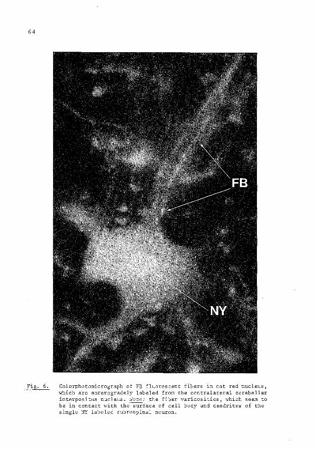
64
Fig. 6. Colorphotomicrograph of FB fluorescent fibers in cat red nucleus, which are anterogradely labeled from the contralateral cerebellar interpositus nucleus. Note: the fiber varicosities, which seem to be in contact with the surface of cell body and dendrites of the single NY labeled rubrospinal neuron.

65
(Vander Kooy and Wise, 1980). The retrograde fluorescent tracers TB, PI and
EB are most suited for retrograde labeling of neurons in combination with mono
amine fluorescence histochemistry, because these tracers and the monoamine
fluorophores can be visualized independently in the same cell. This is facili
tated by the fact that, as has been pointed out earlier, in freeze dried
material TB and PI show a granular distribution throughout the cytoplasm (Fig.
4) while the monoamine fluorophores, show a more diffuse distribution. Retro
grade double labeling through divergent axon collaterals by means of PI or EB
in combination with TB can be combined with monoamine histofluorescence in
which case three fluorescent markers (i.e. two retrograde tracers and one
transmitter derived fluorophore) are visualized in one and the same cell
(Bjorklund and Skagerberg, 1979a+b).
B). Acetylcholinesterase histochemistry
The enzyme acetylcholinesterase (AChE) when present in neurons can be
demonstrated histochemically (Koelle, 1954; Butcher et al., 1975). This method
can be combined with retrograde HRP labeling, which makes it possible to study
the efferent connections of AChE neurons (Hardy et al., 1976). Recently it was
shown that the procedure may be simplified by using retrograde fluorescent
tracers instead of HRP, because under such circumstances only one histochemical
procedure is required. Both the AChE brown reaction products and the fluorescent
tracer can be observed in one and the same cell body by using bright field
illumination and fluorescence microscopy, respectively (Albanese and Benti
voglio, 1982a). Applying this combined method in the rat substantia nigra it
was shown that after injecting TB or FB (360 nm excitation wavelength) or EB
(550 nm excitation wavelength) in the striatum some of the retrogradely labeled
nigrostriatal neurons also contain AChE reaction products (Albanese and Benti
voglio, 1982a).
C. Immunohistochemical technique
Hokfelt and collaborators (Hokfelt et al., 1979a+b, 1980) were the first to
combine the immunohistochemical technique with fluorescent retrograde tracers.
Employing this technique Sawchenko and Swanson favor in particular TB out of
the group of fluorescent tracers (Sawchenko and Swanson, 1981, 1982). However,
some pitfalls are present, because during the immunohistochemical procedure
several of the fluorescent tracers diffuse out of the retrogradely labeled
neuron. This is probably due to the fact that the light fixation as used for
the immunohistochemical procedure does not adequately anchor the tracer in the
retrogradely labeled neurons (Sawchenko and Swanson, 1981). Under these cLrcum-

66
stances it ~s therefore necessary to photograph the retrogradely labeled neu
rons before the immunohistochemistry is applied. By means of this combined
technique the transmitter histochemistry of several pathways has now been
demonstrated (Hokfelt et al., 1979a+b, 1980; Dalsgaard et al., 1982; Sawchenko
and Swanson, 1982; Chesselet et al., 1982). When PI, Primuline or DY.
2HCI are used as retrograde tracers it is not necessary to photograph the
retrogradely labeled neurons before the immunohistochemical procedure, because
these tracers withstand the immunohistochemistry (Steinbusch et al., 1981;
Brann and Emson, 1980; Dalsgaard et al., 1982; Skirboll et al., in preparation;
Steward, 1981). However, when using TB a longer fixation is necessary to
prevent TB from leaking out of the retrogradely labeled cell body during the
immunohistochemical procedure (Sawchenko and Swanson, 1981). Skirboll et al.
(in preparation) prefer a short fixation, because of the immunohistochemistry,
which implies that the TB retrogradely labeled neurons have to be photographed
before this procedure. Moreover, according to these investigators (Skirboll
et al., in preparation) all data about retrogradely labeled neurons are pre
served by photographing and plotting of the material prior to the immunohisto
chemical procedure since during this procedure the tracer may disappear out of
the labeled neurons, which may cause a loss of 5% - 15% of the labeled neurons
(Sawchenko and Swanson, 1981; Vander Kooy and Sawchenko, 1982).
D). Simu~taneous steroid autoradiography and retrograde ~abe~ing of neurons
The steroid autoradiography makes it possible to localize the steroid
target cells in the brain (Kim et al., 1977; Morrell et al., 1975). Combining
the steroid autoradiography with the retrograde tracer technique makes it
possible to establish both the location of steroid target cells and their
fiber connections. For this purpose the retrograde fluorescent tracers
Primuline, True Blue and Granular Blue may be used, since these tracers sur
vive the autoradiographic procedures (Arnold, 1980; Morrell and Pfaff, 1982).

67
VII. Methods for using the fluorescent tracers Evans Blue (EB), DAPI,
Primuline (Pr), DAPI-Primuline mixture, Propidium Iodide (PI), Granular
Blue (GB), True Blue (TB), Fast Blue (FB), Nuclear Yellow (NY), Diami
dino Yellow (DY).
When one begins to use the fluorescent tracers it is of importance first
to gain some experience with the type of retrograde labeling they produce. For
this purpose the rat nigrostriatal system can be used as a reliable testing
ground. Therefore when starting to use the fluorescent tracers one should first
in a few rats inject 0,4 ~1 to 0,6 ~1 of the tracer in the head of the caudate
followed by 3 days survival time. Under these circumstances the neurons of the
substantia nigra pars compacta are consistently labeled.
The tracers are dissolved or suspended in distilled water with the aid
of an ultrasonic waterbath (for % solution or suspension used for the diffe
rent tracers, see table I). In general each time 0. I ml of a solution or sus
pension is made up which may be used for 2 weeks, and which is stored in a
small phial at 4°C. However, the use of fresh solutions or suspensions is
preferred.
The tracers are injected either by means of a glass micropipette (tip
diameter 40-80 ~m), which is connected to an oil filled pressure system, or by
means of a Hamilton microsyringe equipped with a 22 Gauge needle. When sus
pensions are injected by means of a micropipette a pipette with a tip diameter
of more than 60 ~m should be used, because with smaller tip diameters the
pipette tends to become clogged. The smallest injection area obtained after
injecting 0,1 ~1 TB in rat spinal cord grey matter with a micropipette of a
tip diameter of 60 ~m measured in mm in diameter (chapter III, Fig. 12). The
same result has been obtained after injections of FB and DY in the cortex
(Bullie~ personal communications). However, smaller injection areas (diameter
of 0.5 mm) can be obtained by means of iontophoresis or chronic implantations
of micropipettes (Adelheid and Carlsen, 1982).
The micropipette or Hamilton microsyringe with which the tracers are in
jected is held a few minutes in situ after injection. This helps to minimize
spreading of the tracer liquid up into the needle track. For each tracer a
separate pipette or needle is used in order to avoid contamination which may
produce false double labeling.
All tracers are transported retrogradely from terminal fields, but they
are also transported retrogradely from damaged axons. Dissolving the tracers in
2% dimethylsulfoxide (DMSO) enhances their transport from damaged axons, pas
sing through the injection area (Huisman et al., 1982). In order to detect
retrograde neuronal labeling by means of the fluorescent tracers, the survival

68
Code number of Filter Mirror Filter Mirror the tracers System A System N2
providing exci- providing exci-tation light of tation light of 360 nm wavelength 550 nm wavelength
Evans Blue cytoplasm + (EB) nucleus: red
DAPI/ Serva,Heidelberg cytoplasm + Primuline 18860/Eastman, nucleus: blue/
1039 cytoplasm : light golden granules
Propidium Sigma; P-5264 cytoplasm + Iodide nucleolus: (PI) orange-red
Granular Diamidino X
cytoplasm: blue, Blue (GB) Compound 186j134 with silver
golden granules
True Blue Diamidino X cytoplasm +
(TB) Compound 150/129 nucleolus: Blue
Fast Blue Diamidino2~3 cytoplasm + (FB) Compound /50 blue with fine
silver granules
Nuclear Benzimidazole XX nucleus + Yellow (NY) Compound Hoechst nucleolar ring:
S769121 golden yellow (granular)
Diamidino Diamidino X nucleus +
Yellow (DY) Compound 288f26 nucleolar ring: golden yellow (diffuse)
x The code numbers were given by the institute of Phamacy and Food chemistry of the Friedrich-Alexander University in Erlangen (F.R.G.) where these substances have been synthetized. For research purposes samples can be obtained from Prof.Dr. Illing, Warthweg 14-18 Postfach !ISO D-6114 Gross Umstadt Germany
XX For research purposes small samples of NY can be obtained from Dr. H. Loewe, Hoechst Aktiengesellschaft, Postfach 800320, 6230 Frankfurt am Main 80, F.R.G.
I I
J I I I '
i
i
I
Table I: shows for the tracers, which are still used, the code numbers, the label characteristics at 360 and 550 nm, the percentages of tracer solutions, and the survival times, necessary for proper labeling. Note the relatively short survival times, which are used for Nuclear Yellow (NY).

69
SURVIVAL TH1E, necessary for proper labeling
RAT RAT RAT CAT CAT
% tracer Caudate/Put spinal C5 spinal Tl spinal C5 >Pinal Til j solution to Nigra to Red to cortex to red to
1
( w/v ) (SNC) Nucleus Nucleus cortex
I 10% I 24-48h
! I 12,5%/10% 4d
I I i
3% 2d 7d (7d) light labeling
5% i 2-4d 5-7d 7-9d I I I
2% I 2-4d 5-7d 7-9d
3% I 2d 4d 4d 3-4w 3-4w I l l
1% I 6h 24h 40h +46h +70h i - -I
2% 2-3d 7d !Od 3w 4w

70
time has to be chosen such that enough tracer has accumulated in the cell
body. Therefore two factors determine the optimal survival time, i.e. the type
of tracer and the transport distance. Moreover, proper retrograde labeling
with the same tracer over the same distance may require different survival
times ~n different fiber systems. Therefore when applying the tracers in a
given fiber system, it is necessary to do a few preliminary experiments in
order to determine the optimal survival time (see table I for survival times
in striatal-nigral system and descending pathways in rat and cat). The survi
val times for NY must be relatively short, because at longer survival times NY
migrates out of the retrogradely labeled neurons which produces bright fluo
rescence of the nuclei of adjoining glial cells and may result in false retro
grade labeling of surrounding neurons (Bentivoglio et al., 1980b). Therefore
the NY survival times are titrated such that at the most a dull glial staining
occurs around the retrogradely labeled neurons. This implies that when NY is
used in combination with tracers which require a longer survival time e.g. TB
or FB, they are injected first and NY is injected later a short time before
the animal is sacrificed. In this way a relatively long TB or FB survival time
is combined with a relatively short NY survival time. This somewhat compli
cated procedure may be avoided by using DY instead of NY, since during the
relatively long survival times required by TB or FB, DY does not leak out of
retrogradely labeled neurons (Keizer et al., in preparation). In general TB is
used in rat because it is more blue fluorescent than FB. On the other hand FB
is preferred above TB in cat and monkey, because FB is more effectively tran
sported over long distances as required in these animals (c.f. table I).
After the appropriate survival time the animals are sacrificed with an
overdosis Nembutal (6%) and are transcardially perfused with saline followed
by formalin. When the fluorescent tracers are combined with HRP, the perfusion
with glutaraldehyde diminishes drastically the intensity of the fluorescence
and changes its characteristics. Therefore, under these circumstances a forma-
line perfusion should be used as advocated by Kevetter and Willis (1982). In
general the perfusion solutions are pumped through the circulation with a
speed of 80 cc a minute. Rats are perfused with 0.5 liter hypertonic NaCI
(1.5%) followed by I liter cacodylate or citrate buffered formalin (10%, pH
7,2) and the brain and spinal cord are then stored overnight in cacodylate or
citrate buffered sucrose (30%, pH 7,2) at 4°C. Cats and monkeys are trans
cardially perfused with 2 liter hypertonic NaCl (2.7%) followed by 3 liter
cacodylate or citrate buffered formaline. In general a solution of 10% forma
line (pH 7,2) is used. However, with FB 30% formaline (pH 7,2) is used since
this stronger fixation enhances the FB fluorescence. Brain and spinal cord of

71
cats and monkeys are not stored overnight in sucrose. These animals, following
the formaline perfusion, are perfused with cacodylate or citrate buffered
sucrose (8%, pH 7,2) instead and the brain and spinal cord are dissected and
immediately cut in frozen sections. In our studies the tissue is frozen to
approximately -30°C on a freezing microtome and then cut in frozen sections of
30 vm thick. In order to prevent freeze artefacts the temperature is kept con
stant during cutting of the sections. The material does not have to be cut
immediately but first can be stored at very low temperature (-80°C). Injected
spinal segments of a rat spinal cord are coated with carboxymethylcellulose in
order to preserve the sections containing the needle tracks. Especially in the
case in which NY is injected a rapid processing of the material is important
because when the sections are kept in water for some time NY may migrate out
of retrogradely labeled neurons (Bentivoglio et al., 1980b). Therefore the
sections, after being cut, are immediately mounted from distilled water. The
mounted sections are air dried and not coverslipped. The material ~s studied
with a Leitz Ploemopack fluorescence microscope with a high pressure lamp of
100 Watt. The microscope is equipped with a filter mirror system A, D and N2,
providing excitation wavelength of 360 nm, 390 nm and 550 nm, respectively.
The retrogradely labeled neurons are only clearly visible with objectives of
25x or higher. These objectives require the use of immersion oil, which is
applied directly to the air dried sections. The distributions of the single
and double labeled neurons in the sections are plotted with the aid of an X-Y
plotter, which is connected with transducers attached to the microscope stage.
In order to localize exactly the plotted neurons the sections after being
plotted are counterstained with cresyl violet. Fluorescent counterstains may
also be used (Schmued et al., 1982). Pictures (black and white or color) are
taken with the aid of a Leitz-vario-orthomat which regulates the exposure time
automatically. For black and white pictures an Agfapan 100 film is used and
for color pictures a Kodak Ektachrome 200 film is used. Also a Kodak Ekta
chrome 400 or 800 film can be used. When illuminating the sections the fluo
rescence in the retrogradely labeled neurons gradually fades. This is especial
ly obvious in lightly TB or FB labeled neurons. The fluorescence also dimi
nishes with time, especially in slides covered with oil. In order to slow down
this fading of the fluorescence the sections are stored in the dark and at 4°C.
In this way sections without oil may be kept for several weeks. After long
storage the tissue starts to display a diffuse glistering fluorescence, which
tends to obscure the fluorescence of the retrogradely labeled neurons.

72
Acknowledgements
The authors wish to thank Prof.Dr. 0. Dann (Friedrich-Alexander Universitat,
Erlangen) and Dr. H. Loewe (Hoechst Company, Frankfurt) for their collaboration
in developing the fluorescent retrograde tracers. They thank Mr. E. Dalm and
Mr. H. Klink for their technical assistance, Ms. P. van Alphen for the photo
graphy and Ms. G. van Gelder and Ms. E. Klink for typing the manuscript. The
authors also thank Dr. M. Bentivoglio, Dr. C. Catsman-Berrevoets, Dr. D. Van
Der Kooy, Dr. R. Lemon, Dr. T. Hokfelt, Dr. M. Illert for reading it and for
their constructive comments.

VIII. References
I. Albanese, A., Bentivoglio, M. (1982a) Retrograde fluorescent
neuronal tracing combined with acetylcholinesterase
histochemistry. J. Neurosci. Meth. 6: 121-127.
73
2. Albanese, A., Bentivoglio, M. (1982b) The organization of dopa
minergic and non-dopaminergic mesencephalo-cortical
neurons in the rat. Brain Res. 238: 421-425.
3. Alheid, G.F., Carlsen, J. (1982) Small injections of fluorescent
tracers by iontophoresis or chronic implantation of
micropipettes. Brain Res. 235: 174-178.
4. Arnold, A.P. (1980) A technique for simultaneous steroid auto
radiography and retrograde labeling of neurons. Brain
Res. 194: 210-212.
5. Aschoff, A.O., Hollander, H. (1982) Fluorescent compounds as
retrograde tracers compared with horseradish peroxidase
(HRP). A parametric study in the central visual system
of the albino rat. Journal of Neurosci. Methods 6:
179-198.
6. Axelsson, S.A., Bjorklund, A., Falck, B., Lindvall, 0., Svensson,
L.A. (1973) Glyoxilic acid concentration : a new
fluorescence method for the histochemical demonstration
of biogenic monoamines. Acta Physiol. Scand. 87: 57-62.
7. Bentivoglio, M., Vander Kooy, D., Kuypers, H.G.J.M. (1979a) The
organization of the efferent projections of the sub
stantia nigra in the rat. A retrograde fluorescent
double labeling study. Brain Res. 174: 1-17.
8. Bentivoglio, M., Kuypers, H.G.J.M., Catsman-Berrevoets, C.E.,
Dann, 0. (1979b) Fluorescent retrograde neuronal labe
ling in rat by means of substances binding specifically
to adeninethymine rich DNA. Neurosci. Lett. 12: 235-240.
9. Bentivoglio, M., Kuypers, H.G.J.M., Catsman-Berrevoets, C.E.,
Loewe, H., Dann, 0. (1980a) Two new fluorescent retro
grade neuronal tracers which are transported over long
distances. Neurosci. Lett. 18: 25-30.
10. Bentivoglio, M., Kuypers, H.G.J.M., Catsman-Berrevoets, C.E.
(1980b) Retrograde neuronal labeling by means of bis
benzimide and nuclear yellow (Hoechst S 769121).
11easures to prevent diffusion of the tracers out of
retrogradely labeled neurons. Neurosci. Lett. 18: 19-24.

74
II. Bentivoglio, M., Macchi, G., Albanese, A. (1981) The cortical projections
of the thalamic intralaminar nuclei, as studied in cat and
rat with the multiple fluorescent retrograde tracing techni
que. Neurosci. Lett. 26: 5-10.
12. Bentivoglio, M., Molinari, M. (1982) Fluorescent retrograde triple labe
ling of brain stem reticular neurons with ascending and
spinal projections. Neurosci. Lett. Suppl. 10: S71.
13. Bharos, T.B., Kuypers, H.G.J.M., Lemon, R.N., Muir, R.B. (1981) Divergent
collaterals from deep cerebellar neurons to thalamus and
tectum, and to medulla oblongata and spinal cord: retrograde
fluorescent and electrophysiological studies. Exp. Brain Res.
42: 399-410.
14. Bjorklund, A., Falck, B., Hakanson, R. (1968) Histochemical demonstration
of tryptamine properties of the formaldehyde-induced fluoro
phores of tryptamine and related indole compounds in models.
Acta Physiol. Scand. 74 Suppl. 318.
15. Bjorklund, A., Lindvall, 0., Svensson, L.A. (1972) Mechanisms of fluoro
phore formation in the histochemical glyoxilic acid method
for monoamines. Histochemie 32: 113-131.
16. Bjorklund, A., Skagerberg, G. (J979a) Simultaneous use of retrograde
fluorescent tracers and fluorescence histochemistry for con
venient and precise mapping of monoaminergic projections and
collateral arrangements in the CNS. J. Neurosci. Meth. 1:
261-277.
17. Bjorklund, A., Skagerberg, G. (1979b) Evidence for a major spinal cord
projection from the diencephalic All dopamine cell group in
the rat using transmitter-specific fluorescent retrograde
tracing. Brain Res. 177: 170-175.
18. Brann, M.R., Emson, P.C. (1980) Microiontophoretic injection of fluo
rescent tracer combined with simultaneous immunofluorescent
histochemistry for the demonstration of efferents from the
caudate-putamen projecting to the globus pallidus. Neurosci.
Lett. 16: 61-65.
19. Brodal, A. (1940) Modification of Gudden method for study of cerebral
localization. Arch. Neural. Psychiatry 43: 46-58.
20. Brodal, A. (1957) The reticular formation of the brain stem. Anatomical
aspects and functional correlations. London: Oliver & Boyd.

75
21. Burde, R.M., Parelman, J.J., Luskin, M. (1982) Lack of unity of Edinger
Westphal nucleus projections to the ciliary ganglion and spinal
cord: a double-labeling approach. Brain Res. 249: 379-382.
22. Butcher, L.L., Marchand, R. (1978) Dopamine neurons in pars compacta of
the substantia nigra contain acetylcholinesterase: Histochemical
correlations on the same brain section. Europ. J. Pharmacal. 52:
415-417.
23. Cajal, S.R. (1952) Histologie du systeme nerveux de l'Homme et des verte
bres. Madrid. Instituto Ramon y Cajal.
24. Catsman-Berrevoets, C.E., Lemon, R.N., Verburgh, C.A., Bentivoglio, M.,
Kuypers, H.G.J.M. (1980) Absence of callosal collaterals derived
from rat corticospinal neurons. A study using fluorescent retro
grade tracing and electrophysiological techniques. Exp. Brain Res.
39: 433-440.
25. Catsman-Berrevoets, C.E., Kuypers, H.G.J.M. (1981) A search for cortico
spinal collaterals to thalamus and mesencephalon by means of
multiple retrograde fluorescent tracers in cat and rat. Brain Res.
218: 15-33.
26. Cesaro, P., Nguyen-Legros, J., Berger, B., Alvarez, C., Albe-Fessard, D.
(1979) Double labelling of branched neurons in the central nervous
system of the rat by retrograde axonal transport of horseradish
peroxidase and iron dextran complex. Neurosci. Lett. 15: 1-7.
27. Chesselet, M.F., Reisine, T.D., Glowinski, J., Graybiel, A.M. (1982)
Striatal somatostatin: immunohistochemistry and effects on dopa
minergic transmission. Society for neurosci. abstracts p.287,
79. 10.
28. Cowan, W.M., Gottlieb, D.I., Hendrickson, A.E., Price, J.L., Woolsey, T.A.
(1972) The autoradiographic demonstration of axonal connections in
the central nervous system. Brain Res. 37: 21-51.
29. Dahlstrom, A., Fuxe, K. (1964) Evidence for the existence of monoamine
containing neurons in the central nervous system I. Demonstration
of monoamines in the cell bodies of brain stem neurons. Acta
Physiol. Scand. 62 Suppl. 232: 5-55.
30. Dahlstrom, A., Fuxe, K. (1965) Evidence for the existence of monoamlne
neurons in the central nervous system II. Experimentally induced
changes ln the intraneuronal amine levels of bulbospinal neuron
systems. Acta Physiol. Scand. 64, Suppl. 247: 6-36.

76
31. Dalsgaard, C.-J., Hokfelt, T., Elfvin, L.-G., Skirboll, L., Emson, P.
(1982) Substance P-containing sensory neurons projecting to
the inferior mesenteric ganglion: evidence from combined retro
grade tracing and immunohistochemistry. Neuroscience 7: 647-655.
32. Falck, B., Hillarp, N.A., Thieme, G., Torp, A. (1962) Fluorescence of
catecholamines and related compounds condensed with formal
dehyde. J. Histochem. Cytochem. 10: 348-354.
33. Fink, R.P., Heimer, L. (1967) Two methods for selective silver impreg
nation of degenerating axons and their synaptic endings in the
central nervous system. Brain Res. 4: 369-374.
34. Flumerfelt, B.A., Gwyn, D.G. (1974) The red nucleus of the rat: its orga
nization and interconnections. J. Anat. (Lond.) 118: 374-376.
35. Geisert, E.E. Jr. (1976) The use of tritiated horseradish peroxidase for
defining neuronal pathway. Brain Res. 117: 130-135.
36. Glees, P. (1946) Terminal degeneration within the central nervous system
as studied by a new silver method. J. Neuropathol. Exp. Neural.
5: 54-59.
37. Grafstein, B., Forman, D.S. (1980) Intracellular transport ln neurons.
Physiol. Reviews 60: 1167-1283.
38. Graham, R.C., Karnovsky, M.J. (1966) The early stage of absorption of
injected horseradish peroxidase in the proximal tubules of
mouse kidney: ultra-structural cytochemistry by a new technique.
J. Histochem. Cytochem. 14: 291-302
39. Hardy, H., Heimer, L., Switzer, R., Watkins, D. (1976) Simultaneous demon
stration of horseradish peroxidase and acetylcholinesterase.
Neurosci. Lett. 3: 1-5.
40. Hayes, N.L., Rustioni, A. (1979) Dual projections of single neurons are
visualized simultaneously: use of enzym~tically inactive (3H)
HRP. Brain Res. 165: 321-326.
41. Hayes, N.L., Rustioni, A. (1981) Differential and collateral descending
projections to spinal enlargements. Exp. Brain Res. 41: 89-107.
42. Hendrickson, A.E. (1972) Electromicroscopic distribution of axoplasmic
transport. J. Neural. 144: 381-398.
43. Hokfelt, T., Philipson, 0., Kuypers, H.G.J.M., Bentivoglio, M., Catsman
Berrevoets, C.E., Dann, 0. (1979a) Tracing of transmitter
histochemically identified neurons projections: immunohisto
chemistry combined with fluorescent retrograde labelling.
Neurosci. Lett. 342: S342.

44. Hokfelt, T., Terenius, L., Kuypers, H.G.J.M., Dann, 0. (1979b) Evidence
for enkephalin immunoreactive neurons in the medulla oblongata
projecting to the spinal cord. Neurosci. Lett. 14: 55-60.
77
45. Hokfelt, T., Skirboll, L., Rehfeld, J.F., Goldstein, M., Markey, K., Dann,
0. (1980) A subpopulation of mesencephalic dopamine neurons
projecting to limbic areas contains a cholecystokinin-like
peptide: evidence from immunohistochemistry combined with retro
grade tracing. Neuroscience 5: 2093-2124.
46. Holstege, G., Kuypers, H.G.J.M., Boer, R.C. (1979) Anatomical evidence
for direct brainstem projections to the somatic motoneuronal cell
groups and autonomic preganglionic cell groups in cat spinal cord.
Brain Res. 171: 329-333.
47. Holstege, G., Kuypers, H.G.J.M. (1982) The anatomy of brain stem pathways
to the spinal cord in cat. A labeled aminoacid tracing study.
Progress in Brain Res. 57: 145-177.
48. Huisman, A.M., Kuypers, H.G.J.M., Verburgh, C.A. (1981) Quantitative
differences in collateralization of the descending spinal path
ways from red nucleus and other brain stem cell groups in rat as
demonstrated with the multiple fluorescent retrograde tracer
technique. Brain Res. 209: 271-286.
49. Huisman, A.M., Kuypers, H.G.J.M., Verburgh, C.A. (1982) Differences in
collateralization of the descending spinal pathways from red
nucleus and other brain stem cell groups in cat and monkey.
Progress in Brain Res. 57: 185-218.
50. Huisman, A.M., Kuypers, H.G.J.M., Conde, F., Keizer, K. Collaterals of
rubrospinal neurons to the cerebellum in rat. A retrograde fluo
rescent double labeling study. Brain Res. in press.
51. Illert, M., Fritz, N., Aschoff, A., Hollander, H. (1982) Fluorescent
Compounds as Retrograde Tracers Compared with Horseradish Peroxi
dase (HRP). II. A parametric study in the peripheral motor system
of the cat. Journal of Neuroscience Methods 6: 199-218.
52. Innocenti, G.M. (1981) Growth and reshaping of axons in the establishment
of visual callosal connections. Science 212: 824-826.
53. Ivy, G.O., Akers, R.M., Killackey, H.P. (1979) Differential distribution
of callosal projection neurons in the neonatal and adult rat.
Brain Res. 173: 532-537.
54. Ivy, G.O., Killackey, H.P. (1981) The ontogeny of the distribution of
callosal projection in the rat parietal cortex. J. comp. Neural.
195: 367-389.
78
55. Keizer, K., Kuypers, H.G.J.M., Huisman, A.M. Diamidino Yellow (DY)
a fluorescent retrograde neuronal tracer, which migrates only
very slowly out of the cell and can be used in a combination with
TB and FB in double labeling experiments. Submitted to Experi
mental Brain Research.
56. Kevetter, G.A., Willis, W.D. (1982) Spinothalamic cells in the rat lum
bar cord with collaterals to the medullary reticular formation.
Brain Res. 238: 181-185.
57. Kim, Y.S., Stumpf, W.E., Sar, M., Martinez-Vargas, M. (1979) Estrogen and
androgen target cells in the brain of fishes, reptiles and birds
phylogeny and ontogeny. Aroer. Zool. 18: 425-433.
58. Koelle, G.B. (1954) The histochemical localization of cholinesterase ln
the central nervous system of the rat. J. camp. Neural. 100:
211-228.
59. Vander Kooy, D., Kuypers, H.G.J.M., Catsman-Berrevoets, C.E. (1978)
Single mammillary body cells with divergent axon collaterals.
Demonstration by a simple, fluorescent retrograde double labeling
technique in the rat. Brain Res. 158: 189-196.
60. Vander Kooy, D., Kuypers, H.G.J.M. (1979) Fluorescent retrograde double
labeling: Axonal branching in the ascending raphe and nigral
projections. Science 204: 873-875.
61. Vander Kooy, D., Wise, R.A. (1980) Retrograde fluorescent tracing of
substantia nigra neurons combined with catecholamine histofluo
rescence. Brain Res. 183: 447-452.
62. Vander Kooy, D., Hattori, T. (1980) Bilaterally situated dorsal raphe
cell bodies have only unilateral forebrain projections in rat
Brain Res. 192: 550-554.
63. Vander Kooy, D., Carter, D.A. (1981) The organization of the efferent
projections and striatal afferents of the entopeduncular nucleus
and adjacent areas in the rat. Brain Res. 211: 15-36.
64. Vander Kooy, D., Sawchenko, P.E. (1982) Characterization of serotoner
gic neurons using concurrent fluorescent retrograde axonal tracing
and immunohistochemistry. The Journal of Histochem. and Cytochem.
Vol. 30: 794-798.
65. Kristensson, K. (1970) Transport of fluorescent protein tracer in peri
pheral nerves. Acta neuropath. (Berl.) 16: 293-300.
66. Kristensson, K., Olsson, Y. (1971a) Retrograde axonal transport of
protein. Brain Ee~:. 29: 363-365.

79
67. Kristensson, K., Olsson, Y., Sjostrand, J. (I97I) Axonal uptake and retro
grade transport of exogenous proteins in the hypoglossal
nerve. Brain Res. 32: 399-406.
68. Kuypers, H.G.J.M., Catsman-Berrevoets, C.E., Padt, R.E. (I977) Retrograde
axonal transport of fluorescent substances in the rat's fore
brain. Neurosci. Lett. 6: I27-I35.
69. Kuypers, H.G.J.M., Bentivoglio, M., Vander Kooy, D., Catsman-Berrevoets,
C.E. (I979) Retrograde transport of bisbenzimide and propi
dium iodide through axons to their parent cell bodies. Neuro
scL Lett. I2: I-7.
70. Kuypers, H.G.J.M., Bentivoglio, M., Catsman-Berrevoets, C.E., Bharos, A.T.
(I980) Double retrograde neuronal labeling through divergent
axon collaterals, using two fluorescent tracers with the same
excitation wavelength which label different features of the
cell. Exp. Brain Res. 40: 383-392.
7I. Kuypers, H.G.J.M., Huisman, A.M. (I982) The neuroanatomy of the descending
brain pathways. In: "Brain Stem Control of Spinal Mechanisms" .
. Fernstrom Foundation Series No. I New York-Oxford (in press).
72. Kuzuhara, S., Kanazawa, I., Nakanishi, T. (I980) Topographical localiza
tion of the Onuff's nuclear neurons innervating the rectal
and vesical striated sphincter muscles: A' retrograde fluo
rescent double labeling in cat and dog. Neurosci. Lett. I6:
125-130.
73. Lasek, R., Joseph, B.S., Whitlock, D.G. (I968) Evaluation of a radioauto
graphic neuroanatomical tracing method. Brain Res. 8: 319-336.
74. LaVail, J.H., LaVail, M.M. (I972) Retrograde axonal transport in the
central nervous system. Science I76: I4I6-I4I7.
75. Martin, G.F., Humbertson, A.O., Laxson, C., Panneton, W.M. (I979) Evidence
for direct bulbospinal projections to laminae IX, X and the
intermediolateral cell column. Studies using axonal transport
techniques in the North American opossum. Brain Res. I70:
I 65- I 71.
76. Martin, G. F., Cabana, T., DiTorro, F.J., Ro, R.H., Humbertson, A.O. (1982)
Reticular and raphe projections to the spinal cord of the
North American opossum. Evidence for connectional hetero
geneity. In: Progress in Brain Research. Vol. I5: I09-I3I.
Elsevier Biomedical Press, Amsterdam.

80
77. Mesulam, M.-M. (1978) Tetramethyl benzidine for horseradish peroxidase
neurohistochemistry: a non-carcinogenic blue reaction-product
with superior sensitivity for visualizing neural afferents and
efferents. J. Histochem. Cytochem. 26: 106-117.
78. Mesulam, M.-M., Rosene, D.L. (1979) Sensitivity in horseradish peroxidase
neurohistochemistry: A comparative and quantitative study of nine
methods. J. Histochem. Cytochem. 27: 763-773.
79. Mesulam, M.~M., Bishop, G.A., Carson, K.A., King, J.S. (1982) Tracing
nedr~l connections with horseradish peroxidase. M.M. Mesulam
(Ed.) IBRO Handbook Series: Methods in the Neurosciences.
80. Miceli, D., Reperant, J. (1982) Thalamo-hyperstriatal projections in the
pigeon (Columbo livia) as demonstrated by retrograde double labe
ling with fluorescent tracers. Brain Res. 245: 365-371.
81. Morrell, J.I., Kelley, D.B., Pfaff, D.W. (1975) Sex steroid binding in
the brains of vertebrates. In: K.M. Knigge and D.E. Scott (Eds.)
Brain-Endocrine Interaction II. The Ventricular System. Karger,
Basel, pp. 230-256.
82. Morrell, J.I., Pfaff, D.W. (1982) Characterization of estrogen-concentra
ting hypothalamic neurons by their axonal projections. Science
217: 1273-1276.
83. Nauta, W.J.H., Gygax, P.A. (1954) Silver impregnation of degenerating
axons in the central nervous system: a modified technique. Stain
Techno!. 29: 91-94.
·84. Niijima, K., Yoshida, M. (1982) Electrophysiological evidence for bran
ching nigral projections to pontine reticular formation, superior
colliculus and thalamus. Brain Res. 239: 279-282.
85. O'Leary, D.D.M., Stanfield, B.B., Cowan, W.M. (1981) Evidence that the
early postnatal restriction of the cells of origin of the callosal
projection is due to the elimination of axonal collaterals rather
than to the death of neurons. Developmental Brain Res. 1: 607-617.
86. Olmos, De., J., Heimer, L. (1980) Double and triple labeling of neurons
with fluorescent substances: the study of collateral pathways in
the ascending raphe system. Neurosci. Lett. 19: 7-12.
87. Olsson, T., Kristensson, K. (1978) A simple histochemical method for double
labeling of neurons by retrograde axonal transport. Neurosci. Lett.
8: 265-268.
88. Parent, A., de Bellefeuille, L., (1982) Organization of efferent projections
from the internal segment of Globus Pallidus in Primate as revealed
by fluorescence retrograde labeling method. Brain Res. 245: 201-213.

81
89. Pompeiano, 0., Brodal, A. (1957) Experimental demonstration of a somato
topic origin of rubrospinal fibers in the cat. J. camp. Neural.
108: 225-251.
90. Rosina, A. Rapid anterograde movement of the fluorescent tracer fast blue:
A new method for tracing central connections. In preparation.
91. Rosina, A., Provini, L., Bentivoglio, M., Kuypers, H.G.J.M. (1980) Ponto
neocerebellar axonal branching as revealed by double fluorescent
retrograde labeling. Brain Res. 195: 461-466.
92. Sawchenko, P.E., Swanson, L.W. (1980) Immunohistochemical identification
of neurons in the paraventricular nucleus of the hypothalamus
that project to the medulla or to the spinal cord in the rat.
J. camp. Neural. 205: 260-272.
93. Sawchenko, P.E., Swanson, L.W. (1981) A method for tracing biochemically
defined pathways in the central nervous system using combined
fluorescence retrograde transport and immunohistochemical
techniques. Brain Res. 210: 31-51.
94. Schmued, L.C., Swanson, L.W. (1982) SITS: a covalently bound fluorescent
retrograde tracer that ··d0es~·not appear to be taken up by fibers
of-passage. Brain Res. 249: 137-141.
95. Schmued, L.C., Swanson, L.W., Sawchenko, P.E. (1982) Some fluorescent
counterstains for neuroanatomical studies. J. Histochem. and
Cytochem. Vol. 30: 123-128".
96. Sidman, R.L. (1970) Autoradiographic methods and principles for study of 3
the nervous system with thymidine H . In: W.J.H. Nauta and
S.O.E. Ebbesson (Eds.). Contemporary Research Methods in Neuro
anatomy, Berlin-Springer. pp. 252-274.
97. Skirboll, L., Hokfelt, T., Kuypers, H.G.J.M., Bentivoglio, M., Catsman
Berrevoets, C., Goldstein, M., Steinbusch, H., Verhofstad, J.,
Jeffcoate, S., Phillipson, 0., Dokray, G., Brownstein, M.,
Norell, G.A. A method for specific transmitter identification
of retrogradely labeled neurons: Immunocytochemistry combined
with fluorescence tracing. In preparation.
98. Steinbusch, H.H.M., Nieuwenhuys, R., Verhofstad, A.A.J., Vander Kooy, D.
(1981) The nucleus raphe dorsalis of the rat and its projection
upon the caudate-putamen. A combined cytoarchitectonic and
immunohistochemical and retrograde transport study. J. Physiol.
(Paris) 77: 157-174.

82
99. Steward, 0., Scoville, S.A. (1976) Retrograde labeling of central ner
vous pathways with tritiated or Evans Blue-labeled bovine serum
albumin. Neurosci. Lett. 3: 191-196.
100. Steward, 0. (1981) Horseradish peroxidase and fluorescent substances and
their combination with other techniques. Neuroanatomical tract
tracing methods. Heimer, L., and Robards, M.J. (eds).
101. Swanson, L.W., Kuypers, H.G.J.M. (1980a) A direct projection from the
ventromedial nucleus and retrochiasmatic area of the hypothalamus
to the medulla and spinal cord of the rat. Neurosci. Lett. 17:
307-312.
102. Swanson, L.W., Kuypers, H.G.J.M. (1980b) The paraventricular nucleus of
the hypothalamus: Cytoarchitectonic subdivisions and organization
of projections to the pituitary, dorsal vagal complex, and spinal
cord as demonstrated by retrograde fluorescence double-labeling
methods. J. comp. Neurol. 194: 555-570.
103. Swanson, L.W., Sawchenko, P.E., Cowan, W.M. (1980) Evidence that the
commissural associational and septal projections of the regio
inferior of the hippocampus arise from the same neurons. Brain
Res. 197: 207-212.
104. Voshart, K., Vander Kooy, D. (1981) The organization of the efferent
projections of the parabrachial nucleus to the forebrain in the
rat: a retrograde fluorescent double-labeling study. Brain Res.
212: 271-286.
I 05. Weiss, P., Hiscoe, H. B. ( 1948) Experiments on the mechanism of nerve
growth. J. Exp. Zool., 107: 315-395.
106. Wise, S.P., Jones, E.G. (1976) The organization and postnatal development
of the commissural projection of the red somatic sensory cortex.
J. comp. Neurol. 168: 313-344.


84
CHAPTER III: QUANTITATIVE DIFFERENCES IN COLLATERALIZATION
OF THE DESCENDING SPINAL PATHWAYS FROM RED
NUCLEUS AND OTHER BRAINSTEM CELL GROUPS IN RAT
AS DEMONSTRATED WITH THE MULTIPLE FLUORESCENT
RETROGRADE TRACER TECHNIQUE
A. Margriet Huisman, H.G.J.M. Kuypers and
C.A. Verburgh
Brain Res. 209 (1981) 271-286

Brain Research, 209 (1981) 271-286 ©Elsevier/North-Holland Biomedical Press
QUANTITATIVE DIFFERENCES IN COLLATERALIZATION OF THE DESCENDING SPINAL PATHWAYS FROM RED NUCLEUS AND OTHER BRAIN STEM CELL GROUPS IN RAT AS DEMONSTRATED WITH THE MULTIPLE FLUORESCENT RETROGRADE TRACER TECHNIQUE
A. MARGRIET HUISMAN, H. G. J. M. KUYPERS and C. A. VERBURGH
Erasmus Universiteit Rotterdam, Facu!teit der Geneeskunde, Afdeling Anatomie, Postbus 1738,3000 DR Rotterdam (The Netherlands)
(Accepted September 4th, 1980)
Key words: red nucleus - nucleus raphe magnus - ventrolateral pontine tegmentum - multiple fluorescent tracers- collateralization
SUMMARY
In 8 rats 'True Blue' was injected into dorsal half of C5-C8 spinal grey, 5 days later 'Nuclear Yellow' was injected in midthoracic, upper lumbar, lumbosacral and sacral cord respectively. The animals were sacrificed about 43 hours after NY injections. The distribution of retrogradely labeled neurons was studied in Red Nucleus, in Ventrolateral Pontine Tegmentum and in Nucleus Raohe Magnus, all of which project to spinal dorsal grey.
In Red Nucleus large populations of single TB-labeled neurons and single NYlabeled ones occurred in the dorsomedial and ventrolateral part, respectively. In addition, about 8 "" of the neurons labeled with TB from C5-C8 were double labeled with NY from L5-S I, and 35 ~ ,; from T7-8, which percentages resemble those of electrophysiological studies~5 . However, in ipsilateral Nucleus Raphe Magnus about 40 o ~ of the TB-labeled neurons were double labeled from L5-S I. This percentage resembles the 66 o" obtained in electrophysiological studies of reticulospinal collaterals~0. These findings in rat support electrophysiological findings in cat and show, that rubrospinal neurons distribute their fibers primarily to the grey matter of specific groups of spinal segments, while many of the raphe spinal neurons distribute fibers throughout the spinal cord.
INTRODUCTION
Classic anatomical studies using the Golgi technique1 ~ demonstrated that many axons in the central nervous system give off axon collaterals along their trajectory
85
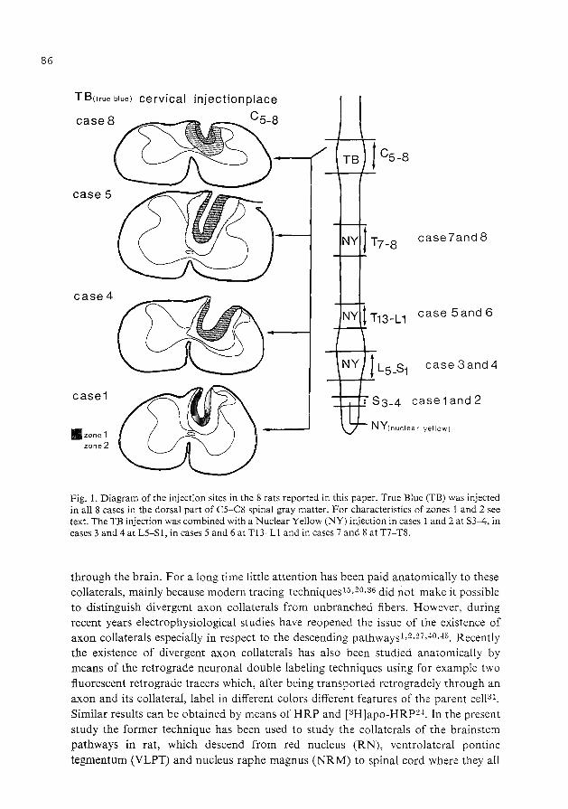
86
T B(true blue) cervical
case 5
case4
case1
lllzone1 zone2
TB t C5-8
case7and 8
case 5and 6
1 Ss-4 case 1 and 2
NY(nuclear yellow)
Fig. 1. Diagram of the injection sites in the 8 rats reported in this paper. True Blue (TB) was injected in all 8 cases in the dorsal part of C5-C8 spinal gray matter. For characteristics of zones 1 and 2 see text. The TB injection was combined with a Nuclear Yellow (NY) injection in cases 1 and 2 at S3-4, in cases 3 and 4 at L5-S1, in cases 5 and 6 at T13-Ll and in cases 7 and 8 at T7-T8.
through the brain. For a long time little attention has been paid anatomically to these collaterals, mainly because modern tracing techniquesl5,Z0,36 did not make it possible to distinguish divergent axon collaterals from unbranched fibers. However, during recent years electrophysiological studies have reopened the issue of the existence of axon collaterals especially in respect to the descending pathwaysl,2,27,-10,-15. Recently the existence of divergent axon collaterals has also been studied anatomically by means of the retrograde neuronal double labeling techniques using for example two fluorescent retrograde tracers which, after being transported retrogradely through an axon and its collateral, label in different colors different features of the parent cel131.
Similar results can be obtained by means of HRP and [3H]apo-HRP2~. In the present study the former technique has been used to study the collaterals of the brainstem pathways in rat, which descend from red nucleus (RN), ventrolateral pontine tegmentum (VLPT) and nucleus raphe magnus (NRM) to spinal cord where they all

terminate in the dorsal half of the spinal gray matter7 ,12,18,26,34,37 ,41,50. The retrograde fluorescent tracers True Blue (TB)9 and Nuclear Yellow (NY)10,u were used. TB produces a blue fluorescent labeling of the cytoplasm and the nucleolus while NY, after short survival times relative to the transport distancell, produces only a golden yellow fluorescence of the nucleus both tracers at 360 nm excitation wavelength. In all animals first TB was injected in the dorsal half of the C5-8 spinal gray. Subsequently in the various animals NY was injected at more caudal levels in the cord (Fig. 1). In all cases in which the TB deposit was restricted to the gray matter, the number of TBlabeled neurons and the number of TB-NY double-labeled ones in the above brain stem cell groups was counted. These data made it possible to approximate both the number of neurons in the three different brain stem cell groups in rat which distribute fibers to the dorsal half of C5-8 spinal gray and the percentages of these cells, which distribute fibers to the various, progressively more caudal portions of the neuraxis.
MATERIALS AND METHODS
In 8 rats, anesthetized with Nembutal (6 %), low cervical1aminectomies were made and in each rat 5 glass micropipette injections of 0.1 ,ul2 %True Blue (TB) ( =0.5 ,ul in total)9 were made in a rostrocaudal row into the dorsal horn and the dorsal part of the intermediate zone of the C5, C6, C7 and C8 segments (Figs. 1 and 2). After 5 days the animals were operated for a second time. Under Nembutal, new laminectomies were made and I ,ul1% Nuclear Yellow (NY)lO,ll was injected in the spinal white and gray matter at more caudal levels of the cord. These NY injections were made by means of multiple micropipette penetrations to damage a large number of fibers. This was in order to obtain NY transport by fibers passing through the injected segment and damaged by the penetrations, as well as by fibers terminating there. In two rats NY injections were made in the sacral cord (cases 1 and 2), in two rats in the lumbosacral cord (cases 3 and 4), in two rats in the upper lumbar cord (cases 5 and 6) and in two rats in the midthoracic cord (cases 7 and 8) (Fig. 1). NY injections at the different levels were made 45, 44, 43 and 40 h prior to the perfusion of the animals, respectively. These short survival times were used in order to avoid migration of NY from the retrogradely labeled neurons in the brain stemll. During the entire postoperative period the animals were given morphine 0.5 mgfday.
The rats were sacrificed with an overdose of Nembutal (6 %). They were transcardially perfused with 1 liter NaCl (1.5 %) followed by 1 liter cacodylatebuffered formaline (10 %, pH 7.2). The brains and the injected segments were stored overnight in cacodylate-buffered sucrose (30 %, pH 7.2) at 4 oc and were cut transversally in frozen sections 30 ,urn thick on a freezing microtome. The injected segments, before being cut, were coated with carboxymethylcellulose in order to keep the sections containing the needle tracks intact. The sections, after being cut, were immediately mounted from distilled water and air dried at room temperature. In general one out of every three sections was mounted.
The material was studied with a Leitz Ploemopack fluorescence microscope equipped with a filter mirror system A providing excitation wavelength of 360 nm wavelength. Subsequently some of the sections were counterstained with cresyl violet.
87

88
Fig. 2. Photomicrograph of one half of a cross-section between C5 and C8 with TB injection site in the spinal gray matter. Note that fluorescence extends through ipsilateral spinal gray matter but that no fluorescence is present in the dorsolateral funiculus.
The distribution of the single TB- and single NY -labeled neurons and of the TB-NY double-labeled one in the contralateral red nucleus (RN), the contralateral ventrolateral pontine tegmentum (VLPT) and the ipsilateral nucleus raphe magnus (NRM) was charted with the aid of an X-Y plotter connected with transducers attached to the microscope stage.
At 360 nm excitation wavelength, the retrograde single TB-labeled neurons (Fig. 3) showed a deep blue fluorescent cytoplasm and a blue fluorescent nucleolus, the retrograde single NY-labeled neurons after short survival time relative to the transport distance (Fig. 3) showed a golden yellow fluorescent nucleus with a clear fluorescent
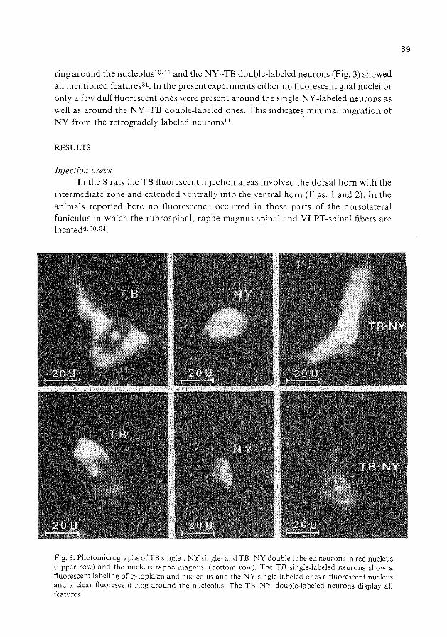
ring around the nucleolus10•11 and the NY-TB double-labeled neurons (Fig. 3) showed all mentioned features31. In the present experiments either no fluorescent glial nuclei or only a few dull fluorescent ones were present around the single NY-labeled neurons as well as around the NY-TB double-labeled ones. This indicates minimal migration of NY from the retrogradely labeled neurons11 .
RESULTS
Injection areas In the 8 rats the TB fluorescent injection areas involved the dorsal horn with the
intermediate zone and extended ventrally into the ventral horn (Figs. I and 2). In the animals reported here no fluorescence occurred in those parts of the dorsolateral funiculus in which the rubrospinal, raphe magnus spinal and VLPT-spinal fibers are located6•30•34.
Fig. 3. Photomicrogrdphs ofTB single-, NY single- and TB-NY double-labeled neurons in red nucleus (upper row) and the nucleus raphe magnus (bottom row). The TB single-labeled neurons show a fluorescent labeling of cytoplasm and nucleolus and the NY single-labeled ones a fluorescent nucleus and a clear fluorescent ring around the nucleolus. The TB-NY double-labeled neurons display all features.
89

90
The TB injection area around the needle tracks showed two concentric fluorescent zones with different features. The first zone, immediately surrounding the needle tracks, contained a bright blue fluorescent cellular debris and a dense accumulation of fluorescent glial nuclei. In counterstained sections this area showed no further cytoarchitectural structure. The second zone contained fluorescent glial nuclei, which decreased in number and brightness towards the periphery of this zone. Peripherally in this zone also blue-labeled neurons and fibers occurred. The second zone faded into the area of the normal tissue with little or no fluorescence. In the present cases zone II extended laterally up to the border of the gray matter (Figs. 1 and 2), rostrally to the rostral border of C5 and caudally to the caudal border of C8.
The NY injection area displayed 3 concentric zones around the needle tracks. The first zone was very narrow and contained yellow tissue fluorescence and a dense accumulation of bright yellow-white glial nuclei. The second zone, which was much wider than the first, also displayed yellow tissue fluorescence but contained fewer fluorescent glial nuclei. The third zone showed no tissue fluorescence but did contain dull fluorescent glial nuclei. The first and second zones involved the injected half of the cord, while the third zone extended into the contralateral half of the spinal cord. Rostro-caudally the first and second zone involved the injected segments, while the third zone extended into the segments rostrally and caudally bordering the injected ones.
Red nucleus In all rats a population of single TB-labeled rubrospinal neurons was present in
the contralateral red nucleus and, in addition, a separate population of single NYlabeled ones occurred. In the caudal portion of the RN the single TB-labeled neurons were present mainly in the dorsomedial part, while the single NY-labeled ones were present in its ventrolateral part (Figs. 4 and 5). In the more rostral part of the nucleus the population oflabeled rubrospinal neurons formed a shell in which the populations of single TB- and of single NY -labeled neurons to some degree became interdigitated (Fig. 4). Some TB-NY double-labeled neurons were present caudally in the red nucleus, between the dorsomedial TB-labeled part and the ventrolateral NY-labeled part. However, the bulk of the double-labeled neurons was observed rostrally in the nucleus intermixed with the single TB- and the single NY -labeled ones. In general, when the NY injections were made progressively more rostrally in the spinal cord the number of TB-NY double-labeled neurons in the RN increased in number (Table I, histogram 1 ). In order to compare the present findings with earlier electrophysiological observations44 the number ofTB-fluorescent neurons in the contralateral red nucleus, which were labeled from C5 to C8, were counted and the percentages of these neurons which were TB-NY double-labeled were computed. For this purpose the following procedure was followed. In each case the TB-labeled neurons as well as the TB-NY double-labeled ones were counted in every mounted section; these numbers were added and then treated according to the following formula:
TB-NY double-labeled neurons X 100%
single TB+ TB-NY double-labeled neurons

rostral case3 TB Cs-8 NY L5-S1
. . . . 0 •• . **0
.. ·. . . . .. .•. • •• ·• •• ·*:·.·. •. ·•· . . : · . . ··:: oo :a* •
. . . .. · ...... ·.· .·. ·.·.: . . .... . .... •• • • 0 0 0
oo o o a· 00
. . . . :. : .· . • • • • • 0
• 0 ·. _Q_b_X10Q;Q37. TB+DL .•
• •* •*o•oo eo ·*.:. *0 0 * 0
.. . ·:. :;. • . • . . • ·.-:*·· ... :· : ... •*• :. ··*·.::::::·:.* .: :a:·;~~oo:.·:
• • 0 0
~X100;67. TB+DL •
TB ::; NY abo 0 0
caseS
TB C5-8 NY T?-8
.... • • • 0
·.*.•o•o•. ~o·oo;~o •
0 •o o **** • ooOooo .• *• 0 ••
**
~X100=35Z TB+DL
Fig. 4. Semi diagrammatic representation of the distribution of retrogradely labeled neurons in the red nucleus after spinal injections in case 2: True Blue (TB) in C5-8 and Nuclear Yellow (NY) in S3-4; in case 3: TB in C5-8 and NY in L5-Sl; in case 8: TB in C5-8 and NY in T7-8. Abbreviations: CMP, posterior commissure; CP, cerebral peduncle; DCP, decussation of superior cerebellar pedurcles; GM, medial geniculate body; IP, interpeduncular nucleus; ML, medial lemniscus; mlf, medial longitudinal fasciculus; NC, cochlear nuclei; NIII, oculomotor nucleus; niii, oculomotor nervus; R, red nucleus; RF, reticular formation; SC, superior colliculus; SNR, substantia nigra, pars reticulata; snc, substantia nigra, pars compacta.
The total number of TB-labeled neurons, including the TB-NY double-labeled ones, in the different cases ranged from 300 to 683. Thus in the two rats with NY injections at S3-S4 the percentages were 0% and 0.3 %, in the two rats with NY injections at L5-S 1 they were 6 ~~ and 9. 7 %, in the two rats with NY injections at T13-Ll they were 15% and 25% and in the two cases with NY injections at T7-T8 the percentages were
91

92
35% and 35% (Table I, histogram I). These findings imply that the red nucleus neurons, which project to the C5-C8 spinal gray matter, also distribute branches to more caudal parts of the spinal cord. They further indicate that a high proportion of these neurons gives rise to branches which pass through or terminate in mid thoracic cord (35 %), that a small proportion of neurons give branches which pass through or terminate in lumbosacral cord (6 ~~and 9.7 %), and that only one out of 300 neurons gives rise to branches which pass through or terminate in low sacral cord.
Nucleus raphe magnus ( N RM) and ventrolateral pontine tegmentum (VLPT) Nucleus raphe magnus. This nucleus, which contains many serotonergic neu
ronsl6,17 also projects by way of the dorsolateral funiculus30,33,49 to the dorsal horn and the dorsal part of the intermediate zone6,34. This nucleus comprises a population of neurons in the ventral part of the rostral medullary tegmentum immediately
Fig. 5. Photomicrograph of caudal part of red nucleus. The True Blue (TB) single-labeled neurons are located in the dorsomedial part and the Nuclear Yellow (NY) single-labeled ones in the ventrolateral part. The oculomotor nerve (III) passes through red nucleus at this level.

TABLE I
( TB-NY double-labeled neurons )
Th? percentages x 100% TB single + TB-NY double-labeled neurons
Histogram I: contralateral red nucleus; histogram 2: ipsilateral nucleus raphe magnus; histogram 3: contralateral ventrolateral pontine tegmentum.
HISTOGRAM 1
RED NUCLEUS
HISTOGRAM 2 NUCLEUS RAPHE
MAGNUS
"" ~ .~ (I) "' ..0 c: ro 2 >-::::J z~ CD 1--
"0 (]):--...: a; c: ..0 ·-
ro "' c:
~~ a:\ (I) 1--c:
93

94
adjoining the medial lemniscus and the pyramidal tract at the level of the facial nucleus. On the basis of the distribution of the serotonergic neuronsl6,17, as well as on the basis of the retrograde HRP labeling after HRP injections in the dorsolateral funiculus33,48,49, the nucleus appears to be less restricted to the raphe than suggested on the basis of the cytoarchitecture47. Thus, on the basis of these findings, the nucleus raphe magnus extends laterally along the pyramidal tract, where it includes the nucleus reticularis magnocellularis ventralis. In the present study therefore the nucleus raphe magnus was defined as that accumulation of neurons in the ventral part of the medullary tegmentum at the level of the facial nucleus, which is labeled retrogradely by HRP injections in the dorsolateral funiculus7,33_
the spinal ·injections of case 2 and s were on this side
case 2 TB c 5_8 NY s 3_4 . . . .
0 • * . • 0 •• . . . . .
• 0 • 0
* .. ..
DL x1nr'=8"' TB+DL -- '·
. * * . "* 0.
DL ., TB+DL X100:27t.
. TB •• •:
case 8 TB c5 _8 NY T7-8
0 * * 0
DL X1 4 "1
TB+DL 00
= 4'·
* DL **
Fig. 6. Semidiagrammatic representation of the distributions of retrogradely labeled neurons in the ventrolateral pontine tegmentum and the nucleus raphe magnus after spinal injections. In case 2: True Blue (TB) in C5-C8 and Nuclear Yellow (NY) in S3-S4 and in case 8: TB in C5-C8 and NY in T7-T8. Note the relatively high number of double-labeled neurons especially in nucleus raphe magnus, even in case 2 with NY injection at S3-4. Abbreviations: AC, cerebral aquaduct; ACU, area cuneiformis; BC, brachium conjunctivum; CI, inferior colliculus; dr, nucleus dorsalis raphe; FLM, medial longitudinal fasciculus; FPT, transverse pontine fibers; LL, lateral lemniscus; LM, medial lemniscus; NLL, nucleus of lateral lemniscus; NTS, nucleus of spinal V tract; NVL, lateral vestibular nucleus; NVM, medial vestibular nucleus; N7, facial nucleus; n.7, facial nerve; PCI, inferior cerebellar peduncle; RM, nucleus raphe magnus; S, nucleus and tractus solitarius; TCS, corticospinal tract; TR, rubrospinal tract; VLPT, ventrolateral pontine tegmentum; vt, ventral tegmental.nucleus; VIV, fourth ventricle.

Ventrolateral pontine tegmentum (VLPT). Retrograde HRP studies revealed a group of neurons adjoining the area of the rubrospinal tract (TRS)7,29,33, which gives rise to mainly crossed spinal fibers which descend in the dorsolateral funiculus throughout the spinal cord30,4s. These fibers seem to correspond to the crossed pontospinal tract of Busch13 and the lateral reticulospinal tract of Papez39. Anterograde labeled amino acid transport studies26 ,34 showed that this crossed descending pathway, which extends throughout the whole length of the spinal cord, terminates in the dorsal part of the spinal gray.
Retrograde labeling in nucleus raphe magnus and ventrolateral pontine tegmentum. The distribution of the single TB-labeled neurons and the TB-NY double-labeled ones in the NRM and the VLPT was also charted in the 8 rats reported here. The findings in these nuclei showed that, in contrast to the arrangement in the RN, the single TB-, single NY- and the TB-NY double-labeled neurons in the NRM and the VLPT did not display any topographical distribution but were randomly intermixed with one another (Fig. 6). In these nuclei the number of the TB-fluorescent neurons labeled from C5-C8, including the TB-NY double-labeled ones, were counted. In the NRM ipsilateral to the spinal injections they ranged from 66 to 378 and in the VLPT contralateral to the spinal injections they ranged from 43 to 150. Subsequently, the percentages of the neurons which were double-labeled with NY in these nuclei were calculated in the same way as in the red nucleus. Histograms 2 and 3 in Table I show that the percentages in these nuclei after NY injections at different spinal levels displayed an entirely different distribution than in the RN (histogram 1, Table I) such that in all cases a considerable percentage of the TB-labeled neurons in NRM and VLPT was double labeled. This was particularly striking in the NRM. Moreover, in this nucleus the percentages of the TB-NY double-labeled neurons were always roughly of the same magnitude, independent of the spinal level of the NY injections. Comparing histogram 1 with histograms 2 and 3 (Table I) clearly demonstrates that this arrangement is strikingly different from that in the RN, which is especially clear when comparing the percentages of the double-labeled neurons in the different nuclei after sacral and lumbosacral NY injections.
The present findings therefore suggest that different groups of RN neurons to a large extent project preferentially to certain levels of the spinal cord and distribute only few fibers to the other levels, while many of the NRM neurons distribute descending fibers throughout the length of the spinal cord, an arrangement which also seems to prevail in the VLPT.
DISCUSSION
The use of multiple retrograde fluorescent tracers seems to be one of the few anatomical techniques suitable to study the existence of collaterals in brain pathways. Therefore this technique has been used to try to clarify anatomically possible quantitative differences in collateralization in the rat rubrospinal tract, raphe spinal tract from the nucleus magnus and crossed pontospinal tract, all of which descend
95

96
from the brain stem through the dorsolateral funiculus and terminate in the dorsal half of the spinal gray7,12,18,26,34,37,41,50_ However, applying this technique to the
descending pathways in spinal cord is difficult, especially in rat, due to the close proximity between the area of termination of the collaterals in the gray matter and the fibers of the descending pathways in the adjoining funiculus. It is therefore imperative that the tracer injection is entirely restricted to the gray matter, because injection of some of the tracer in the lateral funiculus may result in its retrograde transport through damaged fibers. This in turn will lead to retrograde labeling of neurons in the red nucleus and the other brain stem cell groups which may not distribute collaterals to the injected part of the spinal gray. This difficulty only applies to the TB injections in C5-C8 segments. The NY injections at more caudal spinal levels were intentionally made to damage many fibers of the lateral funiculus in order to obtain NY retrograde transport through a maximum number of fibers descending in this funiculus at the NY injected levels.
In light of the above considerations, in all cases each of the 5 TB injection areas in C5-C8 gray were studied histologically in detail and only those cases were selected in which the TB fluorescent zones did not involve the lateral funiculus, while the remainder of the cases (about 20) were discarded. As a consequence the present study is based on the 8 selected cases in which no involvement of the lateral funiculus had occurred (Figs. I and 2). Sparing of the lateral funiculus in these 8 cases was ccmfirmed by the fact that the population of TB-labeled neurons in the caudal part of the contralateral red nucleus was always restricted to its dorsomedial part, while the population of NY-labeled neurons was always located separately in the ventrolateral part (Figs. 4 and 5). This is in keeping with retrograde degeneration and retrograde HRP findings 21,35_ However, in the discarded cases with involvement of the lateral funiculus many either TB single- or TB-NY double-labeled neurons were also present in the ventrolateral part of the red nucleus.
Sparing of the rubrospinal tract also implies sparing of the two other descending pathways, since their fibers are grouped together with the rubrospinal tract in the dorsolateral funiculus, where the raphe-spinal tract is located in the most peripheral zone. Correspondingly, in the discarded cases with TB injections in the lateral funiculus ~ot only the contralateral red nucleus, but also the ipsilateral nucleus raphe magnus and the contralateral ventrolateral pontine tegmentum contained an exceptionally Targe number of TB single- or TB-NY double-labeled neurons.
Earlier electrophysiological studies~ 5 of the collaterals of the descending pathways reported the number of red nucleus neurons which could be antidromically invaded from the C3-8 cervical gray matter, as well as their percentage that could also be antidromically invaded by stimulation at other spinal levels (i.e. T3 and L I). In order to facilitate a comparison with these earlier electrophysiological data, the present data were expressed in the similar manner, such that the number of TBIabeled neurons in the respective brain stem cell groups was counted and the percentage of these neurons which were double labeled was computed (Table 1).
The retrograde labeling findings in the red nucleus (Figs. 4 and 5) confirm its somatotopic organization as demonstrated by means of the retrograde degeneration

technique in rat and cat21,42 and the retrograde HRP transport te.:.:hnique in rat35_ However, the present findings also demonstrate some degree of somatotopic organization in the rostral shell-shaped portion of the red nucleus. This rostral portion consistently contained the highest number of double-labeled neurons (Fig. 4), which indicates that it harbors the bulk of the neurons distributing collaterals to different levels throughout the spinal cord. The presence of this considerable number of branching neurons in the rostral part of the red nucleus probably explains the fact that its somatotopic organization has escaped detection by means of retrograde degeneration and HRP transport technique21,35,42_
The electrophysiological findings45 and the present anatomical findings appear quantitatively roughly of the same magnitude. In fact, the electrophysiological findings show that 5 ~~ of neurons projecting to C3-C8 distribute collaterals to L I or beyond and 50% to T3 or beyond, while the present findings show that 15-25 ~~ of the neurons projecting to C5-C8 distribute collaterals to L I or beyond and 35 ~-~ to T7-8 or beyond. The slight differences between these physiological and anatomical findings may be due to the fact that different cervical and thoracic segments were studied, that possibly a different rostrocaudal extent of the red nucleus was explored in the two studies and that the two techniques are of a different nature. However, the differences in the two sets of findings may also reflect differences between cat and rat, as suggested by some of our preliminary findings in the former animal.
It may be assumed that the red nucleus neurons projecting to the lumbosacral enlargement behave in the same way as those projecting to the cervical enlargement, which were the main subject of the present study. If this is correct, the percentage distribution of the double-labeled neurons in the present cases (Table I, histogram I) indicate that the population of red nucleus neurons which project to a certain group of segments distribute only relatively few collaterals to other groups of segments in the spinal cord.
The retrograde labeling findings in red nucleus thus suggest that the rubrospinal connections represent a highly focussed system. This would be in keeping with functional findings22-28,32,..J6 which indicate that in cat and monkey this system is especially involved in controlling relatively fractionated extremity movements. The highly focussed nature of the rubrospinal system is further emphasized by a comparison oft he findings in red nucleus with those in nucleus raphe magnus at the level of the facial nucleus. Thus, in the latter nucleus a relatively small population of single NY- and single TB-labeled neurons occurred which were largely intermingled and which also were intermixed with a large number of double-labeled neurons, such that 31-50% of the TB-fluorescent neurons labeled from C5-C8 were NY double-labeled from the lumbosacral enlargement or beyond. In this respect, the raphe spinal neurons in the nucleus raphe magnus resemble the reticulospinal ones because, according to electrophysiological findings-1°, 66 ~~ of the reticulospinal neurons projecting to the cervical cord also distribute collaterals to the lumbar cord or below. In the present study the high percentage of double-labeled neurons in the NRM was found to be independent of the level of the NY injections. This indicates that a large percentage of the raphe spinal fibers projecting from nucleus raphe magnus to the cervical cord distributes
97

98
branches throughout the length of the spinal cord. This arrangement may explain the difficulties encountered in demonstrating a somatotopic organization in the nucleus raphe magnus51. It may also suggest that the raphe spinal system in question comprises a focussed component consisting of neurons projecting to the two enlargements, respectively, and a diffusely organized component which distributes throughout the length of the spinal cord and probably gives off collaterals at various spinal levels. This idea of a subdivision of the raphe spinal system would be strengthened if it could be shown that only the latter component mainly comprises serotonergic ~eurons. In that case this component may represent the anatomical substratum of the pain modulating function of the raphe spinal pathway3-S,l9,23,25,39, which function presumably is subserved by serotonergic neuronsl6,17 •43,44. However, the diffuse component may also represent the raphe spinal pathway to the autonomic cell groups throughout the spinal cord, which also are known to receive serotonergic raphe spinal fibers6•
Finally the VLPT, neurons of which give rise to the crossed pontospinal pathway29,33 and which has been regarded as the pontine component of the rubrospinal systeml3, seems to occupy a position between the rubrospinal and the raphe spinal system. Thus, it shows little somatotopic organization and also contains a high percentage of double,.labeled neurons in the population projecting to the C5-C8 gray matter, i.e. in the same way as the neurons of the raphe spinal pathway. However, in contrast to the findings in the nucleus raphe magnus this percentage tend to show some decline when the NY injections are placed progressively more caudally in the cord. This suggests that the crossed pontospinal system displays a certain degree of specialization in that the bulk of the collaterals of the neurons projecting to C5-C8 tend to distribute to thy ~lroracic and lumbosacral levels but not to the sacral cord. Further studies, however, are necessary to confirm this impression and to give clues to the functional distribution of this system, e.g. the control of movements.
ACKNOWLEDGEMENTS
The authors thank Prof. Dr. 0. Dann (Friedrich-Alexander Universitat, Erlangen) and Dr. H. Loewe (Hoechst Company, Frankfurt) for providing small q1lantities of TB and NY. They also thank Mr. E. Dalm, Mr. H. Klink, Miss P. van Alphen and Mr. W. van den Oudenalder for their technical assistance, Dr. R. Lemon for reading the manuscript and Miss G. van Gelder and Miss E. Klink for typing it.
REFERENCES
1 Abzug, C., Maeda, M., Peterson; B. W. and Wilson, V. J:, Branching of individual lateral vestibulospinal axons at different spinal cord levels, Brain Research, 56 (1973) 327-330.
2 Abzug, C., Maeda, M., Peterson, B. W. and Wilson, V. J., Cervical branching oflumbar vestibulospinal axons, J. Physiol. (Land.), 243 (1974) 499-522.
3 Basbaum, A. I., Marley, N. and O'Keefe, J., Spinal cord pathways involved .in the production of analgesia by brain stimulation. In J . .T. Bonica and D. Albe-Fessard (Eds.), Advances in Pain Research and Therapy, Vol. I, Raven Press, New York, 1976, pp. 511-515.
4 Basbaum, A. I., Clanton, C. H. and Fields, H. L., Opiate and stimulus-produced analgesia: func-

tiona! anatomy of a medullospinal pathway, Proc. nat. A cad. Sci. (Wash.), 73 (1976) 4685--4688. 5 Basbaum, A. I., Marley, N.J. E., O'Keefe, J. and Clanton, C. H., Reversal of morphine and stimu-.
Ius-produced analgesia by subtotal spinal cord lesions, Pain, 3 (1977) 43-56. 6 Basbaum, A. 1., Clanton, C. H. and Fields, H. L., Three bulbospinal pathways from the rostral
medulla of the cat: an autoradiographic study of pain modulating systems, J. comp. Neurol., 178 (1978) 209-224.
7 Basbaum, A. I. and Fields, H. L., The origin of descending pathways in the dorsolateral funiculus of the spinal cord of the cat and rat: further studies on the anatomy of pain modulation, J. comp. Neurol., 187 (1979) 513-532.
8 Beall, J. E., Martin, R. F., Applebaum, A. E. and Willis, W. D., Inhibition of primate spinothalamic tract neurons by stimulation in the region of the nucleus raphemagnus, Brain Research, 114 (1976) 328-333.
9 Bentivoglio, M., Kuypers, H. G. J. M., Catsman-Berrevoets, C. E. and Dann, 0., Fluorescent retrograde neuronal labeling in rat by means of substances binding specifically to adenine-thymine rich DNA, Neurosci. Lett., 12 (1979) 235-240.
10 Bentivoglio, M., Kuypers, H. G. J. M., Catsman-Berrevoets, C. E., Loewe, H. and Dann 0., Two new fluorescent retrograde neuronal tracers, which are transported over long distances, Neurosci. Lett., 18 (1980) 25-30.
11 Bentivoglio, M., Kuypers, H. G. J. M. and Catsman-Berrevoets, C. E., Retrograde neuronallabeling by means of Bisbenzimide and Nuclear Yellow (Hoechst S-769121). Measures to prevent diffusion of the tracers out of retrogradely labeled neurons, Neurosci. Lett., 18 (1980) 11-18.
12 Brown, L. T., Rubrospinal projections in the rat, J. comp. Neurol., 154 (1974) 169-188. 13 Busch, H. F. M., Anatomical aspects of the anterior and lateral funiculi at the spinobulbar junction.
In J. C. Eccles and J.P. Schade (Eds.), Organization of the Spinal Cord, Progress in Brain Research, Vol. 11, Elsevier, Amsterdam, 1964, pp. 223-235.
14 Cajal, S. Ramon y, Histologie du Systeme Nerveux de !'Homme et des Vertebres. lnstituto Ramon y Cajal, Madrid, 1952.
15 Cowan, W. M., Gottlieb, D. I., Hendrickson, A. E., Price, J. L. and Woolsey, T. A., The autoradiographic demonstration of axonal connections in the central nervous system, Brain Research, 37 (1972)21-51.
16 Dahlstrom, A. and Fuxe, K., Evidence for the existence of monoamine-containing neurons in the central nervous system. I. Demonstration of monoamines in the cell bodies of brain stem neurons, Actaphysiol. scand., 62, Suppl. 232 (1964) 1-55.
17 Dahlstrom, A. and Fuxe, K., Evidence for the existence of monoamine neurons in the central nervous system. II Experimentally induced changes in the intraneuronal amine levels of bulbospinal neuron systems, Actaphysiol. scand., 64, Suppl. 247 (1965) 1-38.
18 Edwards, S. B., The ascending and descending projections of the red nucleus in the cat: an experimental study using an autoradiographic tracing method, Brain Research, 48 (1972) 45-63.
19 Fields, H. L., Basbaum, A. 1., Clanton, C. H. and Anderson, S.D., Nucleus raphe magnus inhibition of spinal cord dorsal horn neurons, Brain Research, 126 (1977) 441-453.
20 Fink, R. P. and Heimer, L., Two methods for selective silver impregnation of degenerating axons and their synaptic endings in the central nervous system, Brain Research, 4 (1967) 369-374.
21 Flumerfelt, B. A. and Gwyn, D. G., The red nucleus of the rat: its organization and interconnections, J. Anat. (Lond.), 118 (1974) 374-376.
22 Gorska, T. and Sybirska, E., Effects of red nucleus and pyramidal lesions on forelimb movements in cats, Neurosci. Lett., Suppl. 1 (1978) 127.
23 Guilbaud, G., Oliveras, J. L., Giesler, Jr., G. and Besson, J. M., Effects induced by stimulation of the centralis inferior nucleus of the raphe on dorsal horn interneurons in eat's spinal cord, Brain Research, 126 (1977) 355-360.
24 Hayes, N. L. and Rustioni, A., Dual projections of single neurons are visualized simultaneously: use of enzymatically inactive [3 H]HRP, Brain R(!search, 165 (1979) 321-326.
25 Hayes, R. L., Price, D. D., Bennett, G. J., Wilcox, G. L. and Mayer, D. J., Differential effects of spinal cord lesions on narcotic and non-narcotic suppression of nociceptive reflexes: further evidence for the physiologic multiplicity of pain modulation, Brain Research, 155 (1978) 91-101.
26 Holstege, G., Kuypers,H. G. J. M. and Boer, R. C., Anatomical evidence for direct brain stem projections to the somatic motoneuronal cell groups and autonomic preganglionic cell groups in cat spinal cord, Brain Research, 171 (1979) 329-333.
27 Illeri, M., Lundberg, A., Padel, Y. and Tanaka, R., Convergence on propriospinal neurones which
99

100
may mediate disynaptic corticospinal excitation to forelimb motoneurones in the cat, Brain Research, 93 (1975) 530-534.
28 Kuypers, H. G. J. M., The descending pathways to the spinal cord, their anatomy and function. In J. C. Eccles and J. P. Schade (Eds.), Organization of the Spinal Cord, Progress in Brain Research, Vol. 11, Elsevier, Amsterdam, 1964, pp. 178-200.
29 Kuypers, H. G. J. M. and Maisky, V. A., Retrograde axonal transport of HRP from spinal cord to brain stem cell groups in the cat, Neurosci. Lett., 1 (1975) 9-14.
30 Kuypers, H. G. J. M. and Maisky, V. A., Funicular trajectories of descending brain stem pathways in cat, Brain Research, 136 (1977) 159-165.
31 Kuypers, H. G. J. M., Bentivoglio, M., Catsman-Berrevoets, C. E. and Bharos, T. B., Double retrograde neuronal labeling through divergent axon collaterals, using two fluorescent tracers with the same excitation wavelength which label different features of the cell, Exp. Brain Res., in press.
32 Lawrence, D. G. and Kuypers, H. G. J. M., The functional organization of the motor system in the monkey. II. The effects of lesions of the descending brain-stem pathways, Brain, 91 (1968) 15-36.
33 Leichnetz, G. R., Watkins, L., Griffin, G., Murfin, R. and Mayer, D. J., The projection from nucleus raphe magnus and other brain stem nuclei to the spinal cord in the rat: a study using the HRP bluereaction, New·osci. Lett., 8 (1978) 119-124.
34 Martin, G. F., Humbertson, Jr., A. 0., Laxson, L. C., Panneton, W. M. and Tschismadia, I., Spinal projections from the mesencephalic and pontine reticular formation in the North American opossum: a study using axonal transport techniques, J. camp. Neural., 187 (1979) 373-400.
35 Murray, H. M. and Gurule, M. E., Origin of the rubrospinal tract of the rat, New·osci. Lett., 14 (1979) 19-23.
36 Nauta, H. J. W. and Gygax, P. A., Silver impregnation of degenerating axons in the central nervous system: a modified technique, Stain Tee/mol., 29 (1954) 91-94.
37 Nyberg-Hansen, R. and Broda!, A., Sites and mode of termination of rubrospinal fibres in the cat. An experimental study with silver impregnation methods, J. A nat. (Land.), 98 (1964) 235-253.
38 Oliveras, J. L., Redjemi, F., Guilbaud, G. and Besson, J. M., Analgesia induced by electrical stimulation of the inferior centralis nucleus of the raphe in the cat, Pain, 1 (1975) 139-145.
39 Papez, J. W., Reticulospinal tracts in the cat. Marchi method, J. camp. Neural., 41 (1926) 365-399. 40 Peterson, B. W., Maunz, R. A., Pitts, N. G. and Machel, R. G., Patterns ofprojectionand branching
ofreticulospinal neurons, Exp. Brain Res., 23 (1975) 333-351. 41 Petras, J. M., Cortical, tecta! and tegmental fiber connections in the spinal cord of the cat, Brain
Research, 6 (1967) 275-324. 42 Pompeiano, 0. and Broda!, A., Experimental demonstration of a somatotopical origin of rubro
spinal fibers in the cat, J. camp. Neural., 108 (1957) 225-251. 43 Proudfit, H. K. and Anderson, E. G., Influence of serotonin antagonists on bulbospinal systems,
Brain Research, 61 (1973) 331-341. 44 Proudfit, H. K. and Anderson, E. G., New long latency bulbospinal evoked potentials blocked by
serotonin antagonists, Brain Research, 65 (1974) 542-546. 45 Shinoda, Y., Ghez, C. and Arnold, A., Spinal branching of rubrospinal axons in the cat, Exp.
Brain Res., 30 (1977) 203-218. 46 Smith, A.M., Effects ofrubral area lesions and stimulation on conditioned forelimb flexion respons
e;; in the cat, Brain Research, 24 (1970) 549. 47 Taber, E., Broda!, A. and Walberg, F., The raphe nuclei of the brain stem in the cat. I. Normal
topography and cytoarchitecture and general discussion, J. camp. Neural., 114 (1960) 161-188. 48 Tohyama, M., Sakai, K., Sal vert, D., Touret, M. and Jouvet, M., Spinal projections from the lower
brain stem in the cat as demonstrated by theHRP technique. I. Origins of the reticulospinal tracts and their funicular trajectories, Brain Research, 173 (1979) 383-403.
49 Tohyama, M., Sakai, K., Touret, M., Sal vert, D. and Jouvet, M., Spinal projections from the lower brain stem in the cat as demonstrated by the HRP technique. II. Projections from the dorsolateral pontine tegmentum and raphe nuclei, Brain Research, 176 (1979) 215-231.
50 Waldron, M.A. and Gwyn, D. G., Descending nerve tracts in the spinal cord of the rat. I. Fibers from the midbrain, J. camp. Neural., 137 (1971) 143-154.
51 Watkins, L. R., Griffin, G., Leinetz, G. R. and Mayer, D. J., The somatotopic organization of the nucleus raphe magnus and surrounding brain stem structures as revealed by HRP slow-release gels, Brain Research, 181 (1980) 1-15.

tiona! anatomy of a medullospinal pathway, Proc. nat. Acad. Sci. (Wash.), 73 (1976) 4685-4688. 5 Basbaum, A. I., Marley, N.J. E., O'Keefe, J. and Clanton, C. H., Reversal of morphine and stimu
lus-produced analgesia by subtotal spinal cord lesions, Pain, 3 (1977) 43-56. 6 Basbaum, A. I., Clanton, C. H. and Fields, H. L., Three bulbospinal pathways from the rostral
medulla of the cat: an autoradiographic study of pain modulating systems, J. camp. Neural., 178 (1978) 209-224.
7 Basbaum, A. I. and Fields, H. L., The origin of descending pathways in the dorsolateral funiculus of the spinal cord of the cat and rat: further studies on the anatomy of pain modulation, J. camp. Neural., 187 (1979) 513-532.
8 Beall,J.E., Martin, R. F.,Applebaum, A. E. and Willis, W. D., Inhibition of primate spinothalamic tract neurons by stimulation in the region of the nucleus raphe magnus, Brain Research, 114 (1976) 328-333.
9 Bentivoglio, M., Kuypers, H. G. J. M., Catsman-Berrevoets, C. E. and Dann, 0., Fluorescent retrograde neuronal labeling in rat by means of substances binding specifically to adenine-thymine rich DNA, Neurosci. Lett., 12 (1979) 235-240.
10 Bentivoglio, M., Kuypers, H. G. J. M., Catsman-Berrevoets, C. E., Loewe, H. and Dann 0., Two new fluorescent retrograde neuronal tracers, which are transported over long distances, Neurosci. Lett., 18 (1980) 25-30.
11 Bentivoglio, M., Kuypers, H. G. J. M. and Catsman-Berrevoets, C. E., Retrogradeneuronallabeling by means of Bisbenzimide and Nuclear Yellow (Hoechst S-769121). Measures to prevent diffusion of the tracers out of retrogradely labeled neurons, Neurosci. Lett., 18 (1980) 11-18.
12 Brown, L. T., Rubrospinal projections in the rat, J. comp. Neurol., 154 (1974) 169-188. 13 Busch, H. F. M., Anatomical aspects of the anterior and lateral funiculi at the spinobulbar junction.
In J. C. Eccles and J.P. Schade (Eds.), Organization of the Spinal Cord, Progress in Brain Research, Vol. 11, Elsevier, Amsterdam, 1964, pp. 223-235.
14 Cajal, S. Ramon y, Histologie du Systeme Nerveux de !'Homme et des Vertebres. Instituto Ramon y Cajal, Madrid, 1952.
15 Cowan, W. M., Gottlieb, D. I., Hendrickson, A. E., Price, J. L. and Woolsey, T. A., The autoradiographic demonstration of axonal connections in the central nervous system, Brain Research, 37 (1972)21-51.
16 Dahlstrom, A. and Fuxe, K., Evidence for the existence of monoamine-containing neurons in the central nervous system. I. Demonstration of monoamines in the cell bodies of brain stem neurons, Acta physiol. scand., 62, Suppl. 232 (1964) 1-55.
17 Dahlstrom, A. and Fuxe, K., Evidence for the existence of monoamine neurons in the central nervous system. II Experimentally induced changes in the intraneuronal amine levels of bulbospinal neuron systems, Actap!zysiol. scand., 64, Suppl. 247 (1965) 1-38.
18 Edwards, S. B., The ascending and descending projections of the red nucleus in the cat: an experimental study using an autoradiographic tracing method, Brain Research, 48 (1972) 45-63.
19 Fields, H. L., Basbaum, A. I., Clanton, C. H. and Anderson, S.D., Nucleus raphe magnus inhibition of spinal cord dorsal horn neurons, Brain Research, 126 (1977) 441-453.
20 Fink, R. P. and Heimer, L., Two methods for selective silver impregnation of degenerating axons and their synaptic endings in the central nervous system, Brain Research, 4 (1967) 369-374.
21 Flumerfelt, B. A. and Gwyn, D. G., The red nucleus of the rat: its organization and interconnections, J. Anat. (Land.), 118 (1974) 374-376.
22 Gorska, T. and Sybirska, E., Effects of red nucleus and pyramidal lesions on forelimb movements in cats, Neurosci. Lett., Suppl. 1 (1978) 127.
23 Guilbaud, G., Oliveras, J. L., Giesler, Jr., G. and Besson, J. M., Effects induced by stimulation of the centralis inferior nucleus of the raphe on dorsal horn interneurons in eat's spinal cord, Brain Research, 126 (1977) 355-360.
24 Hayes, N. L. and Rustioni, A., Dual projections of single neurons are visualized simultaneously: use of enzymatically inactive [3H]HRP, Brain Research, 165 (1979) 321-326.
25 Hayes, R. L., Price, D. D., Bennett, G. J., Wilcox, G. L. and Mayer, D. J., Differential effects of spinal cord lesions on narcotic and non-narcotic suppression of nociceptive reflexes: further evidence for the physiologic multiplicity of pain modulation, Brain Research, 155 (1978) 91-101.
26 Holstege, G., Kuypers, H. G. J. M. and Boer, R. C., Anatomical evidence for direct brain stem projections to the somatic motoneuronal cell groups and autonomic preganglionic cell groups in cat spinal cord, Brain Research, 171 (1979) 329-333.
27 Illeri, M., Lundberg, A., Padel, Y. and Tanaka, R., Convergence on propriospinal neurones which
99

100
may mediate disynaptic corticospinal excitation to forelimb motoneurones in the cat, Brain Research, 93 (1975) 530-534.
28 Kuypers, H. G. J. M., The descending pathways to the spinal cord, their anatomy and function. In J. C. Eccles and J.P. Schade (Eds.), Organization of the Spinal Cord, Progress in Brain Research, Vol. 11, Elsevier, Amsterdam, 1964, pp. 178-200.
29 Kuypers, H. G. J. M. and Maisky, V. A., Retrograde axonal transport of HRP from spinal cord to brain stem cell groups in the cat, New·osci. Lett., 1 (1975) 9-14.
30 Kuypers, H. G. J. M. and Maisky, V. A., Funicular trajectories of descending brain stem pathways in cat, Brain Research, 136 (1977) 159-165.
31 Kuypers, H. G. J. M., Bentivoglio, M., Catsman-Berrevoets, C. E. and Bharos, T. B., Double retrograde neuronal labeling through divergent axon co !laterals, using two fluorescent tracers with the same excitation wavelength which label different features of the cell, Exp. Brain Res., in press.
32 Lawrence, D. G. and Kuypers, H. G. J. M., The functional organization of the motor system in the monkey. II. The effects of lesions of the descending brain-stem pathways, Brain, 91 (1968) 15-36.
33 Leichnetz, G. R., Watkins, L, Griffin, G., Murfin, R. and Mayer, D. J ., The projection from nucleus raphe magnus and other brain stem nuclei to the spinal cord in the rat: a study using the HRP bluereaction, Neurosci. Lett., 8 (1978) 119-124.
34 Martin, G. F., Humbertson, Jr., A. 0., Laxson, L. C., Panneton, W. M. and Tschismadia, I., Spinal projections from the mesencephalic and pontine reticular formation in the North American opossum: a study using axonal transport techniques, J. camp. Neural., 187 (1979) 373-400.
35 Murray, H. M. and Gurule, M. E., Origin of the rubrospinal tract of the rat, New·osci. Lett., 14 (1979) 19-23.
36 Nauta, H. J. W. and Gygax, P. A., Silver impregnation of degenerating axons in the central nervous system: a modified technique, Stain Tee/mol., 29 (1954) 91-94.
37 Nyberg-Hansen, R. and Broda!, A., Sites and mode of termination of rubrospinal fibres in the cat. An experimental study with silver impregnation methods, J. A nat. (Land.), 98 (1964) 235-253.
38 Oliveras, J. L., Redjemi, F., Guilbaud, G. and Besson, J. M., Analgesia induced by electrical stimulation of the inferior centralis nucleus of the raphe in the cat, Pain, 1 (1975) 139-145.
39 Papez, J. W., Reticulospinal tracts in the cat. Marchi method, J. camp. Neural., 41 (1926) 365-399. 40 Peterson, B. W., Maunz, R. A., Pitts, N. G. and Machel, R. G., Patterns of projection and branching
ofreticulospinal neurons, Exp. Brain Res., 23 (1975) 333-351. 41 Petras, J. M., Cortical, tecta! and tegmental fiber connections in the spinal cord of the cat, Brain
Research, 6 (1967) 275-324. 42 Pompeiano, 0. and Broda!, A., Experimental demonstration of a somatotopical origin of rubro
spinal fibers in the cat, J. camp. New·o!., 108 (1957) 225-251. 43 Proudfit, H. K. and Anderson, E. G., Influence of serotonin antagonists on bulbospinal systems,
Brain Research, 61 (1973) 331-341. 44 Proudfit, H. K. and Anderson, E. G., New long latency bulbospinal evoked potentials blocked by
serotonin antagonists, Brain Research, 65 (1974) 542-546. 45 Shinoda, Y., Ghez, C. and Arnold, A., Spinal branching of rubrospinal axons in the cat, Exp.
Brain Res., 30 (1977) 203-218. 46 Smith, A.M., Effects of rubra1 area lesions and stimulation on conditioned forelimb flexion respons
e> in the cat, Brain Research, 24 (1970) 549. 47 Taber, E., Broda!, A. and Walberg, F., The raphe nuclei of the brain stem in the cat. I. Normal
topography and cytoarchitecture and general discussion, J. camp. New·o!., 114 (1960) 161-188. 48 Tohyama, M., Sakai, K., Sal vert, D., Touret, M. and Jouvet, M., Spinal projections from the lower
brain stem in the cat as demonstrated by the HRP technique. I. Origins of the reticulospinal tracts and their funicular trajectories, Brain Research, 173 (1979) 383-403.
49 Tohyama, M., Sakai, K., Touret, M., Sa!vert, D. and Jouvet, M., Spinal projections from the lower brain stem in the cat as demonstrated by the HRP technique. II. Projections from the dorsolateral pontine tegmentum and raphe nuclei, Brain Research, 176 (1979) 215-231.
50 Waldron, M.A. and Gwyn, D. G., Descending nerve tracts in the spinal cord of the rat. I. Fibers from the midbrain, J. camp. New·o!., 137 (1971) 143-154.
51 Watkins, L. R., Griffin, G., Leinetz, G. R. and Mayer, D. J., The somatotopic organization of the nucleus raphe magnus and surrounding brain stem structures as revealed by HRP slow-release gels, Brain Research, 181 (1980) 1-15.


102
CHAPTER IV: DIFFERENCES IN COLLATERALIZATION OF THE
DESCENDING SPINAL PATHWAYS FROH RED NUCLEUS
AND OTHER BRAINSTEH CELL GROUPS IN CAT AND
HONKEY
A. Hargriet Huisman, H.G.J.H. Kuypers and
C.A. Verburgh
Progress in Brain Res. 57 (1982) 185-218

Descending Pathways to the Spinal Cord,
Progress in Brain Research, Vol. 57
H.G.J.M. Kuypers and G.F. Martin (Eds.) © 1982 Elsevier Biomedical Press
Differences in Collateralization
103
of the Descending Spinal Pathways from Red Nucleus and Other Brain Stem Cell Groups in Cat and Monkey
A. MARGRIET HUISMAN, H.G.J.M. KUYPERS and C.A. VERBURGH
Erasmus Universiteit Rotterdam, Faculteit der Geneeskunde, Afdeling Anatomie, Postbus 1738,3000 DR Rotterdam (The Netherlands)
INTRODUCTION
Classic anatomical studies using the Golgi technique (Cajal, 1952) demonstrated that many axons in the central nervous system give off collaterals along their trajectory through the brain. For a long time little attention has been paid anatomically to these collaterals, mainly because the available tracing techniques (Nauta and Gygax, 1954; Fink and Heimer, 1967 ; Cowan et al., 1972) did not make it possible to distinguish divergent axon collaterals from unbranched fibers. However, a few years ago electrophysiological studies have reintroduced the issue especially in respect to the descending pathways (Abzug et al., 1973, 1974; Illert et al., 1975; Peterson et al., 1975; Shinoda et al., 1977). Recently it has also become possible to study divergent axon collaterals anatomically by injecting two fluorescent tracers which, after being transported retrogradely through an axon and its collateral, label in different colors different features of the parent cell (Kuypers et al., 1980). Similar results may be obtained by using HRP and [3H]apo-HRP (Hayes and Rustioni, 1979). However, this technique seems to produce relatively fewer double labeled neurons (cf., Hayes and Rustioni, 1981; Huisman et al., 1981). In the present study, which deals with the collateralization in several descending brain stem pathways in cat and monkey the former technique was therefore employed.
In a previous fluorescent double labeling study the degrees of collateralization of the .mbrospinal, raphe spinal and crossed pontospinal pathways were compared in rat (Huisman et al., 1981). The results of this anatomical study in rat have much in common with the earlier -electrophysiological findings (Peterson et al., 1975; Shinoda et al., 1977) in cat. However, a relatively larger number of rubrospinal neurons projecting to both the cervical and the lumbar cord have been demonstrated anatomically in rat than was found electrophysiologically in cat. This may reflect differences in the technique, but may also reflect interspecies differences in the degree of rubrospinal collateralization such that in rat a larger percentage of rubrospinal fibers distributes collaterals to different levels of the cord than in cat. The existence of such interspecies differences in degree of rubrospinal collateralization is suggested by the fact that injections of two fluorescent tracers at different spinal levels in opossum result in a relatively larger number of double labeled rubrospinal neurons than in rat (Huisman eta!., 1981 ; Martin et al., 1982) (see Fig. I). In order to detect a possible trend in the interspecies differences in degree of collateralization of the rubrospinal tract, in the present experiments the degree of collateralization of the rubrospinal tract was studied in cat and monkey and the findings were

104
OPOSSUM rostral
p661 caudal
TB
NY
TB: C5 (grey) NY: Lumb
RAT
.. .... . · .. ·.
!~ifj~\ffj;i: TBD+LDLX100::6/.
Fig. I. Semidiagrammatic representation of the distributions of retrogradely labeled neurons in the red nucleus of the opossum (Martin. 1982) and of the rat (Huisman. 1981). True Blue (TB) was injected in the cervical grey matter in opossum at CS and in the rat at C5-C8. Nuclear Yellow (NY) was injected both in opossum and rat in the lumbar cord. Note the difference in somatotopic organization and in the degree of collateralization between opossum and rat. Abbreviations: CMP. posterior commissure: CP. cerebral peduncle: DCP. decussation of superior cerebellar peduncles: GM. medial geniculate body: IP. interpeduncular nucleus: ML. medial lemniscus: mlf. medial longitudinal fasciculus: N IlL oculomotor nucleus: n IlL oculomotor nerve: NC. cochlear nuclei: ped. cerebral peduncle; R, red nucleus: RF. reticular formation: rfl. fasciculus retroflexus: SC. superior colliculus: snc, substantia nigra pars
compacta: SNR. substantia nigra pars reticulata.
compared with those obtained earlier in rat (Huisman et al., 1981). Moreover, as in rat, the collateralization of the rubrospinal tract in cat and monkey was compared with that of the raphe spinal tract and of the crossed pontospinal tract. This was prompted by the fact that these two tracts. as the rubrospinal tract. project by way of the dorsolateral funiculus to the spinal dorsal grey (Nyberg-Hansen and Brodal. 1964; Petras, 1967; Edwards, 1972; Kuypers and Maisky, 1977; Basbaum and Fields, 1978; Basbaum et al.. 1979; Holstege et al., 1979; Martin et al., 1979) but in rat (Huisman et al .. 1981) display a much higher degree of collateralization than the rubrospinal tract. Therefore. if the collateralization of these two tracts in cat and monkey was of the same magnitude as in rat. the possible differences in the collateralization of the rubrospinal tract in these animals would be more convincing.
In the present experiments in cat and monkey the fluorescent tracers ''Fast Blue·' (FB) and "Nuclear Yellow·· (NY) have been used since both are transported relatively effectively over long distances (Bentivoglio et al.. 1980a. b; Kuypers et al., 1980), although FB more slowly than NY, and since they produce a fluorescent double labeling which can be observed at one excitation wavelength (i.e. 360nm).

105
In cat and other species the red nucleus projects also to the ipsilateral inferior olive (Walberg, 1956; Edwards, 1972; Courville and Otabe, 1974; Martinet a!., 1975, 1980; Strominger et al_, 1979) _ Therefore, in two additional cats the relative number of rubrospinal neurons which distribute co !laterals to the inferior olive was determined by means of the same technique.
The results of these experiments indicate that in cat and monkey as compared to rat a smaller percentage ofthe rubrocervical neurons distributes collaterals to more caudal levels. Further, it was found that in cat only very few rubrospinal neurons distribute collaterals to the inferior olive.
FAST BLUE (FB) INJECTION AREA IN THE DORSAL PART OF THE GREY MATTER (CAT)
no FB labeled cells 1n red nucleus
Ty-T8 case4,5and6
zone I
zone :n::
Fig. 2. Diagram of the injection areas in the 11 cats reported in this paper. Fast Blue (FB) was injected in 10 cats in the dorsal part of C5-C8 spinal grey matter and in 1 cat in the dorsal columns of C5-C8 spinal cord. For characteristics of zones. 1, 2 and 3- see text. The FB injection was combined with a Nuclear Yellow (NY) injection in cases 1, 2 and 3 at
L3-SL in cases 4. 5 and 6 at T7-T8 and in cases 7, 8, 9 and 10 at T3-T4.

106
FLUORESCENT INJECTION
AREA IN THE DORSAL
PART OF THE GREY MATTER (CAT)
Fig. 3. Photomicrograph of a part of a cross-section between CS and C8 with FB injection area in the dorsal part of the spinal grey matter of the cat. Note that zones 1 and ll are restricted to the grey matter of the dorsal horn and intermediate
zone.

107
MATERIALS AND METHODS
In 10 cats, anesthetized with Nembutal (6%), low cervical laminectomies were made. In each cat a rostrocaudal row of 7 glass micropipette injections of 3% "Fast Blue" (FB) (Bentivoglio et al., 1980a; Kuypers et al., 1980) were made into the dorsal horn and the dorsal part of the intermediate zone ofC5, C6, C7 and CS (see Fig. 3 and Table II). In one cat FB was injected in the C5-C8 dorsal columns (see Fig. 2). This case served as a control.
After 4 weeks the animals were reoperated. Under Nembutal, new laminectomies were made at more caudal levels (see Fig. 2) and 1% "Nuclear Yellow" (NY) either dissolved in water (Bentivoglio eta!., 1980a,b; Kuypers et al., I 980) or in 2% dimethylsulfoxide (DMSO) was injected in the spinal white and grey matter in the different cats at different spinal levels (see Fig. 2 and Table II). DMSO was used because in the present experiments it was found that the admixture of DMSO in cat improves NY transport from damaged fibers, passing through the injected segments (Keefer, 1978). In the one cat, with FB injections in the dorsal columns, NY was injected at T7-T8.
In order to avoid migration of NY from the retrogradely labeled neurons relatively short survival times were used (Bentivoglio et al., l980b). The appropriate survival times for labeling of red nucleus neurons at each of the various transport distances were determined empirically. They were found to display a more or less linear relationship with the NY transport distance (see Table I). The NY injections at L3 were therefore made 70 h prior to the perfusion ofthe animals, those at TS: 58 h prior and those at T4: 53 h prior (see Tables I and II). Using these survival times either no fluorescent glial nuclei or only a few dull fluorescent ones were present immediately around the single NY-labeled neurons as well as around the FB-NY double labeled ones. This indicates that minimal migration of NY from the retrogradely labeled neurons has occurred. Therefore the NY fluorescent neurons could with confidence be
TABLE l
In order to avoid migration of "Nuclear Yellow·· (NY) out of the labeled neurons. the NY survival time should be restricted. The graph shows the appropriate NY survival times relative to the transport distance. The points which construct the line are found empirically in rubrospinal fibers in cat. Point I is found empirically in propriospinal fibers in cat. (I) The NY survival ofT7-C6 in the cat. which distance is± 8.5 em. amounts to 30 h. (2) The NY survival time ofC6 to the red nucleus in the cat. which distance is± I 0.5 em. amounts to46 h. (3) The NY survival time ofT3 to the red nucleus in the cat. which distance is± 15 em. amounts to 53 h. (4) The NY survival time ofT7 to the red nucleus in the cat. which distance is± 19 em. amounts to 58 h. \5) The NY survival time of L2 to the red nucleus in the cat. which
distance is ± 28 em. amounts to 69 h. Abbreviations: ps. propriospinal: rs. rubrospinal.
NY survival time in hours 70
60
50
40
30
20
10 20 30 40
NY transport distance in em

108
TABLE II
This table shows: (I) the amount in ,ul of Fast Blue 3% (FB) and Nuclear Yellow 1% (NY) used in the different cases; (2) the use of dymethylsulfoxide 2% (DMSO); (3) the injection areas ofFB and NY in the different cases; (4) the FB
and NY survival times.
,ul FB (3 o/c) Injection area Surv. time ,ul NY (I%) lnj. area Surv. time (weeks) use of (hemiin- (h)
DMSO (2%) filtr.)
Case I. cat 2.1 C5-C8 dorsal grey 4 3.6 L3 ipsilat. 70 matter to FB inj.
Case 2. cat 2.I C5-C8 dorsal grey 4 3.6 L3 ipsilat. 70 matter to FB inj.
Case 3, cat 2.I C5-C8 dorsal grey 4 3.6 L3-L6 70 matter in DMSO ipsilat.
to FB inj.
Case 4, cat 2.I C5-C8 dorsal grey 4 3.6 T8 ipsilat. 58 matter to FB inj.
Case 5, cat 2.I C5-C8 dorsal grey 4 3.6 T8 ipsilat. 58 matter to FB inj.
Case 6, cat 2.1 C5-C8 dorsal grey 4 3.6 T7-T8 58 matter in DMSO ipsilat.
to FB inj.
Case 7, cat 2.1 C5-C8 dorsal grey 4 3.6 T4-T5 53 matter ipsilat.
to FB inj.
Case 8, cat 2.1 C5-C8 dorsal grey 4 3.6 T4-T5 53 matter ipsilat.
to FB inj.
Case 9. cat 2.1 C5-C8 dorsal grey 4 3.6 T4 ipsilat. 53 matter in DMSO to FB inj.
Case 10. cat 2.1 C5-C8 dorsal grey 4 3.6 T3-T4 53 matter ipsilat.
to FB inj.
Case I I. cat 2.8 inferior olive 2 3.6 C5-C8 46 contralat. to FB inj.
Case 12. cat 2.8 inferior olive 2 7.5 L3-L5 46 in DMSO contralat. after the
to FB inj. C5-C8 inj.
7.5 C5-C8 in DMSO contralat.
to FB inj.
Case 13. monkey 2...! C5-C8 dorsal grey 4 8.2 Sl-S3 70 matter in DMSO ipsilat.
to FB inj.
Case 14. monkey 4.3 C5-C8 dorsal grey 4 8.2 L2-L5 72 matter in DMSO
Case 15. monkey 4.4 C5-C8 dorsal grey 4 12.8 T8-T9 60 matter

109
TABLE III
This table shows: (I) the% formalin used at the perfusion; (2) the exact amount of single FB, single NY and FB-NY double labeled neurons in red nucleus of the different cases. In addition the single FB + FB-NY double labeled cells
and the single NY+ FB-NY double labeled neurons in red nucleus were calculated.
Formaline Single FB- Single NY- FB-NY cells Single FB + Single NY+ perfusion labeled labeled in red FB-NY cells FB-NY cells
(%) cells in cells in nucleus in red nucleus in red nucleus red nucleus red nucleus
Case 1, cat 10 1320 1622 28 1348 1650
Case 2, cat 10 1268 2792 30 1298 2822
Case 3, cat 30 2328 1896 93 2421 1989
Case 4, cat 30 1704 1428 156 1860 1584
Case 5, cat 30 1893 2415 213 2106 2628
Case 6, cat 30 1623 2268 207 1830 2475
Case 7, cat 30 1383 1005 231 1614 1236
Case 8, cat 30 1437 1146 312 1749 1458
Case 9, cat 30 1712 5294 426 2138 5720
Case 10, cat 10 786 3789 240 1026 4029
Case II, cat 30 2442 0 2442
Case 12. cat 30 2886 2887
Case 13, monkey 30 1330 817 1331 818
Case 14, monkey 30 1236 1635 87 1323 1722
Case 15, monkey 30 1170 1524 120 1290 1644
regarded as genuinely retrogradely labeled. However, in all experiments always faint NY staining of glial nuclei occurred diffusely through the area of NY labeled neurons (see Fig. 9). This may have resulted from migration of NY out the rubrospinal axons.
In the two cats (cases 11 and 12), in which the rubrospinal collaterals to the inferior olive were studied, FB was injected in the latter structure through a hole drilled in the base of the skull and NY was injected contralaterally in the cervical spinal cord through a laminectomy (see Table II and Fig. 5).
All cats were sacrificed with an overdose of Nembutal (6%). They were transcardially perfused with 2liters NaCl (2. 7%) followed by 3 liters cacodylate-buffered (pH 7 .2) formalin ( 10 o/c or 30 o/c. see Table III) followed by 2 liters cacodylate-buffered (pH 7 .2) sucrose (8 o/c).
In 3 monkeys (cases 13, 14 and 15) similar FB injections were made as in the I 0 cats (cases 1-10), i.e., in the dorsal part of the C5-C8 spinal grey matter (see Figs. 4 and 13). After 4 weeks the animals were re-operated and NY either dissolved in water or dissolved in DMSO was injected in the spinal white and grey matter at different spinal levels caudal to T3 (see Table II). The animals were sacrificed at the appropriate NY survival times (see Table II) based on the relationship between transport distance and transport time found in the rubrospinal fibers in cat (see Table I). The monkeys were transcardially perfused with 2liters N aCl (2. 7%) followed by 3 liters cacodylate-buffered (pH 7 .2) formalin (1 0% or 30%, see Table III), followed by 2 liters cacodylate-buffered (pH 7 .2) sucrose (8% ).
The brains and the injected segments of the cats and monkeys were cut transversally in frozen sections, 30 ,urn thick, on a freezing microtome. The sections, after being cut, were
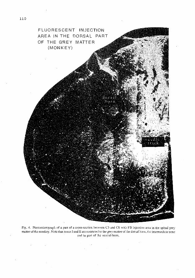
110
FLUORESCENT INJECTION
AREA IN THE DORSAL PART
OF THE GREY MATTER (MONKEY)
Fig. 4. Photomicrograph of a part of a cross-section between CS and CS with FB injection area in the spinal grey matter of the monkey. Note that zones I and II are restricted to the grey matter ofthe dorsal hom. the intermediate zone
and in part of the ventral· horn.

-=-~---zone 3
,..------- zone 2
:\---'.------ zone 1
INJECTION AREA IN THE INFERIOR OLIVE IN CAT (CASE 13)
111
Fig. 5. Diagram of the injection area in inferior olive in cat. For characteristics of zones 1. 2 and 3. see text. Abbreviations: A. nucleus ambiguus: DAO. dorsal accessory olive: dl. dorsal lamella of principal olive: FLM, medial longitudinal fasciculus: G +C. nucleus gracilis and cuneatus: K. cap of Kooy: MAO. medial accessory olive: N VII. facial nucleus: N XII. hypoglossal nucleus: n XII. hypoglossal nerve: N Xllp. nucleus prepositus hypoglossi: NC. cochlear nucleus: NCE. external cuneate nucleus: NRL. lateral reticular nucleus: NTSV. nucleus of spinal V tract: NVL. lateral vestibular nucleus: NVM. medial vestibular nucleus: PC!. inferior cerebellar penduncle: RFl, lateral reticular formation: RS. rubrospinal tract: S. nucleus and tractus solitarius: TCS. corticospinal tract: TS V,
spinal V tract; vi. ventral lamella of principal olive: X, dorsal motor nucleus of vagus.
B Fig. 6. A shows the FE-labeled neurons with their dendrites of the nucleus of Darkschewitsch after FB injections in the cat inferior olive. B shows the FB labeling of the hypoglossal neurons with their axons (nervus hypoglossus) after FB injections in the cat inferior olive. which also damaged the axons of the hypoglossal nerves. at their exit from the medulla oblongata. Note that after a perfusion with 30% formalin instead of 10% the FB labeling is very strong. and
the proximal dendrites and axons are also labeled.

112
Fig. 7. Photomicrographs of single FB. single NY and FB-NY double labeled neurons in red nucleus (upper row) and the nucleus raphe magnus (bottom row) of the cat. The FB single labeled neurons show a fluorescent labeling of the cytoplasm with silver fluorescent granules and the NY single labeled ones a fluorescent nucleus and a clear fluorescent
ring around the nucleolus. The FB-NY double labeled neurons display all features.
immediately mounted from distilled water and air dried at room temperature. In general, one out of every three sections was mounted. The material was studied with a Leitz Ploemopack fluorescence microscope equipped with a filter mirror system A (360 nm excitation wavelength). At this excitation wavelength single FB labeled neurons show a blue fluorescent labeling of the cytoplasm of cell body and proximal dendrites with silver fluorescent granules. Perfusion with 30% formalin produced a more intense FB fluorescence than with 10 o/c formalin and also resulted in labeling of axons and proximal dendrites of the labeled cells (see Fig. 6). Single NY-labeled neurons, after short survival times (Bentivoglio et al., 1980b), show mainly a golden yellow fluorescence of the nucleus with a clear ring around the nucleolus. Double labeled neurons show all mentioned features (see Figs. 7 and 15) at the same 360 nm excitation wavelength (Kuypers et aL 1980).
RESULTS
(A) Injection areas
The FB injection area around the needle tracks in the spinal cord and inferior olive in cat and monkey showed 3 concentric zones (Bharos et al., 1981). Zone I, surrounding the needle track, was brilliantly white-blue fluorescent and contained few cellular elements but many orange fluorescent granules. In cresyl violet sections, it represented a pale area witli a few neurons or glial cells. Zone II was characterized by a dense accumulation of blue fluorescent

113
glial cells with a blue fluorescent nucleus. In cresyl violet sections this zone was darkly stained because of many glial nuclei. Zone III contained some fluorescent glial nuclei and neurons as well as blue fluorescent fibers. Towards its periphery the fluorescent glial nuclei progressively diminished in number and zone III thus faded into an area of normal tissue with little fluorescence.
FB transport occurs from zones I and II, but not from zone III, since in the control cat with FB injections in the dorsal columns in which only zone III involved the rubrospinal termination area (see Fig. 2) no FB-labeled cells occurred in red nucleus. In the cases (cats 1-10 and the 3 monkeys) in which FB was injected in the C5-C8 grey matter, zone II extended laterally up to the border of the grey matter and ventrally into the ventral hom (Figs. 2, 3, 4 and 13). Rostrocaudally it extended up to the rostral border of C5 and down to the caudal border of C8. No FB fluorescence occurred in those parts of the dorsolateral funiculus containing the rubrospinal, raphe spinal and crossed pontospinal tracts (Nyberg-Hansen and Broda!, 1964; Petras, 1967; Edwards, 1972; Kuypers and Maisky, I 977; Basbaum and Fields, 1978, 1979; Holstege et al., 1979; Martinetal., 1979). In the two cats (cats II and 12), with FB injections in the inferior olive, zone II extended from the medial to the lateral border of the inferior olive. It extended dorsally 0.5 mm beyond its dorsal border (see Fig. 5) and extended rostrocaudally throughout the length of the inferior olive.
The NY injection areas also displayed 3 concentric zones (Bharos et al., 1981). Zone I was very narrow and contained yellow tissue fluorescence and a dense accumulation of bright yellow-white glial nuclei. Zone II was much wider than the first. It also displayed yellow tissue fluorescence but contained fewer fluorescent glial nuClei. Zone III contained only dull fluorescent glial nuclei. The zones I and II involved both the grey and white matter of the injected half of the cord, while zone III extended into the contralateral half. Rostrocaudally, zones I and II involved the injected segments, while zone III extended into the rostrally and caudally adjacent ones. However, retrograde transport over long distances does not take place from zone III since no NY retrograde labeling was present in the red nucleus contralateral to the non-injected half of the spinal grey which was involved by zone III only.
(B) Measures taken to increase the number of labeled neurons (Table III)
From some of the findings in the present study the impression wa~ gained that in cat NY is much less effectively transported from damaged axons than in rat (Huisman et al., 1981). In order to determine whether this transport in cat could be improved by the admixture of DMSO: in one cat I o/c NY dissolved in water was injected in the C5--C8 dorsolateral funiculus while in another cat I o/c NY dissolved in 2 o/c DMSO (Keefer, 1978) was injected. Only in the latter cat a substantial number of NY-labeled neurons were present contralaterally in the ventrolateral part of the red nucleus, which neurons project to the lumbosacral cord (Pompeiano and Broda!, 1957; Nyberg-Hansen and Broda!, 1964). The same was observed in other animals of the present study in which NY dissolved in DMSO was injected (see number of NY-labeled neurons in Table III). It was therefore concluded that in cat an effective NY transport from damaged fibers, which pass through the injection area, only occurs when NY is dissolved in DMSO*.
* The facilitatory influence of DMSO on this transport probably results from the fact that it increases the permeability of the membranes. In this respect it is of interest to recall that during bathing the intact vagus nerve bisbenzimide (Bb) remained in solution. while True Blue (TB) adhered directly to the sheath of the nerve and resulted in retrograde
transport (Sawchenko and Swanson. 1981 ).

114
In order to determine whether the FB fluorescence could be improved by a stronger fixation a 30% formalin perfusion was used in cat and monkey instead of 10%. This measure increased the intensity of FB fluorescence and thus also increased the total number of cells which could be recognized as FB-labeled (see numbers of FB-labeled neurons in Table III).
(C) Red nucleus in cat and monkey
Cytoarchitecture The red nucleus in cat and monkey may be subdivided into a caudal (magnocellular) part and
a rostral part. The magnocellular part in cat occupies the caudal 2/3 of the nucleus but in monkey occupies the caudal1/2. In both species the caudal portion of the magnocellularpart of the red nucleus is round in cross-section and contains large neurons which are dispersed between the tegmental fiber bundles. The rostral portion of the magnocellular red nucleus in monkey is also round in cross-section but contains mainly medium sized neurons. In cat this portion of the magnocellular part as seen in cross-section has the shape of a flattened disk and contains both large and small neurons. According to earlier retrograde degeneration findings these various types of neurons in the magnocellular part of the red nucleus in cat and monkey all give rise to rubrospinal fibers (Pompeiano and Broda!, 1957; Poirier and Bouvier, 1966; Kuypers and Lawrence, 1967).
The rostral part of the red nucleus in monkey is rather sharply delineated against the surrounding reticular formation and contains relatively small neurons. Therefore, as in other higher primates. this part of the red nucleus is called the parvicellular part. In the dorsal portion of this parvicellular part the cells form a loose network but in the ventral parts they are rather densely packed. The parvicellular part in monkey extends rostrally close to the mesodiencephalic junction, i.e., just beyond the level where the fasciculus retroflexus (FR) traverses in full length the mesencephalic tegmentum. At these levels the parvicellular part is mainly located lateral to the FR. In monkey, a small group of medium sized multipolar neurons extends from the caudal part of the magnocellular part rostrally over a short distance along the lateral aspect of the parvicellular part, up to the level where the cross-section through the fasciculus retroflexus (FR) is located immediately ventral to the red nucleus.
The rostral part of the red nucleus in cat is also situated lateral to the FR. However, in the cat this part of the nucleus is not truly parvicellular as in monkey but consists of a loose network of small multipolar neurons which is difficult to delineate from the surrounding reticular formation. Yet, it seems reasonable to regard this portion of the red nucleus in cat as the counterpart of the parvicellular part in monkey since both structures give rise to the ipsilaterally descending rubro-olivary fibers (Walberg. 1956; Edwards, 1972; Courville and Otabe, 1974; Strominger et al., 1979; Conde and Conde, 1982).
Rubrospinal neurons in cat and monkey In all 10 cats and in the 3 monkeys a population of single FB-labeled and single NY-labeled
neurons was present in the caudal portion of the magnocellular red nucleus. In both groups of animals the single FB-labeled neurons were concentrated dorsomedially while the single NY-labeled ones were concentrated ventrolaterally (see Figs. 8, 9 and 13). In the rostral portion of the magnocellular part of the nucleus in monkey this arrangement was roughly maintained but in the rostral portion of the magnocellular red nucleus in cat the two populations of single FB- and single NY-labeled neurons which comprised both large and small cells became interdigitated to some degree as observed also in rat (Huisman et al., 1981). It was quite striking to note that the population of labeled rubrospinal neurons in monkey was not

CASE 2
• 0
0 0 0
• 0 if:. 0 0
0 0
CASE 4 CASE 10
F:~DlX100=10,17. FB~~LX100=21,57.
.. o ooo o o 0 0 0 0
0 0 0
~ ... * * o ooo 0 0 0 0
0 0 00 0
0 0 0 0
0 0 oO• oaoo
oooooooaoo oooOOo o oo o o o 0 o
oo 0 oo
FB: IN F. OLIVE NY:C 5_c8
115
Fig. 8. Semidiagrammatic representation of the distributions of retrogradely labeled neurons in the red nucleus of the cat in case 2: FB in C5-C8 and NY in L3: case 4: FB in C5-C8 and NY in T8; case I 0: FB in C5-C8 and NY in T3; ca:-.e II: FB in the ipsilateral inferior olive and NY in the contralateral C5-C8 spinal cord. Abbreviations: CG. central grey: CP. cerebral peduncle; DCP. decussation of superior cerebellar pend uncles; IP. interpeduncular nucleus; LM, medial lemniscus: N III. oculomotor nucleus: MB. mammillary bodies; Ill. oculomotor nerve; R, red nucleus; Rp. parvicellular part of red nucleus: SNC. substantia nigra pars compacta; SNR. substantia nigra pars reticulata; III V.
third ventricle.

116
Fig. 9. Photomicrograph of dorsomedial part of caudal red nucleus in cat.labeled with FB from C5-C8 contralateral spinal cord. which part passes into the ventrolateral part.labeled with NY from TS-T9 contralateral spinal cord. Note the faint staining of glial nuclei diffuse through the ventrolateral part and the FB-NY double labeled neuron located at
the transition of the dorsomedial FB-labeled part to the ventrolateral NY-labeled part.
restricted to the magnocellular part of the red nucleus but extended rostrally as a thin shell alongside the lateral aspect of the parvicellular part up to the level where the cross-section of the fasciculus retroflexus (FR) is located immediately ventral to the nucleus. This is in keeping with the retrograde HRP findings of Kneisley et aL ( 1978) as inferred from their illustrations. In this lateral shell of rubrospinal neurons both single FB- and single NY-labeled neurons were present but the NY-labeled rubrospinal neurons were much more numerous than the FB-labelcd ones and the latter were located more dorsally than the former (see Fig. 13). This is in keeping with the HRP findings (Kneisley et aL. 1978) that the rubrolumbar neurons in monkey extend more rostrally than the rubrocervical ones.
In the cats and monkeys double labeled neurons were also present in the red nucleus. In all cases (cats and monkeys) some FB-NY double labeled neurons were present in the caudal portion of the magnocellular part of the red nucleus. i.e., between the dorsomedially located FB-labeled neurons and the ventrolaterally located NY-Iabe!ed ones. However, as in rat (Huisman et aL, 1981). the majority of the double labeled neurons were observed in the rostral portion of the magnocellular part of the nucleus. where they were intermixed with the single FB- and single NY-Iabeled ones. In monkey double labeled neurons were also observed in the lateral shell alongside the parvicellular part, where they were in general situated dorsally.
When comparing the findings in cats and monkeys it appeared that when the NY injections were made progressively more rostrally in the spinal cord starting at the sacral levels the FB-NY double labeled neurons in red nucleus increased in number. In order to compare the present findings in cat and monkey with those in rat (Huisman et a!.. 1981) and with earlier electrophysiological findings in cat (Shinoda eta!.. I 977) the numbers of single FE-labeled

The percentages
TABLE IV FB-NY double labeled neurons
FB single+ FB-NY double labeled neurons
117
X 100%
Histogram 1 : contriliateral red nucleus of the cat; histogram 2: ipsilateral nucleus raphe magnus of the cat; histogram 3 : contralateral ventrolateral pontine tegmentum of the cat.
CAT HISTOGRAM I
RED NUCLEUS
HJSTOGRAM:U:
NUCL. RAPHE MAGNUS
HISTOGRAMJI[ VENTROLAT. PONT.
TEGMENTUM
2,1%
0 (f)
" 0
CASES
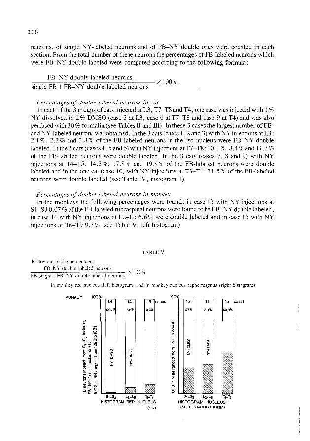
118
neurons, of single NY-labeled neurons and of FB-NY double ones were counted in each section. From the total number of these neurons the percentages ofFB-labeled neurons which were FB-NY double labeled were computed according to the following formula:
FB-NY double labeled neurons ~~~~~~~~~~~~--------XIOO%. single FB + FB-NY double labeled neurons
Percentages of double Labeled neurons in cat In each of the 3 groups of cats injected at L3, T7-T8 and T4, one case was injected with 1%
NY dissolved in 2% DMSO (case 3 at L3, case 6 at T7-T8 and case 9 at T4) and was also perfused with 30% formalin, (see Tables II and III). In these 3 cases the largest number of FBand NY -labeled neurons was obtained. In the 3 cats (cases I, 2 and 3) with NY injections at L3: 2. I o/c, 2. 3 % and 3. 8 % of the FB-labeled neurons in the red nucleus were FB-NY double labeled. In the 3 cats (cases 4, 5 and 6) with NY injections at T7-T8: 10.1 %, 8.4% and 11.3% of the FB-labeled neurons were double labeled. In the 3 cats (cases 7, 8 and 9) with NY injections at T4-T5: 14.3%, 17.8% and 19.8% of the FB-labeled neurons were double labeled and in the one cat (case I 0) with NY injections at T3-T4: 21.5% of the FB-labeled neurons were double labeled (see Table IV, histogram 1).
Percentages of double labeled neurons in monkey In the monkeys the following percentages were found: in case 13 with NY injections at
S I-S3 0.07% of the FE-labeled rubrospinal neurons were found to be FB-NY double labeled, in case 14 with NY injections at L2-L5 6.6o/c were double labeled and in case 15 with NY injections at T8-T9 9.3% (see Table V, left histogram).
Histogran1 of the percentages FB-NY double labeled neurons
TABLE V
==~~--==~~~~~~~----- X 1007c FB single+ FB-NY double labeled neurons
in monkey red nucleus (left histogram) and in monkey nucleus raphe magnus (right histogram).
MONKEY 100
6.6%
S1-S3 L2-Ls
HISTOGRAM RED NUCLEUS
(RN)
14
27%
HISTOGRAM NUCLEUS RAPHE MAGNUS (NRM)

119
Percentages of double labeled neurons in cat and monkey compared The findings in this entire group of cats and monkeys thus imply that some of the red nucleus
neurons, which project to the C5-C8 spinal grey matter, also distribute branches to more caudal parts of the spinal cord. They further indicate that in cat a relatively high proportion of these neurons gives rise to branches which terminate in or pass through the T4-T5 spinal segments ( ± 20%); a smaller proportion gives branches which terminate in or pass through T7-T8 ( ± 10%), and only a very few neurons give branches which terminate in or pass through the lumbosacral cord ( ± 3%). The arrangement in monkey seems to be similar to that in cat, such that a small percentage of rubrocervical neurons gives rise to collaterals which terminate in or pass through T8-T9 (9.3 %), an even smaller percentage gives rise to collaterals which terminate in or pass through L2-L5 (6.6%) while extremely few rubrocervical neurons (0.07%) give rise to co !laterals to the sacral cord.
Rubro-olivary neurons in cat In the two cats (cases II and 12) with FB injections in inferior olive and NY injections
contralaterally in spinal cord the following findings were obtained. In case II in which NY dissolved in water was injected in the cervical cord (C5-C8) the population of single NY labeled neurons was located in the dorsomedial part of the red nucleus. In case 12 in which NY dissolved in DMSO was injected in both the cervical (C5-C8) and the lumbar cord (L3-L5) the single NY-labeled population occupied both the dorsomedial and the ventrolateral parts of the red nucleus. It was therefore concluded that in the former case (II) there was only NY transport from the C5-C8 spinal grey matter, while in the latter case (12) there was NY transport from the C5-C8 and the L3-L5 spinal grey matter and probably also from damaged fibers passing through the injected segments. Both cases were perfused with 30 o/c formalin which resulted in a high intensity of the FB fluorescence in retrogradely FB-labeled cells (see Figs. I 0 and II).
Fig. I 0. Photomicrograph of the single FE-labeled neurons in the parvicellular red nucleus of the cat. immediately lateral to the fasciculus retroflexus (FR). Abbreviations: CP, cerebral peduncle: FR. fasciculus retroflexus; Rp,
parvicellular part of the red nucleus: III V, third ventricle.

120
Fig. 11. Photomicrograph of the single FB-labeled neurons in the parvicellular red nucleus of the cat, ventrally to the fasciculus retroflexus (FR). Abbreviations: CP, cerebral peduncle: FR. fasciculus retroflexus; MB, mammillary
bodies: Rp, parvicellular part of the red nucleus: III V, third ventricle.
the spinal injections of case 2 and 6 were on this side
CASE 2 FB c,.c. NY L3
.. . • 0* 0 ..
* ~*
DL l DL+FB X100=26.
DL z· DL+ FBXlOO =53,2.
CASE 10 FB c,.c8
0 NY T3
* 0
0
0 0 *0
ooo* ~ 00 0
0 • *0
DL~\6 Xl00=54Z
Fig. 12. Semidiagrammatic representation of the distributions of retrogradely labeled neurons in the ventrolateral pontine tegmentum (VLPT) and the nucleus raphe magnus (RM) after spinal injections in cat. In case 2: Fast Blue (FB) in C5-C8 and Nuclear Yellow (NY) in L3 and in case 10 FB in C5-C8 and NY in T3. Note the relatively high number of double labeled neurons especially in RM, even in case 2 with NY injected in L3. Abbreviations: BC. brachium conjunctivum: C. nucleus coeruleus: FLM, medial longitudinal fasciculus: LL. lateral lemniscus: LM. medial lemniscus: NC, cochlear nucleus: NCI, nucleus of inferior colliculus: NCu. cuneate nucleus: NP. pontine nuclei: NRP, reticular nucleus of pontine tegmentum: NTS V. nucleus of spinal V tract: NVL. lateral vestibular nucleus: NVM, medial vestibular nucleus: N VII. facial nucleus: n VII. facial nerve: PCL inferior cerebellar peduncle: PCM. medial cerebellar peduncle: RF. reticular formation: RM. nucleus raphe magnus: RS. rubrospinal tract: SC, nucleus
su'Jcoeruleus: TSC. corticospinal tract: TSV. spinal V tract: VLPT. ventrolateral pontine tegmentum.

121
In the caudal one-third of the red nucleus in both cases only single NY-labeled neurons occurred, but many FE-labeled neurons were present in the central grey at these levels. In the middle one-third a large population of NY-labeled neurons was present but some single FE-labeled cells occurred, i.e., in the most dorsomedial part of the nucleus. This group of red nucleus neurons was continuous with a group of single FE-labeled cells in the dorsomedially adjoining interstitial nucleus of Cajal (INC) and many FB-labeled neurons were also present in the nucleus of Darkschewitsch (see Fig. 6A) (cf., Henkel et al., 1975; Brown et al., 1977; Cintas et al., 1980; Martinet al., 1980; Saint-Cyt and Courville, 1980). Finally in the rostral one-third of the red nucleus only single FB-labeled neurons were present, which were located medial, ventral and lateral to the fasciculus retroflexus (see Figs. 8, 10 and 11). This
MONKEY
CAUDAL DL~LFB x1Q0%:=:6,6%
FB :;:
CASE 15 FB Cs-Cs NY T 8 -T 9
___l?..lc_ x100lb9,3% DL+FB
FAST BLUE(FB) INJECTION AREA IN THE DORSAL PART OF THE GREY MATTER ( C5 .Cs)
Fig. 13. Semidiagrammatic representation of the distributions of retrogradely labeled neurons in the red nucleus after spinal injections in monkey; in case 14: Fast Blue (FB) in C5-C8 and Nuclear Yellow (NY) in L2-L5 and in case 15: FB in C5-C8 and NY in T8-T9. Note the rostral extension of rubrospinal neurons in the lateral part of red nucleus. particularly the neurons which project to lower levels in the spinal cord. Abbreviations: G. central grey; CP, cerebral peduncle: DCP. decussation of superior cerebellar peduncles; IP. interpeduncular nucleus: ML, medial lemniscus; mlf. medial longitudinal fasciculus; N III oculomotor nucleus; n III. oculomotor nerve; Rm, red nucleus, magnocellular part; Rp. red nucleus. parvicellular part; SNC, substantia nigra. pars compacta; SNR, substantia nigra, pars
reticulata.

122
population of FB-labeled neurons, which gradually tapered down at the level of the mammillary bodies, was continuous dorsomedially with a population of single FE-labeled cells in the nucleus ofDarkschewitsch. The group of small FE-labeled cells in the rostral part of the red nucleus could be effectively delineated on the lateral and ventral sides and in part also on the medial side (see Figs. 10 and 11). It resembled in its configuration the parvicellular part of the red nucleus as observed in monkey (Kuypers and Lawrence, 1967).
In these two cats hardly any double labeled neurons were observed. Thus, in case 11 no FB-NY double labeled cells occurred and in case 12 only one FB-NY double labeled neuron was present, at the rostral border of the single NY-labeled population. These findings indicate that the rubro-olivary fibers in cat are derived virtually only from neurons in the rostral part of the red nucleus and that extremely few rubrospinal neurons distribute collaterals to the inferior olive (cf. Anderson, 1971).
(D) Nucleus raphe magnus (NRM) and ventrolateral pontine tegmentum (VLPT) in cat and monkey
Nucleus raphe magnus (NRM) The NRM distributes fibers to the dorsal part of the spinal grey matter including the dorsal
hom, by way of the dorsolateral funiculus (Basbaum et al., 1978; Leichnetz et al., 1978; Basbaum and Fields, I979; Tohyama et al., I979a,b; Goode et al., 1980; Martinet al., I 98 I a). However, the retrograde! y labeled neurons in the area of the NRM after HRP injections in the dorsolateral funiculus (Leichnetz et al., 1978; Basbaum and Fields, 1979; Tohyama et al., I 979a) are not restricted to the NRM but extend laterally through the ventral part of the reticular formation dorsal to the pyramidal tract. In the present study the NRM projecting to the spinal dorsal grey was therefore defined according to the distribution of the retrogradely labeled neurons. This area corresponds to the distribution area of serotonergic neurons in rat (Dahlstrom and Fuxe, I 964, I 965), cat (Persson et al., 1978 ; Wiklund et al., I982) and monkey (Hubbard and DiCarlo, I974; Schofield and Everitt, 1981) at this level, because they extend laterally from the nucleus raphe as defined by Taber (I 960, I 961) into the nucleus gigantocellularis lateralis, i.e., dorsal and lateral to the pyramidal tract.
Ventrolateral pontine tegmentum (VLPT) Retrograde HRP studies showed that a group of neurons in the ventrolateral pontine
tegmentum adjoining the area of the rubrospinal tract (TRS) (Kuypers and Maisky. I 975; Leichnetz et a!., I 978; Basbaum and Fields, 1979) give rise to mainly crossed pontospinal fibers which descend in the dorsolateral funiculus throughout the spinal cord (Kuypers and Maisky, I 977; Holstege et al., I 979; Tohyama et al.. I 979). These fibers seem to correspond to the crossed pontospinal tract of Busch ( 1964) and the lateral reticulospinal tract of Papez (I 926). Anterograde labeled amino acid transport studies (Holstege et al.. 1979; Martinet al., I 979) showed that this crossed descending pathway terminates in roughly the same portion of the spinal grey as the rubrospinal tract. i.e .. in the dorsal part of the spinal intermediate zone. but also terminates in the spinal dorsal hom.
Retrograde labeling in nucleus raphe magnus (NRM) and rentrolateral pontine tegmentum (VLPT) (a) Cat. In the 10 cats with FB and NY spinal injections the single FB. single NY and the
FB-NY double labeled neurons in the NRM and the VLPT did not display any topographical distribution but were randomly intermixed with one another (see Fig. 12). This is in keeping

123
Fig. 14. Photomicrograph of dorsomedial part of caudal red nucleus in monkey, labeled with FB from C5-C8 contralateral spinal cord. Note the reticulated structure. in which the single FB-labeled neurons are arranged.
Fig. 15. Photomicrographs of single FB. single NY and FB-NY double labeled neurons in red nucleus (upper row) and nucleus raphe magnus (bottom row) of the monkey. The single FE-labeled neurons show a fluorescent labeling of the cytoplasm with silver fluorescent granules and the single NY-labeled ones a fluorescent nucleus and a clear ring
around the nucleolus. The FB-NY double labeled neurons display all features.
with the anatomical findings in rat (Huisman et a!., 1981) and opossum (Martin et a!., 1981a,b). In the different experiments in cat the total number of neurons which were FB labeled from C5-C8 in the ipsilateral NRM ranged from 591 to 2943. In the contralateral VLPT the number ranged from Ill to 525. The percentages of the FB-labeled neurons in these nuclei which were FB-NY double labeled after injections at different spinal levels were

124
entirely different from those obtained in red nucleus (see histograms l, 2 and 3 in Table IV). Thus in all cases a large percentage of the FB-labeled neurons in NRM and VLPT was double labeled. This was particularly striking in the NRM, as observed also in rat (Huisman et· al., 1981) and opossum (Martinet al., 1981b). Moreover, the percentages of the FB-NY double labeled neurons in the NRM were always of roughly the same magnitude, varying from 55% to 60%, no matter at which spinal level NY was injected. Only in cases 5 and 7, they were lower: 24.5% and 30.5%, respectively, in which cases the percentages ofFB-NY double labeled neurons in the VLPT were also lower than in the other cases (Table IV).
The findings in the red nucleus, the nucleus raphe magnus and the ventrolateral pontine tegmentum therefore suggest that in red nucleus to a large extent different groups of neurons project preferentially to different levels of the spinal cord and distribute only few collaterals to the other levels, while a large portion of NRM neurons as well as of VLPT ones distribute collaterals throughout the length of the spinal cord. Since always roughly the same percentage of double labeled neurons .occurred in NRM and VLPT independent of the level of NY injections, it seems likely that many NRM and VLPT neurons give rise to long descending fibers proceeding throughout the length of the spinal cord giving off collaterals to the spinal grey at many different levels.
(b) Monkey. The distribution of the labeled cells in NRM and VLPT in monkey was the same as observed in cat (see Fig. 16). The numbers of single FB-labeled neurons including
MONKEY
the spinal injections of case 14 and 15 were on this side
CASE 14 FB Cs-Cs NY L2- Ls
DL ~LFB x100 =31,1%
CASE 15 FB C5 _c8 NY Ts-Tg
o* .....
DLD+\s x100=43,9%
DLf*
Fig. 16. Semidiagrammatic representation of the distributions of retrogradely labeled neurons in the ventrolateral pontine tegmentum (VLPT) and the nucleus raphe magnus (NRM) after spinal injections in monkey. In case 14: Fast Blue (FB) in C5-C8 and Nuclear Yellow (NY) in L2-L5 and in case 15 FB in C5-C8 and NY in T8-T9. Note the relatively high number of double labeled neurons. especially in NRM. even in case 14 with NY injected in L2-L5. Abbreviations: BC brachium conjunctivum: C. nucleus coeruleus: LL. lateral lemniscus: LM. medial lemniscus: N VIII. facial nucleus: n VII. facial nerve: NP. pontine nuclei: NRM. nucleus raphe magnus: NRP. reticular nucleus of pontine tegmentum: NTS V. nucleus of spinal V tract: NVL. lateral vestibular nucleus: NVM. medial vestibular nucleus; PCI. inferior cerebellar peduncle: PCM. medial cerebellar peduncle: RF. reticular formation: RS. rubrospinal tract; SC, nucleus subcoeruleus; ST. spinothalamic tract: TCS. corticospinal tract: tr V. tract of mesencephalic V
nucleus; TS V. spinal V tract: VLPT. ventrolateral pontine tegmentum.

125
FB-NY double labeled ones in the ipsilateral NRM ranged from 1220 to 2344. In the contralateral VLPT these numbers were 321 in case 14 and 525 in case 15. In case 13 the number of such labeled neurons in the contralateral VLPT was very low, therefore no percentages were computed in this case. The percentages of FE-labeled neurons which were FB-NY double labeled in NRM were 27% (case 13), 31.1% (case 14) and 43.9% (case 15) (see Table V). In VLPTthesepercentages were29% (case 14) and30.3% (case 15). Therefore in the NRM, VLPT and the red nucleus in the monkeys the same differences in the percentages of the double labeled neurons were observed as in the cats (compare the histograms in Tables IV and V). This indicates that also in monkey different groups of RN neurons to a large extent project preferentially to certain levels of the spinal cord and distribute only few collaterals to the other levels, while many of the NRM and VLPT neurons distribute collaterals throughout the length of the spinal cord.
DISCUSSION
The present study tried to elucidate in cat and monkey differences in the degree of collateralization in the spinal cord between 3 brain stem pathways, i.e., the rubrospinal tract, the rostral medullary raphe spinal tract and the crossed pontospinal tract, all of which descend through the dorsolateral funiculus and terminate in the dorsal part of the spinal grey (NybergHausen and Brodal, 1964; Petras, 1967; Edwards, 1972; Basbaum and Fields, 1978, 1979; Holstege et al., 1979; Martinet al., 1979). On the basis of the present findings the degree of collateralization of the rubrospinal tract in cat and monkey could also be compared with that in other species. This latter comparison appeared to be of interest because earlier findings suggested that the degree of collateralization of the rubrospinal tract differs in different animals (Huisman et al., 1981; Martinet al., 1982).
Technique
The multiple retrograde fluorescent tracer technique (Kuypers et al., 1980) and the HRP-[3H]apo-HRP technique (Hayes and Rustioni, 1979, 1981) seem to be most suitable for demonstrating the existence of collaterals in brain pathways. However, a comparison of the results obtained by means of these two techniques, e.g., in respect to the raphe spinal projections (cf., Hayes and Rustioni, 1981; Huisman et al., 1981), suggested that the retrograde fluorescent tracer technique produces a larger proportion of double labeled neurons. Therefore this method was chosen. This choice was reinforced by the fact that in the present study a larger number of double labeled neurons were found in raphe and red nucleus in cat than seems to have been obtained by means of the HRP-[3H] apo-HRP method (Hayes and Rustioni, 1981) in the same animal. This difference may be explained by the fact that in the autoradiographic material silver grains will only be elicited in the emulsion by sources of radioactivity which are located in the upper 3 ,urn layer of the 40 ,urn sections (Sidman, 1970). As a consequence, even under optimal circumstances, only a small percentage of the total population of HRP-labeled neurons observed in each 40 ,urn section can be double labeled. The fluorescent tracers are also more sensitive than HRP, as suggested by the findings of Sawchenko and Swanson (1981) that the fluorescent tracers label approximately twice as many neurons than the HRP-TMB technique (Mesulam and Rosene, 1979). This is supported by a comparison of the present findings with those obtained by Conde and Conde (personal communications) which shows that after Fast Blue (FB) injections in inferior olive the number

126
of labeled rubro-olivary neurons at a given level in the red nucleus was significantly larger than obtained with the HRP-BDHC technique (Mesulam, 1976; Conde and Conde, 1982) at approximately the same level (i.e., in a 30 j.lm section about 80 FB-labeled neurons and in a 60 ftm section about 40 HRP-labeled neurons).
In order to obtain reliable quantitative data concerning single FB- and NY-labeled neurons and FB-NY double labeled neurons several requirements had to be fulfilled. First the FB injections in C5-C8 had to be restricted to the grey matter and must not have involved the fiber bundles of the 3 tracts in the dorsolateral funiculus. Under the present circumstances with 4 weeks survival time FB was only transported from zones I and II (see Results). Therefore only those cases have been selected for study (I 0 cats and 3 monkeys) in which zones I and II were restricted to the grey matter and did not involve the dorsolateral funiculus. The remaining cases (I 0 cats) were discarded. In order to obtain reliable quantitative data it was also necessary to label retrogradely a very large number of neurons. For this purpose two measures were taken. First perfusion with 30 Cflc formalin instead of with I 0 o/c formalin was used, because the former results in a much more intense FB labeling than the latter. As a consequence a much larger number of neurons could be recognized as FB labeled. Moreover, in the more recent cases a NY -DMSO mixture was injected in cat since this mixture in cat strongly increased the number of neurons which were retrogradely labeled from damaged axons passing through the injected segments (see Results).
Earlier electrophysiological studies have dealt with the collaterals of descending pathways. Shinoda et a!. ( 1977) reported the number of red nucleus neurons which could be antidromically invaded from C3-C8 grey matter and the percentage of these neurons which could be antidromically invaded by stimulation of the fibers passing through different more caudal segments of the cord. In order to facilitate a comparison between our anatomical data and these physiological data the anatomical experiments were set up in a comparable manner. Thus the number of FB-labeled neurons which project to the C5-C8 dorsal grey matter was counted and the percentages of these neurons which were also labeled with NY were computed. This procedure also allowed a comparison between the present findings in cat and monkey and those obtained earlier in rat (Huisman et a!.. 1981 ).
Somatotopic organi::;ation (!l reel nucleus
In cat the injections of the two tracers in C5-C8 and in more caudal spinal segments in essence labeled two different populations of rubrospinal neurons. The distribution of the total population of these retrogradely labeled rubrospinal neurons confirmed previous findings (Pompeiano and Broda!. 1957; Hayes and Rustioni. 1981) and showed that the rubrospinal neurons are mainly present in the caudal two-thirds of the nucleus. The findings in the experiments with FB injections in the inferior olive and NY injections in the cervical cord further showed that in cat the red nucleus neurons. which distribute fibers to the ipsilateral inferior olive (Walberg. 1956; Courville and Otabe. 1974; Martin et a!.. 1975, 1980; Strominger et al.. 1979). represent a separate population which is largely restricted to the rostral part of the nucleus (see Fig. 8). This is in keeping with other HRP findings (Saint-Cyr and Courville. 1980; Conde and Conde. 1982).
The distribution of the single FB- and single NY-labeled rubrospinal neurons confirmed the somatotopic organization of this nucleus as demonstrated earlier by means of other techniques (Pompeiano and Broda!. 1957; Tsukahara et a!.. 1967; Gwyn. 1971 ; Hayes and Rustioni. 1981). Thus, in the caudal half of the magnocellular part the rubrocervical, the rubrothoracic and the rubrolumbar neurons are located in the dorsomedial, intermediate and ventrolateral
127
part of the nucleus, respectively. On the other hand in the rostral half of the magnocellular part these neurons become intermixed (see Fig. 8).
In monkey retrograde degeneration studies (Poirier and Bouvier, 1966; Kuypers and Lawrence, 1967) showed that the rubrospinal neurons are largely restricted to the magnocellular part. However, the present findings showed that the population of rubrospinal neurons extends further rostrally and continues along the lateral aspect of the parvicellular part (see Fig. 13) up to the level, where the cross-section through the fasciculus retroflexus is located ventral to the nucleus. This seems in keeping with the retrograde HRP finding of Kneisley et al. ( 1978) as inferred from their illustrations. The location of these rubrospinal neurons appears to correspond with that of neurons with coarse Nissl bodies (Miller and Strominger, 1973). This supports the idea that rubrospinal neurons are characterized by their internal configuration more or less independent of the size of the neurons (King et al., 1971).
The present findings support the earlier reportc:d somatotopic organization in the caudal magnocellular part of the monkey red nucleus (Kneisley et al., 1978). They also indicate that in the rostral extension of the population of rubrospinal neurons along the lateral aspect of the parvicellular part the rubrocervical neurons are located dorsal to the rubrolumbar ones and that the latter continue more rostrally than the former (see Fig. 13).
Collateralization in rubrospinal tract The percentages of the rubrocervical neurons in cat projecting to C5-C8 which also
distribute collaterals to more caudal levels, as computed from the present anatomical findings, are of the same order of magnitude as those inferred from the earlier electrophysiological findings in this animal. Thus according to the electrophysiology (Shinoda eta!., I 977) 5% of the rubrocervica1 neurons projecting to C3-C8 distribute collaterals to segments caudal to L I , and 50% distribute collaterals to segments caudal to T2. According to the anatomical findings about 3% of the rubrocervical neurons projecting to C5-C8 distribute collaterals to the segments caudal to L2 and about 20% distribute collaterals to the segments caudal to T3 (see Table IV). In monkey a similar arrangement was found anatomically: 9.3% of the rubrocervical neurons projecting to C5-C8 were found to distribute collaterals to the segments caudal to T8 and 6.6% to the segments caudal toLl (see Table V).
The slight differences between the percentages obtained by means of the electrophysiological and the anatomical techniques may be due to the fact that in the two types of experiments slightly different cervical segments were studied and slightly different thoracic and lumbar segments were stimulated and injected respectively. The differences may also be due to differences in the rostrocaudal extent of the nucleus, explored in the two types of experiments.
Rubrospinal collateralization in different animals A comparison of the present anatomical findings in cat and monkey with those obtained in
rat (Huisman eta! .. 1981) indicates that the rubrospinal system in rat displays a higher degree of collateralization than in cat and monkey. Thus in rat 20% of the rubrocervical neurons projecting to C5-C8 distribute collaterals to segments caudal to L I versus only about 3% in cat and about 6 o/c in monkey. Further, in rat about 35% of the rubrocervical neurons distribute collaterals to segments caudal to T7 versus about 10% in cat and monkey. The rubrospinal tract in cat and monkey therefore appears to be more focussed than in rat such that in the former species a larger proportion of the rubrospinal neurons distributed fibers to a restricted part of the spinal cord than in the latter. This is further emphasized by the fact that the high degrees of collateralization of the raphe spinal and the crossed pontospinal tract are of the same order of magnitude in the 3 species (see below).

128
The comparison of the anatomical findings in opossum and rat (Huisman et al., 1981 ; Martin et al., 1982) strongly suggests that the rubrospinal tract in opossum shows an even higher degree of collateralization than in rat. This would imply that the rubrospinal tract in rat, in tum, is more focussed than in opossum. The fact that the red nucleus in opossum displays a much less rigid somatotopic organization than in rat (Martinet al., 197 4; Huisman et al., 1981) suggests that with an increase in collateralization the somatotopic organization becomes less sharply defined. If this is correct the absence of any somatotopic organization in the red nucleus in pigeon (Wild et al., 1979) and in reptiles (ten Donkelaar and de Boer-van Huizen, 1978) would imply that in these species the rubrospinal tract displays an even more pronounced collateralization than in opossum. In snakes this tract seems to be lacking (ten Donkelaar, 1976a,b). It therefore appears that the rubrospinal tract, as a highly focussed fiber brain stem system, emerges only gradually during phylogeny. It would be of interest to determine whether in respect to the rubrospinal collateralization the ontogeny mimics the phylogeny, such that e.g. in newborn kittens a higher degree of collateralization occurs than in adult cat. This question is prompted by the fact that in other structures such an ontogenetic decrease in col!ateralization has been established (Innocenti, 1981; O'Leary et a!.. I 98 I).
In respect to the phylogenetic decrease in rubrospinal collateralization, it is of interest to recall that in cat and monkey (Kuypers, 1964; Lawrence and Kuypers, 1968; Smith, 1970; Gorska and Sybirska, 1978; Kohlerman et al., 1980) this tract together with the corticospinal tract contributes to the capacity to execute relatively independent movements of the distal parts of the limbs. This is probably based on the fact that in these animals a relatively limited degree of collateralization occurs such that many of the rubrospinal neurons project to very restricted parts of the spinal cord. The suggested decrease in collateralization of this tract from reptiles to pigeon, opossum, rat, cat and monkey makes it likely that the capacity of the rubrospinal tract to provide the facility for the execution of relatively independent movements of the individual extremities also gradually emerges during phylogeny. This notion is strengthened by the fact that such a capacity is clearly present in monkey and cat, in that sense that it seems to be lacking in reptiles.
Collateralization in the three tracts compared The relatively limited degree of collateralization of the rubrospinal tract is emphasized by
the contrast between the number of double labeled neurons in red nucleus and in nucleus raphe magnus (NRM) at the level of the facial nucleus. The NRM in cat and monkey contained a relatively small population of FB and NY single labeled neurons which project to the cervical and the more caudal segments respectively, but contained a relatively large population of double labeled neurons, i.e., 55-60% of the raphe cervical neurons in cat and about 40 o/c in monkey. These high percentages occurred more or less independent of the level of the caudal NY injection (see Tables IV and V). Moreover, no clear-cut somatotopic organization in the distribution of these 3 types of neurons was observed. These findings in cat and monkey are in keeping with those in rat (Huisman et al., 1981) in which about 40 o/c of the raphe cervical neurons were always double labeled and with those in opossum (Martinet al., 1981a,b). The raphe spinal system therefore resembles the reticulospinal system since according to an electrophysiological study (Peterson et al., 1975), 66o/c of the reticulocervical neurons distribute collaterals to the segments caudal to L 1. The anatomical findings suggest that a large percentage of the raphe spinal neurons in NRM distribute collaterals throughout the length of the spinal cord, but that the remainder project to restricted parts of the cord. This would imply that the raphe spinal system comprises both a focussed component and a diffuse component.
In rat, cat and monkey many NRM neurons contain serotonin (Dahlstrom and Fuxe, 1964.

129
RAT TB anterior interpositus NY C5_c8
ROSTRAL
TB~LDLX 100 = 20,4/.
CAUDAL • NY,Jo DL *':~ Ts::: oo ~
Fig. 17. Semidiagrammatic representation of the distributions of retrogradely labeled neurons in the red nucleus in rat after True Blue (TB) injections in the interpositus nucleus and Nuclear Yellow (NY) injections in the spinal cord. ipsilateral to the interpositus injection. Note that almost all TB-labeled rubrocercbellar neurons arc double labeled with NY from the spinal cord. Abbreviations: CMP. posterior commissure: CP. cerebral peduncle: DCP. decussation of superior cerebellar peduncles: GM. medial geniculate body: IP. nucleus interpositus: !Pcd. interpeduncular nucleus: L. lateral nucleus of the cerebellum: M. medial nucleus of the cerebellum: ML. medial lemniscus: mlf. medial· longitudinal fasciculus: NC. cochlear nuclei: N III. oculomotor nucleus: n III. oculomotor nerve: R. red nucleus: RF. reticular formation: SC. superior colliculus: SNC. substantia nigra. pars compacta: SNR. substantia nigra. pars
reticulata: V. fourth ventricle: vest. com pl.. vestibular complex.

130
1965; Hubbard and DiCarlo, 1974; Persson et a!., 1978; Schofield and Everitt, 1981; Wiklund et a!., 1982) and in rat about 45% of the raphe spinal neurons contain serotonin (Bowker et a!., 1981). It would therefore be of interest to determine whether either the focussed or the diffuse component of the raphe spinal system would be preferentially derived from serotonergic neurons. Raphe spinal neurons also contain substance P and enkephalin (Hokfelt et a!., 1979; Bowker et a!., 1981) probably in some neurons in coexistence with serotonin (Hokfelt et a!., 1977, 1978; Bowker eta!., 1981). If the serotonergic neurons in the NRM give rise either to the focussed or the diffuse component of the raphe spinal system one of these two components would in particular sub serve the pain modulating function of the raphe spinal pathway (Oliveras eta!., 1975. 1977; Basbaum eta!., 1976a,b, 1977; Beall et al., 1976; Fields et a!., 1977; Guilbaud eta!., I 977; Basbaum and Fields, I 978, I 979; Fields and Basbaum, I 978; Giesler eta! .. 1981) since this function is presumably provided by serotonergic raphe spinal neurons (Dahlstrom and Fuxe, 1965; Proudfit and Anderson, 1973, 1974; Proudfit eta! .. 1980). However. the focussed or the diffuse component may also represent the serotonergic raphe spinal fibers to the autonomic cell groups throughout the spinal cord (Dahlstrom and Fuxe. 1965; Basbaum eta!.. 1978; Steinbusch. 1981).
The crossed pontospinal tract ji·mn VLPT neurons The crossed pontospina1 tract from VLPT neurons in cat and monkey (Kuypers and Maisky,
I 975) seems to occupy a position between the rubrospinal and the raphe spinal system as also observed in rat. Thus. it shows little somatotopic organization and a relatively high degree of collateralization (see Figs. 12 and I 6).
Rubro-olivary connections The rubro-olivary neurons in cat were found in the rostral portion ofthe red nucleus which is
in keeping with the HRP findings (Saint-Cyr and Courville. I 980; Conde and Conde. 1982). This part probably corresponds with the pars parvicellularis in monkey (Kuypers and Lawrence, !967). Moreover. from the present findings it may be concluded that the rubrospinal neurons and the rubro-olivary ones constitute two separate populations as suggested also by HRP findings (Conde and Conde. I 980. 1982) and that rubrospinal neurons distribute virtually no collaterals to the inferior olive (see Fig. 8).
Rubrocerebellar connections In view of the above discussed rubrospinal and rubro-olivary connections it is of interest to
recall that red nucleus neurons also distribute fibers to the crossed cerebellar interpositus nucleus (Courville and Broda!. I 966). Recently another series of double labeling experiments in rat was initiated in order to detem1ine whether the rubrocerebellar fibers represent col laterals of rubrospinal neurons (Huisman. Kuypers. Conde and Keizer. in preparation). In thi~ ~tudy "True Blue" and "Nuclear Yellow" were used as retrograde tracers. Preliminary double labeling findings in this study show that at least 25 '7c of rubrospinal neurons in rat distribute collaterals to the cerebellar interpositus nucleus (see Fig. I 7). The existence of these collaterals was suggested earlier by retrograde degeneration findings in cat (Broda! and Gogstad. 1954). Electrophysiological studies in cat showed that all rubrocerebellar fibers are collaterals of rubrospinal fibers (Anderson. 197 I). This is in agreement with our preliminary anatomical findings that in rat almost all red nucleus neurons labeled from the cerebellum were also labeled from the spinal cord.

131
ACKNOWLEDGEMENTS
The authors thank Prof. Dr. 0. Dann (Friedrich-Alexander Universitat, Erlangen) and Dr. H. Loewe (Hoechst Company, Frankfurt) for providing small quantities ofFB, TB and NY. They also thank Mr. E. Dalm and Mr. H. Klink for their technical assistance, Miss P. van Alphen and Mr. P. Smaal for the photography, Dr. F. Conde for constructive comments on the manuscripts and Miss E. Klink and Miss G. van Gelder for typing it.
REFERENCES
Abzug. C.. Maeda. M .. Peterson. B.W. and Wilson. V.J. (1973) Branching of individual lateral vestibulospinal axons at different spinal cord levels. Brain Res .. 56: 327-330.
Abzug. C.. Maeda. M .. Peterson. B. W. and Wilson. V .J. (I 974) Cervical branching of lumbar vestibulospinal axons. J. Physiol. (Lond.). 243: 499-522.
Anderson. M.E. (I 971) Cerebellar and cerebral inputs to physiologically identified efferent cell groups in the red nucleus of the cat. Brain Res .. 30: 49--66.
Basbaum. A.!. and Fields. H .L. ( 1978) Endogenous pain control mechanisms: review and hypothesis. Ann. Neurol., 4: 451-462.
Basbaum. A.!. ami Fields. H.L. I I '-J7'-J) The origin of descending pathways in the dorsolateral funiculus of the spinal cord of the cat and rat: further studies on the anatomy of pain modulation. J. comp. New·ol .. 187: 513-532.
Basbaum. A.!.. Marley. N. and O'Keefe. J. ( 1976a) Spinal cord pathways involved in the production of analgesia by brain stimulation. In: Admnces in Pain Research and Therap-'·· Vol. I, J.J. Bonica and D. Albc-Fessard (Eds.). Raven Press. New York. pp. 511-515.
Basbaum. A.!.. Clanton. C .H. and Fields. H.L. ( 1976b) Opiate and stimulus-produced analgesia: functional anatomy of a medullospinal pathway. Proc. IWI. Acad. Sci. (Wasil.), 73: 4685-4688.
Basbaum. A.!.. Marley. N.J.E .. O'Keefe. J. and Clanton. C. H. ( 1977) Reversal of morphine and stimulus-produced analgesia by subtotal spinal cord lesions. Pain. 3: 43-56.
Basbaum. A.!.. Clanton. C .H. and Fields. H .L. (I 978) Three bulbospinal pathways from the rostral medulla of the cat: an autoradiographic study of pain modulating systems. J. comp. Neurol .. 178:209-224.
Beall. J.E .. Martin. R.F .. Applebaum. A.E. and Willis. W.O. (1976) Inhibition of primate spinothalamic tract neurons by stimulation in the region of the nucleus raphe magnus. Brain Res .. 114: 328-333.
Bentivoglio. M .. Kuypers. H.G.J.M .. Catsman-Bcrrcvocts. C.E .. Loewe. H. and Dann. 0. (1980a) Two new fluorescent retrograde neuronal tracers. which arc transported over long distances. Neurosci. Lett .. I 8: 25-30.
Bentivoglio. M .. Kuypers. H.G.J.M. and Catsman-Bcrrcvoets. C. E. (I 980bJ Retrograde neuronal labeling by means of bisbenzimide and Nuclear Yell ow ( Hoechst S-76'-J 121 ). Measures to prevent diffusion of the tracers out of retrogradely labeled neurons. N<'llmsci. Lei/ .. I X: I 1-1 X.
Bharos. T.B .. Kuypers. H.G.J .M .. Lemon. R.N. and Muir. R.B. (I 981 J Divergent col laterals from deep cerebellar neurons to thalamus and tectum. and to medulla oblongata and spinal cord: retrograde fluorescent and clectrophysiological studies. Exp. Brain Res .. 42: 39'-J--410.
Bowker. R.M .. Stcinbusch. H. W.M. and Coulter. J.D. I I '-JX I) Serotonergic and pcptidergic projections to the spinal cord demonstrated by a combined retrograde HRP histochemical and immunocytochemical staining method. Brain Res .. 21 I: 412-417.
Broda!. A. and Gogstad. A.C. (I 954 I R ubrocerebcllar connections an experimental study in the cat. Anal. Rec., I I 8: 455-485.
Brown. J. T .. Chan-Palay. V. and Palay. S .L. ( 1977) A study of afferent input to the inferior olivary complex in the rat by retrograde axonal transport of horseradish peroxidase. J. comp. Neurol .. 176: 1-22.
Busch. H.F.M. ( 196-:1-) Anatomical aspects of the anterior and lateral funiculi at the spinobuibar junction. In: Organbuion of' the Spinal Cord. Progress in Brain Research. Vol. II. J.C. Eccles and J.P. Schade (Eds.). Ebevier. Amsterdam. pp. 223-235.
Cajal. S. Ramon y 1 1952) Histologi~ du SYst2me Nen·eux de /'Homme et des Vertehris. lnstituto Ramon y Cajai.
Madrid. Cintas. H.:vJ .. Rutherford. J .G. and Gwyn. D.G. ( 1980) Some midbrain and diencephalic projections to the inferior
olive in the rat. In: The Inferior Olimn· Nucleus: AnatomY and Physiology. J. Courville et al. (Eds.), Raven Press. New York. pp. 73-96.
Conde. F. and Conde. H. (I 980) Demonstration of a rubrothalamie projection in the cat. with some comments on the origin of the rubrospinal tract. Neuroscience, 5: 789-802.

132
Conde, F. and Conde, H. ( 1982) The rubro-olivary tract in the cat, as revealed by retrograde transport of horseradish peroxidase. Neuroscience. 7: 715-724.
Courville, J. and Brodal. A. ( 1966) Rubro-olivary connections in the cat: an experimental study with silver impregnation methods. J. comp. New·o/ .. 126: 471-486.
Courville, J. and Otabe. S. ( 1974) The rubro-olivary projection in the macaque: an experimental study with silver impregnation methods. J. comp. Neurol .. !58: 479-494.
Cowan, W.M., Gottlieb. D.l., Hendrickson. A.E., Price, J.L. and Woolsey, T.A. (1972) The autoradiographic demonstration of axonal connections in the central nervous system. Brain Res., 37: 21-5 I.
Dahlstrom, A. and Fuxe. K. ( 1964) Evidence for the existence of monoamine-containing neurons in the central nervous system. I. Demonstration of monoamines in the cell bodies of brain stem neurons. Acta physio/. sca!l(/ .. 62 (Suppl. 232): 1-55.
Dahlstr(im, A. and Fuxe. K. ( 1965) Evidence for the existence of monoamine neurons in the central nervous system. II. Experimentally induced changes in the intraneuronal amine levels of bulbospinal neuron system. Acta phvsiol. scwul .. 64 (Suppl. 247): 1-38.
Donkelaar, H.J. ten ( 1976a) Descending pathways from the brainstem to the spinal cord in some reptiles. I. Origin. J. comp. Neurol .. 167: 421-442.
Donkelaar. H.J. ten ( 1976b) Descending pathways from the brainstem to the spinal cord in some reptiles. II. Course and site of termination. J. comp. Neurol .. 167: 443-464.
Oonkclaar. H.J. ten and de Boer-van Huizen. R. ( 1978) Cells of origin of pathways descending to the spinal cord in a lizard (Larcellla gal/mi). Neurosci. Lert.. 9: 123-128.
Edwards. S.B. ( 1972) The ascending and descending projections of the red nucleus in the cat: an experimental study using an autoradiographic tracing n1cthod. Brain Res .. 48: 45-63.
Fields. H.L. and Basbaum, A.!. ( 1978) Brainstem control of spinal pain-transmission neurons. Ann. Rev. Physiol .. 40: 217-248.
Fields. H.L.. Basbaum. A.!.. Clanton. C.H. and Anderson. S.D. ( 1977) Nucleus raphe magnus inhibition of spinal cord dorsal horn neurons. Brain Res., 126: 441-453.
Fink. R.P. and Heimer. L. ( 1967) Two methods for selective silver impregnation of degenerating axons and their synaptic endings in the central nervous system. Brain Res., 4: 369-374.
Giesler. G.J.. Gerhart. K.D., Yezierski. R.P .. Wilcox. T.K. and Willis, W.D. ( 1981) Postsynaptic inhibition of primate spinothalamic neurons by stimulation in nucleus raphe magnus. Brain Res .. 204: 184-188.
Goode. G.E., Humbertson. A.O. and Martin. G.F. ( 1980) Projections from the brain stem reticular formation to laminae I and II of the spinal cord. Studies using light and electron microscopic techniques in the North American opossum. Brain Res., 189: 327-342.
Gorska. T. and Sybirska. E. ( 1978) Effects of red nucleus and pyramidal lesions on forelimb movements in cats. Neurosci. Lerr., I: 127.
Guilbaud. G., Oliveras, J .L.. Giesler. G. and Besson. J. M. ( 1977) Effecb induced by stimulation of the centralis inferior nucleus of the raphe on dorsal interneurons in eat's spinal cord. Brain Res., 126: 355-360.
Gwyn, D.G. ( 1971) Acetylcholinesterase activity in the red nucleus of the rat. Effects of rubrospinal tractotomy. Brain Res., 35:447-461.
Hayes, N .L. and Rustioni. A. ( 1979) Dual projections of single neurons are visualized simultaneously: use of enzymatically inactive [3H]HRP. Brain Res., 165: 321-326.
Hayes. N.L. and Rustioni, A. ( 1981) Differential and collateral descending projections to spinal enlargements. Exp. Brain Res., 41: 89-107.
HenkeL C.K .. Linauts, M. and Martin. G.F. ( 1975) The origin of the annulo-olivary tract with notes on other mesencephalo-olivary pathways. A study by the horseradish peroxidase method. Brain Res .. I 00: 145-150.
Hokfelt. T., Ljungdahl. A., Terenius. L.. Elde. R. and Nilsson. G. ( 1977) Immunohistochemical analysis of peptide pathways possibly related to pain and analgesia: enkephalin and substance P. Proc. nat. A cad. Sci. (Wash.). 74: 3081-3085.
Hokfelt. T .. Ljungdahl. A., Steinbusch. H .. Verhofstad. A .. Nilsson. G .. Brodin, E .. Pernow. B. and Goldstein. M. ( 1978) Immunohistochemical evidence of substance P-like immunoreactivity in some 5-hydroxytryptaminecontaining neurons in the rat central nervous system. Neuroscience. 3: 517-538.
Hokfelt, T .. Terenius. L., Kuypers. H.G .J .M. and Dann. 0. ( 1979) Evidence for enkephalin immunoreactive neurons in the medulla oblongata projecting to the spinal cord. Neurosci. Lerr .. 14: 55-60.
Holstege, G., Kuypers. H.G.J .M. and Boer. R.C. ( 1979) Anatomical evidence for direct brain stem projections to the somatic motoneuronal cell groups and autonomic preganglionic cell groups in cat spinal cord. Brain Res .. 171: 329-333.
Hubbard, J.E. and DiCarlo. V. ( 1974) Fluorescence histochemistry of monoamine-containing cell bodies in the brain stem of the squirrel monkey (Saimiri sciureus). III. Serotonin-containing groups. J. comp. New·ol.. !53: 385-398.

133
Huisman. A.M .. Kuypers. H.G .J .M. and Verbrugh. C.A. (1981) Quantitative differences in collateralization of the descending spinal pathways from red nucleus and other brainstem cell groups in rat as demonstrated with the multiple fluorescent retrograde tracer technique. Brain Res., 209: 271-286.
Illert, M., Lundberg. A., Padel, Y. and Tanaka, R. ( 1975) Convergence on propriospinal neurons which may mediate disynaptic corticospinal excitation to forelimb motoneurones in the cat. Brain Res., 93: 530--534.
Innocenti, G.M. ( 1981) Growth and reshaping of axons in the establishment of visual callosal connections. Science, 212: 824-827.
Keefer. D.A. ( 1978) Horseradish peroxidase as a retrogradely transported. detailed dendritic marker. Brain Res., 140: 15-32.
King. J.S., Schwyn, R.C. and Fox. C.A. (1971) The red nucleus in the monkey (Macaca mulatta). A Golgi and EM study. J. comp. Neurol .. 142: 75-108.
Kneisley. L.W .. Biber, M.P. and La Vail, J.H. (1978) A study of the origin of brain stem projections to monkey spinal cord using the retrograde transport method. Exp. Neurol .. 60: 116-139.
Kohlerman, N.J .. Gibson, A.R. and Houk, J.C. (1980) Unit activity in monkey red nucleus during skilled movements. Soc. Neurosci. Ahstr., 6: 675.
Kuypers, H.G.J .M. ( 1964) The descending pathways to the spinal cord. their anatomy and function. In: Or!(ani~ation of the Spinal Cord, Progress in Brain Research, Vol. II. J.C. Eccles and J.P. Schade (Eds.), Elsevier, Amsterdam, pp. 178-200.
Kuypers, H.G.J .M. and Lawrence, D.G. ( 1967) Cortical projections to the red nucleus and the brainstem in the rhesus monkey. Brain Res., 4: 151-188.
Kuypers. H.G.J.M. and Maisky, V.A. (! 975) Retrograde axonal transport ofHRP from spinal cord to brain stem cell groups in the cat. Neurosci. Lett., I: 9-14.
Kuypers, H.G .J .M. and Maisky. V .A. ( 1977) Funicular trajectories of descending brain stem pathways in cat. Brain Res .. 136: 159-165.
Kuypers, H.G.J.M., Bentivoglio, M .. Catsman-Berrevoets, C.E. and Bharos, T.B. (1980) Double retrograde neuronal labeling through divergent axon collaterals, using two fluorescent tracers with the same excitation wavelength which label different features of the cell. Exp. Brain Res., 40: 383-392.
Lawrence, D.G. and Kuypers, H.G.J.M. ( 1968) The functional organization of the motor system in the monkey. II. The effects of lesions of the descending brain stem pathways. Brain, 91 : 15-36.
Leichnetz, G.R .. Watkins. L.. Griffin, G., Murfin, R. and Mayer, D.J.. (1978) The projection from nucleus raphe magnus and other brain stem nuclei to the spinal cord in the rat: a study using the HRP blue-reaction. Neurosci. Lett.. 8 : I I 9-124.
Martin. G.F .. Dom. R., Katz, S. and King, J.S. ( 1974) The organization of projection neurons in the opossum red nucleus. Brain Res., 78: 17-34.
Martin. G.F .. Dom. R .. King, J.S .. Robards, M. and Watson, G.R.R. ( 1975) The inferior olivary nucleus of the opossum (Didelphis marsupia/is virginiana). its organization and connections. J. comp. Neurol., 160: 507-534.
Martin, G.F., Humbertson. Jr., A.O., Laxson. L.C., Panneton, W.M. and Tschimadia, l. ( 1979) Spinal projections from the mesencephalic and pontine reticular formation in the North American opossum: a study using axonal transport techniques. J. comp. New·of .. 187: 373-400.
Martin. G .F .. Culberson. J., Laxson. C., Linauts. M., Panneton. M. and Tschimadia. l. ( 1980) Afferent connections of the inferior olivary nucleus with preliminary notes on their development: studies using the North American opossum. In: The Inferior Olivary Nucleus: Anatomy and Phvsiology. J. Courville eta!. (Eds.). Raven Press, New York, pp. 35-72.
Martin, G.F., Cabana, T .. Humbertson. A.O .. Laxson. L.C. and Panneton, W.M. ( 198la) Spinal projections from the medullary reticular formation of the North American opossum: evidence for connectional heterogeneity. J. comp. Neuro/.. 196: 663-682.
Martin. G .F .. Cabana, T. and Humbertson, A.O. ( 1981 b) Evidence for collateral innervation of the cervical and lumbar enlargements ofthe spinal cord by single reticular and raphe neurons: studies using fluorescent markers in double-labeling experiments on the North American opossum. Neurosci. Lett., 24: 1-6.
Martin. G.F .. Cabana. T. and Humbertson. A.O. ( 1981) Evidence for a lack of distinct rubrospinal somatotopy in the North American opossum and for collateral innervation of the cervical and lumbar enlargements by single rubral neurons. J. comp. Neuro!., 201: 255-263.
Mesulam, M.M. ( 1976) The blue reaction product in horseradish peroxidase neurohistochemistry: incubation parameters and visibility. J. Histochem. Cytochem., 24: 1273-1280.
Mesulam. M.M. and Rosene, D.L. ( 1979) Sensitivity in horseradish peroxidase neurohistochemistry: a comparative and quantitative study of nine methods. J. Histochem. Cytochem., 27: 763-773.
Miller. R.A. and Strominger. N.L. ( 1973) Efferent connections of the red nucleus in the brainstem and spinal cord of

134
the rhesus monkey. J. comp. Neural., 152: 327-346. Nauta, W.J.H. and Gygax, P.A. ( 1954) Silver impregnation of degenerating axons in the central nervous system: a
modified technique. Stain Techno/., 29: 91-94.
Nyberg-Hansen, P. and Broda!, A. ( 1964) Sites ami mode of termination of rubrospinal fibers in the cat. An experimental study with silver impregnation methods. J. Anat. (Lond.), 98: 235-253.
O'Leary, D.D.M., Stanfield, B.B. and Cowan, W.M. ( 1981) Evidence that the early postnatal restriction of the cells of origin of the callosal projection is due to the elimination of axonal collaterals rather than to death of neurons. Develop. Brain Res., 1: 607-617.
Oliveras, 1 .L., Redjemi. F., Guilbaud, G. and Besson, 1 .M. ( 1975) Analgesia induced by electrical stimulation of the inferior centralis nucleus of the raphe in the cat. Pain, 1: 139-145.
Oliveras. J .L., Hosobuchi, Y., Redjemi. R., Guilbaud, G. and Besson, 1 .M. ( 1977) Opiate antagonist, naloxone, strongly reduces analgesia induced by stimulation of a raphe nucleus (centralis inferior). Brain Res .. 120: 221-229.
Papez. J.W. (1926) Reticulospinal tracts in the cat. Marchi method. J. comp. New·of .. 41: 365-399. Persson, M., Wiklund, L. and Leger. L. ( 1978) Distribution of monoaminergic neurons in cat brain stem, with special
reference to indolaminergic cell groups outside the raphe nuclei. Neurosci. Lett., Suppl. I: S271. Peterson, B.W., Maunz, R.A., Pitts. N.G. and Machel, R.G. (1975) Patterns of projection and branching of
reticulospinal neurons. Exp. Brain Res .. 23: 333-351. Petras, J.M. (1967) Cortical, tecta! and tegmental fiber connections in the spinal cord of the cat. Brain Res .. 6:
275-324. Poirier. L.1. and Bouvier, G. ( 1966) The red nucleus and its efferent nervous pathways in the monkey. J. comp.
Neurol .. 128: 223-244. Pompeiano, 0. and Broda!, A. ( 1957) Experimental demonstration of a somatotopical origin of rubrospinal fibers in
the cat. J. comp. Neurol .. 108: 225-251. Proudfit, H.R. and Anderson. E.G. ( 1973) lntluence of serotonin antagonists on bulbospinal systems. Brain Res .. 61 :
331-341. Proudfit. H. K. and Anderson, E.G. ( 1974) New long latency bulbospinal evoked potentials blocked by serotonin
antagonists. Brain Res .. 65: 542-546. Proudfit. H.K .. Larson. A.A. and Anderson. E.G. ( 1980) The role of GABA and serotonin in the mediation of
raphe-evoked spinal cord dorsal root potentials. Brain Res .. 195: 149-165. Saint-Cyr. J.A. and CourVille. 1. ( 1980) Projections from the motor cortex. midbrain. and vestibular nuclei to the
inferior olive in the cat: anatomical organization and functional correlates. In: The li!f'erior 0/imn· Nucleus: AnatomY and Phn"iology, J. Courville eta!. (Eds.). Raven Press. New York. pp. 97-124.
Sawchenko. P.E. and Swanson. L.W. ( 1981) A method for tracing biochemically defined pathways in the central nervous system using combined t1uorescence retrograde transport and immunohistochemical techniyucs. BrainRes .. 210:~1-51.
Schofield. S. M.P. and E n~ritt. B .J. ( ILJX I 1 The organization of indolamine neunms in the brain of the rhesus mun ~cy (Macaca mulllltu). J. comp. Neurol., 197: 369-383.
Shinoda, Y .. Chez. C. and Arnold. A. ( 1977) Spinal branching of rubrospinal axons in the cat. Exp. Brain Re.1., 30: 203-218.
Sidman. R.L. ( 1970) Autoradiographic methods and principles for study of the ner\'ous system with thymidine- 3H. In: Com~mporan· Res~arch Methods in Neuroanatomy. W.J.H. Nauta and S.O.E. Ebbesson 1Eds.1. Springer. Berlin, pp. 252-274.
Smith, A.M. ( 1970) Effects of rubral area lesions and stimulation on conditioned forelimb tlexion responses in the cat. Brain Res .. 24: 549.
Steinbusch. H. W .M. ( 1981) Distribution of sen,tonin-immunoreacti\'it) in the central ner\'llliS S) stem of the rat-cell bodies and terminals. N~uros,·i~nce. 6: 557--618.
Strominger. N.L.. Truscott. C .. Miller. R.A. and Royce. G.J. ( IY7YI An autoradiographic stud) of the rubrnoli,·ar) tract in the rhesus monkey. J. COl/If'. c\'eurol .. 83: 3J-t6.
Taber. E. (1961) The cyotoarchitccture of the brain stem of the cat. J. co1np . .\'~um! .. 116: 27-69. Taber. E., Broda!. A. and Walberg. F. ( 19601 The raphe nudei nf the brain stem in the cat. I. Normal topograph) and
cytoarchitecture and general discussiLm. J. comt'· .\'eurl'l .. 114: 161-ll-:8. Tohyama. M., Sakai. K .. Sal vert. D .. Tourct. \1. and JoU\et. \1. 1 I Y7Yal Spinal projcc·tion, fn,m the lLl\\ er brain stem
in the cat as demonstrated by the HRP technique. I. Origins nf the reticulospinal tracts and their funicular trajectories. Brain Res .. I 73: 383--1-03.
Tohyama. M .. Sakai. K .. Touret. M., Salwr1. D. and 1nun~t. l\1. 1 !979b 1 Spinal pmjectinns fwm the ILl\\ er brain stem in the cat as demonstrated by the HRP tec·hnique. II. Pwjec·tiL'n' fwm the dnrsal pnntine tegmentum and raphe nuclei. Brain Res .. 176: 215-231.

135
Tsukahara, N., Toyama, K. and Kosaka, K. (1967) Electrical activity of red nucleus neurones investigated with intracellular microelectrodes. Exp. Brain Res., 4: 18-33.
Walberg, F. (1956) Descending connexions to the inferior olive. J. camp. Neurol .. 104: 77-173. Wiklund, L., Leger, L. and Persson, M. (1981) Monoamine cell distribution in the cat brain stem. A fluorescence
histochemical study with quantification of indolaminergic and locus coeruleus cell groups. J. comp. Neurol .. 203:613-647.
Wild, J.M., Cabot, J.B., Cohen, D.H. and Karten, H.J. ( 1979) Origin, course and terminations of the rubrospinal tract in the pigeon (Columba Iivia). J. camp. Neurol., 187: 639-654.

136
CHAPTER V: COLLATERALS OF RUBROSPINAL NEURONS TO THE
CEREBELLUM IN RAT. A RETROGRADE FLUO
RESCENT DOUBLE LABELING STUDY
A. Margriet Huisman, H.G.J.M. Kuypers,
F. Conde and K. Keizer
Brain Research: in press
Key words: red nucleus - cerebellar inter
positus nucleus - multiple fluo
rescent tracers - collateralization.

137
Summary
In a previous study the collateralization of the rubrospinal tract in the
spinal cord of rat, cat and monkey was studied by means of the fluorescent
retrograde double labeling technique. In the present study the existence of
rubrospinal collaterals to the cerebellar interpositus nucleus (NI) has been
studied using the same technique. In rat 'True Blue' (TB) was injected in the
cerebellar NI and 'Nuclear Yellow' (NY) was injected ipsilaterally in white
and grey matter of CS-CS spinal segments. In some cases a new fluorescent
retrograde tracer was used instead of NY, ~.e. 'Diamidino Yellow' (DY) which
produces retrograde labeling similar to NY but which migrates only very slowly
out of the retrogradely labeled neurons. In these experiments only very few
single TB labeled rubrocerebellar neurons occurred, but many (~ 90%) of the
TB fluorescent rubrocerebellar neurons were TB-NY or TB-DY double-labeled
from the spinal cord. At least 37% of the NY and DY fluorescent rubrospinal
neurons were NY-TB and DY-TB double labeled from the cerebellum. These fin-
dings indicate that, in rat, almost all rubrocerebellar fibers represent col
laterals of rubrospinal neurons, and that at least 37% of the rubrospinal
neurons give rise to such cerebellar collaterals.
Introduction
Anatomical studies demonstrate that the red nucleus projects not only to
the lower brain stem and tl1e spinal cord, but also to the cerebellum.6
'12
. . . . 6 . . I Earl~er anatom~cal f~nd~ngs supported by some electrophys~olog~cal data
suggested that the rubrocerebellar fibers represent collaterals of rubrospinal
neurons. In the present study an attempt has been made to clarify this point
with the aid of the multiple fluorescent retrograde labeling technique~ 7
Classic anatomical studies using the Golgi technique8
demonstrated that
many axons in the central nervous system give off collaterals along their
trajectory through the brain. For a long time little attention has been paid
anatomically to these collaterals, mainly because the available anterograde
d . d h . 31,16,28,13 "d k . "bl egenerat~on an transport tee n~ques d~ not rna e ~t pass~ e
to distinguish divergent axon collaterals from unbranched fibers. Recently it
has become possible to study divergent axon collaterals anatomically, either
by using two retrograde fluorescent tracers, which for example label diffe-27 . 3 20
rent features of the parent cell, or by us~ng HRP and H-apo-HRP. However,
it seems that with the latter technique fewer double labeled neurons are
demonstrated than with the former~2 , 23 ' 21 Therefore the multiple fluorescent

case 1
case 2
case 3
case 4
case 5
case 6
case 7
case 8
Table I
TB (2%) injections in cerebellum TB survival times NY (I%) or DY (2%) NY and DY sur-till perf us ion injections in vival times
cs-cs spinal till perfusion segments
1, 2 ul 2% TB 7 days 2 ul 1% NY 24 hours
1,2 ul 2% TB 7 days 2 ul 1% NY 24 hours
2,4 ul 2% TB 7 days 2 ul 2% DY 7 days
1,6 ul 2% TB 7 days 2 ul 2% DY 7 days
1,2ul2%TB 7 days 2 ul 1% NY 24 hours
1 ,6 ul 2% TB 7 days 2 ul 2% DY 7 days
1,2 ul 2% TB 7 days 2 ul 1% NY 24 hours
I , 2 ul 2% TB 7 days 2 ul 2% DY 7 days
shaHs: the amount of 2% TB injected in the cerebellum (column 1), the TB survival times (column 2), the amount of 1% NY and 2% DY, injected in the spinal cord (column 3) and the NY and DY survival times (column 4).
1--' w co

139
retrograde tracing technique was used in the present study. In rats, 'True
Blue' (TB)2
was injected in the cerebellar interpositus nucleus and 'Nuclear
Yellow' (NY) 3 •4 was injected ipsilaterally in the spinal cord. In frozen
section material, TB produces a blue fluorescent retrograde labeling of neuro
nal cytoplasm, while NY (after short survival time) produces mainly a yellow
fluorescence of the nucleus. On the basis of earlier findings, double retro
gradely labeled neurons in the red nucleus which are labeled from the cerebel
lum as well as from the spinal cord would display a blue fluorescent cytoplasm 27
and a yellow fluorescent nucleus when viewed at 360 nrn excitation wavelength.
In some cases of the present study a new fluorescent retrograde tracer was
used instead of NY. This new tracer, which will be described separately, is a . . . ' . . . ' ( ) 24 d d~arn~d~no compound: D~am~d~no Yellow DY . It produces a retrogra e neuro-
nal labeling somewhat similar to that obtained with NY, but it migrates more
slowly out of the retrogradely labeled neurons.
In the present experiments, the distribution of the single and double
labeled neurons in the contralateral red nucleus was studied, after injections
of the two tracers in the cerebellum and the spinal cord, respectively. More
over, the neurons labeled from the spinal cord were counted and the percentage
of these neurons which were double labeled from the cerebellum was computed.
Thus the proportion of rubrospinal neurons which distribute collaterals to the
cerebellum could be approximated. 22,23
In earlier fluorescent double labeling experiments the collaterali-
zation of the rubrospinal and raphespinal fibers in the spinal cord was studied
in rat, cat and monkey. The fluorescent retrograde labeling findings regarding
the rubrospinal collateralization to the cerebellum in rat were compared with
the earlier findings regarding the collateralization of the rubrospinal fibers
in the spinal cord.
Materialsand Methods
Injections
In 29 rats anaesthetized with Nembutal (intrahepatic, 6%), 3 to 6 glass ?
micropipette injections of 0.4 ~l 2% 'True Blue' (TB)- were made (1.2- 2.4 ~l
in total) in the anterior two thirds of the cerebellar interpositus nucleus
(NI) (Table I). In one of the rats no other injections were made. This animal
was sacrificed after 7 days survival time and served as a control. In some of
the rats, immediately after the cerebellar TB injections, laminectomies were
made at C5-C8 and in each of these rats a spinal hemiinfiltration (i.e.
140
multiple penetrations in one half of the spinal cord) was made with 2 ~1 2%
'Diamidino Yellow' (DY)24
ipsilateral to the interpositus injections. These
animals survived for one week. The other rats, after they had survived the TB
injections for 6 days, were reoperated under Nembutal anaesthesia. In these
animals laminectomies were made at C5-C8 and in each of these rats a hemi
infiltration was made with 2 ~1 1% 'Nuclear Yellow' (NY) 3 ' 4 ' 27 dissolved in 2%
dimethylsulfoxide (DMSO) (Table I). DMSO was used because it improves NY 23 transport from damaged fibers passing through the injected segments. These
animals were sacrificed 24 hours after the NY injections. Thus, in these cases
a 7 days TB survival time and a 24 hours NY survival time was used (see Table I).
The short NY survival time was necessary ln order to prevent in vivo
migration of NY from the retrogradely labeled neurons in red nucleus~ The NY
and DY spinal hemiinfiltrations were made by means of multiple micropipette
penetrations to damage a large number of passing fibers. This was in order to
obtain NY and DY transport by fibers passing through the injected segments and
damaged by the penetrations, as well as by fibers terminating there.
Of the 29 rats only 9 were used because in the others the TB injections
were either located too medially and the needle had penetrated the fourth
ventricle or the injection areas extended too deeply and involved the brain
stem including the area of the rubrospinal tract. In 4 of the 9 rats the TB
cerebellar injections were centered in the anterior interpositus nucleus (NIA)
(cases 1,2,3 and 4). Two of these cases (I and 2) had a spinal hemiinfiltra
tion with NY and the other two cases (3 and 4) had a spinal hemiinfiltration
with DY (Table I). In the one control rat with only the TB injection in the
cerebellum, the injection area was also centered in the NIA. In the remaining
4 rats (cases 5,6,7 and 8) TB cerebellar injections were centered outside the
NI. In case 5 it was located just medial and rostral to the nucleus, in cases
6 and 7 dorsal to it and in case 8 dorsal and medial to it. Cases 5 and 7 had
a spinal hemiinfiltration with NY while cases 6 and 8 had a spinal hemi
infiltration with DY. (Table I)
Perfusion, Fixation
At the end of the survival period the animals were deeply anaesthetized
with Nembutal and were perfused transcardially with 1.00 1. Na Cl (1.5%)
followed by 1.00 1. cacodylate buffered formaline (10%, pH 7,2). The brains
and the injected segments were dissected and stored overnight in cacodylate
buffered sucrose (30%, pH 7,2) at 4°C. They were cut transversally in frozen
sections, 30 ~m thick, on a freezing microtome. The injected segments, before

Fig. 1.
CASE 2 injectionarea in hem i-infi It ration
CASE 3 TB injectionarea in interpos.ant. DY hemi-infiltration in C
zone II
Upper row: Diagrams of TB injection areas centered in anterior interpositus nucleus in case 2 (left diagram) and case 3 (right diagram). For characteristics of zones I and II see text. Abbreviations: IP, interpositus nucleus; L, lateral cerebellar nucleus; LVN, lateral vestibular nucleus; M, medial cerebellar nucleus; MVN, medial vestibular nucleus; PCI, inferior cerebellar peduncle; V, fourth ventricle. Bottom row: Diagrams of the NY hemiinfiltration at C5-C8 in case 2 (left diagram) and of DY hemiinfiltration at C5-C8 in case 3 (right diagram). For characteristics of zones I, II and III see text. Note: after injecting the same quantity of 2% DY and 1% NY the DY injection area is more restricted than the NY injection area.
141
being cut, were coated with carboxymethyl-cellulose ~n order to keep the
sections containing the needle tracks intact. After being cut, the sections
through the brain stem and spinal cord were immediately mounted from distilled 4 water and air dried at room temperature. In general every fourth brain stem
section was studied ~n detail.
Data analysis
The material was studied with a Leitz Ploemopack fluorescence microscope
142
equipped with a filter mirror system A providing excitation light of 360 nm
wavelength. The fluorescent injection areas were outlined and the distribu
tions of the single TB- and single NY or DY-labeled neurons and of the TB-NY
or TB-DY double labeled ones in the contralateral red nucleus (RN) were charted
with the aid of an X-Y plotter connected with transducers attached to the
microscope stage. The sections through the cerebellum were counterstained with
cresyl violet and the relationship between the injection areas and the cere
bellar nuclei was determined.
At 360 nm excitation wavelength the single TB labeled neurons showed a
deep blue fluorescent cytoplasm and a blue fluorescent nucleolus but no labe
ling of the nucleus, which is in keeping with earlier observations~ The single
NY labeled rubrospinal neurons, in the present cases showed mainly a golden
yellow granular fluorescence of the nucleus with a clear fluorescent ring
around the nucleolus as described earlier~ At this same excitation wave-
length, single DY labeled neurons showed a rather diffuse golden yellow fluo
rescent nucleus, which appeared smaller than the NY labeled ones~4 The fluo
rescent ring around the nucleolus in the DY labeled nuclei was less pronounced
than in the NY labeled ones. In addition, the cytoplasm of both the cell body
and proximal dendrites of the single DY labeled neurons frequently showed some
diffuse yellow fluorescence with golden yellow fluorescent granules. Many neu
rons ~n the red nucleus showed a blue TB fluorescent cytoplasm and a yellow
NY fluorescent nucleus at the same 360 nm excitation wavelength. These neurons
were therefore regarded as TB-NY double labeled~] TB-DY double labeled neurons
showed a rather diffuse golden DY fluorescent nucleus with a slightly fluo
rescent ring around the nucleolus and a blue TB fluorescent cytoplasm. In some
cases the blue TB fluorescent cytoplasm showed some slight admixture of yellow ?4
DY fluorescence and contained some golden yellow fluorescent granules:
Results
A) Injection areas
Cerehellwn
The TB fluorescent injection areas ~n the cerebellum consisted of two con
centric fluorescent zones. Zone I, immediately surrounding the needle track,
contained a bright blue fluorescent cellular debris and a dense accumulation
of fluorescent glial nuclei, but otherwise had lost its normal histological
texture. Zone II contained much fewer fluorescent glial nuclei, which decreased
in number and brightness towards the periphery of this zone. In the periphery

Fig. 2.
FLUORESCENT I~JJ ECTION AREA !N THE CEREBELLAR
~JUCLEUS INTERPOSITUS MJTERIOR
143
Composite photomicrograph of a part of a cross section through cerebellum at the level of the anterior interpositus nucleus (NIA) with TB injection area centered in the (NIA)(case 2). Note: no fluorescence occurs in inferior cerebellar peduncle or in lateral vestibular nucleus and no ependymal cells of the fourth ventricle are fluorescent. For location of TB zones I and II see left diagram Fig. 1.

144
Fig. 3. Diagrams of the TB injection areas in case 5 (left diagram) and in case 6 (right diagram). Note: in case 5 only TB zone II involves the anterior interpositus nucleus (NIA) while in case 8 both TB zones I and II are located dorsomedially to the NIA. Abbreviations: L, lateral cerebellar nucleus; LVN, lateral vestibular nucleus; M, medial cerebellar nucleus; MVN, medial vestibular nucleus; NIA, anterior interpositus nucleus; PCI, inferior cerebellar peduncle; V, fourth ventricle.
of this zone blue fluorescent neurons and fibers also occurred. Zone II gradu
ally faded into the surrounding normal tissue with little or no fluorescence.
In cases I, 2, 3 and 4 as well as in the control animal the TB zone was
centered in the rostral part of the NI, referred as NIA (Figs. I and 2) with
some involvement of the caudal part (NIP). The involvement of the deep cere
bellar nuclei by the injection areas was studied in cresyl violet sections, in 26
which the nuclei were defined Ln keeping with the descriptions of Korneliussen
and of Courville and Brodal!2
In case I zone II extended ventrally only into
the most dorsal portion of the vestibular complex, but spared the inferior
cerebellar peduncle. In case 2 zone II spared also the vestibular complex (Fi~
I and 2). In case 3, zone I extended medially up to the border of the fourth
ventricle and zone II extended ventrally only into the dorsal portion of the
vestibular complex (Fig. 1). In case 4, zone II extended ventrally both into
the vestibular complex and into the inferior cerebellar peduncle. In this case
fluorescent fibers could be traced caudally through the inferior cerebellar
peduncle lateral to the descending tract of the spinal trigeminal nerve. How
ever, no blue fluorescent fibers were present in the area of the rubrospinal

145
tract. The ependymal cells lining the fourth ventricle were not fluorescent
except in case 3 in which fluorescent ependymal cells were present but only
in the area where zone II extended up to the ventricle (Fig. 1).
In the cases 5, 6, 7 and 8 zone I did not involve the NI (Fig. 3). In
case 5 it occupied an area media-rostral to the NI in cases 6 and 7 an area
dorsal to it and in case 8 an area dorsal and medial to it (Fig. 3). In cases
5 and 6 zone II involved a major part of the NI (Fig. 3). On the other hand,
~n case 7 zone II extended just up to the dorsal border of the NI and in case
8 up to its dorsal and medial border (Fig. 3).
Spinal cord
The NY injection area in the C5-C8 spinal segments (case I, 2, 5 and 7)
displayed 3 concentric zones (Fig. 1). Zone I was very narrow and showed
yellow tissue fluorescence and a dense accumulation of bright yellow-white
glial nuclei. Zone II, which was much wider than zone I, also displayed yellow
tissue fluorescence but contained much fewer fluorescent glial nuclei and some
fluorescent neuronal nuclei. In contrast to the findings in the TB injection
areas, this central part of the NY injection area showed relatively normal
tissue texture in the cresyl violet sections. Zone III showed no tissue fluo
rescence and contained only dull fluorescent glial and neuronal nuclei. Zones
I and II involved the injected half of the cord, while zone III extended into
the contralateral half (Fig. 1). Rostra-caudally zones I and II involved the
injected segments, while zone III extended into the segment rostrally and
caudally bordering the injected ones.
The DY injection area in the C5-C8 spinal segments (cases 3, 4, 6 and 8)
was smaller than the NY injection area and showed the following characteristi~
(Fig. 1). The ends of the needle tracks were enlarged by a mass of brown
yellow fluorescent material and pieces of yellow fluorescent necrotic tissue
containing many brightly labeled cellular nuclei (Fig. 1). The end of the
needle tracks were surrounded by two concentric zones in the same way as ~n
the TB injection area. Zone I displayed yellow blueish tissue fluorescence
(360 nm excitation wavelength) and contained a dense accumulation of bright
glial nuclei. In cresyl violet sections this zone had lost its normal histolo
gical texture. Zone II was wider than zone I and showed no tissue fluorescence
but only yellow fluorescent glial and neuronal nuclei, which decreased peri
pherally ~n number and brightness. This zone II faded into the area of normal
tissue with little or no fluorescence. Zone I of the DY injection area ~n
volved the injected half of the cord and zone II extended from the injected
half of the cord over a short distance into the contralateral half (Fig. 1).

146
Fig. 4. Upper row: Photomicrographs of NY (or DY) labeled rubrospinal neurons, double labeled with TB from cerebellum. Note golden yellow granules in the blue cytoplasm of the TB-DY double labeled neurons. Middle row:Photomicrographs of a single NY and a single DY labeled rubrospinal neuron. Note: the DY labeled nucleus is more diffusely fluorescent than the NY labeled one and the fluorescent ring around the nucleolus in the DY labeled nucleus is less pronounced than in the NY labeled one. Bottom row: Photomicrographs of single TB labeled rubro-cerebellar neurons. Note: they display only a blue TB fluorescent cytoplasm and nucleolus and do not display any yellow fluorescence of the nucleus.

145
tract. The ependymal cells lining the fourth ventricle were not fluorescent
except in case 3 in which fluorescent ependymal cells were present but only
ln the area where zone II extended up to the ventricle (Fig. 1).
In the cases 5, 6, 7 and 8 zone I did not involve the NI (Fig. 3). In
case 5 it occupied an area medio-rostral to the NI in cases 6 and 7 an area
dorsal to it and in case 8 an area dorsal and medial to it (Fig. 3). In cases
5 and 6 zone II involved a major part of the NI (Fig. 3). On the other hand,
in case 7 zone II extended just up to the dorsal border of the NI and in case
8 up to its dorsal and medial border (Fig. 3).
Spinal cord
The NY injection area in the C5-C8 spinal segments (case I, 2, 5 and 7)
displayed 3 concentric zones (Fig. 1). Zone I was very narrow and showed
yellow tissue fluorescence and a dense accumulation of bright yellow-white
glial nuclei. Zone II, which was much wider than zone I, also displayed yellow
tissue fluorescence but contained much fewer fluorescent glial nuclei and some
fluorescent neuronal nuclei. In contrast to the findings in the TB injection
areas, this central part of the NY injection area showed relatively normal
tissue texture in the cresyl violet sections. Zone III showed no tissue fluo
rescence and contained only dull fluorescent glial and neuronal nuclei. Zones
I and II involved the injected half of the cord, while zone III extended into
the contralateral half (Fig. 1). Rostra-caudally zones I and II involved the
injected segments, while zone III extended into the segment rostrally and
caudally bordering the injected ones.
The DY injection area in the C5-C8 spinal segments (cases 3, 4, 6 and 8)
was smaller than the NY injection area and showed the following characteristi~
(Fig. 1). The ends of the needle tracks were enlarged by a mass of brown
yellow fluorescent material and pieces of yellow fluorescent necrotic tissue
containing many brightly labeled cellular nuclei (Fig. 1). The end of the
needle tracks were surrounded by two concentric zones in the same way as 1n
the TB injection area. Zone I displayed yellow blueish tissue fluorescence
(360 nm excitation wavelength) and contained a dense accumulation of bright
glial nuclei. In cresyl violet sections this zone had lost its normal histolo
gical texture. Zone II was wider than zone I and showed no tissue fluorescence
but only yellow fluorescent glial and neuronal nuclei, which decreased peri
pherally 1n number and brightness. This zone II faded into the area of normal
tissue with little or no fluorescence. Zone I of the DY injection area in
volved the injected half of the cord and zone II extended from the injected
half of the cord over a short distance into the contralateral half (Fig. I).
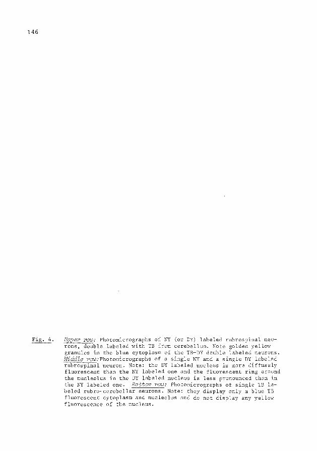
146
Upper row: Photomicrographs of NY (or DY) labeled rubrospinal neurons, double labeled with TB from cerebellum. Note golden yellow granules in the blue cytoplasm of the TB-DY double labeled neurons. Middle row:Photomicrographs of a single NY and a single DY labeled rubrospinal neuron. Note: the DY labeled nucleus is more diffusely fluorescent than the NY labeled one and the fluorescent ring around the nucleolus in the DY labeled nucleus is less pronounced than in the NY labeled one. Bottom row: Photomicrographs of single TB labeled rubro-cerebellar neurons. Note: they display only a blue TB fluorescent cytoplasm and nucleolus and do not display any yellow fluorescence of the nucleus.

147

148
Upper row: Photomicrographs of single NY (or DY) labeled rubrospinal neurons and of NY (or DY) labeled rubrospinal neurons double labeled with TB from cerebellum. Note: the fluorescence of the DY labeled nucleus is more diffuse and the TB-DY double labeled neuron contains golden yellow granules in the blue labeled cytoplasm. Bottom row: For comparison: photomicrographs of single TB-labeled rubrospinal neurons with TB fluorescent cytoplasm and nucleolus. Note: the absence of nuclear fluorescence.
149
Rostrocaudally the first and second zones were restricted to the injected
segments.
The DY injection area was more restricted than the NY injection area in
both the transverse and in longitudinal directions. Therefore it may be con
cluded that DY, which is less water soluble than NY, diffuses less through the
tissue at the injection site.
Red Nucleus
In the 8 rats (cases 1-8) in which NY or DY was injected ~n the spinal
cord, many NY- or DY-labeled rubrospinal neurons were present in the dorsa
medial, intermediate and ventrolateral parts of the contralateral red nucleus.
These neurons, when single labeled3
'24
showed a yellow fluorescence of the
nucleus with some yellowish fluorescence of the cytoplasm (Figs. 4 and 5). In
the one control rat with only a TB injection in the cerebellum, many single
TB labeled neurons were present ~n the red nucleus. They were located through
out the red nucleus both in the transverse and longitudinal directions. They
showed only a blue cytoplasmic and nucleolar labeling without any indication
of a yellow labeling of the nucleus (Fig. 4). In the cases 1-8 with injections
in the cerebellum and in the spinal cord relatively few single TB labeled
neurons (~ 70) were present in the contralateral red nucleus. However, a
relatively large proportion of the NY- or DY-labeled rubrospinal neurons were
double labeled with TB from the cerebellum, such that they showed a yellow
fluorescent nucleus and a blue TB fluorescent cytoplasm (Figs. 4 and 5).
In general, the double labeled neurons were present throughout the rostra
caudal extent of the red nucleus (Fig. 6). However, in the most caudal end
of the nucleus mainly single NY- or DY-labeled rubrospinal neurons were
present and only relatively few double labeled neurons occurred (Fig. 6). The
double labeled neurons were more or less evenly distributed throughout the
cross-sectional profile of the red nucleus, except in cases 2 and 3 in which
more double labeled neurons were present in the intermediate and lateral
portions of the nucleus, including the lateral horn described by Reid et al.
197535
than in the medial parts (Fig. 6). In order to determine the percentage
of the NY- or DY-labeled rubrospinal neurons which were TB double labeled from
the cerebellum the following procedure was adopted. In each case the NY- or
DY-labeled neurons as well as the TB-NY or TB-DY double labeled ones were
counted in every fourth section through the red nucleus. These numbers were
then added, multiplied by four and treated according to the following formula.
TB-NY double-labeled neurons single NY + TB-NY double-labeled neurons X 100%
( In the animals in which DY was used read DY for NY )

150
Semidiagrammatic representations of the distributions of retrogradely labeled neurons in red nucleus of cases 2 and 3 after TB injections contralaterally in the anterior interpositus nucleus (NIA) and a NY (case 2) or DY (case 3) hemiinfiltration contralaterally at C5-C8. Note: the double labeled neurons are more or less evenly distributed throughout the rostrocaudal extend of the red nucleus; in case 3 the double labeled neurons show a preponderance laterally in the nucleus. Abbreviations: CP, cerebral peduncle; DCP, decuss~1tion of superior cerebellar peduncles; GM, medial geniculate body; I Ped, interpeduncular nucleus; ML, medial lemniscus; mlf, medial longitudinal fasciculus; NC, cochlear nuclei; N III, ocul,Jmo tor nucleus; n III oculomotor nerve; R, red nucleus; RF, reticular formation; SNR, substantia nigra, pars reticulata; SNC, substania nigra, pars compacta.

ROSTRAL
CAUDAL
CASE 2 TB anterior interpositus NY c5-c 8
0
0
*
~X100 =26,97. DL+NY
TB •:•:•
CASE 3 TB anterior interpositus DY c5 -c8
151

r-------------------.------------------------------r--------------------------,-------------------~---------------~
TB-NY and TB-DY percentages of ' TB injection area NY and DY labeled rubro- double labeled yellow (NY or DY) (zones I and II) in spinal neurons, including neurons in the labeled neurons, the cerebellum double labeled ones red nucleus which \vere double
labeled with TB
CASE 1 zone I involved the anterior interpositus nucleus 2380 (NY) 484 20,4% (NIA)
CASE 2 zone I involved NIA 2168 (NY) 584 26,9%
CASE 3 zone I involved NIA 2468 (DY) 1036 42 %
CASE 4 zone I involved NIA 2824 (DY) 812 28,7%
CASE 5 zone I b~rderi~g on NIA 2184 (NY) 33 6 15 4% zone II lnvolvlng NIA '
CASE 6 zone I b~rderi~g on NIA 1996 (DY) 31 6 16 % _____ zone II lnvolvlng NIA
CASE 7 zones I and II dorsal 2246 (NY) 46 2 %
I to NIA o
CASE 8 zones I and II dorsomedlal 2344 (DY) S2 2 % to NIA o
Table II shmis:
--- -- -
the localization of the TB injection area in the cerebellum (column 1), the numbers and DY labeled rubrospinal neurons (column 2), the number of NY and DY rubrospinal double labeled \vith TB from the cerebellum (column 3) and the percentages of NY and labeled rubrospinal neurons, double labeled from the cerebellum (column 4).
of NY neurons, DY
>--' Ul N

153
In the four cases (cases 1,2,3,4) in which zone I of the TB injection
was centered in the NIA the following findings were obtained. In the cases ln
jected with NY, the total numbers of NY-labeled rubrospinal neurons, including
the NY-TB double labeled ones were: 2380 (case I) and 2168 (case 2). In the
cases injected with DY, the total numbers of DY-labeled neurons including the
DY-TB double labeled ones were 2468 (case 3) and 2824 (case 4)(Table II). In
the two rats with the NY injections 20.4% (case I) and 26.9% (case 2) of the
NY labeled rubrospinal neurons were double labeled from the cerebellum. In the
two rats with the DY injections, 42% (case 3) and 28.7% (case 4) of the DY
labeled neurons were double labeled from the cerebellum (Fig. 6, Table II).
The higher percentage in case 3 as compared to cases I, 2 and 4 may be explain
ed by the fact that in case 3 a large quantity of TB was injected in the cere
bellum than in the other cases (cf. Table I) and correspondingly a larger in
jection area was found.
In the four other cases (cases 5,6,7 and 8), ln which the TB zone I was
located outside the NI the following findings were obtained. In the red nucleus
approximately the same number of NY or DY labeled rubrospinal neurons was found
as in cases 1,2,3 and 4 (Table II), and these neurons displayed the same distri
bution as in the cases I to 4. However, the percentages of the NY and DY la
beled neurons which were NY-TB or DY-TB double labeled varied according the
location of the cerebellar injection area. Thus, in cases 5 and 6, in which
only zone II involved the NI (Table II), 15.4% and 16% of the rubrospinal neu
rons was double labeled from the cerebellum. However, in cases 7 and 8 in which
neither zone I nor zone II involved the NI, virtually no single TB labeled
neurons were found in the red nucleus and in both cases only 2% of the rubro
spinal neurons were double labeled (Table II).
These retrograde double labeling findings lead to the conclusion that
almost all (~90%) of the rubro-cerebellar fibers to the anterior interpositus
nucleus represent collaterals of rubrospinal neurons. In the 8 cases a maxlmum
number of 2824 rubrospinal neurons were labeled (case 4, Table II). In case 3
with the largest cerebellar TB injection area the maximum number of rubrospinal
neurons was double labeled from the cerebellum (i.e. 1036, Table II). According
to the formula used in the present study 37% of the rubrospinal neurons were
double labeled from the cerebellum. Within the restraint of the present method
these findings suggest that at least 37% of the rubrospinal neurons give rise
to rubrocerebellar collaterals.
In some of the cases, in which the TB injections were centered in the
NIA, the efferent fibers from the cerebellum to the contralateral red nucleus

154
Fig. 7. Photomicrographs of FB fluorescent fibers in cat red nucleus, which are anterogradely labeled from the contralateral cerebellar interpositus nucleus. Note: the fiber varicosities, which seem to be in contact with the surface of cell body and dendrites of the single NY labeled rubrospinal neurons.
were anterogradely labeled and appeared as very thin blue threads in the red
nucleus. However, in some additional experiments in cat with ,.Fast Blue" (FB) 3
injections in the NIA and NY injections in the spinal cord, such anterograde
labeling of cerebello-rubral fibers was much more pronounced. Thus in these
cases, which will be reported separately, blue FB fluorescent fibers could be
traced from the cerebellum to the cell body and dendrites of retrogradely NY
labeled rubrospinal neurones (Fig. 7).
155
Discussion
Earlier anatomical studies10
'12
showed that in cat fibers from the red
nucleus descend through the rubrospinal decussation to the level of the pontine
trigeminal nuclei15
and then proceed along the fibers of the brachium conjunc
tivum into the cerebellum. These rubrocerebellar fibers approach the deep cere
bellar nuclei from the ventral side~ 2 Brodal and Gogstad (1954)
6 on the basis
of retrograde changes concluded that in the cat the bulk of these fibers ter-12
minate in the lateral cerebellar nucleus. However, Courville and Brodal (1966)
on the basis of anterograde degeneration demonstrated that the termination
area of the rubrocerebellar fibersis largely restricted to the interpositus
nucleus (NI) chiefly its anterior part (NIA) and that only a few fibers termi
nate in the posterior interpositus nucleus (NIP). This discrepancy in regard
to the termination of rubrocerebellar fibers may be due to differences in the
definition of the border between the interpositus and lateral nucleus used in
these two studies and to differences in technique. In view of the anterograde
degeneration findings of Courville and Brodal12
it has been assumed in the
present study that the bulk of the rubrocerebellar fibers both in cat and rat
terminate in the rostral portion of the NI (i.e. NIA). In this study an attempt
was made to determine whether these crossed rubrocerebellar fibers represent
collaterals of rubrospinal neurons. For this purpose in rats 'True Blue' (TB)
was injected in the cerebellum and 'Nuclear Yellow' (NY) was injected ipsi
laterally in the spinal cord. In some cases a new tracer 'Diamidino Yellow'
(DY) was used instead of NY. This new tracer produces roughly the same retro
grade labeling as NY but has the advantage that it migrates much more slowly
out of the retrogradely labeled neurons than NY. Therefore DY does not require
a short survival time4
and can be injected with TB in the same session.
The present study essentially comprises 9 cases. In one of them, injec
tions (TB) were made in only the cerebellum and in the other 8 cases the in
jections were made both in the cerebellum (TB) and the spinal cord (NY or DY).
In the one case with only a TB injection in the cerebellum, many single TB la
beled neurons were present throughout the contralateral red nucleus. These neu
rons displayed the typical TB labeling described earlier2
and contained only a
blue TB fluorescent cytoplasm and a bright blue fluorescent nucleolus. However,
they clearly did not display any yellow fluorescence of the nucleus. In the
other eight cases only few such single TB labeled neurons were present while
the majority (~90%) of the neurons with a TB fluorescence of the cytoplasm
were double labeled such that they also showed a pronounced yellow fluorescence
of the nucleus. On the basis of these double labeling findings it has been con
cluded that the rubrocerebellar fibers mainly represent collaterals of rubro-

156
spinal neurons. This is in keeping with the earlier anatomical and electro-. l . . d" . 6, I physlo oglcal fln lngs ln cat.
In order to determine the percentage of rubrospinal neurons which give
rise to collaterals to the cerebellar interpositus nucleus it was necessary
to obtain retrograde labeling of a maximum number of rubrospinal neurons as
well as of a maximum number of rubrocerebellar neurons.
In order to obtain retrograde labeling of a maximum number of rubrospinal
neurons use was made of the fact that NY and DY are transported retrogradely
both from terminal areas and from damaged axons?'24
Therefore, the NY and DY
injections in CS-CS were made by means of multiple pipette penetrations both
into the grey matter and the white matter. The injections in the grey matter
were expected to produce retrograde labeling of rubrocervical neurons, while
those in the white matter were expected to produce retrograde labeling of
rubrospinal neurons from damaged rubrospinal fibers passing through the CS-CS
dorsolateral funiculus to more caudal levels of the cord;' 40 , 32 , 33 This
strategy appeared to have been successful since in all cases a large number of
rubral neurons was retrogradely labeled. Moreover, they were distributed over
the dorsomedial, intermediate and ventrolateral parts of the cross-sectional
profile of the red nucleus, which parts project to the cervical, the thoracic . IS 30 19 34
and the lumbosacral cord respectlvely. ' ' ' In view of these findings, it
may be concluded that, within the restraints of the present method, a maximum
number of rubrospinal neurons was retrogradely labeled.
In order to judge whether the cerebellar TB injections produced a maximum
number of TB labeled rubrocerebellar neurons the following points should be
taken into account. The TB injection areas consist of two concentric zones, i.e.
zone I and II, the former of which seems to give rise to the highest degree of
retrograde labeling. The NI injections were always made from above, while accor-. . . . . 12 l . dlng to the earller anatomlcal flndlngs the rubrocerebe lar flbers penetra-
ted the nucleus from below, i.e. through its hilar region. As a consequence it
was expected that the TB injections which were restricted to the NI produce
retrograde labeling of rubrocerebellar neurons mainly from the termination area
of the rubrocerebellar fibers, while injections which also involved the hilar
region of the NI would in part produce retrograde labeling of rubral neurons
from damaged rubrocerebellar fibers. In keeping with these expectations it was
found that in the cases 7 and 8 in which the TB injection area (zones I and II)
was located dorsal or medial to the NI and largely spared the nucleus, only a
very small percentage of rubrospinal neurons was double labeled. However, in
the cases 5 and 6 in which zone I was located outside the NI, but zone II in
volved the nucleus, a larger percentage of rubrospinal neurons was double

157
labeled, and in the cases I to 4 in which zone I was centered in the NIA a much
larger percentage of rubrospinal neurons was double labeled. Moreover, in case
3, in which zone I was also centered in the NIA, but in which the TB injection
area most extensively involved the hilar region of the nucleus, the maximum
number of rubrospinal neurons was double labeled, i.e. 1036 (Table II).
In the 8 cases a maximum number of 2824 rubrospinal neurons were labeled
(case 4, Table II). In case 3 with the largest cerebellar TB injection area
involving both the nucleus and the hilar region a maximum number of 1036 rubro
spinal neurons was double labeled from the cerebellum (Table II). According to
the formula used in the present study 37% of the rubrospinal neurons were
double labeled from the cerebellum.
With the restraint of the present method these findings suggest that at least
37% of the rubrospinal neurons give rise to rubrocerebellar collaterals.
The double labeled neurons in the red nucleus, distributing divergent
axon collaterals to both the cerebellar interpositus nucleus and the spinal
cord, were more or less evenly distributed throughout the red nucleus both in
the transverse and longitudinal directions except in cases 2 and 3 in which
more double labeled neurons were present in the intermediate and lateral parts
of the nucleus than in the medial parts (Fig. 6). This is in general accor-6
dance with the findings of Brodal and Gogstad (1954) that after cerebellar
lesions retrograde changes occur in large and medium sized neurons, which are
located in the same parts of the red nucleus as the retrogradely affected
neurons after spinal lesions. However, after cerebellar lesions Brodal and
Gogstad found a slight preponderance of retrogradely affected neurons medially
in the nucleus, while in the present study the double labeled neurons showed a
slight preponderance laterally in the nucleus which in cases 2 and 3 was more
pronounced than in the other cases (Fig. 6). This difference may be due to 6
the following. In the study of Brodal and Gogstad mainly large to intermediate
neurons were taken into account, because in such neurons the retrograde changes
can be recognized most reliably. However, this may produce a bias, because in
the rostral part of the red nucleus such neurons tend to be concentrated in
the medial and intermediate parts. On the other hand, ~n the present study
also many relatively small rubrospinal neurons were double labeled, not only
in the medial and intermediate parts of the nucleus, but also in the ventro
lateral parts, including the lateral horn described by Reid et a1 .. 35
In respect to the present findings it is of interest to note that the
interpositus nucleus represents the main source of afferents to the rubrospinal . II 17 14 neurons which projection in addition is somatotopically organ~zed. ' '
The rubrospinal collaterals to the interpositus nucleus therefore appear to

158
Fig. 8.
cerebellum
M. I.
rubrospinal tract
Diagram of the interconnections between red nucleus and contralateral cerebellar anterior interpositus nucleus. Note that the rubro-interpositus connections are established by collaterals of rubrospinal neurons.

159
represent a direct, tightly coupled return projection to cells of origin of
the afferents of these rubral neurons (Fig. 8). The precise functional role
of these return projections is still unclear. However, they must be of criti
cal importance since such return projections occur in several places in the
cerebellar circuitry. Thus, for example the efferent fibers from the deep cere
bellar nuclei to the red nucleus and the thalamus display a similar arrangement
. . h b 11 37 ' 38 f and glve off dlrect return collaterals to t e cere e ar cortex, rom
where they receive the bulk of their afferents. Moreover, the inferior olive,
which gives rise to projections to the cerebellar cortex and the deep cere
bellar nuclei39 also receives return projections from these deep cerebellar
nuclei. In this context it is of interest to note that in parallel with the
return collaterals from the rubrospinal neurons to the interpositus nucleus
also another return projection from the red nucleus to the cerebellar cortex
exists. This return projection is established by way of rubral fibers to the . 41 10 . 1 5 lateral retlcular nucleus, ' which nucleus proJects to the cerebe lum. In
light of the present findings the question arises as to whether these rubro
lateral reticular connections are also established by collaterals of rubro
spinal neurons. 22 23 .
In a preceding fluorescent retrograde labeling study ' lt was demon-
strated that in rat, cat and monkey the rubrospinal and raphe spinal fibers
give rise to collaterals along their trajectory throughout the spinal cord.
However, the rubrospinal system was found to produce much fewer of such col
laterals than the raphe spinal system. Thus, in rat 20% and in cat and monkey
3.8% and 6.6% of the rubrocervical neurons were found to provide collaterals
to segments caudal to 11, while ln all these species 31-48% of the raphe cervi
cal neurons provide collaterals to the segments caudal to 11 and only a slight-
ly smaller percentage (27%-35%) to segments caudal to Sl (cf. Histograms! and
2, Fig. 9). It was therefore concluded that the rubrospinal tract represents a
relatively focussed system, which distributes its individual fibers to restric
ted groups of spinal segments, while the raphe spinal tract represents a
diffuse system, which distributes almost half of its fibers throughout the
length of the spinal cord. Moreover, the degree of collateralization of the
rubrospinal system in rat appeared to be much higher than in cat and monkey,
Slnce ln rat as compared to cat and monkey a much higher percentage of rubro
cervical neurons give rise to collaterals to the spinal cord caudal to 11 29
(Fig. 9, histogram I). This, together with observations in pigeon42
and opossum
suggests that the rubrospinal tract as a focussed system emerges only gradually
during phylogeny.
In view of the above interspecies differences ln the degree of collaterali-

160
100Z HISTOGRAM I RED NUCLEUS
in rat (R) cat {C)
monkey(M) uro
'l!l U-o
E"*"< ~.oO - ~~ ~~~ Q).CQl .c :J c ~..go
Cl (J} c c ·-0"0 ~ :J
~u c:: .!::
10 HISTOGRAM II
N. RAPHE MAGNUS
in rat (R) cat (C)
monkey (M)
oro c,l!)2 EJl'b e~ o -Q.)Ii ~:g~ Q) 0 c .c "0 0
"' -C) c "0 :J (3
·'= sacral I umbo·
sacral high
lumbar
Fig. 9. Histogram I, showing the percentages of rubrocervical neurons, which distribute descending collaterals passing through more caudal levels of the cord in rat (R), cat (C) and monkey (M). Note that in cat and monkey a smaller percentage of the rubrocervical neurons distribute collaterals through more caudal levels of the cord than in rat (c.f. Huisman et al., 22, 23). Histogram II, showing the percentages of raphe-cervical neurons from Taberset al. 35 Nucleus Raphe Magnus and adjoining reticular formation, which distribute collaterals passing through more caudal levels of the cord in rat (R), cat (C) and monkey (M). llote that a large portion (approx. Ij3) of the rubrocervical neurons give rise to collaterals which descend throughout the length of the spinal cord.

161
zation of the rubrospinal system in the cord the question arose as to whether
such interspecies differences also exist in respect to the rubrospinal collate
rals to the cerebellum. Therefore a further interspecies comparison of these
rubrospinal collaterals would be of interest. However, it would seem unlikely
that the degree of rubrospinal collateralization to the cerebellum would run
parallel with that of the rubrospinal collateralization in the cord, since
these two types of rubrospinal collaterals probably subserve an entirely diffe
rent function.
Acknowledgements
The authors thank Prof.Dr. 0. Dann (Friedrich-Alexander Universitat,
Erlangen) and Dr. H. Loewe (Hocchst Company, Frankfurt) for providing small
quantities of TB, DY and NY. They also thank Mr. E. Dalm, Mr. H. Klink and
Miss P. van Alphen for their technical assistance, Dr. R. Lemon for reading
the manuscript and Miss G. van Gelder and Miss E. Klink for typing it.

162
References
I. Anderson, M.E., Cerebellar and cerebral inputs to physiologically identi
fied efferent cell groups in the red nucleus of the cat. Brain Res., 30
(1971) 49-66.
2. Bentivoglio, M., Kuypers, H.G.J.M., Catsman-Berrevoets, C.E. and Dann, 0.,
Fluorescent retrograde neuronal labeling in rat by means of substances
binding specifically to adenine-thymine rich DNA. Neurosci. Lett., 12 (1979)
235-240.
3. Bentivoglio, M., Kuypers, H.G.J.M., Catsman-Berrevoets, C.E., Loewe, H., and
Dann, 0., Two new fluorescent retrograde neuronal tracers, which are tran
sported over long distances. Neurosci. Lett., 18 ( 1980) 25-30.
4. Bentivoglio, M., Kuypers, H.G.J.M. and Catsrnan-Berrevoets, C.E., Retrograde
neuronal labeling by means of Bisbenzimide and Nuclear Yellow (Hoechst
S-769121). Measures to prevent diffusion of the tracers out of retrogradely
labeled neurons. Neurosci. Lett., 18 (1980) 11-18.
5. Brodal, A.,The cerebellar connections of the nucleus reticularis lateralis
(nuclei funiculi lateralis) in rabbit and cat. Experimental investigations.
ActaPsychiat. etNeur., 18 (1943) 171-233.
6. Brodal, A. and Gogstad, A.C., Rubro-cerebellar connections. Anat. Rec., 118
(1954) 455-482.
7. Brown, L.T., Rubrospinal projections in the rat. J. Camp. Neural., 154
(1974) 169-188.
8. Cajal, S., Ramon, Y., Histologie du Systeme Nerveux de l'homme et des
vertebres. Instituto Ramon Y Cajal, Madrid, 1952.
9. Catsman-Berrevoets, C.E. and Kuypers, H.G.J.M., A search for corticospinal
collaterals to thalamus and mesencephalon by means of multiple retrograde
fluorescent tracers in cat and rat. Brain Res., 218 (1981) 15-33.
10. Courville, J., Rubrobulbar fibers to the facial nucleus and the lateral
reticular nucleus (nucleus of the lateral funiculus). An experimental study
in the cat with silver impregnation methods. Brain Res., I (1966) 317-337.

163
11. Courville, J., Somatotopical organization of the projection from the nucleus
interpositus anterior of the cerebellum to the red nucleus. An experimental
study in the cat with silver impregnation methods. Exp. Brain Res., 2 (1966)
191-245.
12. Courville, J. and Brodal, A., Rubrocerebellar connections ln the cat: an
experimental study with silver impregnation methods. J. Camp. Neural., 126
(1966) 471-486.
13. Cowan, W.M., Gottlieb, D.I., Hendrickson, A.E., Price, J.L. and Woolsey,
T.A., The autoradiographic demonstration of axonal connections in the cen
tral nervous system. Brain Res., 37 (1972) 21-51.
14. Dekker, J.J., Anatomical evidence for direct fiber projections from the
cerebellar nucleus interpositus to rubrospinal neurons. A quantitative EM
study in the rat combining anterograde and retrograde intra-axonal tracing
methods. Brain Res., 205 (1981) 229-244.
15. Edwards, S.B., The ascending and descending projections of the red nucleus
in the cat: an experimental study using an autoradiographic tracing method.
Brain Res., 48 (1972) 45-63.
16. Fink, R.P. and Heimer, L., Two methods for selective silver impregnation of
degenerating axons and their synaptic endings in the central nervous system.
Brain Res., 4 (1967) 369-374.
17. Flumerfelt, B.A., Otabe, S., and Courville, J., Distinct projections to the
red nucleus from the dentate and interposed nuclei in the monkey. Brain
Res., 50 (1973) 408-414.
18. Flumerfelt, B.A. and Gwyn, D.G., The red nucleus of the rat: its organiza
tion and interconnexions. J. Anat., 188 (1974) 374-376.
19. Gwyn, D.G., Acetylcholinesterase activity ln the red nucleus of the rat.
Effects of rubrospinal tractomy. Brain Res., 35 (1971) 447-461.
20. Hayes, N.L. and Rustioni, A., Dual projections of single neurons are visua
lized simultaneously: use of enzymatically inactive (3H) HRP. Brain Res.,
165 (1979) 321-326.

164
21. Hayes, N.L. and Rustioni, A., Differential and collateral descending
projections to spinal enlargements. Exp. Brain Res., 41 (1981) 89-107.
22. Huisman, A.M., Kuypers, H.G.J.M. and Verburgh, C.A., Quantitative diffe
rences in collateralization of the descending spinal pathways from red
nucleus and other brain stem cell groups in rat as demonstrated with the
multiple fluorescent retrograde tracer technique. Brain Res., 209 (1981)
271-286.
23. Huisman, A.M., Kuypers, H.G.J.M., Verburgh, C.A., Differences in collatera
lization of the descending spinal pathways from red nucleus and other brain
stem cell groups in cat and monkey. Progress in Brain Research 57 (1982) 185-219.
24. Keizer, K., Kuypers, H.G.J.M., Huisman, A.M., Diamidino-Yellow (DY), a
fluorescent retrograde neuronal tracer which migrates only very slowly out
of the cell and can be used in combination with TB and FB in double labe
ling experiments, in preparation.
25. King, J.S., Dom, R.M., Conner, J.B. and Martin, G.F., An experimental light
and electron microscopic study of cerebellorubral projections in the opos
sum, Didelphis Marsupialis Virginiana. Brain Res. 52 (1973) 61-78.
26. Korneliussen, H.K., On the morphology and subdivisions of the cerebellar
nuclei of the rat. J. Hirnforsch., 10 (1968) 109-122.
27. Kuypers, H.G.J.M., Bentivoglio, M., Catsman-Berrevoets, C.E. and Bharos,
T.B., Double retrograde neuronal labeling through divergent axon collatera~
using two fluorescent tracers with the same excitation wavelength, which
label different features of the cell. Exp. Brain Res. 40 (1980) 383-392.
28. Lasek, R., Joseph, B.S., Whitlock, D.G., Evaluation of a radioautographic
neuroanatomical tracing method. Brain Res., 8 (1968) 319-336.
29. Martin, G.F., Cabana, T. and Humbertson, A.D., Evidence for a lack of
distinct rubrospinal somatotopy in the north american opossum and for col-
lateral innervation in the north american opossum and for co11ateral inner-
vation of the cervical and lumbar enlargements by single rubral neurons.
J. Camp. Neural., 201 (1981) 255-263.
30. Murray, H.M. and Gurule, M.E., Origin of the rubrospinal tract of the rat.
Neurosci. Lett., 14 (1979) 19-23.

165
31. Nauta, H.J.W., and Gygax, P.A., Silver impregnation of degenerating axons
in the central nervous system: a modified technique. Stain Technol., 29
(1954) 91-94.
32. Nyberg-Hansen, R. and Brodal, A., Sites and mode of termination of rubro
spinal fibers in the cat. An experimental study with silver impregnation
methods. J. Anat. (Land.) 98 (1964) 235-253.
33. Petras, J.M., Cortical, tectal and tegmental fiber connections ~n the
spinal cord of the cat. Brain Res., 6 (1967) 275-324.
34. Pompeiano, 0., Brodal, A., Experimental demonstration of a somatotopical
origin of rubrospinal fibers in the cat. J. Camp. Neural., 108 (1957)
225-251.
35. Reid, J.M., Gwyn, D.G., and Flumerfelt, B.A., Cytoarchitectonic and Golgi
Study of the Red Nucleus in the rat. J. Camp. Neural., 162 (1975) 337-362.
36. Taber, E., Brodal, A., and Walberg, F., The Raphe nuclei of the brain stem
in the cat. I. Normal topography and cytoarchitecture and general discus
sion. J. Camp. Neural., 114 (1960) 161-188.
37. Tolbert, D.L., Bantli, H. and Bloedel, J.R., The intra-cerebellar nucleo
cortical projection. Exp. Brain Res. 30 (1977) 425-434.
38. Tolbert, D.L., Bantli, H. and Bloedel, J.R., Multiple branching of cere
bellar efferent projections in cats. Exp. Brain Res., 31 (1978) 305-316.
39. Voogd, J. and Bigare, F., Topographical distribution of olivary and
corticonuclear fibers in the cerebellum: a review. In: J. Courville et al.
(Eds.). The inferior Olivary Nucleus: Anatomy and Physiology, Raven Press,
New York, 1980, pp. 207-234.
40. Waldron, H.A. and Gwyn, D.G., Descending nerve tracts ~n the spinal cord
of the rat. I. Fibers from the midbrain. J. Camp. Neural., 137 (1971)
143-154.
41. Walberg, F., Descending connections to the lateral reticular nucleus. An
experimental study in the cat. J. Camp. Neural., 109 (1958) 363-390.
42. Wild, J.M., Cabot, J.B., Cohen, D.H. and Karten, H.J., Origin, course and
terminations of the rubrospinal tract ~n the pigeon (Columba Livia). J.
Camp. Neural., 187 (1979) 639-654.

166
CHAPTER VI: GENERAL DISCUSSION
VI.!.
VI.2.
VI.3.
Technique
The rubrospinal tract ~n rat, cat and monkey
VI.2.a. Somatotopic organization of red
nucleus
VI.2.b. Collateralization of the rubrospinal
tract in rat, cat and monkey
VI.2.c. Collateralization of the rubrospinal
tract compared with collaterali
zation of the raphe-spinal and
ventrolateral pontine tegmentum
spinal tracts in rat, cat and monkey
VI.2.d. Collateralization of the rubrospinal
tract to the cerebellum
References

VI.
VI.1.
167
GENERAL DISCUSSION
Technique
Classic anatomical studies using the Golgi technique (Cajal, 1952)
demonstrated that many axons in the central nervous system give off
axon collaterals along their trajectory through the brain. However,
with the available tracing techniques, which are based on degenera
tion or intra-axonal transport (Cowan, 1972; Fink and Heimer, 1967;
Kristensson, 1970; Kristensson et al., 1971; LaVail and LaVail,
1972; Nauta and Gygax, 1954) it is very difficult to distinguish
divergent axon collaterals from unbranched fibers. Therefore in the
late seventies several attempts were made to demonstrate the exi
stence of axon collaterals by developing different retrograde tracers,
which are transported retrogradely through the stem axon and through
a collateral and which can be demonstrated independently in the
parent cell body.
At the moment the following 3 methods for such retrograde
double labeling of neurons by way of divergent axon collaterals are
available.
A) The HRP-tritiated apo-HRP technique (Hayes and Rustioni,
1979 and 1981). This technique is based on the fact that both the 3
enzyme HRP and the H apo-HRP are transported retrogradely to the
cell body (Geisert, 1976; Kristensson and Olsson, 1971; LaVail and
LaVail, 1972). The presence of HRP is demonstrated histochemically,
while the presence of the tritiated-apo HRP is then demonstrated by
means of autoradiography. Thus, this technique requires a combined
histochemical-autoradiographic procedure. However, because of the
fact that by means of the autoradiographic technique the presence of
label can only be detected in the upper 3 pm of the section, the
presence of the double labeled neurons can only be demonstrated in
these upper 3 f-!m. (Sidman, 1970).
E) The HRP-Iron dextran technique (Cesaro et al., 1979; Olsson
and Kristensson, 1978). The presence of these two tracers in the
parent cell body may be demonstrated histochemically by first pro
cessing the sections for HRP and then demonstrating the presence of
ferric ions in the cell body by means of the Perl's reaction. How
ever, double labeling may be underestimated when few granules of
iron dextran or HRP are present in the cell body, which are over
shadowed by a massive storage of the other reaction product. This

168
can be prevented by adapting the survival times to the fiber systems under
study.
C) The fluorescent double labelinq technique. This technique makes use of
the fact that several fluorescent substances may be transported retrogradely
through divergent axon collaterals to the same parent cell body where they can
be visualized independently by means of fluorescence microscopy (Kuypers et
al., 1977, 1980). Since this "fluorescent" technique requires only a relati
vely simple procedure (see Chapter II) and is one of the few anatomical tech
niquessuitable to study quantitatively the existence of collaterals in brain
pathways, this technique has been used to try to clarify anatomically possible
quantitative differences in collateralization ~n the different brainstem path
ways, which descend through the dorsolateral funiculus i.e. the rubrospinal
pathway, the descending pathways from the ventrolateral pontine tegmentum and
from the raphe magnus including the adjoining ventral reticular formation.
The retrograde fluorescent tracers, used in this study (Chapter III and
IV), were 11True Blue" (TB) and 11 Nuclear Yellow" (NY) in rats (Chapter III)
and 11 Fast Blue" (FB) and 11 Nuclear Yellow" (NY) in cats and monkeys (Chapter
IV)(BentivoBlio et al., 1979; Bentivoglio et al., 1980a+b; Kuypers et al.,
1980). These tracers are transported relatively effectively over long distan
ces, although TE and FE more slowly than NY, and produce a fluorescent double
labeling which can be observed at one excitation wavelength (i.e. at 360 nm a
blue TB or FB labeling of the cytoplasm and a yellow NY labeling of the
nucleus).
After relatively long survival times NY may migrate out of retrogradely
labeled neurons into surrounding neurons, as signalled by the presence of NY
labeled fluorescent glial nuclei (c.f. Chapter II). In order to avoid false
retrograde labeling relatively short survival times should be used (Benti
voglio et al., 1980b). Therefore first TE or FE was injected in the C5-C8
dorsal spinal grey and subsequently in a later operation NY was injected at
more caudal levels of the cord, a short time before the animal was sacrificed.
Following this procedure, in all cases only dull NY glial labeling was present
around the single NY- and double TE-NY or FB-NY labeled neurons. This indi
cates that only minimal migration of NY out of retrogradely labeled neurons
occurred i.e. without any risk of false retrograde labeling. In exceptional
cases single FE-labeled neurons in the central nervous system show some blue
to white FE labeling of the nucleus which could make such neurons difficult
to distinguish from FE-NY double labeled ones. In this respect it should be
emphasized however, that in our experiments in the brain in which a relati
vely low percentage of FB was dissolved in distilled water only very seldom

169
a strong FB labeling of the neuronal nucleus was obtained (Bharos et al.,
1981; Huisman et al., unpublished observations). Moreover, in this material
the exceptional blue to white FB labeling of the nucleus could always be
clearly distinguished from the yellow to green NY labeling of the nucleus.
Therefore the combination of FB and NY has been consistently used in double
labeling experiments in cats and monkeys. Applying this technique to the
descending pathways in spinal cord (Chapter III and IV) is difficult, espe
cially in rat, due to the close proximity between the area of termination of
the fibers in the grey matter and their trajectory in the adjoining funiculus
In order to obtain reliable results it is therefore imperative that the tracer
injection is entirely restricted to the grey matter, because injection of some
of the tracer in the lateral funiculus may result in its retrograde transport
through damaged fibers. This in turn will lead to retrograde labeling of
neurons ln the red nucleus and the other brain stem cell groups which may not
distribute collaterals to the injected part of the spinal grey This diffi
culty only applies to the injections in C5-C8 segments, because the injections
at more caudal spinal levels were intentionally made in white and grey matter
in order to damage many fibers in the lateral funiculus which would result in
retrograde transport of the tracer through a maximum number of fibers descen
ding in this funiculus at the injected levels. In light of the above consl
derations, in all cases each of the injection areas in C5-C8 grey were studied
histologically in detail and only those cases were selected for study in which
the fluorescent zones, from which retrograde transport occurs to the parent
cell bodies, did not involve the lateral funiculus. The sparing of the fibers
in the lateral funiculus was confirmed by the fact that the population of
retrogradely labeled neurons from the cervical grey matter in the caudal part
of the contralateral red nucleus was always restricted to its dorsomedial
part, while the population of neurons which were retrogradely labeled from
lower levels of the cord was always located separately in the ventrolateral
part of the nucleus. This is in keeping with retrograde degeneration and
retrograde HRP findings (Flumerfelt and Gwyn, 1974; Gwyn, 1971; Kneisley et
al., 1978; Murray and Gurule, 1979; Pompeiano and Brodal, 1957). Sparing of
the rubrospinal tract also implies sparing of the two other descending path
ways, since their fibers are grouped together with the rubrospinal tract in
the dorsolateral funiculus, where the raphe-spinal tract is located in the
most peripheral zone (Basbaum and Fields, 1978 and 1979; Brown, 1967;
Edwards, 1972; Holstege et al., 1979; Kuypers and Maisky, 1977; Martinet al.,
1978 and 1979; Miller and Strominger, 1973; Nyberg-Hansen and Brodal, 1964;
Petras, 1967; Tohyama et al;, 1979a+b; Waldron and Gwyn, 1971).

170
A comparison of the results obtained by the multiple retrograde fluo-3
rescent tracer technique on one hand and the HRP- H-apo-HRP technique (Hayes
and Rustioni, 1979, 1981) on the other hand suggested that the retrograde
fluorescent tracer technique produces a larger proportion of double labeled
neurons (Hayes and Rustioni, 1981; c.f. Huisman et al., 1981; Huisman et al.,
1982). This difference may be explained by the fact that in the autoradio
graphic material silver grains will only be elicited in the emulsion by
sources of radioactivity which are located in the upper 3 pm layer of the 40
pm sections (Sidman, 1970). As a consequence, even under optimal circumstan
ces, only a small percentage of the total population of HRP labeled neurons
observed in each 40 pm section can be double labeled.
The fluorescent tracers are suggested to be more sensitive than HRP, as
pointed out by the findings of Sawchenko and Swanson (1981) that the fluo
rescent tracer TB in rat labels approximately twice as many neurons than the
HRP-TMB technique using HRP-polyacrylamide gel implants (Mesulam and Rosene,
1979). This is supported by a comparison of the present findings with those
obtained by Conde and Conde (1982) (personal communications) which show that
in our experiments after Fast Blue (FB) injections ~n inferior olive of the
cat the number of FB labeled rubro-olivary neurons at a given level in the
red nucleus was significantly larger than obtained with the HRP-BDHC techni
que (Conde and Conde, 1982; Mesulam, 1976,197~,1979) at approximately the
same level (i.e. in a 30 pm section about 80 FB labeled neurons and in a 60
pm section about 40 HRP labeled neurons). On the other hand comparing the
efficacies of FB and HRP in retrograde labeling of cortical neurons in cat
after injections in the cervical cord including the white matter showed that
twice as many cortical neurons were labeled with HRP than with FB (Keizer et
al., in preparation b). However, this is true only for FB transport from
damaged fibers, since after FB injections in termination areas i.e. in the
spinal grey instead of in the dorsolateral funiculus, in which the bulk of
the corticospinal fibers descend (Armand and Kuypers, 1980; Armand, 1982;
Chambers and Liu, 1957) approximately the same number of FB labeled neurons
are present in the corresponding cortical area as obtained with HRP. After FB
injections in the dorsolateral funiculus much less FB labeled neurons were
obtained than HRP (Keizer et al., in preparation b) even if FB is dissolved
in dimethylsulfoxide (Huisman et al., 19~2; Keefer, 1978). The efficacy of the
fluorescent dye ,Nuclear Yellow" (NY), which is related to ,Bisbenzimide" (Bb)
turned out to be comparable to that of HRP (TMB procedure) (Keizer et al., in
preparation b; Sawchenko and Swanson, 1981). The fact that a larger number
of cortical neurons is retrogradely labeled from damaged fibers with HRP than

171
with FB, may be explained by the fact that FB gives much more necro
sis at the injection site than HRP, which necrosis may interfere with
uptake and retrograde transport of the tracer. However, NY gives much
less necrosis at the injection site than FB and TB. Yet ~n cat and
monkey optimal retrograde NY transport from damaged fibers is only
obtained when NY is dissolved in dimethylsulfoxide (Huisman et al.,
1982). Remarkably enough this ph~nomenon was not observed in rat in
which a very effective retrograde NY labeling of neurons was obtained
both from damaged fibers as well as from termination areas (Huisman
etal., 1981).
The fact that in all cases a difference in degree of collatera
lization was found between the rul,rospinal tract on one hand and the
raphe- and ventrolateral pontine tegmentospinal tracts on the other
hand supports the reliability of this double labeling technique.
Furthermore, the quantitative anatomical data of the present studies
were all in good agreement with earlier electrophysiological studies,
which will be discussed below.
In chapter V the use of a new retrograde tracer is reported:
11Diamidino Yellow"(DY), which migrates only very slowly out of the
cell and can be used in combination with TB or FB in double labeling
experiments (Keizer et al., ~n preparation a).DY when combined with
TB or FB is as effective as NY in double labeling of neurons by way
of divergent axon collaterals. However, DY migrates much more slowly
out of the retrogradely labeled neurons than G~ and therefore does
not require a short survival time. As a consequence DY may be inj ec
ted at the same time as TB or FB, without the risk that DY leaks out
of retrogradely labeled neurons into surrounding glial and neuronal
nuclei, which could produce false double labeling.
After injecting DY in the cervical cord in rat and cat many
single DY labeled neurons were present in both the dorsomedial and
ventrolateral parts of the red nucleus, which indicates that DY as NY
is transported both from axon terminals and from passing fibers
(Keizer et al., in preparation; Huisman et al., in press ) . More
over, in double labeling experiments in rat in which the collaterali
zation of rubrospinal neurons to the cerebellum was studied quanti
tatively, the combination TB-NY was compared with the combination
TB-DY (Chapter V). The findings in these cases led to the conclusion
that in quantitative double labeling studies DY may be used instead
of NY, since the combination TB-DY results approximately in the same

172
VI.2.
VI. 2.a.
percentage of double labeled neurons as the combination TB-NY. This
was confirmed by a more elaborate quantitative study in rat and cat,
in which DY was compared with NY in the combination with TB in rat
and with FB in cat (Keizer et al., in preparation a).
The rubrospinal tract in rat, cat and monkey
Somatotopic organization of red nucleus
In rat the retrograde labeling findings ln the red nucleus (Chapter
III Figs. 4 and 5) confirm its somatotopic organization as demon
strated by means of the retrograde degeneration technique (Flumer
felt and Gwyn, 1974) and the retrograde HRP transport technique
(Murray and Gurule, 1979). However, contrary to these studies the
present findings also demonstrate some degree of somatotopic organi
zation in the rostral shell-shaped portion of the red nucleus. This
rostral portion consistently contained the highest number of double
labeled neurons (Chapter III Fig. 4) which indicates that it harbors
the bulk of the neurons distributing collaterals to different levels
throughout the spinal cord. The presence of this considerable number
of branching neurons in the rostral part of the red nucleus probably
explains the fact that its somatotopic organization has escaped
detection by means of other techniques (Flumerfelt and Gwyn, 1974;
Gwyn, 1971; Murray and Gurule, 1979).
In cat the retrograde labeling findings in red nucleus confirm
previous findings (Hayes and Rustioni, 1981; Pompeiano and Brodal,
19572 and showed that the rubrospinal neurons are mainly present in
the caudal two-thirds of the nucleus. The findings ln the experi
ments with FB injections in the inferior olive and NY injections in
the cervical cord further showed that in cat the red nucleus neurons,
which distribute fibers to the ipsilateral inferior olive (Courville
and Otabe, 1974; Martinet al., 1975, 1980; Strominger et al., 1979;
Walberg, 1956) represent a population, which is seperate from rubro
spinal neurons and which is largely restricted to the rostral part
of the nucleus including the area laterally adjoining the fasciculus
retroflexus (Chapter IV Figs. 8, 10 and II). This is in keeping
with other HRP findings (Conde and Conde, 1982; Saint-Cyr and Cour
ville, 1980 and 1981; Walberg and Nordby, 19BI). The distribution
of the rubrospinal neurons, which are retrogradely labeled from the
cervical, thoracic and lumbar cord confirmed the somatotopic organi-

VI.2.b.
173
zation of this nucleus as demonstrated earlier by means of other
techniques (Hayes and Rustioni, 1981; Pompeiano and Brodal, 1957;
Tsukahara et al., 1967). Thus, in the caudal half of the magnocellu
lar part of the rubrocervical, the rubrothoracic and the rubrolumbar
neurons are located in the dorsomedial, intermediate and ventro
lateral parts of the nucleus, respectively. On the other hand in the
rostral half of the magnocellular part these neurons become inter
mixed (Chapter IV Fig. 8).
In monkey retrograde degeneration studies (Kuypers and Lawrence,
1967; Poirier and Bouvier, 1966) showed that the rubrospinal neurons
are largely restricted to the magnocellular part. However, the
present findings showed that the population of rubrospinal neurons
extends further rostrally and continues along the lateral aspect of
the parvicellular part (Chapter IV Fig. 13) up to the level, where
the cross-section through the fasciculus retroflexus is located
ventral to the nucleus. This seems in keeping with the retrograde
HRP finding of Kneisley and collaborators (1978) as inferred from
their illustrations. The location of these rubrospinal neurons
appears to correspond with that of neurons with coarse Nissl bodies
(Miller and Strominger, 1973). This supports the idea that rubro
spinal neurons are characterized by their internal configuration
more or less independent of the size of the neurons (King et al.,
1971). The present findings support the earlier reported somatotopic
organization in the caudal magnocellular part of the monkey red
nucleus (Kneisley et al., 1978). They also indicate that in the
rostral extension of the population of rubrospinal neurons along the
lateral aspect of the parvicellular part, the rubrocervical neurons
are located dorsal to the rubrolumbar ones and that the latter neu
rons continue more rostrally than the former (Chapter IV Fig. 13).
CoUateraZ-iza<;ion o."' the ri/J:rospinal tr·act in rat, cat and monkey
Earlier electrophysiological studies in cat have dealt with the
collaterals of the rubrospinal pathway. Shinoda et al. (1977), re
ported the number of red nucleus neurons, which could be antidromi
cally invaded from C3-C8 grey matter and the percentage of these
neurons, which could be antidromically invaded by stimulation of the
fibers passing through different more caudal segments of the cord.
In order to facilitate a comparison between our anatomical data and

174
these physiological data the anatomical experiments were set up in a comparable
manner. Thus the number of TB or FB labeled neurons which project to the C5-C8
dorsal grey matter were counted and the percentages of these neurons which were
also labeled with NY from lower levels of the cord were computed. These percen
tages as computed from the anatomical findings in cat, were found to be of the
same order of magnitude as those inferred from the earlier electrophysiological
findings in this animal. Thus according to the electrophysiology in cat (Shino
da et al., 1977) 5% of the rubrocervical neurons projecting to C3-C8 distri
bute collaterals to segments caudal to Ll and 50% distribute collaterals to
segments caudal to T2. According to the anatomical findings about 3% of the
rubrocervical neurons projecting to C5-C8 distribute collaterals to the seg
ments caudal to L2 and about 20% distribute collaterals to the segments caudal
to TJ (Chapter IV Table IV). In monkey a similar arrangementwas found anatomi
cally: 9,3% of the rubrocervical neurons projecting to C5-C8 were found to
distribute collaterals to the segments caudal to T8 and 6,6% to the segments
caudal toLl (Chapter IV Table V). The slight differences between the percen
tages obtained by means of the electrophysiological and the anatomical techni
ques may be due to the fact that in the two types of experiments slightly
different cervical segments were studied and slightly different thoracic and
lumbar segments were stimulated and injected, respectively. The differences may
also be due to differences in the rostra-caudal extent of the nucleus explored
ln the two types of experiments.
A comparison of the anatomical findings in cat and monkey with those ob
tained in rat (Chapter IV and III) respectively, indicates that the rubrospinal
system ln rat displays a higher degree of collateralization than in cat and
monkey. Thus in rat 20% of the rubrocervical neurons projecting to C5-C8 distri
bute collaterals to segments caudal to Ll versus only about 3% in cat and about
6% in monkey. Further, in rat about 35% of the rubrocervical neurons distribute
collaterals to segments caudal to T7 versus about 10% ln cat and monkey. The
rubrospinal tract in cat and monkey therefore appears to be more focussed than
in rat, such that in the former species a larger proportion of the rubrospinal
neurons distribute fibers to a restricted part of the spinal ccrd than in the
latter. This conclusion is further strengthened by the fact that the high
degrees of collateralization of the raphe-spinal and the crossed pontospinal
tract are of the same order of magnitude in the three species (see VI.2.c.).
The comparison of the anatomical findings in opossum and rat (Huisman et
al., 1981; Martinet al., 1981a) strongly suggests that the rubrospinal tracl
in opossum shows an even higher degree of collateralization than in rat. This
would imply that the rubrospinal tract ln rat, in turn, is more focussed than

175
1n opossum. The fact that the red nucleus in opossum displays a much
less rigid somatotopic organization than in rat (Huisman et al.,
1981; Martinet al., 1974, 1981) implies that with an increase in
collateralization the somatotopic organization becomes less sharply
defined. If this ~correct the absence of any somatotopic organiza
tion in the red nucleus in pigeon (Wild et al., 1979) and in reptiles
(ten Donkelaar and De Boer-van Huizen, 1978) would imply that in
these species the rubrospinal tract displays an even more pronounced
collateralization than in opossum. In the tadpole this tract develops
just at the stage when limbs are developing (ten Donkelaar, 1982),
while in snakes this tract seems to be lacking (ten Donkelaar, 1976a+
b). It therefore appears that the rubrospinal tract, as a highly
focussed fiber brain stern system, is only present in higher mammals.
It would be of interest to determine whether in respect to the rubro
spinal collateralization the 11 ontogeny rn1rn1cs the phylogeny", such
that e.g. in new-born kittens a higher degree of collateralization
occurs than in adult cat. This question is prompted by the fact that
in other structures such an ontogenetic decrease in collateralization
has been established (Innocenti, 1981; O'Leary et al., 1981).
In respect to the decrease in rubrospinal collateralization
from higher to lower mammals it is of interest to recall that in cat
and monkey (Gorska and Sybirska, 1978; Kohlerrnan et al., 1980;
Kuypers, 1964; Lawrence and Kuypers, 1968; Smith, 1970) this tract
together with the corticospinal tract contributes to the capacity to
execute relatively independent movements of the extremities in parti
cular their distal parts. This is probably a reflection of the fact
that in these animals a relatively limited degree of rubrospinal col
lateralization exists such that many of the rubrospinal neurons
project to very restricted parts of the spinal cord. The suggested
decrease in collateralization of this tract from reptiles to pigeon,
opossum, rat, cat and monkey makes it likely, that the capacity of
the rubrospinal tract to assist in the execution of relatively in
dependent movements of the individual extremities is present only in
the higher mammals. This is exemplified by the fact that this capa
city which 1s clearly present in monkey and cat, seems to be lacking
in reptiles.

176
VI. 2 • c..::. _ _::_C..::o:..:l:..:l..::a:..:t--=e--=r--=a:..:l:..:i..::z:..:a:..:t:..:i..::o..::n:___::o_,_f_::_t..::h:..:e_::_r..::u::::b..c:r--=o-=s"'p--'7.--'-. n:..:a:..:l..:::..:t:..:r:..:a:..:c:..:t..:::..:c:..:o..::m:J;.p:..:a=r-=e-=d.:____::w:..:?.--=· t..::h.:__:t:..:.h:.::.._e
collateralization of the raphe spinal and ventrolateral pontine
tegmentum spinal tracts in rat, cat and monkey.
The descending pathway from the nucleus raphe magnus (NRM)(i.e.
the nucleus raphe magnus as defined by Taber (1960,1961) and the
lateral adjoining ventral reticular formation dorsal to the pyrami
dal tract) and the crossed descending pathway from the ventrolateral
pontine tegmentum (VLPT) descend together with the rubrospinal tract
through the dorsolateral funiculus. Moreover as the rubrospinal tract
they distribute fibers to the dorsal spinal grey (Basbaum et al.,
1978; Basbaum and Fields, 1979; Goode et al., 1980; Holstege et al.,
1979; Kuypers and Maisky, 1975 and 1977; Leichnetz et al., 1978;
Martin et al., 1979; Martin et al., 19i:l1; Tohyama et al., 1979a+b).
This arra~gement provided the opportunity to compare the collaterali
zation o{ these three tracts in the same experiment. As compared to
the findings in the red nucleus a relatively large proportion of NRM
cervical neurons was found to be double labeled from more caudal
levels of the cord in all three species (c.f. Chapter III Table I;
Chapter IV, Table IV and V; Chapter V Fig. 9). Thus in rat about 40%,
in cat about 55-60% and in monkey about 40% of raphe-cervical neurons
were double labeled from several more caudal levels of the cord.
These high percentages occurred more or less independent of the level
of the caudal injections varying between 30-50% in rat, 40-60% in cat
and 30-40% in monkey. Moreover, no clear-cut somatotopic organiza
tion in the distribution of the single and double labeled neurons was
observed. (Chapter III Fig. 6; Chapter IV Figs. 12 and 16). These
findings in rat, cat and monkey (Huisman et al., 1981; Huisman et al.,
1982; Chapter III and IV) are in keeping with those in opossum
(Martin et al., 1981a+b). The raphe spinal system therefore resembles
the reticulospinal system since according to an electrophysiological
study (Peterson et al., 1975). 66% of the reticulocervical neurons
distribute collaterals to the segments caudal to L1.
In respect to the raphe spinal connections, the anatomical
findings_suggest that a large percentage of the raphe spinal neurons
in NRM distribute collaterals throughout the length of the spinal
cord, but that the remainder project to restricted parts of the cord.
This would imply that the raphe spinal system comprises both a
focussed component and a diffuse component. One of these two compo
nents may possibly subserve the pain modulating function of the

VI.2.d.
177
raphe spinal pathway (Basbaum et al., 1976a+b, 1977; Basbaum and
Fields, 1978 and 1979; Beall et al., 1976; Fields et al., 1977;
Fields and Basbaum, 1978; Giesler et al., 1981; Guilbaud et al.,
1977; Hayes et al;, 1978; Oliveras et al., 1975 and 1977) which
function is presumably provided by serotonergic raphe spinal neurons
(Dahlstrom and Fuxe, 1965; Proudfit and Anderson, 1973 and 1974;
Proudfit et al., 1980). However, the focussed or the diffuse compo
nent may also represent the serotonergic raphe spinal fibers to the
autonomic cell groups throughout the spinal cord (Amendt et al.,
1979; Basbaum et al., 1978; Bowker et al., in press; Coote and
Macleod, 1974; Dahlstrom and Fuxe, 196~; Loewy and McKellar, 1981;
Steinbusch, 1981).
The crossed pontospinal tract (Busch, 1964; Papez, 1926) from
VLPT neurons in rat, cat and monkey (Basbaum and Fields, 1979;
Kuypers and Maisky, 1975; Leichnetz et al., 1978) which tract is
located in the dorsolateral funiculus ventromedial to the rubro
spinal tract (Busch, 1961), seems to occupy a position between the
rubrospinal and the raphe spinal system in respect to the degree of
collateralization. Thus, it shows little somatotopic organization
and a relatively high degree of collateralization (Chapter III Fig.
6 and Table I; Chapter IV Figs. 12 and 16 and Table IV). In contrast
to the findings in the nucleus raphe magnus the percentages of VLPT
cervical neurons, which are double labeled from more caudal levels
of the cord tend to show some decline when the injections are placed
progressively more caudally in the cord. This suggests that the
crossed pontospinal system which has been regarded as the pontine
component of the rubrospinal system (Busch, 1964) displays a certain
degree of specialization in that the bulk of the collaterals of the
neurons projecting to C5-C8 tend to distribute to the thoracic and
lumbosacral levels but not to the sacral cord. Further studies,
however, are necessary to confirm this impression and to give clues
to the function distribution of this system.
Collateralization of the rubrospinal tract to the cerebellum
Since the red nucleus also distributes fibers to the contra
lateral cerebellar anterior interpositus nucleus (NIA)(Courville and
Brodal, 1966) and since these fibers originate from the same rostra
caudal parts of the red nucleus as the rubrospinal fibers (Brodal

178
and Gogstad, 1954) it was of interest to study the collateralization of rubro
spinal neurons to the cerebellum by means of the retrograde fluorescent double
labeling technique and to compare this with the collateralization of the rubro
spinal tract in the spinal cord (Huisman et al., 1981, 1982 and Huisman et al.,
in press., Chapter III, IV and V). It was found that in rat, almost all rubro
cerebellar fibers represent collaterals of rubrospinal neurons (Chapter V Fig.
6), which is in keeping with earlier anatomical and electrophysiological fin
dings in cat (Anderson, 1971; Brodal and Gogstad, 1954) and that at least 37%
of the rubrospinal neurons give rise to such cerebellar collaterals. The double
labeled neurons in the red nucleus, distributing divergent axon collaterals to
both the cerebellar NJAand the spinal cord, were more or less evenly distri
buted throughout the red nucleus both in the transverse are longitudinal direc
tions except in cases 2 and 3 in which more double labeled neurons were present
in intermediate and lateral parts of the nucleus (Chapter V Fig. 6). This is in
accordance with the findings of Brodal and Gogstad (1954) that after cerebellar
lesions retrograde changes occur in neurons, which are located in the same
parts of the red nucleus as the retrogradely affected neurons after spinal
lesions.
In respect to the findings that almost all rubrocerebellar fibers are col
laterals of rubrospinal ones it is of interest to note that the interpositus
nucleus represents the main source of afferents to the rubrospinal neurons
which projection in addition is somatotopically organized (Courville, 1966;
Dekker, 1981; Flumerfelt et al., 1973; King et al., 1973). The rubrospinal
collaterals to the interpositus nucleus therefore appear to represent a direct,
tightly coupled return projection to cells of origin of the afferents of these
rubral neurons (Chapter V Fig. 8). The precise functional role of these return
projections is still unclear. However, they must be of critical importance
since such return projections occur in several places in the cerebellar clr
cuitry. In this context it is of interest to note that in parallel with the
return collaterals from the rubrospinal neurons to the interpositus nucleus
also another return projection from the red nucleus to the cerebellar cortex
exists. This return projection is established by way of rubral fibers to the
supraspinal portion of the lateral reticular nucleus (i.e. the part of the
nucleus not in receipt of fibers from the spinal cord (Brodal, 1943; ~alberg,
1958). This lateral reticular nucleus is also designated as the nucleus of the
lateral funiculus (Brodal, 194J), which name makes it distinct from the reti
cular formation. In light of the present findings the question arises as to
whether these rubro-lateral reticular connections are also established by col
laterals of rubrospinal neurons.
179
As was shown in Chapter III and IV (Huisman et al., 1981 and 1982), the
rubrospinal tract represents a relatively focussed system, which distributes
its individual fibers to restricted groups of spinal segments. Moreover, the
degree of collateralization of the rubrospinal system in rat appeared to be
much higher than in cat and monkey, since in rat as compared to cat and
monkey a much higher percentage of rubrocervical neurons give rise to col
laterals to the spinal cord caudal toLl (Chapter V Fig. 9, histogram I).
This, together with observations in pigeon (Wild, 1979) and opossum (Martin
et al., 1981c) suggests that the rubrospinal tract as a focussed system
emerges only gradually during phylogeny. In view of the above interspecies
differences in the degree of collateralization of the rubrospinal system in
the cord the question arose as to whether such interspecies differences also
occur ln respect to the rubrospinal collaterals to the cerebellum. Therefore
a further interspecies comparison of these rubrospinal collaterals would be
of interest. However, to some extent, it would seem unlikely that the degree
of rubrospinal collateralization to the cerebellum would run parallel with
that of the rubrospinal collateralization in the cord, since these two types
of rubrospinal collaterals probably subserve an entirely different function.

180
VI.3. References
1. Anderson, M.E. (1971) Cerebellar and cerebral inputs to physiolo
gically identified efferent cell groups in the red
nucleus of the cat. Brain Res. 30: 49-66.
2. Amendt, K., Czachurski, J., Dembowsky, K., Sel er, H. (1979)
Bulbospinal projections to the intermediolateral cell
column; a neuroanatomical study. J. of the Autonomic
Nerv. Syst. 1: 103-117.
3. Armand, J., Kuypers, H.G.J.M. (1980) Cells of origin of crossed
and uncrossed corticospinal fibers in the cat. A
quantitative horseradish peroxidase study. Exp. Brain
Res. 40: 23-34.
4. Armand, J. (1982) The origin, course and terminations of corti
cospinal fibers in various mammals. Progress in Brain
Res. 57: 329-360.
5. Basbaum, A.I., Marley, N., O'Keefe, J. (1976a) Spinal cord path
ways involved in the production of analgesis by brain
stimulation. In: J. J. Bonica and D. Albe-Fessard (Eds .)
Advances in Pain Research and Therapy, Vol. 1.
Raven Press, New York pp. 511-515.
6. Basbaum, A. I., Clanton, C.H., Fields, H.L. (1976b) Opiate and
stimulus-produced analgesia: functional anatomy of a
medullospinal pathway. Proc. nat. Acad. Sci. (Wash.)
73, 4685-4688.
7. Basbaum, A.I., Marley, N.J.E., O'Keefe, J., Clanton, C.H. (1977)
Reversal of morphine and stimulus-produced analgesia
by subtotal spinal cord lesions. Pain 3: 43-56.
8. Basbaum, A.I., Clanton, C.H., Fields, H.L. (1978) Three bulbo
spinal pathways from the rostral medulla of the cat:
an autoradiographic study of pain modulating systems.
J. comp. Neurol. 178: 209-224.
9. Basbaum, A.I., Fields, H.L. (1978) Endogenous pain control
mechanisms: review and hypothesis. Ann. Neurol. 4:
451-462
10. Basbaum, A.I., Fields, H.L. (1979) The origin of descending path
ways in the dorsolateral funiculus of the spinal cord
of the cat and rat: further studies on the anatomy of
pain modulation. J. comp. Neural. 187: 513-532.

181
II. Beall, J.E., Martin, R.F., Applebaum, A.E., Willis, W.D. (1976) Inhibi-
tion of primate spinothalamic tract
region of the nucleus raphe magnus.
neurons by stimulation in the
Brain Res. 114: 328-333.
12. Bentivoglio, M., Kuypers, H.G.J.M., Catsman-Berrevoets, C.E., Dann, 0.
(1979) Fluorescent retrograde neuronal labeling in rat by means of
substances binding specifically to adenine-thymine rich DNA. Neuro
sci. Lett. 12: 235-240.
13. Bentivoglio, M., Kuypers, H.G.J.M., Catsman-Berrevoets, C.E., Loewe, H.,
Dann, 0. (1980a) Two new fluorescent retrograde neuronal tracers
which are transported over long distances. Neurosci. Lett. 18:
25-30.
14. Bentivoglio, M., Kuypers, H.G.J.M., Catsman-Berrevoets, C.E. (1980b)
Retrograde neuronal labeling by means of bisbenzimide and nuclear
yellow (Hoechs t S 7 69121) . Measures to prevent diffusion of the
tracers out of retrogradely labeled neurons. Neurosci. Lett. 18:
19-24.
15. Bharos, T.B., Kuypers, H.G.J.M., Lemon, R.N., Muir, R.B. (1981) Divergent
collaterals from deep cerebellar neurons to thalamus and tectum,
and to medulla oblongata and spin11 . .l cord: Retrograde fluorescent
and electrophysiological studies. Exp. Brain Res. 42: 399-410.
16. Bowker, R.M., Westlund, K.N., Sullivan, M.C., Coulter, J.D. (1982) Organi
zation of descending serotonergic projections to the spinal cord.
Progress in Brain Res. 57: 239-267.
17. Brodal, A. (1943) The cerebellar connections of the nucleus reticularis
lateralis (nuclei funiculi lateralis) in rabbit and cat. Experi
mental investigations. Acta Psychiat. et Neur. 18: 171-233.
18. Brodal, A., Gogstad, A.C. (1954) Rubro-cerebellar connections. Anat. Rec.
118: 455-482.
19. Brown, L.T. (1974) Rubrospinal projections ~n the rat. J. camp. Neural.
154: 169-188.
20. Busch, H.F.M. (1964) Anatomical aspects of the anterior and lateral funi
culi at the spinobulbar junction. In J.C. Eccles and J.P. Schade
(Eds.) Organization of the Spinal Cord, Progress ~n Brain Research
vol. 111, Elsevier, Amsterdam, 1964, pp. 223-235.
21. Cajal, S., Ramon Y. (1952) Histologie du Systeme Nerveux de l'Homme et
des Vertebres. Instituto Ramon y Cajal. Madrid.
182
22. Cesaro, P., Nguyen-Legros, J., Berger, B., Alvarez, C., Albe-Fessard, D.
(1979) Double labeling of branched neurons in the central nervous
system of the rat by retrograde axonal transport of horseradish
peroxidase and iron dextran complex. Neurosci. Lett. 15: 1-7.
23. Chambers, W.W., Liu, C.N. (1957) Cortico-spinal tract of the cat. An
attempt to correlate the pattern of degeneration with deficits ln
reflex activity following neocortical lesions. J. camp. Neural.
108: 23-55.
24. Conde, F., Conde, H. (1982) The rubro-olivary tract in the cat, as demon
strated with the method of retrograde transport of horseradish
peroxidase. Neuroscience 7: 715-725.
25. Coote, J.H., Macleod, V.H. (1974) The influence of bulbospinal monoaminer
gic pathways on sympathetic nerve activity. J. Physiol. 241:
453-475.
26. Courville, J. (1966) Somatotopic organization of the projection from the
nucleus interpositus anterior of the cerebellum to the red nucleus.
An experimental study in the cat with silver impregnation methods.
Exp. Brain Res. 2: 191-215.
27. Courville, J., Brodal, A. (1966) Rubrocerebellar connections in the cat.
An experimental study with silver impregnation methods. J. comp.
Neural. 126: 471-486.
28. Cowan, W.M., Gottlieb, D.I., Hendrickson, A.E., Price, J.L., Woolsey, T.A.
(1972) The autoradiographic demonstration of axonal connections in
the central nervous system. Brain Res. 37: 21-51.
29. Dahlstrom, A., Fuxe, K. (1965) Evidence for the existence of monoamine
neurons ln the central nervous system. II. Experimentally induced
changes ln the intraneuronal amine levels of bulbospinal neuron
system. Acta Physiol. Scand. 64 (247): 1-38.
30. Dekker, J.J. (1981) Anatomical evidence for direct fiber projections from
the cerebellar nucleus interpositus to rubrospinal neurons. A
quantitative EM study in the rat combining anterograde and retro
grade intra-axonal tracing methods. Brain Res. 205: 229-244.
31. ten Donkelaar, H.J. (1976a) Descending pathways from the brainstem to the
spinal cord ln some reptiles. I. Origin. J. camp. Neuro 1. 167:
421-442.
32. ten Donkelaar, H.J. (1976b) Descending pathways from the bra ins tern to the
spinal cord ln some reptiles. II. Course and site of termination.
J. camp. Neural. 16 7: 443-464.

183
33. ten Donkelaar, H.J., de Boer-van Huizen, R. (1978) Cells of origin of
pathways descending to the spinal cord in a lizard (larcenta
galloti). Neurosci. Lett. 9: 123-128.
34. ten Donkelaar, H.J., de Boer-van Huizen, R. (1982) Observations on the
development of descending pathways from the brainstem to
the spinal cord in the Clawed Toad Xenopus laevis. Anat.
Embryol. 163: 461-473.
35. Edwards, S.B. (1972) The ascending and descending projections of the red
nucleus in the cat: an experimental study using an auto
radiographic tracing method. Brain Res. 48: 45-63.
36. Fields, H.L., Basbaum, A. I., Clanton, C.H., Anderson, S.D. (1977) Nucleus
raphe magnus inhibition of spinal cord dorsal horn neurons.
Brain Res. 126: 441-453.
37. Fields, H.L., Basbaum, A.I. (1978) Brainstem control of spinal pain trans
mission neurons. Ann. Rev. Physiol. 40: 217-248.
38. Fink, R.P., Heimer, L. (1967) Two methods for selective silver impregnation
of degenerating axons and their synaptic endings in the
central nervous system. Brain Res. 4: 369-374.
39. Flumerfelt, B.A., Otabe, S., Courville, J. (1'373) Distinct projections to
the red nucleus from the dentate and interposed nuclei in
the monkey. Brain Res. 50: 408-414.
40. Flumerfelt, B.A., Gwyn, D.G. (1974) The red nucleus of the rat: its organi
zation and interconnexions. J. Anat. 188: 374-376.
41. Geisert, E.E. Jr. (1976) The use of tritiated horseradish peroxidase for
defining neuronal pathways: a new application. Brain Res.
11 7: 130-135 .
42. Giesler, G.J., Gerhart, K.D., Yezierski, R.P., Wilcox, T.K., Willis, W.D.
(1981) Postsynaptic inhibition of primate spinothalamic
neurons by stimulation in nucleus raphe magnus. Brain Res.
204: 184-188.
43. Goode, G.E., Humbertson, A.O., Martin, G.F. (1980) Projections from the
brain stem reticular formation to laminae I and II of the
spinal cord. Studies using light and electron microscopic
techniques in the north american opossum. Brain Res. 189:
327-342.
44. Gorska, T., Sybirska, E. (1978) Effects of red nucleus and pyramidal
lesions on forelimb movements in cats. Neurosci. Lett. Supp.
1: 127.

184
45. Guilbaud, G., Oliveras, J.L., Giesler, G., Besson, J.M. (1977) Effects
induced by stimulation of the centralis inferior nucleus of the
raphe on dorsal horn interneurons in eat's spinal cord. Brain
Res. 126: 355-360.
46. Gwyn, D.G. (19712 Acetylcholinesterase activity in the red nucleus of the
rat. Effects of rubrospinal tractotomy. Brain Res. 35: 447-461.
47. Hayes, N.L., Rustioni, A. (1979) Dual projections of single neurons are
visualized simulatenously: use of enzymatically inactive (3
H)HRP
Brain Res. 165: 321-326.
48. Hayes, N.L., Rustioni, A. (1981) Differential and collateral descending
projections to spinal enlargements. Exp. Brain Res. 41: 89-107.
49. Hayes, R.L., Price, D.D., Bennett, G.J., Wilcox, G.L., Mayer, D.J. (1978)
Differential effects of spinal cord lesions on narcotic and non
narcotic suppression of nociceptive reflexes: further evidence
for the physiologic multiplic1ty of pain modulation. Brain Res.
155: 91-100.
50. Holstege, G., Kuypers, H.G.J.M., Boer, R.C. (1979) Anatomical evidence for
direct brain stem projections to the somatic motoneuronal cell
groups and autonomic preganglionic cell groups in cat spinal
cord. Brain Res. 171: 329-333.
51. Huisman, A.M., Kuypers, H.G.J.M., Verburgh, C.A. (1981) Quantitative
differences in collateralization of the descending spinal path
ways from red nucleus and other brain stem cell groups in rat
as demonstrated with the multiple fluorescent retrograde tracer
technique. Brain Res. 209: 271-286.
52. Huisman, A.M., Kuypers, H.G.J.M., Verburgh, C.A. (1982) Differences in col
lateralization of the descending spinal pathways from red nucleus
and other brain stem cell groups in cat and monkey. Progress in
Brain Research 57: 185-219.
53. Huisman, A.M., Kuypers, H.G.J.}!., Conde, F., Keizer, K. Collaterals of
rubrospinal neurons to the cerebellum in rat. A retrograde fluo
rescent double labeling study. in press.
54. Innocenti, G.M. (1981) Growth and reshaping of axons in the establishment
of visual callosal connections. Science 212: nr 4496: 824-827.
55. Keefer, D.A. (1978) Horseradish peroxidase as a retrogradely transported
detailed dendritic marker. Brain Res. 140: 15-32.

185
56. Keizer, K., Kuypers, H.G.J.M., Huisman, A.M. Diamidino Yellow (DY) a
fluorescent retrograde neuronal tracer which migrates only very
slowly out of the cell and can be used in combination with TB and
FB in double labeling experiments. In preparation~
57. Keizer, K., Kuypers, H.G.J.M. Corticospinal collaterals to the lower . . b
brain stem ln cat. In preparatlon.
58. King, J.S., Schwyn, R.C., Fox, C.A. (1971) The red nucleus ln the monkey
(Macaca mulatta). A Golgi and E.M. Study. J. comp. Neurol. 142:
75-108.
59. King, J.S., Dom, R.M., Conner, J.B., Martin, G.F. (1973) An experimental
light and electron microscopic study of cerebellorubral projections
in the opossum, Didelphis Marsupialis Virginiana. Brain Res. 52:
61-78.
60. Kneisley, L.W., Biber, M.P., LaVail, J.H. (1978) A study of the origin
of brain stem projections to monkey spinal cord using the retro
grade transport method. Exp. Neural. 60: 116-139.
61. Kohlerman, N.J., Gibson, A.R., Houk, J.C. (1980) Unit activity in monkey
red nucleus during skilled movements. Society for neurosci.
Abstracts vol. 6: 675.
62. Kristensson, K. (1970) Transport of fluorescent protein tracer ln peri
pheral nerves. Acta Neuropath. (Berl.) 16: 293-300.
63. Kristensson, K., Olsson, Y. (1971) Retrograde axonal transport of protein.
Brain Res. 29: 363-365.
64. Kristensson, K., Olsson, Y., Sjostrand, J. (1971) Axonal uptake and retro
grade transport of exogenous proteins in the hypoglossal nerve.
Brain Res. 32: 399-406.
65. Kuypers, H.G.J.M. (1964) The descending pathways to the spinal cord, their
anatomy and function. In: J.C. Eccles and J.P. Schade (Eds.)
Organization of the Spinal Cord, Progress in Brain Research vol. I I
Elsevier, Amsterdam pp. 178-200.
66. Kuypers, H.G.J.M., Lawrence, D.G. (1967) Cortical projections to the red
nucleus and the brainstem in the rhesus monkey. Brain Res. 4:
151-188.
67. Kuypers, H.G.J.M., Maisky, V.A. (1975) Retrograde axonal transport of HRP
from spinal cord to brain stem cell groups in the cat. Neurosci.
Lett.l:9-14.
68. Kuypers, H.G.J.M., Naisky, V.A. (1977) Funicular trajectories of descending
brain stem pathways in cat. Brain Res. 136: 159-165.

186
69. Kuypers, H.G.J.M., Bentivoglio, M., Catsman-Berrevoets, C.E., Bharos, A.T.
(1980) Double retrograde neuronal labeling through divergent axon
collaterals using two fluorescent tracers with the same excitation
wavelength which label different features of the cell. Exp. Brain
Res. 40: 383-392.
70. LaVail, J.H., LaVail, M.M. (1972) Retrograde axonal transport in the cen
tral nervous system. Science 176: 1416-1417.
71. Lawrence, D.G., Kuypers, H.G.J.M. (1968) The functional organization of
the motor system in the monkey. II. The effects of lesions of the
descending brainstem pathways. Brain 91: 15-36.
72. Leichnetz, G.R., Watkins, L., Griffin, G., Murfin, R., Mayer, D.J. (1978)
The projection from nucleus raphe magnus and other brain stem
nuclei to the spinal cord in the rat: a study using the HRP blue
reaction. Neurosci. Lett. 8: 119-124.
73. Loewy, A.D., McKellar, S. (1981) Serotonergic projections from the ventral
medulla to the intermediolateral cell column in the rat. Brain
Res. 211:146-152.
74. Martin, G.F., Dom, R., Katz, S., King, J.S. (1974) The organization of
projection neurons in the opossum red nucleus. Brain Res. 78:
17-34.
75. Martin, R.F., Jordan, L.M., Willis, W.D. (1978) Differential projections
of cat medullary raphe neurons demonstrated by retrograde labe
ling following spinal cord lesions. J. comp. Neurol. 182: 77-88.
76. Martin, G.F., Humbertson, Jr., A.O., Laxson, L.C., Panneton, W.M.,
Tschmismadia, I. (1979) Spinal projections from the mesencephalic
and pontine reticular formation in the North American opossum:
a study using axonal transport techniques. J. comp. Neural. 87:
373-400.
77. Martin, G.F., Cabana, T., Humbertson, A.O., Laxson, L.C., Panneton, W.M.
(1981a) Spinal projections from the medullary reticular formation
of the north american opossum: evidence for connectional hetereo
genity. J. comp. Neurol. 196:663-682.
78. Martin, G.F., Cabana, T., Humbertson, A.O. ( 1981b) Evidence for collateral
innervation of the cervical and lumbar enlarz.ements of the spinal
cord by single reticular and raphe neurons: Studies using fluo
rescent markers in double-labeling experiments on the north
american opossum. Neurosci. Lett. 24: 1-6.

187
79. Martin, G.F., Cabana, T., Humbertson, A.O. (19~1c) Evidence for a lack
of distinct rubrospinal somatotopy in the north american opossum
and for collateral innervation of the cervical and lumbar enlarge
ments by single rubral neurons. J. comp. Neural. 201: 255-263.
80. Mesulam, M.M. (1976) The blue reaction product in horseradish peroxidase
neurohistochemistry: incubation paramaters and visibility. J.
Histochem. Cytochem. 24: 1273-1280.
81. Mesulam, M.M. (1978) Tetramethyl benzidine for horseradish peroxidase
neurohistochemistry: a non-carcinogenic blue reaction-product with
superior sensitivity for visualizing neural afferents and effe
rents. J. Histochem. Cytochem. 26: 106-117.
82. Mesulam, M.H., Rosene, D.L. ( 1979) Sensitivity in Horseradish Peroxidase
Neurohistochemistry: A comparative and quantitative study of nine
methods. J. Histochem. Cytochem. 27: 763-773.
83. Miller, R.A., Strominger, N.L. (1973) Efferent connections of the red
nucleus in the brainstem and spinal cord of the rhesus monkey. J.
comp. Neural. 152: 327-346.
84. Murray, H.M., Gurule, M.E. (1979) Origin of the rubrospinal tract of the
rat. Neurosci. Lett. 14: 19-23.
85. Nauta, H.J.W., Gygax, P.A. (1954) Silver impregnation of degenerating
axons lll the central nervous system: a modified technique. Stain
Technol. 29: 91-94.
86. Nyberg-Hausen, R., Brodal, A. (1964) Sites and mode of termination of
rubrospinal fibers ln the cat. An experimental study with silver
impregnation methods. J. Anat. (Loud.) 98: 235-253.
87. O'Leary, D.D.M., Stanfield, B.B., Cowan, h'.Y!. (1981) Evidence that the
early postnatal restriction of the cells of origin of the callosal
projection is due to the elimination of axonal collaterals rather
than to death of neurons. Dev. Brain Res. 1: 607-617.
88. Oliveras, J.L., Redjemi, F., Guilbaud, G., Besson, J.M. (1975) Analgesia
induced by electrical stimulation of the inferior centralis nucleus
of the raphe in the cat. Pain 1: 139-145.
89. Oliveras, J.L., Hosobuchi, Y., Redjemi, R., Guilbaud, G., Besson, J.M.
(1977) Opiate antagonist, naloxone, strongly reduces analgesia
induced by stimulation of a raphe nucleus (centralis inferior)
Brain Res. 120: 221-229.
90. Olsson, T., Kristensson, K. (1978) A simple histochemical method for double
labelling of neurons by retrograde axonal transport. Neurosci.
Lett. 8: 265-268.

188
91. Papez, J.W. (1926) Reticulospinal tracts in the cat. Marchi method. J.
camp. Neural. 41: 365-399.
92. Peterson, B.W., Maunz, R.A., Pitts, N.G., Machel, R.G. (1975) Patterns of
projection and branching of reticulospinal neurons. Exp. Brain Res.
23: 333-351.
93. Petras, J.M. (1967) Cortical, tectal and tegmental fiber connections ln
the spinal cord of the cat. Brain Res. 6: 275-324.
94. Poirier, L.J., Bouvier, G. (1966) The red nucleus and its efferent nervous
pathways in the monkey. J. camp. Neural. 128: 223-244.
95. Pompeiano, 0., Brodal, A. (1957) Experimental demonstration of a somato
topical origin of rubrospinal fibers in the cat. J. camp. Neural.
108: 225-251.
96. Proudfit, H.R., Anderson, E.G. (1973) Influence of serotonin antagonist on
bulbospinal systems. Brain Res. 61: 331-341.
97. Proudfit, H.K., Anderson, E.G. (1974) New long latency bulbospinal evoked
potentials blocked by serotonin antagonists. Brain Res. 65:
542-546.
98. Proudfit, H.K., Larson, A.A., Anderson, E.G. (1980) The role of GABA and
serotonin in the mediation of raphe-evoked spinal cord dorsal root
potentials. Brain Res. 195: 149-165.
99. Saint-Cyr, J.A., Courville, J. (1980) Projections from the motor cortex,
midbrain, and vestibular nuclei to the inferior olive in the cat:
anatomical organization and functional correlates. The inferior
olivary nucleus: anatomy and physiology. J. Courville (Ed.) pp.
97-124.
100. Saint-Cyr, J.A., Courville, J. (1981) Sources of descending afferents to
the inferior olive from the upper brainstem in the cat as revealed
by the retrograde transport of horseradish peroxidase. J. camp.
Neural. 198: 567-581.
101. Sawchenko, P.E., Swanson, L.W. (1981) A method for tracing biochemically
defined pathways in the central nervous system using combined fluo
rescence retrograde transport and immunohistochemical techniques.
Brain Res. 210: 31-51.
102. Shinoda, Y., Ghez, C., Arnold, A. (1977) Spinal branching of rubrospinal
axons in the cat. Exp. Brain Res. 30: 203-218.
103. Sidman, R.L. (1970) Autoradiographic methods and principles for study of
h . h h "d" 3 t e nervous system Wlt t yml lne-H In: W.J.H. Nauta and S.O.E.
Ebbesson (Eds.), Contemporary Research Methods in Neuroanatomy,
Berlin-Springer, pp. 252-274.

189
104. Smith, A.M. (1970) Effects of rubral area lesions and stimulation on
conditioned forelimb flexion responses in the cat. Brain Res. 24:
549.
105. Taber, E., Brodal, A., Walberg, F. (1960) The raphe nuclei of the brain
stem in the cat. I. Normal topography and cytoarchitecture and
general discussion. J. camp. Neural. 114: 161-188.
106. Taber, E. (1961) The cytoarchitecture of the brain stem of the cat. J.
camp. Neural. 116: 27-69.
107. Tohyama, M., Sakai, K., Salvert, D., Touret, M., Jouvet, M. (1979a) Spinal
projections from the lower brain stem in the cat as demonstrated
by the HRP technique. I. Origins of the reticulospinal tracts and
their funicular trajectories. Brain Res. 173: 383-403.
108. Tohyama, M., Sakai, K., Touret, M., Salvert, D., Jouvet, M. (1979b) Spinal
projections from the lower brain stem in the cat as demonstrated
by the HRP technique. II. Projections from the dorsolateral pontine
tegmentum and raphe nuclei. Brain Res. 176: 215-231.
109. Tsukahara, N., Tohyama, K., Kosaka, K. (1967) Electrical activity of red
nucleus neurons investigated with intracellular microelectrodes.
Exp. Brain Res. 4: 18-33.
110. Walberg, F. (1958) Descending connections to the lateral reticular nucleus.
An experimental study in the cat. J. camp. Neural. 109: 363-390.
Ill. Walberg, F., Nordby, F. (1981) A re-examination of the rubroolivary tract
in the cat, using horseradish peroxidase as a retrograde and antero
grade neuronal tracer. Neurosci. Vol. 6 nr. I I: 2379-2391
112. Waldron, M.A., Gwyn, D.G. (1971) Descending nerve tracts in the spinal
cord of the rat. I. Fibers from the midbrain. J. camp. Neural. 137:
143-154.
113. Wild, J.M., Cabot, J.B., Cohen, D.H., Karten, H.J. (1979) Origin, course
and terminations of the rubrospinal tract in the pigeon (calumba
livia). J. camp. Neural. 187: 639-654.

190
Summary
Classical anatomical studies using the Golgi technique demonstrated that many
axons in the central nervous system give off axon collaterals along their
trajectory through the brain. For a long time little attention has been paid
anatomically to these collaterals mainly because modern tracing techniques did
not make it possible to distinguish divergent axon collaterals from unbranched
fibers. However, during recent years electrophysiological studies have re
opened the issue of the existence of axon collaterals especially in respect to
the descending pathways. Recently the existence of divergent axon collaterals
has also been studied anatomically by means of the retrograde neuronal double
labeling techniques using for example two fluorescent retograde tracers which,
after being transported retrogradely through an axon and its collateral, label
in different colors different features of the parent cell (Kuypers et al.,
1980).
In the present quantitative anatomical study, which deals with the col
lateralization of descending brainstem pathways from red nucleus (RN), raphe
magnus (NRM) and ventrolateral pontine tegmentum (VLPT) in rat, cat and monkey
this retrograde fluorescent double labeling technique was employed (c.f.
chapter III and IV). The descending brainstem pathways from RN, NRM and VLPT
all descend throughout the spinal cord in the dorsolateral funiculus and ter
minate in the dorsal half of the spinal grey matter. In order to study the col
lateralization of these pathways, "True Blue" (TB) in rat or "Fast Blue" (FB)
in cat and monkey was injected in the dorsal half of the C5-C8 spinal grey
avoiding the adjoining dorsolateral funiculus. Subsequently in the various
animals NY was injected at more caudal levels in the cord. TB and FB produce a
blue fluorescent labeling of the cytoplasm while NY after short survival times
relative to the transport distance, produces only a golden yellow fluorescence
of the nucleus. This fluorescent retrograde labeling obtained with all three
tracers can be observed at the same 360 nm excitation wavelength. In all cases
in which the TB or FB deposit was restricted to the grey matter, the number of
TB or FB labeled neurons and the number of TB-NY or FB-NY double labeled ones
~n the above brain stem cell groups was counted. These data made it possible
to approximate both the numbers of neurons ~n the three different brain stem
cell groups ~n rat which distribute fibers to the dorsal half of C5-C8 spinal
grey and to compute the percentages of these cells, which distribute fibers
to the various, progressively more caudal portions of the neuraxis.
The retrograde fluorescent labeling findings in the red nucleus confirm
its somatotopic organization as demonstrated by means of other anatomical and

191
physiological techniques (Kneisley et al., 197~; Murray and Gurule, 1979;
Pompeiano and Brodal, 1957; Tsukahara et al., 1967). The double labeling
findings show a relatively limited degree of collateralization of rubrocervical
neurons to more caudal levels of the cord indicating that the red nucleus
represents a rather focussed system which distributes its individual fibers to
specific groups of spinal segments. On the other hand, the NRM-spinal and
the VLPT-spinal pathways show little somatotopic organization and a relatively
high degree of collateralization. These double labeling findings are in keeping
with electrophysiological findings in cat (Peterson et al., 1975; Shinoda et
al., 1977). A comparison of the anatomical findings in cat and monkey, with
those obtained in rat indicates that the rubrospinal system in rat displays a
higher degree of collateralization than in cat and monkey, while the high degree
of collateralization of the NRM-spinal and the VLPT-spinal pathways are of the
same order of magnitude in all three species (c.f. chapter III and IV). This
indicates that the rubrospinal tract as a highly focussed brain stem fiber
system, emerges only gradually during phylogeny.
In another double labeling study in rat the collateralization of rubro
spinal neurons to the cerebellar anterior interpositus nucleus (NIA) was stu
died and compared with the collateralization of the rubrospinal tract in the
spinal cord (c.f. chapter V). For this purpose TB was injected in the NIA and
NY or 11Diamid ino Yell ow" (DY) in the cervical spinal cord. It was found that
in rat almost all rubrocerebellar fibers represent collaterals of rubrospinal
neurons, which is in keeping with earlier anatomical and electrophysiological
findings in cat (Anderson, 1971; Brodal and Gogstad, 1954) and that at least
37% of the rubrospinal neurons give rise to such cerebellar collaterals. The
double labeled neurons in the red nucleus, distributing divergent axon col
laterals to both the cerebellar NIA and the spinal cord, were more or less
evenly distributed throughout the red nucleus, which is in general accordance
with the retrograde degeneration findings of Brodal and Gogstad (1954). In this
same study DY was compared with NY in double labeling with TB. From the fin
dings it was concluded that in quantitative double labeling studies DY may be
used instead of KY, since the combination TB-DY results in approximately the
same percentage of double labeled neurons as the combination TB-NY. This was
confirmed by a more elaborate quantitative study in rat and cat, in which DY
was compared with 1\Y in the combination with TB in rat and with FB in cat
(Keizer et al., in preparation)
192
References
I. Anderson, M.E. (1971) Cerebellar and cerebral inputs to physiologically
identified efferent cell groups in the red nucleus of the cat.
Brain Res. 30: 49-66.
2. Brodal, A., Gogstad, A.C. (1954) Rubro-cerebellar connections. Anat. Rec.
118: 455-482.
3. Keizer, K., Kuypers, H.G.J.M., Huisman, A.M. Diarnidino Yellow (DY) a
fluorescent retrograde neuronal tracer which migrates only very
slowly out of the cell and can be used in combination with TB and
FB in double labeling experiments. In preparation.
4. Kneisley, L.W., Biber, M.P., LaVail, J.H. (1978) A study of the origin of
brain stern projections to monkey spinal cord using the retrograde
transport method. Exp. Neurol. 60: 116-139.
5. Kuypers, H.G.J.M., Bentivoglio, M., Catsrnan-Berrevoets, C.E., Bharos, A.T.
(1980) Double retrograde neuronal labeling through divergent
axon collaterals using two fluorescent tracers with the same
excitation wavelength which label different features of the cell.
Exp. Brain Res. 40: 383-392.
6. Murray, H.M., Gurule, M.E. (1979) Origin of the rubrospinal tract of the
rat. Neurosci. Lett. 14: 19-23.
7. Peterson, B.W., Maunz, R.A., Pitts, N.G., Machel, R.G. (1975) Patterns of
projection and branching of reticulospinal neurons. Exp. Brain
Res. 23: 333-351.
8. Pornpeiano, 0., Brodal, A. (1957) Experimental demonstration of a somato
topical origin of rubrospinal fibers in the cat. J. cornp. Neurol.
I OS: 225-251 .
9. Shinoda, Y., Ghez, C., Arnold, A. (1977) Spinal branching of rubrospinal
axons in the cat. Exp. Brain Res. 30: 203-218.
10. Tsukahara, N., Toyama, K., Kosaka, K. (1967) Electrical activity of red
nucleus neurones investigated with intracellular rnicroelectrodes.
Exp. Brain Res. 4: 18-33.


194
Samenvatting voor leken
Om mijn studie te plaatsen in bet perspectief van de kennis en concepten over
het centraal zenuwstelsel grijp ik ver in de tijd terug.
In de Griekse oudheid was bet Plato (427-347 voor Christus) die drie ziels
vormen lokaliseerde in het lichaam: bet denken in de hersenen, de levensadem
in het hart en het instinkt in de lever. Het denken werd door de Grieken ge
plaatst in de hersenholtes (=ventrikels), die omgeven werden door hersen
substantie, die niet van betekenis geacht werd. Op grand van deze 11ventrikel
leer" heeft Galenus, die vanaf 162 in Rome praktizeerde, een medisch oevre
geschreven, dat eeuwenlang zijn invloed heeft doen gelden. In die tijd werden
de hersenen dan ook afgebeeld als schematisch afgebakende kompartimenten en
later als bolvormige holtes. De sensus communis, een begrip dat van Aristote
les afkomstig is, en door hem in het hart gelokaliseerd, werd in die tijd
onder andere in een van die holtes geplaatst. Dit begrip is later te her
kennen in het Engelse common sense. In de Renaissance wordt de schematische
hersenholte afbeelding vervangen door meer realistische weergaves zoals o.a.
door Leonardo da Vinci (1452-1519) die behalve kunstenaar ook een wetenschaps
man was. In de 16e eeuw gaat de aandacht in de hersenanatomie verschuiven van
de hersenholtes naar de hersensubstantie zelf, die om de ventrikels been ligt.
Deze verschuiving is met name te bespeuren in de afbeeldingen van de hersenen.
Zo laat Vesalius (1514-1564) in een houtsnede bijzondere strukturen zien, die
wij nu als capsula interna zouden herkennen en laat Varolio (1543-1575) de
pons (brug) zien. Ook Descartes (1596-1650) en Willis (1621-1675) laten natuur
getrouwe afbeeldingen zien. Zij houden echter allen nog vast aan de Griekse
theorie dat de belangrijkste processen plaatsvinden in de hersenholtes en dat
de hersensubstantie zelf een onbelangrijke rol speelt. De Deen Niels Stensen
(1638-1686) onderstreept echter, dat kennis van hersenen alleen te verkrijgen
is via onbevooroordeelde studie van anatomie van het zenuwstelsel, met name
van de vezelstrukturen in de witte stof. Met de komst van de lichtmicroscoop
(van Leeuwenhoek 1632-1723) werd het mogelijk om de struktuur van hersenweefsel
te bestuderen. In de huidige tijd beantwoorden modellen als een 11 telefoon
centrale" of een .. computer" bet meest aan de gestelde vragen over struktuur
en funktie van de hersenen. Deiters (1834-1863) beschreef voor het eerst de
zenuwcellen, waaruit het centraal zenuwstelsel is opgebouwd. Een zenuwcel
(=neuron) bestaat uit een zenuwcellichaam met een groat aantal korte uitlopers
(=dendrieten) en een lange uitloper, (zenuwvezel of axon), die zich kan op
splitsen in zenuwvezelvertakkingen (Fig. 1). De zenuwvezeleindigingen staan in
contact met een volgende zenuwcel of spiercel. De informatie overdracht loopt

c
""" / dendrieten
zenuwvezeleindi ging-
A
I zenuwvezelvertakki ngen
/ (collateralen)
zenuwvezeleindiging I
8
- teruggaand transport van fluorescente merkstoffen
Fig. I.
195
van het zenuwcellichaam via de zenuwvezel naar de zenuwvezeleindiging(en) en
vindt plaats door elektrische veranderingen (potentiaal veranderingen) in de
wand (membraan) van de zenuwvezel. Allerlei technieken zijn sindsdien ont
wikkeld om een zenuwcellichaam met zijn zenuwvezel zichtbaar te maken om zo
doende te bepalen waar die zenuwcel via zijn vezel, zijn informatie naartoe
stuurt. Aanvankelijk werd gebruik gemaakt van de zilver impregnatie techniek
van Golgi (1844-1926) 1 die selectief enkele zenuwcellen in een gebied kleurt
terwijl de rest van de cellen ongekleurd blijft. Met behulp van deze techniek
heeft Cajal (1852-1~34) het zenuwstelsel bestudeerd (Cajal, 1952) waarbij hij
beschreef dat zeer veel zenuwvezels zijtakken afgeven (Fig. 1). Vervolgens
werd gebruikt gemaakt van degeneratieve technieken. Deze technieken zijn ge
baseerd op het feit dat na beschadiging van een zenuwcellichaam, de bijbe
horende zenuwvezel degenereert (afsterft) en dat na beschadiging van een zenuw
vezel, het bijbehorende zenuwcellichaam degenereert. Deze degeneratieve ver
anderingen in zenuwvezel of zenuwcellichaam zijn na een bepaalde behandeling
van bet weefsel zichtbaar onder de microscoop (Marchi en Algeri, 1885;

196
Rasdolsky, 1925; Nauta en Gygax, 1954; Fink en Heimer, 1967). Gedurende de
veertiger jaren werd door Weiss en Hiscoe (1948) aangetoond, dat er in een
zenuwcel een vloeistofstroom bestaat die vanuit het zenuwcellichaam de zenuw
vezel ingaat. Eind zestiger jaren werd ontdekt, dat m.b.v. deze stroom radio
actief gemerkte stoffen door de zenuwvezel getransporteerd kunnen worden en
aldus vezelverbindingen in de hersenen opgespoord kunnen worden (Lasek en
medewerkers, 1968; Cowan en medewerkers, 1972). Kristensson en Olsson (1970,
1971) en de LaVails (1972) toonden aan, dat er in de zenuwvezel ook een terug
gaande vloeistofstroom bestaat, d.w.z. van het vezeluiteinde teruggaand in de
richting van het zenuwcellichaam. Deze stroom is dus tegen de richting in van
de electrische informatie-overdracht. Met behulp van het enzym mierikswortel
peroxidase (horseradish peroxidase: HRP) wat teruggaand getransporteerd wordt
kunnen de zenuwcellichamen van de verschillende zenuwvezels geidentificeerd
worden, waardoor vezelverbindingen bepaald kunnen worden. Behalve HRP zijn er
momenteel vele andere stoffen bekend, die teruggaand in zenuwcellen getran·
sporteerd worden.
Onze onderzoeksgroep onder leiding van Professor Kuypers heeft recent een
aantal merkstoffen beschreven, die met een fluorescentie microscoop zichtbaar
te maken zijn. Een aantal van deze stoffen is zeer geschikt om te onderzoeken
of een individuele zenuwcel vertakkingen (collateralen) naar verschillende
gebieden in de hersenen stuurt. Hiertoe worden twee stoffen, die in de fluo
rescentie microscoop van elkaar te onderscheiden zijn op twee verschillende
plaatsen in de hersenen ingespoten. Als na zo'n dubbele injectie op b.v. punt A
en B, in gebied C door teruggaande stroom dubbelgemerkte zenuwcellen gevonden
worden, dan betekent dit dat die cellen in gebied C een zenuwvezel vertakking
naar punt A en een andere naar punt B sturen (Fig. 1). Ieder van deze cellen
geeft dus tegelijkertijd informatie door naar twee verschillende gebieden in
de hersenen, wat in functioneel opzicht van groot belang is om te weten.
Immers als meer bekend wordt over hoe 11de telefooncentrale" of 11 computer"
geconstrueerd is, wordt daardoor ook duidelijker hoe deze eventueel werkt.
Met behulp van deze techniek heb ik onderzocht in welke mate de 11rubrospinale
vezels" (dit zijn vezcls, die vanuit de celgroep 11nucleus ruber", gelegen in
de hersenstam, door het hele ruggemerg heen afdalen) op zijn traject door het
ruggemerg heen zijtakken afgeeft. De anatomische bevindingen van mijn verge
lijkende studie in diverse diersoorten geven meer inzicht in de functie die
deze 11 rubrospinale vezels" hebben in de besturing van de motoriek. Vereen
voudigd zou geconcludeerd kunnen worden dat de 11rubrospinale vezels" een
functie hebben in de besturing van de middelgrove motoriek van armen en benen,
d.w.z. die motoriek die tussen de grove motoriek van de romp en de fijne
motoriek van de individuele vingers instaat.
197
Literatuur
1. Ariens-Kappers, J. Geschiedenis der geneeskunde; opvattingen over de bouw
en de functie van het ventrikelsysteem in de hersenen, van de Griekse tijd
tot aan het begin van de 19e eeuw. Nederlands Tijdschrift voor Geneeskunde
98 IV 49 (1954) 3569-3587.
2. Braunfels-Esche, S. (1961) Leonardo da Vinci, Das anatomische Werk.
Friedrich-Karl Schattauer-Verlag, Stuttgart.
3. Cajal, S. Ramon, Y. (1952) Histologie du Systeme Nerveux de l'Homme et des
Vertebres. Instituto Ramon y Cajal, Madrid.
4. Cowan, W.M., Gottlieb, D.I., Hendrickson, A.E. Price, J.L., Woolsey, T.A.
(1972) The autoradiographic demonstration of axonal connect~ons in the
central nervous system. Brain Res. 37: 21-51.
5. Fink, R.P., Heimer, L. (1967) Two methods for selective silver impregna
tion of degenerating axons and their synaptic endings in the central
nervous system. Brain Res. 4: 369-374.
6. Groenewegen, H.J., van der Want, J.J.L. (1982) Neuroanatomie: Kleur ~n een
gr~jze wereld, natuur en techniek 7: 538-557
7. Kristensson, K. (1970) Transport of fluorescent protein tracer in peri
pheral nerves. Acta neuropath. (Berl.) 16: 293-300.
8. Kristensson, K., Olsson, Y. (1971) Retrograde axonal transport of protein.
Brain Res. L9: 363-365.
9. Kuypers, H.G.J.M., Catsman-Berrevoets, C.E., Padt, R.E. (1977) Retrograde
axonal transport of fluorescent substances in the rat's forebrain. Neuro
sci. Lett. 6: 127-135.
10. Kuypers, H.G.J.M., Bentivoglio, M., Catsman-Berrevoets, C.E., Bharos, A.T.
(1980) Double retrograde neuronal labeling through divergent axon col
laterals, using two fluorescent tracers with the same excitation wavelength
which label different features of the cell. Exp. Brain Res. 40: 383-392.
11. Lasek, R., Joseph, B.S., \Vhitlock, D.G. (1968) Evaluation of a radioauto
graphic neuroanatomical tracing method. Brain Res. 8: 319-336.
12. LaVail, J.H., LaVail, M.M. (1972) Retrograde axonal transport ~n the cen
tral nervous system. Science 176: 1416-1417.
13. Lyons, A.S., Petrucelli, R.J. Medicine, an illustrated history. Harry N.
Abrahams, Inc., Publishers, New York.
14. Marchi en Algeri (1885) Sulle degenerazioni discendenti consecutive a
lesioni sperimentale in diverse zone della corteccia cerebrale Riv. sper.
freniat. Meds. leg., 11: 492-494.
198
15. Nauta, H.J.W., Gygax, P.A. (1954). Silver impregnation of degenerating
axons in the central nervous system: a modified technique. Stain Technol.
29: 91-94.
16. Nieschlah, E. (1965) Geschichte der Medicin: Otto Deiters (1834-1863)
Med. Welt 4: 222-226.
17. Rasdolsky, I. (1925) Beitrage zur Architektur der grauen substanz des
Riickenmarks (Unter Benutzung einer neuen Methode der Farbung der Nerven
faserm Kollateralen) Virchows Arch. Pathol. Anat. Physiol. 257: 356-363.
18. Schulte, B.P.M., Endtz, L.J. (1977) A short history of neurology in the
Netherlands. Published on the occasion of the eleventh international
congress of neurology. sept. 1977.
19. Weiss, P., Hiscoe, H.B. (1948) Experiments on the mechanism of nerve
growth. J. Exp. Zool. 107: 315-395.

List of abbreviations
A
AC
AChE
ACU
BA
Bb
BC
BDHC
c CG
CI
c~
nucleus ambiguus
cerebral aquaduct
acetylcholinesterase
area cuneiformis
bovine albumine
Bisbenzimide
brachium conjunctivum
benzidine dihydrochloride
nucleus coeruleus
central grey
inferior colliculus
posterior commissure
CP (or Cped) cerebral peduncle
DAO dorsal accessory olive
DAPI/Pr DAPI/Primuline
DCP decussation of superior cerebellar peduncles
DL double labeled
dl dorsal lamella of principal olive
DMSO dimethylsulfoxide
dr nucleus dorsalis raphe
DY Diamidino Yellow
EB Evans Blue
EPSP excitatory postsynaptic potential
FB Fast Blue
FLM medial longitudinal fasciculus
FPT transverse pontine fibers
FR fasciculus retroflexus
G central grey
GB Granular Blue
G+C nucleus gracilis and cuneatus
GM medial geniculate body
HRP Horseradish peroxidase
HRP-TMB Horseradish peroxidase-tetramethylbenzidine dihydrochloride
IP interpeduncular nucleus
IPSP inhibitory postsynaptic potential
K cap of Kooy
L lateral nucleus of cerebellum
199
200
LL
LM
LVN
MAO
M
MB
ML
MLF (or mlf)
MVN
NIII
niii
NVII
nVII
NXII
nXII
NXII p
NC
NCE
NCI
NCu
NIA
NLL
NP
NRL
NRM
NRP
NTS v
NVL
NVM
NY
PCI
PCM
ped
PI
Pr
ps
k (or RN)
RF
RFI
lateral lemniscus
medial lemniscus
lateral vestibular nucleus
medial accessory olive
medial nucleus of the cerebellum
mammillary bodies
medial lemniscus
medial longitudinal fasciculus
medial vestibular nucleus
oculomotor nucleus
oculomotor nerve
facial nucleus
facial nerve
hypoglossal nucleus
hypoglossal nerve
nucleus prepositus hypoglossi
cochlear nuclei
external cuneate nucleus
nucleus of inferior colliculus
cuneate nucleus
anterior interpositus nucleus
nucleus of lateral lemniscus
pontine nuclei
lateral reticular nucleus
nucleus raphe magnus
reticular nucleus of pontine tegmentum
nucleus of spinal V tract
lateral vestibular nucleus
medial vestibular nucleus
Nuclear Yellow
inferior cerebellar peduncle
medial cerebellar peduncle
cerebral peduncle
Propidium Iodide
Primuline
propriospinal
red nucleus
reticular formation
lateral reticular formation

rfl fasciculus retroflexus
RM nucleus raphe magnus
Rm red nucleus, magnocellular part
Rp red nucleus, parvicellular part
RS rubrospinal tract
rs rubrospinal
s nucleus and tractus solitarius
sc nucleus subcoerul eus
SNC (or snc) substantia nigra, pars compacta
SNR substantia nigra, pars reticulata
ST spinothalamic tract
TB True Blue
TCS corticospinal tract
TR ~or TRS) rubrospinal tract
trV
TSV
VIII
VIV
tract of mesencephalic V nucleus
spinal V tract
third ventricle
fourth ventricle
vest. compl. vestibular complex
vl
vt
VLPT
X
ventral lamella of principal olive
ventrAl tc?ment[]l nucleus
ventrolateral pontine tegmentum
dorsal motor nucleus ot vagus
201


203
Dankwoord
Speciale dank ben ik verschuldigd aan mijn ouders, die mij in staat stelden
geneeskunde te studeren. Ret onderzoek, waarop dit proefschrift is gebaseerd
is dankzij vele mensen tot stand gekomen. In de eerste plaats dankzij de bij
zonder goede en enthousiaste begeleiding van mijn promotor Prof. Hans Kuypers.
De coreferenten Prof.Dr. J. Voogd en Prof.Dr. H.F.M. Busch bedank ik voor hun
constructieve commentaar op het manuscript van dit proefschrift. Tevens wil
ik graag bedanken:
Eddie Dalm voor zijn onmisbare hulp bij de operaties en de postoperatieve zorg
van de dieren. Bob Verhoeven voor zijn zorg voor de dieren. Renny Klink voor
zijn enorme bijdrage aan het histologisch bewerken van het materiaal. Paula
van Alphen en Piet Smaal voor hun bijzondere hulp bij de fotografie. Edith
Klink voor het uitstekende type-werk van het manuscript en van het uiteinde
lijke proefschrift. Collega's voor al hun adviezen.
Alle bovengenoemden hebben tevens voor een prettig werkklimaat gezorgd.
"Last, but certainly not least" dank ik Ale, vrienden en familie voor hun
morele steun tijdens het werken aan dit proefschrift.


205
Curriculum Vitae
Anne-Margriet Huisman werd op 27 augustus 1952 te Breda geboren. Zij bezocht
van 1965-1972 het Stedelijk Gymnasium te Breda, waar zij in 1972 het diploma
gymnasium-~ behaalde. Van 1972-1979 studeerde zij geneeskunde aan de Erasmus
Universiteit te Rotterdam; het doctoraalexamen legde zij af in 1978 en het arts
examen, cum laude, in 1979. Naast haar studie heeft zij studentassistent
schappen op de afdelingen pathologische anatomie en maatschappelijke gezond
heidszorg vervuld, waar zij assisteerde bij het pre- en postkandidaats
onderwijs. Vanaf november 1979 is zij als wetenschappelijk medewerkster werk
zaam op de afdeling neuroanatomie van de Erasmus Universiteit, waar zij onder
leiding van Prof.Dr. H.G.J.M. Kuypers onderzoek verrichtte naar de col
lateralisatie van descenderende hersenstambanen. De resultaten van dit onder
zoek staan beschreven in dit proefschrift. In november 1983 begint zij met de
opleiding inwendige geneeskunde in het Academisch Ziekenhuis Dijkzigt te
Rotterdam.


