collagenous enteritis - an alternative cause of
TRANSCRIPT
Ann Colorectal Res 2021;9(2):78-81.
Collagenous Enteritis - An Alternative Cause of Malabsorptive Enteropathy
Ebrahim Mirakhor1*, MD; June Choe1, MD; Robert I. Goodman1, MD
1Department of General Internal Medicine, Cedars-Sinai Medical Center
Case Report
Introduction: Collagenous Enteritis (CE) is a recently described and rare pathological diagnosis characterized by intestinal villus blunting, intraepithelial lymphocytic infiltrate, and expanded subepithelial collagenous bands. Case Presentation: We discuss the case of a 40-year-old woman with prior history of cervical cancer who presented to our center with a two-year history of progressive unintentional weight loss (32 kg), chronic diarrhea, and severe malnutrition. During this time, she was labeled as having celiac disease but despite documented adherence to a prolonged gluten-free diet, she continued to experience diarrhea, fatigue, and weight loss. After extensive workup, pathology from a follow-up push enteroscopy revealed the presence of intestinal intraepithelial lymphocytosis and severe villus blunting in addition to a patchy enlarged subepithelial collagen layer characteristic of CE extending from the duodenum to the terminal ileum. Conclusion: With a better understanding of CE and its response to various treatment modalities, a more favorable outlook has replaced its once grim prognosis. Discontinuation of offending medications and initiation of corticosteroids remain the mainstay of therapy, often with favorable outcomes. In our case, treatment with corticosteroids and maintenance on a strict gluten-free diet resulted in significantly reduced stool output and gradual weight gain.
Please cite this paper as:Mirakhor E, Choe J, Goodman RI. Collagenous Enteritis - An Alternative Cause of Malabsorptive Enteropathy. Ann Colorectal Res. 2021;9(2):78-81. doi: 10.30476/ACRR.2021.91296.1100.
*Corresponding authors: Ebrahim Mirakhor,8700 Beverly Blvd Becker 220, Los Angeles, CA 90048. Tel: +1 310 6910048Email: [email protected]
Received: 20-05-2021Accepted: 05-07-2021
Journal compilation © 2021 Annals of Colorectal Research, Shiraz University of Medical Sciences
Keywords: Collagenous sprue, Malabsorption, Diarrhea
Abstract
Introduction
Collagenous Enteritis (CE) is a recently described and rare pathological diagnosis characterized by
intestinal villus blunting, intraepithelial lymphocytic infiltrate, and expanded subepithelial collagenous bands. Its rarity, clinical resemblance to more commonly encountered causes of enteropathy, and
need for pathology make it an elusive diagnostic challenge.
Case Presentation
A 40-year-old woman with prior history of cervical cancer (treated with chemotherapy and radiotherapy) presented to our academic medical center with a two-

Collagenous enteritis induced malabsorption
http://colorectalresearch.sums.ac.ir/ 79
year history of progressive unintentional weight loss (32 kg), chronic diarrhea, and severe malnutrition.
Esophagogastroduodenoscopy (EGD) and colonoscopy three months prior to admission had revealed duodenal villous blunting with intraepithelial lymphocytes on biopsy; the patient was diagnosed with celiac disease. Despite documented adherence to a prolonged gluten-free diet, she continued to experience diarrhea, fatigue, and weight loss prior to presenting to our center.
Exam revealed an ill-appearing cachectic woman, weighing 36.2 kg (body mass index 12.9 kg/m2), with temporal and proximal muscle wasting and a moderately distended abdomen without tenderness. Initial laboratory workup revealed normocytic anemia (hemoglobin 8.7 g/dL), hypokalemia (potassium 3.1 mmol/L), hypoalbuminemia (albumin 2.0 g/dL), and elevated erythrocyte sedimentation rate (ESR; 19 mm). The total bilirubin was 0.6 g/dL and mild elevations in liver enzyme levels were also noted (aspartate transaminase 103 U/L; alanine aminotransferase 45 U/L; alkaline phosphatase 110 U/L). Infectious workup for enteric pathogens, parasites, Clostridium difficile, Tropheryma whipplei, HIV, CMV, and Vibrio was negative. Inflammatory and secretory diarrhea workup including stool calprotectin, VIP, somatostatin, urine 5-hydroxyindoleacetic acid, and gastrin levels were all within normal limits. Autoimmune assays for immunoglobulins and anti-nuclear antibodies were unremarkable. In the malabsorptive workup, pancreatic elastase and fecal fat were within normal limits. HLA analysis confirmed positive HLA-DQ2
and negative DQ8. Follow-up celiac serologies were negative, although the patient was noted to be on a prolonged gluten-free diet. CT demonstrated enlarged fatty liver, large volume ascites, and terminal ileum wall thickening. A liver biopsy demonstrated severe steatosis and minimal steatohepatitis in line with the patient’s history of malnutrition. Despite the initiation of an elemental diet and
scheduled antidiarrheals, the patient continued to have refractory diarrhea. She subsequently underwent capsule endoscopy notable for diffuse villous blunting along the entire length of the small bowel, scalloping of mucosal folds, mucosal erythema, and granularity. Pathology from a follow-up push enteroscopy revealed the presence of intestinal intraepithelial lymphocytosis and severe villus blunting in addition to a patchy enlarged subepithelial collagen layer characteristic of collagenous enteritis extending from the duodenum to the terminal ileum (Figures 1 and 2). Treatment with high-dose methylprednisolone and maintenance on a strict gluten-free diet resulted in significantly reduced stool output and gradual weight gain. She was subsequently switched to maintenance oral prednisone.
Discussion
Collagenous Enteritis (CE) was initially described in 1947 as an idiopathic malabsorptive syndrome characterized by deposition of subepithelial eosinophilic hyaline material (1). The term collagenous sprue was formally introduced in 1970 in
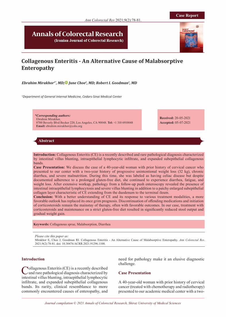
Figure 1: Duodenal biopsy: (A) Severe villous blunting with sloughing of the surface epithelium. (B) High-power view with expanded lamina propria and lymphoplasmacytic infiltrates. (C) Thick subepithelial collagen layer. (D) Trichrome stain.

Mirakhor E et al.
Ann Colorectal Res 2021;9(2)80
recognition of a celiac-like condition unresponsive to a gluten-free diet with the histopathologic hallmark of subepithelial collagen deposition (2). In cases reported thus far, CE shows a 2:1 female-to-male ratio and can affect individuals of any age (3, 4). Reported ages have ranged from 2 to 85 years, with the overwhelming majority occurring in the fifth decade of life (3-6).
Patients with CE often present with a nonspecific clinical picture with symptoms of nausea, bloating, watery diarrhea, and weight loss, with associated laboratory evidence of anemia and hypoalbuminemia (2, 4, 7). Due to clinical overlap with other enteropathies, diagnosis requires endoscopy with biopsy. Endoscopic findings suggestive of CE include diffuse mucosal edema, erythema, and granularity with flattening of villi and scalloping of mucosal folds (4, 6). Clinical severity is directly proportional to the extent of small bowel (especially duodenum and jejunum) involvement and not the severity of mucosal abnormality on biopsy (8).
From a prognostic standpoint, whereas historically CE has been associated with an intractable clinical course characterized by severe malnutrition necessitating parenteral support and ultimately leading to death, newer evidence suggests a heterogenous clinical course with more favorable outcomes (2, 5). In many studies, CE has also been associated with lymphocytic gastritis, collagenous gastritis, microscopic colitis, paraneoplastic syndromes, common variable immunodeficiency, T-cell lymphomas, and a host of other autoimmune conditions (6, 9-13).
The pathophysiology of CE remains largely unknown. Owing to their similar clinical presentation and shared susceptibility alleles (i.e. HLA DQ2 and DQ8), CE was previously thought to represent a refractory form of celiac spectrum disorders (3, 4, 14, 15). However, newer studies have demonstrated HLA DQ6/DQ9 positivity in those with CE in the absence of HLA DQ2/DQ8 (16). Various case series implicate angiotensin II receptor blockers (ARBs; most notably olmesartan), non-steroidal anti-inflammatory drugs (NSAIDs), and proton pump inhibitors (PPIs) as potential triggers of CE (11, 17-19). Taken together, this differentiates CE as a distinct entity arising from the interaction between genes and environmental exposures, with a possible HLA-mediated immune mechanism (4, 16). Furthermore, the absence of celiac-associated antibodies (i.e., anti-TTG, anti-gliadin, and anti-endomysial antibodies) in most cases of CE and lack of response to a gluten-free diet lends additional support to this notion. Interestingly, no associated triggers of CE were identified in this case and our patient continued to have refractory diarrhea despite adherence to a gluten-free diet.With a better understanding of CE and its
response to various treatment modalities, a more favorable outlook has replaced its once grim prognosis. Discontinuation of offending agents (ARBs, NSAIDs, PPIs) and initiation of corticosteroids remain the mainstay of therapy, often with favorable outcomes (4, 6, 7, 12, 19). When CE manifests secondary to a paraneoplastic syndrome, treatment of underlying malignancy
Figure 2: Jejunum/ileum biopsy: (A) Increased intraepithelial lymphocytes. (B) Thickened subepithelial collagen layer. (C) Ileum showing lymphocytosis. (D) Trichome stain for subepithelial collagen.

Collagenous enteritis induced malabsorption
http://colorectalresearch.sums.ac.ir/ 81
References
1. Schein J. Syndrome on non tropical sprue with hitherto undescribed lesions of the intestine. Gastroenterology. 1947;8(4):438-460.
2. Weinstein WM, Saunders DR, Tytgat GN, Rubin CE. Collagenous sprue--an unrecognized type of malabsorption. N Engl J Med. 1970;283(24):1297-1301.
3. Cuoco L, Villanacci V, Salvagnini M, Bassotti G. Collagenous sprue with associated features of refractory celiac disease. Rev Esp Enferm Dig. 2012;104(4):223-225.
4. Lan N, Shen B, Yuan L, Liu X. Comparison of clinical features, treatment, and outcomes of collagenous sprue, celiac disease, and collagenous colitis. J Gastroenterol Hepatol. 2017;32(1):120-127.
5. Karakus E, Ekinci O, Kirsaclioglu CT, Ozaydin E, Atakan C, Dursun A. A rare cause of protein losing enteropathy: collagenous sprue. Fetal Pediatr Pathol. 2015;34(2):133-135.
6. Zhao X, Johnson RL. Collagenous sprue: a rare, severe small-bowel malabsorptive disorder. Arch Pathol Lab Med. 2011;135(6):803-809.
7. Holdstock DJ, Oleesky S. Successful treatment of collagenous sprue with combination of prednisolone and gluten-free diet. Postgrad Med J. 1973;49(575):664-667.
8. Robert ME. Gluten sensitive enteropathy and other causes of small intestinal lymphocytosis. Semin
Diagn Pathol. 2005;22(4):284-294.9. Daniels JA, Lederman HM, Maitra
A, Montgomery EA. Gastrointestinal tract pathology in patients with common variable immunodeficiency (CVID): a clinicopathologic study and review. The American journal of surgical pathology. 2007;31(12):1800-1812.
10. Freeman HJ, Berean KW. Resolution of paraneoplastic collagenous enterocolitis after resection of colon cancer. Can J Gastroenterol. 2006;20(5):357-360.
11. Robert ME, Ament ME, Weinstein WM. The histologic spectrum and clinical outcome of refractory and unclassified sprue. The American journal of surgical pathology. 2000;24(5):676-687.
12. Vakiani E, Arguelles-Grande C, Mansukhani MM, et al. Collagenous sprue is not always associated with dismal outcomes: a clinicopathological study of 19 patients. Mod Pathol. 2010;23(1):12-26.
13. Cellier C, Delabesse E, Helmer C, et al. Refractory sprue, coeliac disease, and enteropathy-associated T-cell lymphoma. French Coeliac Disease Study Group. Lancet. 2000;356(9225):203-208.
14. Nielsen OH, Riis LB, Danese S, Bojesen RD, Soendergaard C. Proximal collagenous gastroenteritides: clinical management. A systematic review. Ann Med. 2014;46(5):311-317.
15. Yau AH, Xiong W, Ko HH. Collagenous enterocolitis manifesting as watery diarrhoea and iron-deficiency anaemia. BMJ Case Rep. 2015;2015.
16. Kung VL, Liu TC, Ma C. Collagenous Enteritis is Unlikely a Form of Aggressive Celiac Disease Despite Sharing HLA-DQ2/DQ8 Genotypes. The American journal of surgical pathology. 2018;42(4):545-552.
17. Desruisseaux C, Bensoussan M, Desilets E, et al. Adding Water to the Mill: Olmesartan-Induced Collagenous Sprue-A Case Report and Brief Literature Review. Can J Gastroenterol Hepatol. 2016;2016:4837270.
18. Nielsen JA, Steephen A, Lewin M. Angiotensin-II inhibitor (olmesartan)-induced collagenous sprue with resolution following discontinuation of drug. World J Gastroenterol. 2013;19(40):6928-6930.
19. Vasant DH, Hayes S, Bucknall R, Lal S. Clinical and histological resolution of collagenous sprue following gluten-free diet and discontinuation of non-steroidal anti-inflammatory drugs (NSAIDs). BMJ Case Rep. 2013;2013.
20. van Gils T, van de Donk T, Bouma G, van Delft F, Neefjes-Borst EA, Mulder CJ. The first cases of collagenous sprue successfully treated with thioguanine. BMJ Open Gastroenterol. 2016;3(1):e000099.
leads to clinical improvement (10). Refractory cases treated with azathioprine, thioguanine, or tumor necrosis factor-α inhibitors have also yielded
notable treatment responses (14, 20).
Conflicts of interests: None declared.