cold-active acid pectinolytic system from psychrotolerant ... · cold-active acidic bacterial...
TRANSCRIPT

495Am. J. Enol. Vitic. 64:4 (2013)
1Research Fellow; 2Researcher, Consejo Nacional de Investigaciones Cientí-ficas y Técnicas (CONICET); Facultad de Ciencias Aplicadas a la Industria, Universidad Nacional de Cuyo, Bernardo de Irigoyen 375 (5600) San Rafael, Mendoza, Argentina.*Corresponding author (email: [email protected]; tel/fax: +54-260-4421947/4430673)Acknowledgments: This research was supported by the following grants: CONICET–PIP N° 11220110100823; SECTyP–UNCuyo N° 06/L116 and I+D (UNCuyo) program; FONCyT–ANPCyT–MINCYT (PICT Bicentenario) Nº 2010-0847. The authors are grateful to Raúl R. Raya, Lucía M. Mendoza, and Mario D. Baigori for their advice on genetic identification.Publication costs of this article defrayed in part by page fees.Manuscript submitted Jan 2013, revised Jun 2013, accepted Jul 2013Copyright © 2013 by the American Society for Enology and Viticulture. All rights reserved.doi: 10.5344/ajev.2013.13002
Cold-Active Acid Pectinolytic System from Psychrotolerant Bacillus: Color Extraction from Red Grape Skin
María C. Martín1 and Vilma I. Morata de Ambrosini2*
Abstract: Cold-active enzymes are potentially relevant to food processing and, among them, pectinases belong to the most important enzymes in the fruit juice and wine industry. Bacillus sp. CH15 from grape berries was screened for the production of cold-active acidic pectinolytic enzymes. Taxonomic studies and 16S rDNA analysis revealed that the isolate was closely related to the Bacillus subtilis group. Maximum pectinolytic activity under acidic condi-tions (pH 5.0) and at low temperature (20°C) was obtained after 24 hr of incubation in medium containing citrus pectin as sole carbon source (0.305 U/mL). Polymethylgalacturonase activity was the predominant pectinase under the given assay conditions, whereas highest levels of pectate lyase activity were found at 60°C. At 5 and 10°C, the enzymatic system maintained 15 and 30% of the maximum activity, respectively. This is the first report on a pectinolytic enzyme system produced by a Bacillus strain and active at 20°C and pH 3.6, conditions similar to those in winemaking. According to classical and CIELab color parameters of short macerations with red grape skins at low temperature, the bacterial pectinolytic system produced a rapid color extraction and the macerates exhibited better chromatic characteristics than those obtained with commercial pectinases or after natural extraction. Total anthocyanin contents of the macerates with Bacillus enzyme system and after natural extraction were significantly different, whereas relative individual pigment quantities did not show any significant difference between the two treatments. In conclusion, Bacillus sp. CH15 could be used as a microbial source of cold-active acidic pectinases in red winemaking.
Key words: pectinolytic system, cold-active enzymes, Bacillus, polyphenols, anthocyanins, red grapes
Bioconversion of thermosensitive or volatile organic com-pounds and enzymatic food processing are preferably per-formed at low temperatures. In the winemaking process, the use of low temperatures (15 to 20°C) can increase the pro-duction and retention of volatile compounds, thus improving the aromatic profile of wines (Molina et al. 2007). However, color extraction in red wines diminishes at low temperatures, and therefore pectinolytic enzymes active at low temperature are required both for extraction and clarification. Although several cold-active enzymes have been identified and studied, there are only a few reports on cold-active pectinases (Cabeza et al. 2011, Merín et al. 2011, Birgisson et al. 2003).
Pectinases are enzymatic systems able to break down pec-tic polymers of plant cell walls. These enzymes comprise two main types: methylesterases, which remove methoxyl groups
from pectin, and depolymerases. The depolymerases include hydrolases and lyases, which cleave the bonds between ga-lacturonate units, the former by hydrolysis of the α-1,4 glyco-sidic bonds and the latter by β-elimination. Pectinases have a wide spectrum of industrial applications. Acidic pectinases are used in extraction and clarification of fruit juices and wines, whereas alkalophilic pectinases are mostly used in degumming and retting of fiber crops and pretreatment of pectic wastewater from fruit juice industries (Kashyap et al. 2001). The major sources of acidic pectinases are Aspergillus niger strains, whereas alkaline pectinases are mainly obtained from alkalophilic bacteria (Pedrolli et al. 2009).
Bacillus spp. are spore-forming Gram-positive bacteria that are preferred in industrial enzyme production. They produce exocellular hydrolytic enzymes, mainly pectinases but also proteases, amylases, and cellulases, and production time is less compared to fungi. In addition, some fungal strains are able to secrete mycotoxins as a secondary metabolite (Bleve et al. 2006) and undesirable enzymes such as β-glucosidases, which are able to hydrolyze anthocyanin compounds, produc-ing a loss of color in red wines (Romero-Cascales et al. 2008). Therefore, GRAS (“generally recognized as safe”) bacteria producing high levels of enzymes could be an interesting pec-tinase source for industrial applications. Furthermore, these enzymes can be easily cloned and overexpressed in other mi-croorganisms. Most of the pectinases reported in Bacillus are alkaline, especially pectate lyases (van Dyk et al. 2010, Zheng et al. 2012, Rehman et al. 2012). There are only a few studies on the production of acidic pectinases from bacteria. Soares et al. (2001) reported five Bacillus species that produced pec-tinases and their application on juice extraction, while our

496 – Martín and Morata de Ambrosini
Am. J. Enol. Vitic. 64:4 (2013)
research group described two Bacillus sp. strains that pro-duced pectinases active at acidic pH (Cabeza et al. 2011).
In the winemaking process, pectinases yield consider-able benefits: faster fermentation start, higher must yield, easier pressing, quicker and more complete clarification, and an important extraction of aromatic and polyphenol com-pounds from grape skins (Doco et al. 2007). Some of these polyphenols, anthocyanins, are the pigments responsible for the color of red grapes and young wines, while tannins are compounds with high antioxidant capacity and responsible for astringency and bitterness (Cheynier et al. 2006). Other polyphenols, especially flavonoids and stilbenes, have sev-eral beneficial physiological effects, mostly because of their antioxidant properties (Basli et al. 2012). The grape skin cell wall constitutes a barrier against diffusion of polyphenols. However, permeability to polyphenols can be increased by partial hydrolysis of their structural polysaccharides (pectin, hemicellulose, and cellulose), a process that can be facili-tated by maceration enzymes. In a previous study, a bacterial pectinolytic preparation was used in the maceration of red grape skins with very good results regarding extraction of both pigments and total polyphenols (Cabeza et al. 2009). In the current study, the aim was to characterize the pectinolytic system produced by Bacillus sp. CH15 and to evaluate its ef-fect on the extraction of pigments and total polyphenols from red grape skins during short macerations at low temperature in order to examine its potential as a cold-active acidic pec-tinase source.
Materials and MethodsMicroorganisms and culture conditions. All bacterial
strains were previously isolated from grape berries from San Rafael, an enological area in the province of Mendoza, Ar-gentina, and subsequently selected for their ability to produce pectinases active at low temperature (Cabeza et al. 2011). Bacterial growth and enzyme production were carried out in pectin medium according to Kobayashi et al. (2001) but with some modifications (w/v): 0.5% pectin from citrus peel (Sigma, St. Louis, MO), 0.5% soy peptone, 0.5% yeast extract, 0.1% K2HPO4, 0.02% MgSO4·7H2O, 0.005% MnSO4·6H2O, 0.005% CaCl2·2H2O and 50 mM acetate buffer (pH 5.0). All chemical reagents were of analytical grade.
Cultures were incubated at 30°C for 48 hr under agitation (100 rpm). Optical density at 560 nm was used to measure cellular growth. After cells were removed by centrifugation (5,000 × g, 15 min at 4°C), supernatants were filtered through a 0.22 μm pore size membrane. Cell-free supernatants were used to assay enzyme activity.
Phenotypic and genotypic identification of bacterial strains. To identify the strain with highest cold-active pec-tinase production, phenotypic and genotypic assays were performed using the following (biochemical) tests: Gram-staining, spore-staining, observation under a light micro-scope, mobility, Proskauer and catalase, anaerobic growth, development at different incubation temperatures and NaCl concentrations, polysaccharide hydrolysis, nitrate reduction, acid formation from glycogen, methyl α-d-mannoside and
starch, and use of glucose, xylose, mannitol, and propionate (Logan and De Vos 2009).
For genotypic characterization, the isolate was geneti-cally characterized according to identification of genes cod-ing for bacterial 16S rRNA. Previously, DNA was extracted according to the method described by Tewari et al. (2005). To amplify the variable region of the 16S rDNA gene the following universal single strand primers were used: 27f, 5´-AGAGTTTGATCMTGGCTCAG-3 ,́ M = C/A (forward) and 1492r, 5´-TACGGYTACCTTGTTACGACTT-3 ,́ Y = C/T (reverse). The extracted genomic DNA was amplified in 25 µL of reaction mixture containing 14.9 µL PCR water, 2.5 µL buffer 10x, 1 µL MgCl2 (50 mM), 0.2 µL of each primer (10 µM), 5 µL dNTPs, 0.2 µL Taq polymerase, and 1 µL DNA. Amplification consisted of an initial denaturation step at 95°C for 5 min, followed by 35 cycles at 95°C for 90 sec, primer union at 57°C for 90 sec, extension at 72°C for 2 min, and a final extension at 72°C for 2 min. The PCR products were separated electrophoretically on 0.8% agarose gels, which were run at 80 V for 2 hr. Band patterns were visualized with ethidium bromide and photographs were taken under UV light. All PCR reagents were from Promega (Madison, WI). The amplified product of the appropriate size (~1.5 kbp) was sequenced at CERELA (CERELA-CONICET, Tucumán, Argentina). 16S rRNA sequence data of Bacillus sp. CH15 reported in this article have been submitted to the GenBank nucleotide sequence database of the NCBI (National Center for Biotechnology Information). 16S rRNA homology was carried out using the BLAST (Basic Local Alignment Search Tool) website (http://blast.ncbi.nlm.nih.gov) and a phyloge-netic tree was constructed using the neighbor-joining method (Tamura et al. 2004).
Enzyme assays. Total pectinolytic activity was assayed by quantification of reducing sugars released from a pectin dispersion (0.25% citrus pectin in 50 mM acetate buffer, pH 5.0) using 3,5-dinitrosalicylic acid (DNS) (Miller 1959). d-(+)-Galacturonic acid was used as standard (Sigma). Reac-tion mixtures contained 0.45 mL of substrate and 0.05 mL of enzymatic extract and were incubated at corresponding temperatures for 30 min. The reaction was stopped by add-ing 0.5 mL of 1% DNS reagent. After cooling down, 1.5 mL distilled water was added and absorbance was measured at 530 nm. One unit of pectinolytic activity (U) was defined as the amount of enzyme required to release 1 μmol of reducing sugar per minute under the given assay conditions. All assays were carried out in triplicate.
Polygalacturonase activity was determined with polyga-lacturonic acid (Sigma) according to Miller (1959).
Pectin lyase and pectate lyase activity were determined spectrophotometrically at 235 nm by monitoring the forma-tion of unsaturated products from pectin and polygalacturonic acid, respectively (Collmer et al. 1988). One unit of lyase ac-tivity (U) was defined as the amount of enzyme that produced 1 μmol of unsaturated product per minute under the given assay conditions. Molar extinction coefficients of galacturonic acid and unsaturated pectin were 4,600 L/mol*cm and 5,500 L/mol*cm, respectively.

Cold-Active Acidic Bacterial Pectinases for Winemaking – 497
Am. J. Enol. Vitic. 64:4 (2013)
Pectinesterase activity was measured by titration of the carboxylic groups released continuously from 0.5% pectin dis-persion in 0.1 M NaCl adjusted to pH 7.5 with 0.5 M NaOH (Moyo et al. 2003). One unit of enzymatic activity (U) was defined as the amount of enzyme required to release 1 μmol of –COOH groups per minute under the given assay conditions.
Xylanase and cellulase activity were assayed with Birch-wood xylan and microgranular cellulase substrates, respec-tively. Substrates were prepared in 50 mM buffers at an ap-propriate pH and reactions were performed according to the standard method by Miller (1959), using xylose or glucose as standards.
β-Glucosidase activity was determined by quantification of glucose released from a 0.5% cellobiose solution using an enzymatic kit (Ghose 1987). One unit of β-glucosidase activ-ity (U) was defined as the amount of enzyme that released 2 μmol of glucose from cellobiose per minute under the given assay conditions.
All enzyme activities described above were assayed under the following two conditions: 50°C/pH 6.0 (50 mM acetate buffer) and 20°C/pH 3.6 (50 mM citrate buffer) (resembling winemaking conditions).
Temperature, pH, and pectinolytic activity. The effects of temperature and pH were assayed using a one variable at a time approach. The temperature effect on the total pecti-nolytic activity was determined assaying different tempera-tures between 5 and 70°C according to standard assay pro-cedures (Miller 1959). Relative activities are expressed as the percentages of the ratio of the activity obtained at a certain temperature and the maximum activity obtained within the temperature range assayed. Thermostability was assayed by measuring the residual activity after incubation of the enzy-matic extract at different temperatures for 30 min. Pectino-lytic activity was determined at pH 3.6 and 20°C (resembling winemaking conditions).
The effect of pH on the enzymatic activity was determined at 20°C by assaying total pectinolytic activity at different pH values ranging from 3.0 to 9.0. The following buffers (50 mM) were used: citrate (from pH 3.0 to 6.0), phosphate (pH 7.0 and 8.0), and Tris-HCl (pH 9.0). Results are expressed as relative activity compared with the highest activity at the different pH values assayed.
Extraction of pigments and polyphenols from grape skins. Extraction of pigments and polyphenols was carried out with Malbec grapes (Vitis vinifera) from San Rafael, southern Mendoza, Argentina. Berries in good sanitary con-ditions (2013 vintage) were harvested at optimum ripeness (25.2% w/v reducing sugar, 5.70 g/L total acidity, pH 3.65). Only the skins were used for analyses.
Grapes were manually destemmed and crushed and skins were separated from the pulp and seeds. Skins were split and unfolded to expose the internal part where the color material is located to increase the contact area for extraction. The frag-mented skins (1.65 g) were introduced into test tubes and sup-plemented with 2.5 mL extraction solution, which contained increasing doses (50, 100, 150, 200, and 250 µL) of cell-free B. sp. CH15 supernatant obtained under standard conditions
in 50 mM citrate buffer (pH 3.6). Reaction blanks without enzyme (natural extraction) were assayed simultaneously. In addition, Extrazyme and Inozyme Terroir commercial pectin-ases (Institut Oenologique de Champagne, Épernay, Marne, France) were used as control, at concentrations indicated by the manufacturer (corresponding to total pectinolytic activ-ity of 1757 and 2516 U/g, respectively). Skins and extraction solution were combined and incubated at 20°C under shaking conditions (130 rpm) for 2 hr. All macerations were carried out in triplicate.
Macerates were centrifuged at 10,000 x g for 10 min and absorbances were measured using a LAMBDA 25 UV/Vis spectrophotometer (PerkinElmer, Waltham, MA), with 0.1 cm path-length glass cells. All data were converted to a path length of 1 cm for calculations.
The color index (CI) was calculated as the sum of absor-bances at 420 nm, 520 nm, and 620 nm, and the shade as the ratio between 420 and 520 nm, according to Glories (1984) and Sudraud (1958). To calculate the CIELab parameters, the whole visible spectrum (380 to 780 nm) was recorded, and the illuminant D65 and 10° observer were used in the calculus. Lightness (L*), chroma (C*), red color intensity (a*), and yel-low color intensity (b*) parameters were obtained using UV WinLab V5 software (PerkinElmer). The CIELab differences (ΔEr,s*) were calculated using the following equation:
ΔEr,s* = [(L* – L*NE)2 + (a* – a*NE)2 + (b* – b*NE)2]1/2
where the subscript (NE) corresponds to natural extraction.Total polyphenol content was quantified using the Folin-
Ciocalteu method with gallic acid as standard (Singleton and Rossi 1965). Total anthocyanins were measured according to the method by Puissant-Léon (Cayla et al. 2002), which consisted in the absorbance at 520 nm after incubation of a wine sample for 30 min in 0.1 N HCl (1:40 ratio, v/v).
HPLC analysis of anthocyanins. HPLC analysis of an-thocyanic compounds was carried out on a Shimadzu (Kyoto, Japan) LC10 HPLC chromatograph, equipped with a SPD-M10Avp UV/Vis Photodiode Array Detector, fitted with Shimadzu software. Macerate samples, previously filtered through a 0.45 μm pore size membrane, were run at a constant temperature of 40°C on a LiChrospher 100 RP-18 reversed-phase column (4.6 mm x 250 mm, 5 μm particle size) (Merck, Darmstadt, Germany) equipped with a precolumn (RP-18; 2 mm x 20 mm, 30 to 40 μm particle size). Two elution sol-vents were used as mobile phase: water/formic acid/acetoni-trile (87:10:3) (A) and water/formic acid/acetonitrile (40:10:50) (B). The flow rate was fixed at 0.8 mL/min with the following linear gradients: from 6 to 30% B in 15 min, from 30 to 50% B in 15 min, from 50 to 60% B in 5 min, and from 60 to 6% B in 6 min, followed by washing and reconditioning of the column. UV-visible spectra (scanning from 200 to 600 nm) were recorded for all peaks. The different anthocyanic com-pounds were identified using authentic standards and by com-parison of the retention times and spectra with those found in the literature (Revilla and Ryan 2000, Vivar-Quintana et al. 2002). Quantification of anthocyanins was based on peak areas at 518 nm. Relative amounts of individual anthocyanins

498 – Martín and Morata de Ambrosini
Am. J. Enol. Vitic. 64:4 (2013)
are expressed as percentages (%) of total anthocyanins, and consequently the total peak area was used to calculate the relative area for individual peaks. HPLC-DAD analysis was performed with a representative sample after each treatment.
Statistical analysis. ANOVA and multiple range tests were applied to analyze all results, using Statgraphics Plus 5.1 software (Manugistics, Rockville, MD). Differences between means were considered statistically significant when p < 0.05 (95.0% confidence level).
Results and DiscussionMicroorganisms producing cold-active pectinase. Mi-
croorganisms showing pectinolytic activity were previously isolated from grape berry surfaces (Cabeza et al. 2011). Grapes represent a natural reservoir of diverse microorgan-isms that may affect production and storage of wine. It is particularly common to find microorganisms that are able to secrete hydrolytic enzymes. From the 400 pectinolytic isolates reported by Cabeza et al. (2011), seven bacterial strains were selected for the present study because of their ability to produce cold-active acidic pectinases with pectin as substrate. All microorganisms belonged to the Bacillus genus according to their phenotypic profile: Gram-positive, catalase-positive, aerobic, motile, spore-forming, and rod-shaped bacteria. Strains producing pectinolytic enzymes ac-tive at low temperature and low pH are required in the wine-making process to improve the aromatic profile and quality of wines. In order to select the most effective cold-active pectinase-producing strain among the isolates, enzymatic activity was assayed at low temperature (15°C) and at pH 5.0 and compared with activity at 30°C (Figure 1). Maxi-mum pectinolytic activity at 15°C was found with isolate CH15 (0.220 U/mL), which was the only strain that showed significantly higher activity at low temperature than at 30°C (typical temperature for acidic pectinases reported in the lit-erature). At 15°C, all other isolates showed a relative activity between 32 and 70% compared with the maximum activity
observed, and consequently, isolate CH15 was selected for further studies.
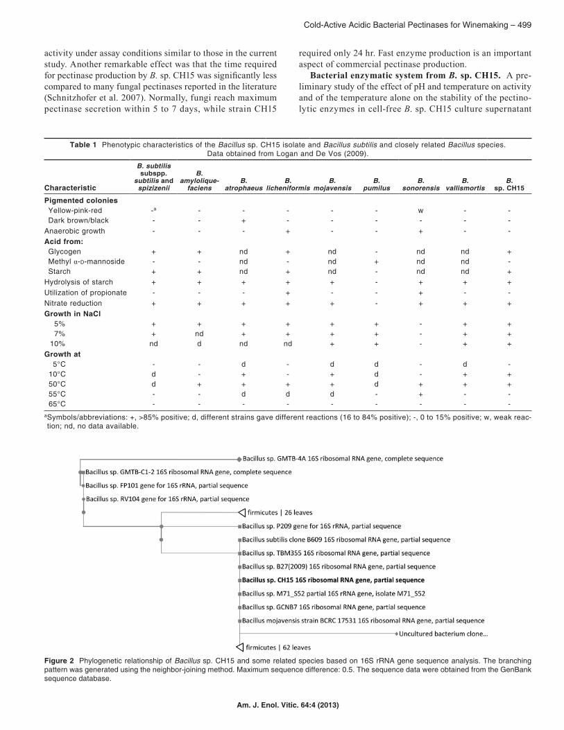
To identify the selected strain, a phenotypic profile was performed according to Logan and De Vos (2009). Some morphological and physiological characteristics of the iso-late and differential characteristics of Bacillus subtilis and closely related Bacillus species are given (Table 1). According to comparative analyses, CH15 showed greatest similarity with B. subtilis subspp. subtilis and spizizenii.
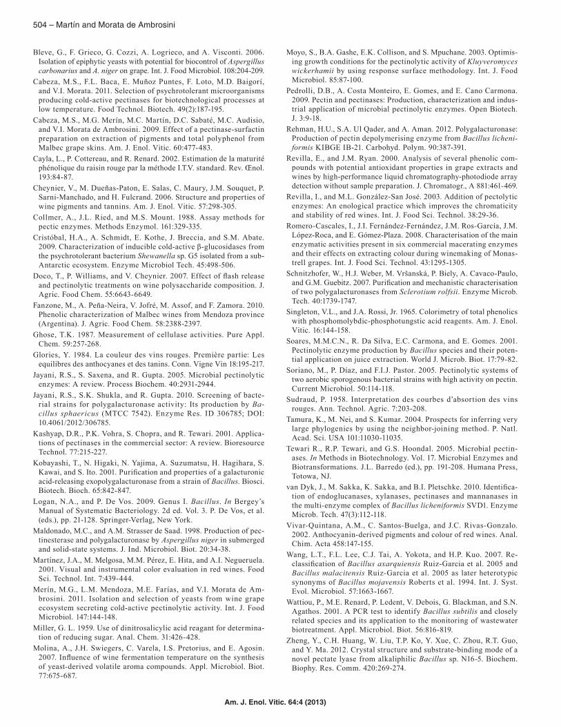
To define the classification, the 16S rDNA sequence (1,390 nucleotides) was almost completely determined and submitted to the GenBank nucleotide sequence database under acces-sion number JF340155 (http://www.ncbi.nlm.nih.gov/nuccore/JF340155). From this 16S rDNA sequence, a phylogenetic tree was constructed using the neighbor-joining method (Figure 2). According to this analysis, CH15 should be classified with-in the Bacillus genus, with species of the B. subtilis group as the closest related species with a 16S rDNA sequence similar-ity of 99%. The Bacillus subtilis group includes the following taxa: B. subtilis subsp. subtilis, B. licheniformis, B. amylo-liquefaciens, B. atrophaeus, B. mojavensis, B. vallismortis, B. subtilis subsp. spizizenii, B. sonorensis, B. velezensis, B. axarquiensis, and B. malacitensis (Wang et al. 2007). Wattiou et al. (2001) also included B. pumilus. This B. subtilis group has the advantage that all strains are nonpathogenic bacteria and they are considered GRAS; consequently, the strains or their metabolites could be safely used in the food industry. However, further studies are necessary to identify the strain at species level.
Secretion of pectinases by Bacillus sp. CH15. The rela-tionship between B. sp. CH15 growth and pectinase production was studied in pectin medium at 30°C for 48 hr under shak-ing conditions (100 rpm). Maximal growth (1.2, OD600nm) was observed after 24 hr of incubation (Figure 3). After this period growth slightly decreased, suggesting cell death and lysis.
In view of the potential application of the culture super-natant as an enzymatic preparation in the winemaking pro-cess, pectinolytic activity was assayed at acidic pH and 20°C. Pectinase production increased immediately, accompanied by cell growth, reaching its maximum after 24 hr of incubation (0.305 ± 0.013 U/mL), at which point the strain had already reached the stationary phase. After 24 hr, pectinolytic activ-ity decreased significantly, probably due to protease activ-ity. Similar results have been found for other Bacillus strains (Jayani et al. 2010).
Maximum pectinolytic activity obtained with B. sp. CH15 was higher than that reported in other Bacillus sp. strains, determined under similar assay conditions (Soriano et al. 2005). Pectinolytic activity was even higher than that re-ported for some strains belonging to Aspergillus niger, the major acidic pectinase-producing microorganism (Blandino et al. 2001, Maldonado and Strasser de Saad 1998); these authors observed levels close to 0.050 U/mL. Jayani et al. (2010) reported acceptable polygalacturonase activity (4.5 U/mL) at acidic pH and at 30°C for Bacillus sphaericus, but op-timal activity was observed at neutral pH. To date, there are very few reports on pectinases from bacterial strains showing
Figure 1 Pectinolytic activity of cell-free supernatants from bacterial isolates at pH 5.0 and at 30°C or 15°C (means ± SD, n = 3). One unit of pectinolytic activity (U) was defined as the amount of enzyme required to liberate 1 μmol of reducing sugar per minute under the given assay conditions (see text).
Cold-Active Acidic Bacterial Pectinases for Winemaking – 499
Am. J. Enol. Vitic. 64:4 (2013)
activity under assay conditions similar to those in the current study. Another remarkable effect was that the time required for pectinase production by B. sp. CH15 was significantly less compared to many fungal pectinases reported in the literature (Schnitzhofer et al. 2007). Normally, fungi reach maximum pectinase secretion within 5 to 7 days, while strain CH15
required only 24 hr. Fast enzyme production is an important aspect of commercial pectinase production.
Bacterial enzymatic system from B. sp. CH15. A pre-liminary study of the effect of pH and temperature on activity and of the temperature alone on the stability of the pectino-lytic enzymes in cell-free B. sp. CH15 culture supernatant
Figure 2 Phylogenetic relationship of Bacillus sp. CH15 and some related species based on 16S rRNA gene sequence analysis. The branching pattern was generated using the neighbor-joining method. Maximum sequence difference: 0.5. The sequence data were obtained from the GenBank sequence database.
Table 1 Phenotypic characteristics of the Bacillus sp. CH15 isolate and Bacillus subtilis and closely related Bacillus species. Data obtained from Logan and De Vos (2009).
Characteristic
B. subtilis subspp.
subtilis and spizizenii
B. amylolique-
faciensB.
atrophaeusB.
licheniformisB.
mojavensisB.
pumilusB.
sonorensisB.
vallismortisB.
sp. CH15
Pigmented coloniesYellow-pink-red -a - - - - - w - -Dark brown/black - - + - - - - - -
Anaerobic growth - - - + - - + - -Acid from:Glycogen + + nd + nd - nd nd +Methyl α-d-mannoside - - nd - nd + nd nd -Starch + + nd + nd - nd nd +
Hydrolysis of starch + + + + + - + + +Utilization of propionate - - - + - - + - -Nitrate reduction + + + + + - + + +Growth in NaCl
5% + + + + + + - + +7% + nd + + + + - + +
10% nd d nd nd + + - + +Growth at
5°C - - d - d d - d -10°C d - + - + d - + +50°C d + + + + d + + +55°C - - d d d - + - -65°C - - - - - - - - -
aSymbols/abbreviations: +, >85% positive; d, different strains gave different reactions (16 to 84% positive); -, 0 to 15% positive; w, weak reac-tion; nd, no data available.

500 – Martín and Morata de Ambrosini
Am. J. Enol. Vitic. 64:4 (2013)
was carried out (Figure 4). A temperature profile of total pectinolytic activity at pH 5.0 showed a maximum at 60°C, optimum temperature, and two other peaks at 20 and 35°C. These results suggest that there was most likely more than one pectinolytic enzyme present in the crude extract. That can be expected because the enzyme activity assayed com-prised various pectin-degrading enzymes that together con-stitute the pectinolytic system. Other authors have reported similar results with a multienzyme complex from Bacillus licheniformis (Van Dyk et al. 2010). While 60°C has been reported previously as the optimum temperature for Bacil-
lus pectinases, bacterial pectinases with a low-temperature optimum have not yet been described.
Thermostability of the B. sp. CH15 pectinolytic system was determined by incubating samples of cell-free culture su-pernatant at a particular temperature for 30 min and at pH 5.0, followed by measuring activity under conditions resembling those in winemaking (20°C and pH 3.6). The pectinolytic system started to lose activity at 30°C and became totally inactive at 50°C (Figure 4), suggesting that it is composed of thermo-sensitive enzymes.
According to these results, the system had important en-zymatic activity at pH 5.0 and at low temperature (a relative activity of about 80, 30, and 15% at 20, 10, and 5°C, respec-tively, compared to maximum activity observed at 60°C) and was inactive at 50°C. This latter feature is very useful for stopping enzyme activity after the processing stage for pro-cesses carried out at low temperature.
A pH profile for total pectinolytic activity at 20°C also dis-played two peaks: at pH 4.0 and 6.0. To our knowledge, there are no previous reports on pectinases from Bacillus strains with activity at low pH. Most of the Bacillus pectinases are alkaline with optimal activity at pH values between 7.0 and 11.0 and usually at temperatures ranging from 40 to 70°C (Pedrolli et al. 2009, Jayani et al. 2005). Features of the differ-ent pectinolytic enzymes that constitute the crude extract are shown (Figure 4), but it is necessary to assay individual en-zymes to reveal any differences in properties and to select the most appropriate enzyme for its application in winemaking.
The pectinolytic system produced by B. sp. CH15 was ac-tive at low pH (3.0 to 6.0) and at low temperatures (from 5°C), a requisite for the processing of most fruit juices and wine. This feature may be attributed to the source where the microorganism was isolated, to the grape, which has an acidic pH, and to the climate of the sampling region (aver-age annual temperatures range from 10 to 22°C), suggesting adaptation of the strain to these conditions. There has been recent growing interest in cold-active enzymes for their ap-plication in food processing. Most studies have focused on research on cold-adapted microorganisms; numerous psy-chrophilic and psychrotolerant microorganisms isolated from cold regions, such as glaciers and sub-Artic and sub-Antarctic regions, have been studied (Cristóbal et al. 2009, Birgisson et al. 2003). However, in the present work, we report a cold-active enzymatic system produced by a microorganism, iso-lated from a temperate region under appropriate selection pressure conditions, as informed previously by Cabeza et al. (2011). Our findings would suggest that even in regions with-out extreme temperatures, microorganisms may acquire the ability to hydrolyze substances at low temperature during cold seasonal periods. In addition, since the microorganism is mesophilic, the culture conditions offer considerable benefits to industrial enzyme production.
Characterization of the individual enzymes of the pecti-nolytic system of the B. sp. CH15 isolate was carried out. Enzymatic activity was determined in the culture superna-tant under different conditions: 50°C/pH 6.0 and 20°C/pH 3.6 (similar to winemaking conditions) (Table 2). Highest
Figure 3 Bacillus sp. CH15 growth curve and pectinase production. Culture medium (250 mL pectin medium) with 2% of B. sp. CH15 in-oculum was incubated for 48 hr at 30°C under shaking conditions (100 rpm) and 2 mL samples were taken periodically. Optical density was at 560 nm; pectinolytic activity was determined with citrus pectin at 20°C and pH 5.0 (means ± SD, n = 3).
Figure 4 Effect of pH (○) and temperature (●) on pectinolytic activity and effect of temperature (--∆--) on enzyme stability. Assays were car-ried out in cell-free supernatant from a B. sp CH15 culture. To determine the effect of the pH on pectinolytic activity, the reaction was carried out at 20°C for 30 min at different pH values, using buffers 50 mM sodium citrate (pH 3.0 to 6.0), 50 mM potassium phosphate (pH 7.0 and 8.0) and 50 mM Tris-HCl (pH 9.0). Maximum activity (100%) was 0.330 U/mL. The effect of temperature on activity was determined by performing the reaction at different temperatures ranging from 5 to 70°C in 50 mM potassium acetate buffer at pH 5.0. Maximum activity (100%) corre-sponded to 0.420 U/mL. Stability was also assayed, determining residual activity under conditions resembling winemaking (20°C and pH 3.6) after incubation at different temperatures for 30 min (means ± SD, n = 3).

Cold-Active Acidic Bacterial Pectinases for Winemaking – 501
Am. J. Enol. Vitic. 64:4 (2013)
enzyme activity corresponded to pectate lyase and was ob-served at 50°C/pH 6.0 (1.827 ± 0.115 U/mL). However, this activity was not detected under conditions resembling wine-making (20°C and pH 3.6). Significant levels of pectin lyase and polygalacturonase activity were also detected at 50°C/pH 6.0. Polymethylgalacturonase activity was significantly higher under conditions resembling winemaking (0.246 ± 0.060 U/mL) than at 50°C/pH 6.0. These results are consistent with those found in the literature, indicating that bacterial lyase enzymes, especially pectate lyase, have optimum activity at neutral or alkaline pH and usually at temperatures ranging from 40 to 70°C, as mentioned before. In contrast, polymeth-ylgalacturonase enzymes generally show optimum activity at acidic pH (from 3.5 to 5.0) and at lower temperatures (from 30 to 50°C) (Jayani et al. 2005). Pectinesterase activity was not detected under either of the conditions assayed. Pectines-terases release methanol as one of the reaction products, and hence pectinolytic preparations used in winemaking should only contain very limited levels of this enzyme.
Pectinolytic enzymes active at a pH similar to that of grapes and wines (pH 3.5 to 4.5) are required to hydrolyze pectic polymers of the grape skin cell wall and release pig-ments and aromatic compounds. Additionally, commercial pectinolytic preparations used in winemaking contain other enzyme activities called secondary activities, which can in-clude cellulases and hemicellulases. The latter enzymes show a synergistic effect with pectinases, facilitating degradation of polysaccharides of grape skin cell walls. Cellulases and hemicellulases hydrolyze cellulose and xylan, respectively, which, together with pectin, are the major constituents of plant cell walls. Bacillus sp. CH15 produced xylanases and cellulases under standard conditions. Xylanase activity was significantly higher at 50°C/pH 6.0 than at 20°C/pH 3.6, whereas cellulase activity did not show any significant dif-ference between the two assay conditions. β-Glucosidase activity was not detected in the crude enzyme extract. This enzyme partially hydrolyzes anthocyanin compounds, the main pigments of red wines, which leads to a reduction in the color intensity. Hence, a lack of this enzyme would be
beneficial to the application of the pectinolytic system in color extraction from red grapes.
Effect of pectinolytic system on extraction of pigments and polyphenols. Removal of pigments and polyphenols from red grape skins by the bacterial pectinolytic system was assayed during short macerations. Assaying different doses of B. sp. CH15 cell-free supernatant obtained under standard conditions (data not shown), revealed an optimum dose of 150 µL of cell-free supernatant in the reaction mixture (2.50 mL total volume), corresponding to 0.00836 U/mL of reac-tion mixture. The effect of the bacterial pectinolytic system at this concentration on color and pigments extraction from red grape skins was studied at pH 3.6 and 20°C. Simultane-ously, a comparative assay with two commercial pectinases, Extrazyme and Inozyme Terroir, was carried out under the same assay conditions. While the former is a pectinolytic preparation especially designed to favor quick extraction of phenolic compounds in red winemaking, the second com-mercial product has been designed for clarification of white musts. Nevertheless, the latter was selected too because it has been shown that some clarification enzymes also increase extraction of phenolic compounds (Revilla and González-San José 2003), which would allow a better comparison.
Natural extraction (without pectinases) produced a col-or index (CI) of 6.24 ± 0.77 and a shade of 0.577 ± 0.032 (Table 3). Total polyphenol content (TPC) was 672.0 ± 17.6 mg GAE/L and anthocyanins totaled 268.0 ± 24.1 mg/L. Color index and anthocyanin concentration were signifi-cantly higher with the bacterial pectinolytic system than with natural extraction or commercial pectinases, achieving levels of 8.22 ± 0.19 and 316.2 ± 25.5 mg/L, respectively. No significant differences were observed in shade between the bacterial pectinolytic system and natural extraction or the Extrazyme commercial pectinase. The highest shade value was obtained for Inozyme Terroir commercial pectinase, indicating a higher extraction of yellow than red pigments. TPC increased ~6% with the bacterial pectinolytic system compared to natural extraction, but the highest level was observed with Extrazyme (725.0 ± 17.6 mg GAE/L), with an increase of ~9%. These results would suggest that the CH15 pectinolytic system produced a high extraction of polyphe-nols, particularly colored compounds (anthocyanins), while Extrazyme selectively extracted certain polyphenols, other than anthocyanins. Consequently, extraction with this com-mercial enzyme did not contribute much to color, which is in agreement with the low levels of anthocyanins and color index observed with this enzyme.
CIELab coordinates were also determined. Lightness (L*) with the bacterial pectinolytic system was significantly lower than that after natural extraction and extraction with Ino-zyme Terroir commercial pectinase, indicating that the first macerate was darker and had a more vivid color. C* and a* (red color intensity) parameters with the bacterial pectino-lytic system significantly increased compared with the other samples. This result indicates a more efficient extraction of red pigments, which could be attributed to the higher concen-tration of anthocyanic compounds. No significant difference
Table 2 Enzyme activity in cell-free supernatant of the Bacillus sp. CH15 bacterial culture and assayed under two pairs
of conditions: 50°C/pH 6.0 and 20°C/pH 3.6 (resembling winemaking conditions).
Enzymatic activity (U/mL)a
Enzyme 20°C/pH 3.6 50°C/pH 6.0Polymethylgalacturonase 0.246 ± 0.060 a 0.175 ± 0.050 bPolygalacturonase 0.057 ± 0.005 a 0.165 ± 0.020 bPectin lyase 0.177 ± 0.050 a 0.431 ± 0.030 bPectate lyase nd 1.827 ± 0.115Pectinesterase nd ndCelullase 0.032 ± 0.005 a 0.055 ± 0.005 aXylanase 0.038 ± 0.005 a 0.347 ± 0.103 bβ-Glucosidase nd ndaValues are means ± standard deviation (n = 3). Different letters within each row indicate significant differences among samples (p < 0.05). nd: not detected.

502 – Martín and Morata de Ambrosini
Am. J. Enol. Vitic. 64:4 (2013)
was observed for the b* parameter (yellow color intensity) among the samples.
The difference in color between macerates with the bacte-rial pectinolytic system and natural extraction, ΔEr,s = ((L* – L*NE)2 + (a* – a*NE)2 + (b* – b*NE)2 )1/2, was 8.37 CIELab units. This value is visually detectable, because it is above the threshold for the human eye regarding distinguishing the color of wines, when trained tasters use standardized wine-tasting glasses (2.7 CIELab units; Martínez et al. 2001). A difference of 0.60 CIELab units was obtained for the Inozyme Terroir, compared to 2.74 CIELab units (threshold) for Extra-zyme. The small difference in color obtained with Inozyme Terroir indicates that this enzyme preparation is not suitable
Table 3 Effect of the B. sp. CH15 enzymatic system and two commercial pectinases, Inozyme Terroir and Extrazyme, on extraction of pigments and polyphenols from red grape skins. Grape skins were incubated at 20°C with shaking (130 rpm) for 2 hr with extraction
solutions (50 mM sodium citrate buffer, pH 3.6), supplemented with pectinolytic enzymes (except for natural extraction).
Parametera Natural extraction Bacillus sp. CH15b Inozyme Terroir ExtrazymeColor index 6.24 ± 0.77 ac 8.22 ± 0.19 b 6.30 ± 0.24 a 6.68 ± 0.68 aShade 0.577 ± 0.032 ab 0.508 ± 0.040 a 0.614 ± 0.053 b 0.585 ± 0.030 abTPC (mg GAE/L) 672.0 ± 17.6 ab 708.2 ± 16.9 bc 656.3 ± 4.1 a 725.0 ± 17.6 cAnthocyanins (mg/L) 268.0 ± 24.1 a 316.2 ± 25.5 b 265.8 ± 20.5 a 264.9 ± 50.2 aL* 81.3 ± 2.2 b 77.2 ± 1.9 a 81.2 ± 0.6 b 79.2 ± 0.5 abC* 21.5 ± 1.5 a 28.7 ± 2.4 b 21.3 ± 1.4 a 23.0 ± 3.4 aa* 21.4 ± 1.4 a 28.6 ± 2.3 b 21.3 ± 1.4 a 23.0 ± 3.4 ab* -0.86 ± 0.08 a -1.97 ± 0.6 a -0.29 ± 0.10 a -0.12 ± 0.10 aΔEr,s (CIELab units) - 8.37 0.60 2.74aTPC (mg GAE/L): total polyphenol content (mg gallic acid equivalent/L). L*: lightness. C*: chroma. a*: red color intensity. b*: yellow color in-tensity. ∆Er,s = ((L* – L*NE)2 + (a* – a*NE)2 + (b* – b*NE)2)1/2; NE = natural extraction. CIELab value of 2.7 units is the threshold for the human eye to distinguish the color of wines, when trained tasters use standardized winetasting glasses (Martínez et al. 2001).
bCell-free supernatant from a 24-hr B. sp. CH15 culture obtained in pectin medium at 30°C under shaking conditions (100 rpm).cValues are means ± standard deviation (n = 3). Different letters within each row indicate significant differences among samples (p < 0.05).
Figure 5 Chromatographic profile of anthocyanins in macerates of grape skins supplemented with the CH15 pectinolytic system. Peak identification: 3-glucoside compounds of delphinidin (1), cyanidin (2), petunidin (3), peonidin (4), and malvidin (5); 3-acetyl-glucoside compounds of peonidin (6) and malvidin (7); and 3-p-coumaroyl-glucoside compounds of peonidin (8) and malvidin (9).
for color extraction, which is in agreement with the nature of this preparation (clarification enzyme). The minor effect shown by Extrazyme commercial pectinase could be due to the fact that this preparation is not specifically designed for extraction of color at low temperature (20°C), a condition assayed in this study.
The composition of the major anthocyanins in the mac-erates was analyzed with HPLC-DAD, and the chromato-graphic profile of anthocyanins in macerates of grape skins supplemented with the CH15 pectinolytic system and mea-sured at 518 nm is shown (Figure 5). Nine different peaks were identified and assigned to 3-glucoside compounds of delphinidin (1), cyanidin (2), petunidin (3), peonidin (4), and

Cold-Active Acidic Bacterial Pectinases for Winemaking – 503
Am. J. Enol. Vitic. 64:4 (2013)
Table 4 Relative individual anthocyanin quantities in macerates of grape skins supplemented with pectinolytic enzymes and after natural extraction. The relative area of individual peaks was calculated as the percentage of the total area. Data are the result of
HPLC-DAD analysis of a representative sample after each treatment.
Relative area (%)Peak Compound Natural extraction Bacillus sp. CH15 Inozyme Terroir Extrazyme
1 Delphinidin-3-glucoside 2.37 2.22 1.66 1.422 Cyanidin-3-glucoside 0.72 0.20 0.35 0.403 Petunidin-3-glucoside 5.42 5.89 4.22 3.904 Peonidin-3-glucoside 7.61 4.54 5.65 6.125 Malvidin-3-glucoside 69.96 73.46 73.52 74.096 Peonidin-3-acetyl-glucoside 0.86 0.65 0.64 0.687 Malvidin-3-acetyl-glucoside 9.24 9.15 10.15 9.848 Peonidin-3-p-coumaroyl-glucoside 0.37 0.32 0.28 0.279 Malvidin-3-p-coumaroyl-glucoside 3.45 3.57 3.53 3.29
Total glycosylated 86.07 86.31 85.41 85.92Total acetylated 10.11 9.80 10.79 10.52Total coumaroylated 3.82 3.89 3.80 3.56
malvidin (5), 3-acetyl-glucoside compounds of peonidin (6) and malvidin (7), and 3-p-coumaroyl-glucoside compounds of peonidin (8) and malvidin (9). All macerates presented similar anthocyanin patterns, and the different enzymatic treatments did not have a selective effect on any of the an-thocyanins identified, compared to natural extraction.
The relative anthocyanin amounts (% of total anthocya-nins determined) in macerates of grape skins supplemented with pectinolytic enzymes and after natural extraction were determined (Table 4). Nonacylated glucosides formed the most abundant group of pigments in all macerates (~86%), compared with acylated forms. The acylated forms showed a higher proportion of acetylglucosides (~10%) than coumar-oylglucosides (~4%), which is in agreement with findings for other Malbec wines from Argentina (Fanzone et al. 2010).
Of the anthocyanins identified in the current study, malvi-din-3-glucoside was the most dominant pigment in all mac-erates, with a relative amount between 69.9 and 74.1%, and malvidin-3-acetyl-glucoside was the second most abundant compound, with a relative quantity between 9.1 and 10.1%. The relative malvidin-3-glucoside content increased 3.5% af-ter extraction with the bacterial pectinolytic system compared with natural extraction, while extraction with Extrazyme and Inozyme Terroir showed an increase of 4.1 and 3.6%, respectively. However, according to the total anthocyanin concentration (Table 3), extraction with the commercial en-zyme preparations was not high enough to produce significant effects on the color index or C* parameters. The higher total anthocyanin extraction produced with the bacterial pectino-lytic system is in agreement with the significantly increased color index and C* values observed with this enzyme. On the other hand, relative contents of petunidin and peonidin glyco-sides, the third most abundant compounds, were lower after treatment with enzymes than after natural extraction, which is in agreement with results previously reported (Cabeza et al. 2009), except petunidin-3-glucoside, which slightly increased with the bacterial pectinolytic system.
According to our results, the bacterial pectinolytic sys-tem allows a rapid extraction of anthocyanin compounds,
resulting in musts with better chromatic characteristics than those obtained with commercial pectinases. Consequently, the use of this cold-active pectinolytic system compensates the limited color extraction in macerations of red grape skins conducted at low temperature. Further experiments under real vinification conditions are currently being carried out to confirm these effects and to propose the potential use of this pectinolytic preparation to improve color extraction in red winemaking at low temperature.
ConclusionsIn the present study, a Bacillus sp. strain was able to se-
crete a multienzyme system with pectinolytic activity at low temperature and under acidic conditions. In addition, it se-creted other polysaccharidases that showed a synergistic ef-fect with pectinases, facilitating degradation of grape skin cell walls. This is the first report on a pectinolytic system pro-duced by a Bacillus strain active at 20°C and pH 3.6, condi-tions similar to those in winemaking. The bacterial enzymatic preparation was assayed at acidic pH and low temperature, resembling winegrapes and winemaking processes. It showed good properties for color extraction from red grape skins, producing macerates with better chromatic characteristics than those obtained with commercial pectinases. Consider-ing the advantages of Bacillus strains over fungi with respect to the enzymatic production, Bacillus sp. CH15 could be an alternative microbial source of cold-active acidic pectinases for its potential use in red winemaking.
Literature CitedBasli, A., S. Soulet, N. Chaher, J.M. Mérillon, M. Chibane, J.P.
Monti, and T. Richard. 2012. Wine polyphenols: Potential agents in neuroprotection. Oxid. Med. Cell. Longev. ID 805762; DOI: 10.1155/2012/805762.
Birgisson, H., O. Delgado, L. García Arroyo, R. Hatti-Kaul, and B. Mattiasson. 2003. Cold-adapted yeasts as producers of cold-active polygalacturonases. Extremophiles 7:185-193.
Blandino, A., K. Dravillas, D. Cantero, S.S. Pandiella, and C. Webb. 2001. Utilization of whole wheat f lour for the production of extracel-lular pectinases by some fungal strains. Process Biochem. 37:497-503.
504 – Martín and Morata de Ambrosini
Am. J. Enol. Vitic. 64:4 (2013)
Bleve, G., F. Grieco, G. Cozzi, A. Logrieco, and A. Visconti. 2006. Isolation of epiphytic yeasts with potential for biocontrol of Aspergillus carbonarius and A. niger on grape. Int. J. Food Microbiol. 108:204-209.
Cabeza, M.S., F.L. Baca, E. Muñoz Puntes, F. Loto, M.D. Baigorí, and V.I. Morata. 2011. Selection of psychrotolerant microorganisms producing cold-active pectinases for biotechnological processes at low temperature. Food Technol. Biotech. 49(2):187-195.
Cabeza, M.S., M.G. Merín, M.C. Martín, D.C. Sabaté, M.C. Audisio, and V.I. Morata de Ambrosini. 2009. Effect of a pectinase-surfactin preparation on extraction of pigments and total polyphenol from Malbec grape skins. Am. J. Enol. Vitic. 60:477-483.
Cayla, L., P. Cottereau, and R. Renard. 2002. Estimation de la maturité phénolique du raisin rouge par la méthode I.T.V. standard. Rev. Œnol. 193:84-87.
Cheynier, V., M. Dueñas-Paton, E. Salas, C. Maury, J.M. Souquet, P. Sarni-Manchado, and H. Fulcrand. 2006. Structure and properties of wine pigments and tannins. Am. J. Enol. Vitic. 57:298-305.
Collmer, A., J.L. Ried, and M.S. Mount. 1988. Assay methods for pectic enzymes. Methods Enzymol. 161:329-335.
Cristóbal, H.A., A. Schmidt, E. Kothe, J. Breccia, and S.M. Abate. 2009. Characterization of inducible cold-active β-glucosidases from the psychrotolerant bacterium Shewanella sp. G5 isolated from a sub-Antarctic ecosystem. Enzyme Microbiol Tech. 45:498-506.
Doco, T., P. Williams, and V. Cheynier. 2007. Effect of flash release and pectinolytic treatments on wine polysaccharide composition. J. Agric. Food Chem. 55:6643-6649.
Fanzone, M., A. Peña-Neira, V. Jofré, M. Assof, and F. Zamora. 2010. Phenolic characterization of Malbec wines from Mendoza province (Argentina). J. Agric. Food Chem. 58:2388-2397.
Ghose, T.K. 1987. Measurement of cellulase activities. Pure Appl. Chem. 59:257-268.
Glories, Y. 1984. La couleur des vins rouges. Première partie: Les equilibres des anthocyanes et des tanins. Conn. Vigne Vin 18:195-217.
Jayani, R.S., S. Saxena, and R. Gupta. 2005. Microbial pectinolytic enzymes: A review. Process Biochem. 40:2931-2944.
Jayani, R.S., S.K. Shukla, and R. Gupta. 2010. Screening of bacte-rial strains for polygalacturonase activity: Its production by Ba-cillus sphaericus (MTCC 7542). Enzyme Res. ID 306785; DOI: 10.4061/2012/306785.
Kashyap, D.R., P.K. Vohra, S. Chopra, and R. Tewari. 2001. Applica-tions of pectinases in the commercial sector: A review. Bioresource Technol. 77:215-227.
Kobayashi, T., N. Higaki, N. Yajima, A. Suzumatsu, H. Hagihara, S. Kawai, and S. Ito. 2001. Purification and properties of a galacturonic acid-releasing exopolygalacturonase from a strain of Bacillus. Biosci. Biotech. Bioch. 65:842-847.
Logan, N.A., and P. De Vos. 2009. Genus I. Bacillus. In Bergey’s Manual of Systematic Bacteriology. 2d ed. Vol. 3. P. De Vos, et al. (eds.), pp. 21-128. Springer-Verlag, New York.
Maldonado, M.C., and A.M. Strasser de Saad. 1998. Production of pec-tinesterase and polygalacturonase by Aspergillus niger in submerged and solid-state systems. J. Ind. Microbiol. Biot. 20:34-38.
Martínez, J.A., M. Melgosa, M.M. Pérez, E. Hita, and A.I. Negueruela. 2001. Visual and instrumental color evaluation in red wines. Food Sci. Technol. Int. 7:439-444.
Merín, M.G., L.M. Mendoza, M.E. Farías, and V.I. Morata de Am-brosini. 2011. Isolation and selection of yeasts from wine grape ecosystem secreting cold-active pectinolytic activity. Int. J. Food Microbiol. 147:144-148.
Miller, G. L. 1959. Use of dinitrosalicylic acid reagant for determina-tion of reducing sugar. Anal. Chem. 31:426-428.
Molina, A., J.H. Swiegers, C. Varela, I.S. Pretorius, and E. Agosin. 2007. Influence of wine fermentation temperature on the synthesis of yeast-derived volatile aroma compounds. Appl. Microbiol. Biot. 77:675-687.
Moyo, S., B.A. Gashe, E.K. Collison, and S. Mpuchane. 2003. Optimis-ing growth conditions for the pectinolytic activity of Kluyveromyces wickerhamii by using response surface methodology. Int. J. Food Microbiol. 85:87-100.
Pedrolli, D.B., A. Costa Monteiro, E. Gomes, and E. Cano Carmona. 2009. Pectin and pectinases: Production, characterization and indus-trial application of microbial pectinolytic enzymes. Open Biotech. J. 3:9-18.
Rehman, H.U., S.A. Ul Qader, and A. Aman. 2012. Polygalacturonase: Production of pectin depolymerising enzyme from Bacillus licheni-formis KIBGE IB-21. Carbohyd. Polym. 90:387-391.
Revilla, E., and J.M. Ryan. 2000. Analysis of several phenolic com-pounds with potential antioxidant properties in grape extracts and wines by high-performance liquid chromatography-photodiode array detection without sample preparation. J. Chromatogr., A 881:461-469.
Revilla, I., and M.L. González-San José. 2003. Addition of pectolytic enzymes: An enological practice which improves the chromaticity and stability of red wines. Int. J. Food Sci. Technol. 38:29-36.
Romero-Cascales, I., J.I. Fernández-Fernández, J.M. Ros-García, J.M. López-Roca, and E. Gómez-Plaza. 2008. Characterisation of the main enzymatic activities present in six commercial macerating enzymes and their effects on extracting colour during winemaking of Monas-trell grapes. Int. J. Food Sci. Technol. 43:1295-1305.
Schnitzhofer, W., H.J. Weber, M. Vršanská, P. Biely, A. Cavaco-Paulo, and G.M. Guebitz. 2007. Purification and mechanistic characterisation of two polygalacturonases from Sclerotium rolfsii. Enzyme Microb. Tech. 40:1739-1747.
Singleton, V.L., and J.A. Rossi, Jr. 1965. Colorimetry of total phenolics with phosphomolybdic-phosphotungstic acid reagents. Am. J. Enol.Vitic. 16:144-158.
Soares, M.M.C.N., R. Da Silva, E.C. Carmona, and E. Gomes. 2001. Pectinolytic enzyme production by Bacillus species and their poten-tial application on juice extraction. World J. Microb. Biot. 17:79-82.
Soriano, M., P. Díaz, and F.I.J. Pastor. 2005. Pectinolytic systems of two aerobic sporogenous bacterial strains with high activity on pectin. Current Microbiol. 50:114-118.
Sudraud, P. 1958. Interpretation des courbes d’absortion des vins rouges. Ann. Technol. Agric. 7:203-208.
Tamura, K., M. Nei, and S. Kumar. 2004. Prospects for inferring very large phylogenies by using the neighbor-joining method. P. Natl. Acad. Sci. USA 101:11030-11035.
Tewari R., R.P. Tewari, and G.S. Hoondal. 2005. Microbial pectin-ases. In Methods in Biotechnology. Vol. 17. Microbial Enzymes and Biotransformations. J.L. Barredo (ed.), pp. 191-208. Humana Press, Totowa, NJ.
van Dyk, J., M. Sakka, K. Sakka, and B.I. Pletschke. 2010. Identifica-tion of endoglucanases, xylanases, pectinases and mannanases in the multi-enzyme complex of Bacillus licheniformis SVD1. Enzyme Microb. Tech. 47(3):112-118.
Vivar-Quintana, A.M., C. Santos-Buelga, and J.C. Rivas-Gonzalo. 2002. Anthocyanin-derived pigments and colour of red wines. Anal. Chim. Acta 458:147-155.
Wang, L.T., F.L. Lee, C.J. Tai, A. Yokota, and H.P. Kuo. 2007. Re-classification of Bacillus axarquiensis Ruiz-Garcia et al. 2005 and Bacillus malacitensis Ruiz-Garcia et al. 2005 as later heterotypic synonyms of Bacillus mojavensis Roberts et al. 1994. Int. J. Syst. Evol. Microbiol. 57:1663-1667.
Wattiou, P., M.E. Renard, P. Ledent, V. Debois, G. Blackman, and S.N. Agathos. 2001. A PCR test to identify Bacillus subtilis and closely related species and its application to the monitoring of wastewater biotreatment. Appl. Microbiol. Biot. 56:816-819.
Zheng, Y., C.H. Huang, W. Liu, T.P. Ko, Y. Xue, C. Zhou, R.T. Guo, and Y. Ma. 2012. Crystal structure and substrate-binding mode of a novel pectate lyase from alkaliphilic Bacillus sp. N16-5. Biochem. Biophy. Res. Comm. 420:269-274.