codelink gene expression system: single-assay bioarray
TRANSCRIPT
728instructions
CodeLink Gene Expression System: Single-Assay Bioarray Hybridization and Detection
Warning
For research use only. Not recommended or intended for the diagnosis of disease in humans or animals. Do not use internally or externally in humans or animals.
i 63005460 (080075-00/Rev. AA/2004-04) ● p1
Page finder System description . . . . . . . . . . . . . . . . . . . . . . . . . . . . . . . . . . . . . . . . . . . . . . . . . . . . 3
Safety warnings and precautions . . . . . . . . . . . . . . . . . . . . . . . . . . . . . . . . . . . . . . . . . . 4
Storage and handling conditions. . . . . . . . . . . . . . . . . . . . . . . . . . . . . . . . . . . . . . . . . . . 4
CodeLink Gene Expression Profiling System: 1-Assay Bioarray Processing
Hybridization and Detection
Protocol overview . . . . . . . . . . . . . . . . . . . . . . . . . . . . . . . . . . . . . . . . . . . . . . . . . . . . . 5
Products used for processing . . . . . . . . . . . . . . . . . . . . . . . . . . . . . . . . . . . . . . . . . . . . . 5
Other materials required for processing. . . . . . . . . . . . . . . . . . . . . . . . . . . . . . . . . . . . . . 6
Protocol for hybridization and detection . . . . . . . . . . . . . . . . . . . . . . . . . . . . . . . . . . . . . 6
Fragmentation of cRNA. . . . . . . . . . . . . . . . . . . . . . . . . . . . . . . . . . . . . . . . . . . . . . . 6
Preparation of hybridization reaction mixtures. . . . . . . . . . . . . . . . . . . . . . . . . . . . . . . 7
Loading of reaction mixtures into bioarray chambers . . . . . . . . . . . . . . . . . . . . . . . . . . 7
Hybridization . . . . . . . . . . . . . . . . . . . . . . . . . . . . . . . . . . . . . . . . . . . . . . . . . . . . . . 7
Post-hybridization wash. . . . . . . . . . . . . . . . . . . . . . . . . . . . . . . . . . . . . . . . . . . . . . . 8
Detection with streptavidin-dye conjugate . . . . . . . . . . . . . . . . . . . . . . . . . . . . . . . . . . 8
Bioarray scanning and analysis . . . . . . . . . . . . . . . . . . . . . . . . . . . . . . . . . . . . . . . . . 9
Appendix 1: Stock solutions . . . . . . . . . . . . . . . . . . . . . . . . . . . . . . . . . . . . . . . . . . . . . 11
Appendix 2: Bacterial mRNA control use interpretation. . . . . . . . . . . . . . . . . . . . . . . . . . 13
Appendix 3: Precipitation of cRNA . . . . . . . . . . . . . . . . . . . . . . . . . . . . . . . . . . . . . . . . 14
Appendix 4: CodeLink Parallel Processing Kit. . . . . . . . . . . . . . . . . . . . . . . . . . . . . . . . . 15
Appendix 5: Sealing the Flex Chamber port . . . . . . . . . . . . . . . . . . . . . . . . . . . . . . . . . . 16
Appendix 6: Process Flow Charts. . . . . . . . . . . . . . . . . . . . . . . . . . . . . . . . . . . . . . . . . . 17
Ordering information . . . . . . . . . . . . . . . . . . . . . . . . . . . . . . . . . . . . . . . . . . . . . . . . . . 18
References . . . . . . . . . . . . . . . . . . . . . . . . . . . . . . . . . . . . . . . . . . . . . . . . . . . . . . . . . 18
● p2
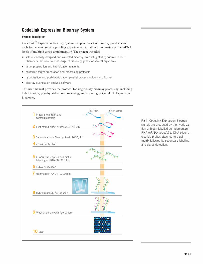
CodeLink Expression Bioarray System System description
CodeLink™ Expression Bioarray System comprises a set of bioarray products and tools for gene expression profiling experiments that allows monitoring of the mRNA levels of multiple genes simultaneously. The system includes:
• sets of carefully designed and validated bioarrays with integrated hybridization Flex Chambers that cover a wide range of discovery genes for several organisms
• target preparation and hybridization reagents
• optimized target preparation and processing protocols
• hybridization and post-hybridization parallel processing tools and fixtures
• bioarray quantitation analysis software
This user manual provides the protocol for single-assay bioarray processing, including hybridization, post-hybridization processing, and scanning of CodeLink Expression Bioarrays.
1 Prepare total RNA and bacterial controls
2 First-strand cDNA synthesis 42 °C, 2 h
3 Second-strand cDNA synthesis 16 °C, 2 h
4 cDNA purification
6 cRNA purification
7 Fragment cRNA 94 °C, 20 min
5 In vitro Transcription and biotin labelling of cRNA 37 °C, 14 h
8 Hybridization 37 °C, 18–24 h
10 Scan
9 Wash and stain with fluorophore
mRNA SpikesTotal RNA
Fig 1. CodeLink Expression Bioarray signals are produced by the hybridization of biotin-labelled complementary RNA (cRNA) target(s) to DNA oligonucleotide probes attached to a gel matrix followed by secondary labelling and signal detection.
● p3
Safety warnings and precautions Consider all chemicals as potentially hazardous. Only persons trained in laboratory techniques and familiar with the principles of good laboratory practice should handle these products. Wear suitable protective clothing such as laboratory coats, safety glasses, and gloves. Exercise caution to avoid contact with skin or eyes; if contact should occur, wash immediately with water. See Material Safety Data Sheet for specific recommendations.
For each reagent required but not provided, follow the manufacturer’s safety requirements.
Handling These procedures involve working with RNA; therefore, exercise great care to avoid RNase contamination. All solutions must be RNase-free and pipette tips must be aerosol-resistant and changed before each step. Use commercially prepared nuclease-free water (rather than water treated with diethylpyrocarbonate (DEPC)) for all nucleic acid steps.
Storage The CodeLink Expression Assay Reagent Kit components used in hybridization and detection should be stored as follows:
• Hybridization Buffer A: ambient temperature
• Hybridization Buffer B and 5× Fragmentation Buffer: 4 °C
Biotin-labelled cRNA target is prepared by a linear amplification method. The poly(A)+
RNA subpopulation (within the total RNA population) is primed for reverse transcription by a DNA oligonucleotide containing the T7 RNA polymerase promoter 5′ to a d(T)24
sequence (Fig 1). After second-strand cDNA synthesis, the cDNA serves as the template for an in vitro transcription (IVT) reaction to produce the target cRNA. The IVT is performed in the presence of biotinylated nucleotides to label the target cRNA. This method produces approximately 1000-fold to 5000-fold linear amplification of the input poly(A)+ RNA.
A set of bacterial control mRNAs is included in CodeLink Expression Assay Reagent Kit as controls for the cDNA synthesis and the IVT reactions. These controls are added to the total RNA sample during target preparation. Each step of the CodeLink Expression Bioarray processing procedure, including target preparation and hybridization, can be monitored using these control mRNAs. Additionally, the bacterial control mRNAs can be used to estimate the sensitivity of RNA detection.
Hybridization is performed overnight in a temperature-controlled shaking incubator. Optimized hybridization buffer components are also included in CodeLink Expression Assay Reagent Kit for use at this step.
Post-hybridization processing includes a stringent wash to remove unbound and non-specifically hybridized target molecules, a staining step with a Cy™5-Streptavidin conjugate, and several non-stringent washing steps to remove unbound conjugate. Following a final rinse, the bioarrays are dried by centrifugation and scanned. Analysis of the bioarrays with the CodeLink Expression Analysis software is described in the Help accessible in the software.
● p4
CodeLink Gene Expression System: Single-Assay Bioarray Hybridization and Detection
Protocol overview
1. Fragment cRNA. In 25 µl total volume, add 10 µg of cRNA to 5 µl of 5× fragmen
tation buffer and incubate at 94 ºC for 20 minutes.
2. Prepare hybridization reaction mixture. Bring 10 µg of fragmented cRNA, 78 µl of hybridization buffer component A, and 130 µl of hybridization buffer component B to a final volume of 260 µl with water. Incubate at 90 ºC for 5 minutes and immediately chill on ice for 5–30 minutes.
3. Loading reaction mixtures into array chambers. Slowly inject 250 µl of hybridization reaction mixture into array input port and seal ports with sealing strips.
4. Hybridization. Set the shaker speed to 300 rpm and incubate slides for 18–24 h at 37 ºC, maintaining a consistent hybridization time for comparative experiments.
5. Post-hybridization wash. Remove the Flex Chamber using the hybridization removal tool to hold the bioarray in place while peeling the Flex Chamber off at a 60º angle. Then, place the bioarrays into the bioarray rack while it sits inside the medium reagent reservoir containing 0.75× TNT. Transfer the bioarray rack to the large reagent reservoir containing preheated 0.75× TNT, and incubate at 46 °C for exactly 1 h. Do not exceed 1 h incubation.
6. Detection with streptavidin-dye conjugate. Fill each slot of the small reagent reservoir with 3.4 ml of Cy5-Streptavidin working solution. Transfer the bioarray rack from the large reagent reservoir into the small reagent reservoir and incubate bioarrays at ambient temperature for 30 minutes. Wash the bioarrays four times with 1× TNT (5 minutes each wash) in large reagent reservoirs. Rinse the bioarrays in 0.1× SSC/0.05% Tween™ for 30 seconds. Immediately follow the rinse with centrifugation to dry bioarrays, and store dried bioarrays in the dark.
7. Bioarray scanning and analysis. Scan bioarrays and analyze with CodeLink Expression Analysis software. Scanning with GenePix™ 4000B is described in this booklet. For use of CodeLink Bioarrays with other scanners, visit the CodeLink website (www.amershambiosciences.com).
Products used for processing CodeLink Expression Assay Reagent Kit, Manual Prep, 24 reactions 320012
5× fragmentation buffer hybridization buffer component A hybridization buffer component B
Cy5-Streptavidin (staining solution is a 1:500 dilution in TNB; see Appendix 1) PA45001
CodeLink Expression Analysis software 310030
CodeLink Shaker Kit, 12 slides 310016
CodeLink Parallel Processing Kit, 12 slides 310010
Innova™ 4080 shaking incubator 310024
Use only high-quality cRNA generated with a CodeLink Expression Assay target preparation protocol for the following steps to achieve optimal results.
Avoid physical contact with the probe-containing area of the bioarray. Handle bioarrays on the edges throughout the procedure.
Exercise caution to avoid RNase contamination. All solutions must be RNase-free; pipette tips must be changed before each step. Use nuclease-free water for all nucleic acid steps. Use aerosol-resistant tips for all pipetting steps.
To avoid fluorophore photobleaching, prepare the Cy5-labelled Streptavidin conjugate in opaque or dark tubes. In addition, minimize bioarray exposure to ambient light, particularly after completing step 6.1.
Ensure that the ambient temperature in the laboratory is 23 ºC ± 2 ºC.
Minimize exposure to particulate matter. Clean all work areas prior to hybridization and analysis.
The list of reagents and equipment is recommended for best performance. Contact Amersham Biosciences technical support for information on use of alternatives.
Caution: Gently vortex and briefly centrifuge all reagent tubes prior to use.
● p5
Storage Store hybridization buffer component A at room temperature. If a precipitate forms upon storage, briefly vortex prior to use to ensure solution homogeneity. If necessary, warm at 37 ºC to redissolve.
Store hybridization buffer component B at 4 ºC until use.
Other materials required for processing All reagents used in the protocol should be molecular biology grade or higher.
• 0.5% NEN blocking reagent (PerkinElmer, FP1020)
• nuclease-free water
• 1 M Tris-HCl, pH 7.6
• 5 M NaCl
• Tween 20
• 20× SSC (Ambion, 9763)
• Isopropanol (to wash reagent container for TNT preparation)
• GenePix 4000B Array Scanner (Axon Instruments) and computer configured for GenePix 4000B Array Scanner or other CodeLink validated scanner (see www.amershambiosciences.com for CodeLink validated scanner application notes.)
• centrifuge, 4-15C (QIAGEN Corp, 81010 or similar) with appropriate centrifuge plate rotor, 2 × 96 (QIAGEN Corp, 81031). For alternative centrifuges, contact Amersham Biosciences technical support or visit our Web site.
• pipette tips, sterile, RNase-free, and aerosol-resistant
• 1.7-ml microcentrifuge tubes, sterile and RNase-free
• 15- and 50-ml conical tubes: sterile, nuclease-free
• wide bore pipette tips: sterile, RNase-free, and aerosol-resistant
• vortex mixer
• water bath (settings 90 ºC and 46 ºC)
• pipettors
• microcentrifuge
• thermal cycler
• powder-free gloves
• microtiter plate lid, black (Corning Corp, 3935)
Protocol for hybridization and detection
●1
Keep 5× fragmentation buffer at 4 ºC until use. Briefly vortex and spin down prior to use to ensure solution homogeneity.
For steps 1–2, use of a 10% volume mixture overage is recommended but not required.
Fragmentation of cRNA
1.1 For each bioarray to be loaded, bring 10 µg of cRNA (from step 6.13 of CodeLink target preparation protocol) to a final volume of 20 µl with nuclease-free water in a thin-walled microcentrifuge tube.
1.2 Add 5 µl of 5× fragmentation buffer for each bioarray. Place tube in a thermal cycler and heat for 20 minutes at 94 ºC using the heated lid feature.
1.3 Cool to 0 ºC in the thermal cycler for at least 5 minutes.
● p6

●2
Preparation of hybridization reaction mixtures
2.1 Set the temperature of the shaker-incubator to 37 ºC for hybridization. Assemble bioarray tray posts from the CodeLink Shaker Kit onto the incubator platform.
2.2 Transfer the cRNA sample from the thin-walled tube to a 1.5-ml microcentrifuge tube. For each bioarray to be processed, prepare 260 µl of hybridization solution containing 10 µg of fragmented target cRNA in a 1.5-ml microcentrifuge tube:
78 µl hybridization buffer component A
130 µl hybridization buffer component B
27 µl nuclease-free water
25 µl fragmented cRNA (from Step 1)
260 µl total volume
2.3 Vortex the solution for 5 seconds at maximum speed. Incubate the hybridization solution at 90 ºC for 5 minutes to denature the cRNA.
2.4 Cool the tube(s) on ice for at least 5 and no more than 30 minutes Load all bioarrays within 30 minutes of denaturing the cRNA.
●3
If performing replicate bioarrays, do not prepare more than 4 reactions (~1 ml) in a single microcentrifuge tube. Larger volumes will not allow good heat transfer.
Loading of reaction mixtures into bioarray chambers
3.1 Set the 12-slide shaker tray on a level surface. Place the bioarrays into the shaker tray with the input/output ports facing up. Load bioarrays in sets of 12 or less.
3.2 Vortex the hybridization reaction mixture for 5 seconds at maximum speed. Centrifuge briefly to gather the liquid at the bottom of the tube. Place the tube back on ice.
Caution: Ensure that the target sample is loaded very slowly and carefully to avoid introduction of bubbles into the chamber.
3.3 For each bioarray chamber, draw 250 µl into a 1-ml wide-bore pipette tip. Place the 1-ml pipette tip containing the hybridization solution over the array input port closest to the right edge of the slide label and press until the tip forms a seal with the chamber (see Appendix 5 for details).
3.4 Slowly inject the entire sample into the Flex Chamber without using the blowout feature of the pipettor. When the Flex Chamber is full, maintain thumb pressure on the pipettor, hold down the bioarray over the tab region with index finger of opposite hand, and remove the pipette tip from the input port. Discard any excess target mix remaining in the pipette tip. Aspirate any excess fluid surrounding the outside of the port with a pipette tip and discard. Use a lint-free wipe to blot residual fluid from around the port, taking care not to actually touch the port.
3.5 After loading up to 12 bioarrays, seal the Flex Chamber ports using the sealing strips and port sealing tool as described in Appendix 5. Do not touch the Flex Chamber or directly depress the port.
●4
Hybridization
4.1 Align the 12-slide shaker tray notches with the front and back posts fixed to the shaker-incubator platform to place the loaded shaker tray into the shaker-incubator. The Flex Chamber should be facing up.
● p7

Refer to Appendix 4 for identification of the processing fixtures.
Filter all TNT buffer solutions (Appendix 1) through a 0.2-µm filter prior to use.
Prepare sufficient volumes of 1× and 0.75× TNT buffers (Appendix 1) for the entire procedure. Approximately one liter of 1× TNT, and 500 µl of 0.75× TNT are needed for 12 bioarrays.
At the beginning of each wash step, gently agitate the bioarray rack.
Avoid contact with the probe-containing surface of the microarray throughout the post-hybridization steps.
Do not allow the bioarrays to dry out during any of the processing steps.
Clean the shaker incubator platform and the shaker tray with deionized water after each hybridization run.
Ensure that the temperature of the 0.75× TNT buffer placed in the large reagent reservoir is 46 ºC.
Take care to prevent contamination of the fluid in the large reagent reservoir with water from the water bath.
4.2 Set the shaker speed to 300 rpm and incubate slides for 18–24 h at 37 ºC. It is critical that arrays used in any form of comparison are hybridized for the same amount of time within the given range.
4.3 To prepare for the next step, fill a large reagent reservoir with 240 ml of filtered 0.75× TNT buffer. Cover the reservoir and incubate in a 46 ºC water bath overnight.
●5
Post-hybridization wash
5.1 Fill each slot in the medium reagent reservoir with 13 ml of filtered 0.75× TNT. Place the bioarray rack into the reservoir. Leave at ambient temperature.
5.2 Remove the 12-slide shaker tray from the shaker incubator, and place on a level surface at ambient temperature. Process only 12 slides at a time per person. Leave the trays in the shaker until ready to process.
5.3 Place the first bioarray to be processed into the Flex Chamber removal tool.
5.4 Remove the Flex Chamber by lifting the tab and slowly pulling it back at a 60° angle without creasing the Flex Chamber.
5.5 Place the bioarray into a slot of the bioarray rack, which was placed into a medium reagent reservoir containing 0.75× TNT in step 5.1. Use the bioarray position tool, tooth-side down, to ensure the bioarrays are properly seated.
5.6 To avoid potential cross-contamination, rinse the surface of the Flex Chamber removal tool with approximately 5 ml of ambient temperature 0.75× TNT buffer dispensed from a squirt bottle. Keep the medium reagent reservoir at ambient temperature until all bioarrays have been processed.
5.7 Repeat steps 5.3–5.6 for each hybridized bioarray.
5.8 Transfer the bioarray rack with bioarrays from the medium reagent reservoir into the pre-warmed, 0.75× TNT-filled large reagent reservoir from step 4.3. Replace the lid on the large reagent reservoir and then on the water bath. Incubate at 46 ºC for exactly 1 h; longer incubation time may significantly reduce signal intensities.
●6
If processing 12 bioarrays or less during secondary labelling, prepare a centrifuge balance unit (medium reagent reservoir with bioarray rack and blank glass slides) for the bioarray drying step.
Recall that ambient temperature is 23ºC ±2ºC.
Detection with streptavidin-dye conjugate
6.1 Fill each slot in the small reagent reservoir with 3.4 ml of Cy5-Streptavidin working solution (Appendix 1). Leave at ambient temperature. Cover the small reagent reservoir with a black lid to prevent photobleaching of the fluorophore by ambient light.
6.2 Remove the bioarray rack with bioarrays from the large reagent reservoir at 46 °C, and place into the small reagent reservoir containing the Cy5-Streptavidin working solution. Cover with the lid and incubate bioarrays at ambient temperature for 30 minutes.
6.3 During incubation, prepare for the wash steps by filling three large reagent reservoirs each with 240 ml of ambient temperature 1× TNT buffer.
● p8

6.4 After the 30 minutes incubation, remove the bioarray rack with bioarrays from the staining solution and place into one large reagent reservoir containing 1× TNT buffer (prepared in 6.3). Do not drain the solution from the bioarrays. Following an initial gentle up-and-down agitation, incubate the bioarrays at ambient temperature for 5 minutes covered from light.
6.5 Remove the bioarray rack from the first large reagent reservoir with 1× TNT buffer and place into a second large reagent reservoir containing 1× TNT buffer. Again, do not attempt to drain the solution from the bioarrays and gently agitate the bioarray rack. Incubate at ambient temperature for 5 minutes, covered from light. Repeat this step with two additional large reagent reservoirs containing fresh 1× TNT buffer for a third and fourth wash.
6.6 During the third wash, thoroughly rinse a large reagent reservoir with distilled water and dry it. Completely fill this reservoir with the final rinse 0.1× SSC/ 0.05% Tween 20 solution (Appendix 1).
6.7 Transfer the bioarray rack into the large reagent reservoir completely filled with 0.1×
SSC/ 0.05% Tween 20 at ambient temperature. Incubate the slides for 30 seconds while continually agitating mildly up and down.
6.8 Remove the bioarray rack from the large reagent reservoir and blot the bottom edge of the bioarrays briefly on an absorbent paper towel. Place the bioarray rack in a clean, dry medium reagent reservoir. Dry the bioarrays by centrifugation in the Qiagen Sigma 4-15C centrifuge with corresponding bucket rotor (2 × 96-well plate) or similar system using following settings:
speed: 2000 rpm (644 × g)
acceleration: 9
deceleration: 9
time: 3 minutes
6.9 To easily remove the bioarrays from the bioarray rack, place the bioarray rack with slides into the bioarray removal tool. Place the dry bioarrays into a light-protected slide box until they are scanned. Bioarrays should be scanned within two days of assay completion.
6.10 Repeatedly rinse all reservoirs with deionized water to clean, and invert to dry.
6.11 Wash the rack with Alconox™ soap, scrubbing between the rails with a pipe cleaner style brush. Rinse thoroughly with deionized water to remove residual soap. Air-dry the rack.
●
Caution: Move into step 6.8 (the drying step) immediately because slides may dry non-uniformly in the air, leading to non-uniform signal loss.
Remember to use a centrifuge balance unit if processing less than 12 bioarrays.
Keep the bioarrays covered until they are scanned to protect them from dust and light and handle them with gloves.
For best results, scan bioarrays within two days of staining.
If bioarrays will be retained for future scanning, they should be stored in a dry box protected from light.
If regions of high background on the bioarray are noted upon scanning, the bioarrays may be rerinsed by repeating steps 6.7 and 6.8 from above.
Bioarray scanning and analysis Not all incidents of high background can be removed by rerinsing.For scanning with a GenePix Array Scanner, use the following steps. For information on
use of alternative scanners with CodeLink bioarrays, visit the CodeLink website Refer to the manufacturer’s manual (www.amershambiosciences.com). for use and maintenance of the
scanner (1).7.1 Turn the scanner on 15 minutes prior to use.
7.2 Open the cover to expose the slide holder.
● p9
7

7.3 Lift the latch of the slide holder and lift the upper clip.
7.4 Wearing latex gloves, load the bioarray into the tray with the label side down and closest to the front of the scanner.
7.5 Pull the clip on the left of the slide out and let the bioarray fall into place. Release the clip to put pressure against the bioarray.
7.6 Grab bioarray by the edges and move the bioarray toward you.
7.7 Lower upper clip and press down on the latch until it clicks.
7.8 Slide the cover to the right to cover the slide holder.
7.9 Open the GenePix software and select the following settings:
expand setting file name: Expression_5um.gps: whole genome products Expression_10um.gps: 10K and 20K products
wavelength: 635 nm
PMT voltage: 600 V
laser power: 100%
pixel size: 5µm for Whole Genome products 10µm for 10K and 20K products
focus position: 0 µm
7.10 In the Report tab, open the scanning script by clicking Scan CodeLink Slide.
7.11 Enter the bioarray serial number and click Next. The Experiment and Scan Information interface is displayed.
7.12 Type in the project name, experiment name, and sample name. The username is automatically captured. When opening this interface for the first time, a message box may ask whether to allow an ActiveX interaction to proceed. Click Yes.
7.13 Select a setting (.gps) file. The standard setting file is CodeLinkExpr.gps. If a settings file was previously selected, the name and path are displayed under Current Settings File. To select a new file, click Browse under Select New Settings File. The values for project name, experiment name, user name, and settings file that were entered for a previous bioarray are retained but may be changed.
7.14 In the Load and Scan Slide screen, the standard TIF file name for the current bioarray is displayed. If desired, the name may be modified, however, the slide number as the first part of the name cannot be altered.
7.15 Click Browse to select the image path or the location where the image files will be stored. If a Security Alert message box is displayed, click Yes.
7.16 Click Scan Slide. The Image tab will display, and the instrument will perform the scan.
7.17 When complete, the view will return to the Report tab.
7.18 Click Save Image to save the scanned image at the appropriate file location.
7.19 Slide the cover to the left and remove the bioarray.
7.20 To scan the next bioarray, click New Slide and enter the serial number for the next bioarray. The setting information previously entered will be retained.
7.21 Analyze the image from each bioarray using CodeLink Expression Analysis software.
● p10
Appendix 1: Stock solutions 1× TNT buffer (10 liters)
0.10 M Tris-HCl, pH 7.6
0.15 M NaCl
0.05% Tween 20
1. Prepare sufficient volumes of 1× TNT buffer for the entire procedure. Approximately 1 liter of 1× TNT is needed for 12 bioarrays.
2. Rinse a 10-liter reagent container with 150 ml isopropanol. Rinse the carboy twice with 2 liters of deionized water and completely drain the reagent container.
3. In a separate container, add the following:
1 liter 1 M Tris-HCl, pH 7.6
300 ml 5 M NaCl
5 ml Tween 20
8.7 liter deionized water
4. Mix well by swirling. Filter TNT through a 0.2 µm filter and transfer to reagent container. This solution can be stored up to two weeks at ambient temperature.
0.75× TNT buffer
1. Prepare a sufficient volume of 0.75× TNT buffer for the entire procedure. Approximately 500 ml of 0.75× TNT is needed for 12 bioarrays.
2. Add 25 ml of deionized water to 75 ml of 1× TNT buffer per 100 ml of buffer required.
3. This solution should be stored similarly to 1× TNT, in a isopronal-rinsed reagent container.
TNB Buffer (0.6 liters)
0.1 M Tris-HCl, pH 7.6
0.15 M NaCl
0.5% NEN blocking reagent (PerkinElmer, FP1020)
1. To a clean 2-liter Erlenmeyer flask with a stir bar, add the following:
522 ml nuclease-free water
60 ml 1 M Tris-HCl, pH 7.6
18 ml 5 M NaCl
2. On a stir plate heated to 60 ºC, slowly add 3 g of NEN blocking reagent in 0.5 g increments until all 3 g of blocking reagent are dissolved.
3. Turn off heat and continue mixing for 30 minutes. While the solution is still warm, filter through a 0.88 µm filter.
4. Aliquot the TNB buffer into 50-µl tubes and store at -20 ºC. TNB can be stored for 12 weeks at -20 ºC.
5. For use, thaw TNB overnight at 4 ºC. Thawed aliquots can be stored at 4 ºC up to one week.
Minimize exposure of dye-conjugate Cy5-Streptavidin stock solution reagents to ambient light.
1. Add 1 ml of nuclease-free water to the 1 mg bottle of lyophilized Cy5-Streptavidin. This reagent already contains buffer components. To mix, pipette gently up and down or vortex with moderate speed. Do not allow foam to form. Place on ice for 5 minutes. Repeat the mixing three additional times, placing on ice between mixings. Protect this solution from light at all times.
● p11
2. Aliquot the Cy5-Streptavidin solution for the number of bioarrays processed in a typical run. For example, dispense 90 µl aliquots for a 12-bioarray run.
3. This solution can be stored up to six months (or until expiry date) at -20 ºC, protected from light. Once thawed, unused portions of aliquots may be used for up to one week if stored protected from light at 4 °C. Avoid multiple freeze-thaw cycles.
1:500 Cy5-Streptavidin working solution
1. Prior to dilution, centrifuge the thawed Cy5-Streptavidin stock for 1 minutes at ≥ 8000 × g to remove any precipitates.
2. Add 6.8 µl of the Cy5-Streptavidin stock solution to 3393.2 µl of filtered TNB buffer for each bioarray to be processed. Mix gently by inversion. Use this working solution within 15 minutes of preparation.
0.1× SSC / 0.05% Tween 20 (1 liter)
20× SSC (Ambion, 9763)
Tween 20
1. Make sure all solutions are molecular biology grade. Prepare sufficient volume of 0.1×
SSC/0.05% Tween buffer for the entire rinsing or re-rinsing procedure. Approximately 250 ml of 0.1X SSC/0.05% Tween buffer is needed for 12 slides.
2. In a clean 1-liter container, add the following:
994.5 ml deionized Water
0.5 ml Tween 20
5 ml 20× SSC buffer
Mix well by swirling. This solution can be stored up to 2 weeks at ambient temperature.
● p12
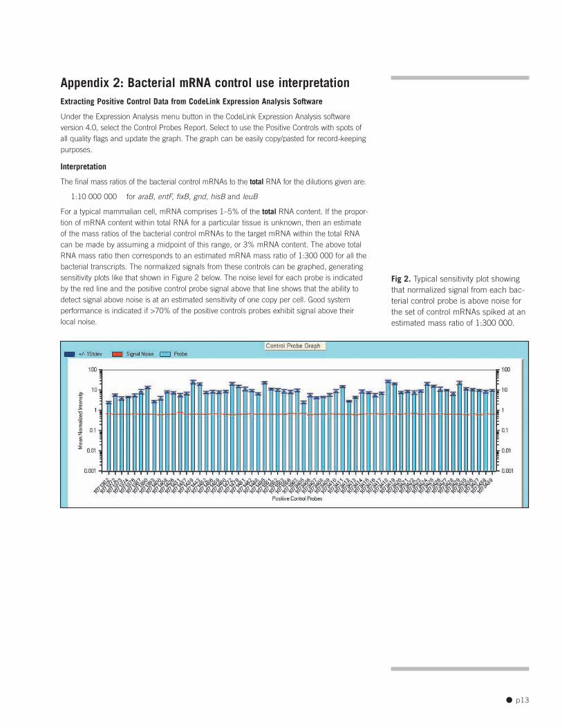
Appendix 2: Bacterial mRNA control use interpretation Extracting Positive Control Data from CodeLink Expression Analysis Software
Under the Expression Analysis menu button in the CodeLink Expression Analysis software version 4.0, select the Control Probes Report. Select to use the Positive Controls with spots of all quality flags and update the graph. The graph can be easily copy/pasted for record-keeping purposes.
Interpretation
The final mass ratios of the bacterial control mRNAs to the total RNA for the dilutions given are:
1:10 000 000 for araB, entF, fixB, gnd, hisB and leuB
For a typical mammalian cell, mRNA comprises 1–5% of the total RNA content. If the proportion of mRNA content within total RNA for a particular tissue is unknown, then an estimate of the mass ratios of the bacterial control mRNAs to the target mRNA within the total RNA can be made by assuming a midpoint of this range, or 3% mRNA content. The above total RNA mass ratio then corresponds to an estimated mRNA mass ratio of 1:300 000 for all the bacterial transcripts. The normalized signals from these controls can be graphed, generating sensitivity plots like that shown in Figure 2 below. The noise level for each probe is indicated by the red line and the positive control probe signal above that line shows that the ability to detect signal above noise is at an estimated sensitivity of one copy per cell. Good system performance is indicated if >70% of the positive controls probes exhibit signal above their local noise.
Fig 2. Typical sensitivity plot showing that normalized signal from each bacterial control probe is above noise for the set of control mRNAs spiked at an estimated mass ratio of 1:300 000.
● p13
Appendix 3: Precipitation of cRNA It may be necessary to remove contaminants in the cRNA solution by ethanol precipitation.
1. Transfer the cRNA from step 6.13 of the target preparation protocol to a new 1.5-ml microcentrifuge tube. Add 50 µl (0.5 volumes) of ice-cold 5 M ammonium acetate, and vortex for 5 seconds at maximum speed. Add 375 µl (2.5 volumes) of ice-cold absolute ethanol to sample and vortex for 5 seconds at maximum speed. Incubate the sample at least 30 minutes at -20 ºC.
2. Centrifuge at ≥ 10 000 × g for 15 minutes at 4 ºC. Carefully remove and discard the supernatant.
3. Add 0.5 ml 70% ethanol and vortex for 5 seconds to wash pellet.
4. Centrifuge at 4 ºC for 10 minutes at ≥ 10 000 × g. Carefully remove and discard supernatant. Remove any recalcitrant drops using a 200-µl pipette tip. Air-dry the pellet.
5. Resuspend cRNA in 20 µl nuclease-free water. Quantify the precipitated cRNA by UV spectrophotometry as detailed in section 7 of the manual target preparation protocol.
6. Store the cRNA at -70 ºC.
● p14
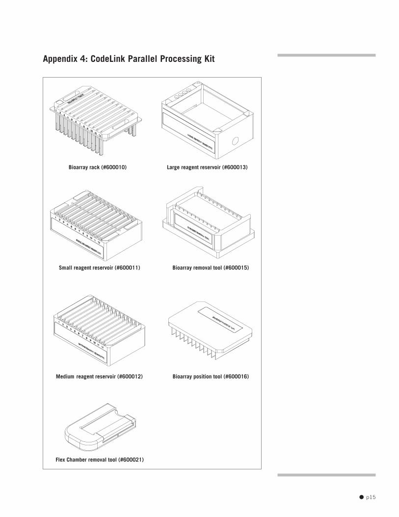
Appendix 4: CodeLink Parallel Processing Kit
Bioarray rack (#600010) Large reagent reservoir (#600013)
Small reagent reservoir (#600011) Bioarray removal tool (#600015)
Medium reagent reservoir (#600012) Bioarray position tool (#600016)
Flex Chamber removal tool (#600021)
● p15

Adhesive will attach firmly at first contact. Use caution to ensure that the Sealing Strip is in the proper orientation before placing it on the chamber. Do not remove a misaligned Sealing Strip. Instead, place a second strip to cover any open ports.
Fig A. Proper placement of the Sealing Strips over the 2-port Flex Chamber ports.
Fig B. Proper placement of the Sealing Strips over the 4-port Flex Chamber ports.
Appendix 5: Sealing the Flex Chamber port 1. Place the Sealing Strips to achieve the seals illustrated in Figure A for the 2-port Flex Chamber style and in Figure B for the 4-port Flex Chamber style.
2. Press the Sealing Strip against the port using the Port Sealing Tool (Figure C).
Loading port
Loading port
Fig C. Proper port sealing technique.
port sealing tool
sealing strip
chamber
glass slide
● p16

Appendix 6: Process Flow Charts Bioarray Hybridization and Detection
Fragment cRNA: add 5× Fragmentation Buffer to cRNA, 94 ºC, 20 min
Prepare Hybridization Mix: mix fragmented cRNA, Hyb Buffer A, and Hyb Buffer B
Load Slides: Carefully pipette 250 µl into chamber; seal ports; 300 rpm shaker-incubator, 37 ºC, 18–24 h
CodeLink Bioarray Do not allow slides to dry outPost-Hybridization during the following steps.
Processing After 18 h or 24 h hybridization at 37 ºC, remove Hybridization Chamber from each slide and place slides in Holding Medium Reservoir.
Holding Reservoir 0.75× TNT, 23 ºC ±2 ºC, ≤ ~10 min
Medium
Stringent Wash: 0.75× TNT, 46 ºC,
Large EXACTLY 1 h
Secondary Labelling: SA-Cy™5 Staining Solution, ambient temperature, 30 min
Small
Large
Wash: 4 separate washes of 1× TNT, ambient temperature, 5 min each
Large Large
Final Rinse: 1 rinse of 0.1× SSC/0.05% Tween, 30 s Move immediately to next step.Large
Spin Dry: Centrifuge at 644 rcf, 3 min
Medium
Medium
● p17

Ordering information Description Size Code number Reference
CodeLink UniSet™ Human I Bioarray 6 bioarrays 25-6700-04 300007-6PK
CodeLink UniSet Human I Bioarray 24 bioarrays 25-6700-05 300007-24PK
CodeLink UniSet Human II Bioarray 6 bioarrays 25-6700-64 300014-6PK
CodeLink UniSet Human II Bioarray 24 bioarrays 25-6700-65 300014-24PK
CodeLink UniSet Human 20K I Bioarray 6 bioarrays 25-6700-66 300019-6PK
CodeLink UniSet Human 20K I Bioarray 24 bioarrays 25-6700-67 300019-24PK
CodeLink UniSet Mouse I Bioarray 6 bioarrays 25-6700-08 300013-6PK
CodeLink UniSet Mouse I Bioarray 24 bioarrays 25-6700-09 300013-24PK
CodeLink UniSet Rat I Bioarray 6 bioarrays 25-6700-10 300012-6PK
CodeLink UniSet Rat I Bioarray 24 bioarrays 25-6700-11 300012-24PK
CodeLink UniSet Mouse 20K Bioarray 6 bioarrays 25-6700-76 300022-6PK
CodeLink UniSet Mouse 20K Bioarray 24 bioarrays 25-6700-77 300022-24PK
CodeLink Whole Human Genome Bioarray 6 bioarrays 25-6700-89 300026-6PK
CodeLink Whole Human Genome Bioarray 24 bioarrays 25-6700-90 300026-24PK
ADME Rat Bioarray 6 bioarrays 25-6700-06 300010-6PK
ADME Rat Bioarray 24 bioarrays 25-6700-07 300010-24PK
CodeLink Expression Assay Reagent Kit, 24 reactions 25-6700-68 320012 Manual Prep
CodeLink Shaker Kit 12 slides 25-6700-13 310016
CodeLink Shaker Kit 48 slides 25-6700-12 310017
CodeLink Parallel Processing Kit 12 slides 25-6700-15 310010
CodeLink Parallel Processing Kit 48 slides 25-6700-38 310011
Cy5-Streptavidin 25-0054-31 PA45001
CodeLink Expression Analysis v4.0 software 25-6700-31 3100030
References
1. GenePix 4000 User’s Guide, Axon Instruments, Inc., 2500–136 Rev E (2001).
● p18
CodeLink, Cy, and UniSet are trademarks of Amersham Biosciences Limited
Amersham and Amersham Biosciences are trademarks of Amersham plc
Alconox is a trademark of Alconox Inc.
GenePix is a trademark of Axon Instruments, Inc.
Innova is a trademark of New Brunswick Scientific Co Inc.
QIAGEN is a trademark of QIAGEN Inc.
Tween is a registered trademark of ICI Americas Inc
Customer shall have the right to perform gene expression analysis using the Bioarrays and to add data generated from such
gene expression analyses to databases created and maintained by Customer for Customer’s internal use or SNP scoring using
the Bioarrays and to use data generated from such SNP scoring for research purposes. Bioarrays sold to Customer will be
used only for Customer’s internal purposes and shall not be resold in their original or any modified form. Amersham Bioarrays
contain oligonucleotide probes that are designed to recognize unique regions of the genome of a target organism. The
sequence information used to design these probes may have been obtained from various public and proprietary databases
and may include sequences covered by various US and foreign patents. Substantial investment has been made in the design
and optimization of these probes. Probe sequences are proprietary to Amersham and protected by various intellectual property
laws. Customer shall not make any attempt to determine the probe sequences deposited by Amersham on the Bioarrays or to
use the Bioarrays or portions thereof for any purpose other than internal use; however, the foregoing shall not prevent
Customer from determining or knowing (i) the gene names and sequences of the entire genes for which the probes are used
as test sequences, (ii) the sequences of the mRNA and cDNA generated by Customer as part of the sample preparation
process, or (iii) sequences of relevant gene fragments (other than the probes themselves).
GRANT OF LIMITED LICENSE FOR RESEARCH FIELD PRODUCTS:
This limited license permits the person or legal entity to which this Nucleic Acid Array ("Array") has been provided to use the Array,
and the data generated by use of the Array, only for its internal research purposes to identify and study the presence and/or
levels of expressed nucleic acid sequences in the ordinary course of Buyer's normal internal research activities, including clinical
trials. In addition, Buyer agrees not and understands it is not licensed to (a) provide commercial data or databases to or on
behalf of any third party that relate to an Array; or (b) use an expression probe array or data therefrom in a clinical diagnostic
setting where data from an individual's sample is given to such individual or their caregiver. Neither Amersham nor its licensors
grants any other licenses, express or implied, to permit the manufacturer, use, sale or importation of the Array, any other
arrays, any nucleic acids on the Array, or any data generated from use of the Array.
© Amersham plc 2004—All rights reserved
All goods and service are sold subject to the terms and conditions of sale of the company within the Amersham group that
supplies them. A copy of these terms and conditions is available on request.
www.amershambiosciences.com
● p19
Amersham Biosciences UK Limited Amersham Place, Little Chalfont, Buckinghamshire, UK HP7 9NA
Amersham Biosciences AB SE-751 84 Uppsala, Sweden
Amersham Biosciences Corp. 800 Centennial Avenue, PO Box 1327, Piscataway, NJ 08855 USA
Amersham Biosciences GmbH Munzinger Strasse 9, D-79111 Freiburg, Germany
Amersham Biosciences (SV) Corp. 928 East Arques Avenue, Sunnyvale CA 94085-4520 USA
● p20