coccidian tales - kanyana wildlife rehabilitation centre€¦ · 2016). the lifecycle of those...
TRANSCRIPT

Page 1 of 8
Coccidian Tales Belinda Brice and Gwyneth Thomas
Kanyana Wildlife Rehabilitation Centre 120 Gilchrist Road, Lesmurdie, 6076, Perth, Western Australia
Email: [email protected] and [email protected] Abstract Coccidia are protozoan parasites that commonly infect a wide variety of vertebrate and invertebrate hosts including birds and reptiles. Even though the coccidian group contains many different species, only a few cause clinical disease. Information regarding the prevalence of these single-celled parasites in wild birds and reptiles is limited. The Kanyana Wildlife Rehabilitation Centre (KWRC) admits more than 3000 animals a year, many of which we screen for intestinal parasites including coccidia. In this presentation, we shall focus on those coccidia infecting wild birds and reptiles. We shall discuss how coccidia are transmitted, the signs they may produce, their treatment and control. Incidence rates of coccidia infection amongst various wild bird and reptile species admitted to KWRC will be detailed. The KWRC is involved in a collaborative study with scientists from Murdoch University to morphologically and genetically characterize the coccidian species that are identified in wildlife admitted to the centre. This has led to the discovery of numerous new coccidian species. Introduction There are many different coccidian species but only a few are responsible for causing clinical disease (coccidiosis) whilst most infections remain asymptomatic. In those cases where infection occurs but no disease results, it is called coccidiasis. Coccidiosis is an economically important disease of domestic animals such as poultry, pigs, sheep, goats and cattle. Coccidia are a common cause of diarrhoea in the chicken-like or game birds, in pigeons, doves, ducks, geese, swans and many other bird species as well (Samour, 2016). There is however, a lack of information about coccidiosis in wild birds worldwide. The Isospora, Eimeria and Caryospora are all genera of the Eimeriidae family and this paper will concentrate on these genera. Epidemiology The coccidia are a diverse group of unicellular protozoan parasites infecting both vertebrates and invertebrates (Fayer, 1980). Coccidia belong to the phylum Apicomplexa as they contain a cluster of organelles known as an apical complex at one end of the cell which helps them to enter the host’s cells (Morrissette and Sibley, 2002). The coccidia are mostly host specific. For example, a coccidian species from the Eimeriidae family that infects a reptile will not infect a bird or a mammal. Coccidia infecting animals are not infectious to humans as we have our own species-specific coccidians. Coccidia are also very specific to which region of the intestines they infect, with Eimeria infections being more specific than Isospora infections (Samour, 2016). Coccidian oocysts are often found in the faeces of both captive and wild birds. Coccidian infections rarely cause a problem in wild birds and other animals but they are a problem in young animals which are housed in small areas like those on production farms. Other factors such as poor nutrition, poor sanitation, the stresses of weaning, moving animals from one area to another and changes in feed all play a role in the coccidian infection process. Most animals will become infected in the first few months of their life and show varying levels of disease severity. Older animals are usually resistant to clinical disease but they may well have sporadic asymptomatic infections which could act as sources of infection for the younger animals. Isospora are the most common coccidian parasites infecting the passerine (perching) birds (Duszynski et al., 1999). Despite the large number of Isospora spp. infecting wild birds, relatively few of these have been genetically characterized (Olson et al., 1998; Carreno and Barta, 1999). Most Isospora are only mildly pathogenic resulting in a mild enteritis but some species have disseminated stages in the blood and tissues which can result in death of the bird.
Page 2 of 8
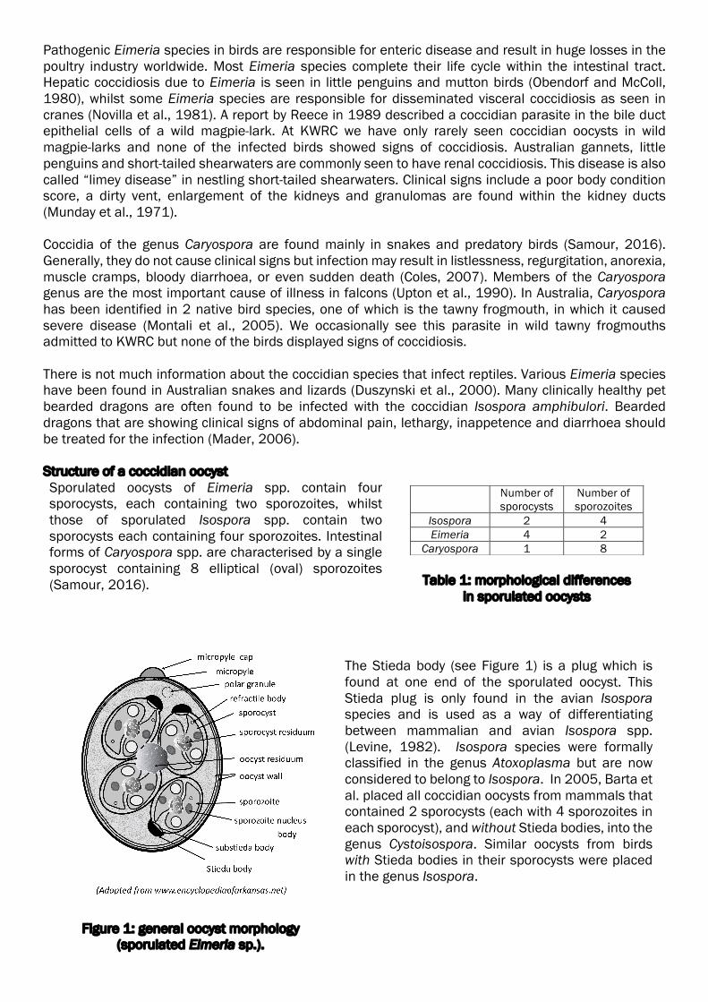
Pathogenic Eimeria species in birds are responsible for enteric disease and result in huge losses in the poultry industry worldwide. Most Eimeria species complete their life cycle within the intestinal tract. Hepatic coccidiosis due to Eimeria is seen in little penguins and mutton birds (Obendorf and McColl, 1980), whilst some Eimeria species are responsible for disseminated visceral coccidiosis as seen in cranes (Novilla et al., 1981). A report by Reece in 1989 described a coccidian parasite in the bile duct epithelial cells of a wild magpie-lark. At KWRC we have only rarely seen coccidian oocysts in wild magpie-larks and none of the infected birds showed signs of coccidiosis. Australian gannets, little penguins and short-tailed shearwaters are commonly seen to have renal coccidiosis. This disease is also called “limey disease” in nestling short-tailed shearwaters. Clinical signs include a poor body condition score, a dirty vent, enlargement of the kidneys and granulomas are found within the kidney ducts (Munday et al., 1971). Coccidia of the genus Caryospora are found mainly in snakes and predatory birds (Samour, 2016). Generally, they do not cause clinical signs but infection may result in listlessness, regurgitation, anorexia, muscle cramps, bloody diarrhoea, or even sudden death (Coles, 2007). Members of the Caryospora genus are the most important cause of illness in falcons (Upton et al., 1990). In Australia, Caryospora has been identified in 2 native bird species, one of which is the tawny frogmouth, in which it caused severe disease (Montali et al., 2005). We occasionally see this parasite in wild tawny frogmouths admitted to KWRC but none of the birds displayed signs of coccidiosis. monoculture and raising b There is not much information about the coccidian species that infect reptiles. Various Eimeria species have been found in Australian snakes and lizards (Duszynski et al., 2000). Many clinically healthy pet bearded dragons are often found to be infected with the coccidian Isospora amphibulori. Bearded dragons that are showing clinical signs of abdominal pain, lethargy, inappetence and diarrhoea should be treated for the infection (Mader, 2006). Structure of a coccidian oocyst Sporulated oocysts of Eimeria spp. contain four sporocysts, each containing two sporozoites, whilst those of sporulated Isospora spp. contain two sporocysts each containing four sporozoites. Intestinal forms of Caryospora spp. are characterised by a single sporocyst containing 8 elliptical (oval) sporozoites (Samour, 2016).
Table 1: morphological differences
in sporulated oocysts
Number of sporocysts
Number of sporozoites
Isospora 2 4 Eimeria 4 2
Caryospora 1 8
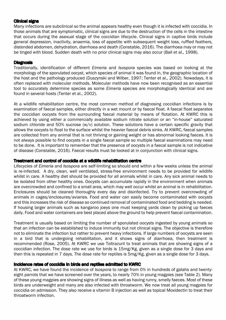
Figure 1: general oocyst morphology
(sporulated Eimeria sp.).
The Stieda body (see Figure 1) is a plug which is found at one end of the sporulated oocyst. This Stieda plug is only found in the avian Isospora species and is used as a way of differentiating between mammalian and avian Isospora spp. (Levine, 1982). Isospora species were formally classified in the genus Atoxoplasma but are now considered to belong to Isospora. In 2005, Barta et al. placed all coccidian oocysts from mammals that contained 2 sporocysts (each with 4 sporozoites in each sporocyst), and without Stieda bodies, into the genus Cystoisospora. Similar oocysts from birds with Stieda bodies in their sporocysts were placed in the genus Isospora.

Page 3 of 8
Taxonomic classification of the coccidia is still controversial and is becoming more reliant on the development of molecular techniques and studies. The KWRC and Murdoch university’s collaborative study has led to the characterization of two complete Isospora mitochondrial genomes, one from a domestic canary and the other from a wild yellow-throated miner. The results from this work further supports the monophyletic nature of Isospora from avian hosts (Yang et al., 2017). Generalised lifecycle of coccidia Coccidians may only parasitise a single host (monoxenous) during their lifecycle (direct) or they may parasitise multiple hosts (heteroxenous) (Fayer, 1980). The best-studied avian coccidians are the Eimeria spp. as they are responsible for causing disease in many commercial poultry species (Samour, 2016). The lifecycle of those coccidians that have a single host lifecycle (Isospora and Eimeria species) shall be described (see Figure 2 and 3). The 3 stages of the lifecycle of those coccidia with a direct lifecycle are the asexual cycle (merogony), the sexual cycle (gametogony) and the formation of spores (sporogony).
Figure 2: general lifecycle of coccidia
(Eimeria sp.) in chickens.
Figure 3: general lifecycle (cellular
perspective) of coccidia (Eimeria sp.) in chickens.
The thick wall of the oocyst enables it to survive for up to 12 months in moist conditions and a temperature between 30°C and 40°C (Constable, 2016). Oocysts become dried out or damaged by being exposed to direct sunlight (Long, 1982). The sporulation process may take a few hours or days depending on the conditions at the time and the specific species of coccidia. An interesting fact about some coccidia is that they generally shed more oocysts in the late afternoon and evening than in the morning. This favors transmission of the parasite amongst birds at roosting time (Dolnik, 2011). The parasites shedding schedule appears to be calibrated by the light-dark cycle (diurnal periodicity) experienced by the bird throughout the day. The reason the coccidian parasite does this is probably an adaptive trait which aims to minimize the exposure of the oocysts to the sun and in so doing decreases their possibility of drying out (Martinaud et al., 2009). At KWRC we have found that this diurnal oocyst shedding occurs in many of the bird species we have sampled, including magpies, doves, pigeons, red wattlebirds, laughing kookaburras and honeyeaters. This should be considered when collecting samples for screening from wild birds.
Page 4 of 8
Clinical signs Many infections are subclinical so the animal appears healthy even though it is infected with coccidia. In those animals that are symptomatic, clinical signs are due to the destruction of the cells in the intestine that occurs during the asexual stage of the coccidian lifecycle. Clinical signs in captive birds include general depression, inactivity, anaemia, loss of appetite with subsequent weight loss, ruffled feathers, distended abdomen, dehydration, diarrhoea and death (Constable, 2016). The diarrhoea may or may not be tinged with blood. Sudden death with no prior clinical signs may also occur (Ball et al., 1998). Diagnosis Traditionally, identification of different Eimeria and Isospora species was based on looking at the morphology of the sporulated oocyst, which species of animal it was found in, the geographic location of the host and the pathology produced (Duszynski and Wilber, 1997; Tenter et al., 2002). Nowadays, it is often replaced with molecular methods. Molecular methods have now been recognised as an essential tool to accurately determine species as some Eimeria species are morphologically identical and are found in several hosts (Tenter et al., 2002). At a wildlife rehabilitation centre, the most common method of diagnosing coccidian infections is by examination of faecal samples, either directly in a wet mount or by faecal float. A faecal float separates the coccidian oocysts from the surrounding faecal material by means of flotation. At KWRC this is achieved by using either a commercially available sodium nitrate solution or an “in-house” saturated sodium chloride and 50% sucrose (w/v) solution. These solutions have a certain specific gravity that allows the oocysts to float to the surface whilst the heavier faecal debris sinks. At KWRC, faecal samples are collected from any animal that is not thriving or gaining weight or has abnormal looking faeces. It is not always possible to find oocysts in a single faecal sample so multiple faecal examinations may need to be done. It is important to remember that the presence of oocysts in a faecal sample is not indicative of disease (Constable, 2016). Faecal results must be looked at in conjunction with clinical signs. Treatment and control of coccidia at a wildlife rehabilitation centre Lifecycles of Eimeria and Isospora are self-limiting so should end within a few weeks unless the animal is re-infected. A dry, clean, well ventilated, stress-free environment needs to be provided for wildlife whilst in care. A healthy diet should be provided for all animals whilst in care. Any sick animal needs to be isolated from other healthy ones. Oocysts can accumulate rapidly in the environment when animals are overcrowded and confined to a small area, which may well occur whilst an animal is in rehabilitation. Enclosures should be cleaned thoroughly every day and disinfected. Try to prevent overcrowding of animals in cages/enclosures/aviaries. Food and water can easily become contaminated with oocysts and this increases the risk of disease so continued removal of contaminated food and bedding is needed. If housing larger animals such as kangaroo joeys one must keeping yards clean by picking up faeces daily. Food and water containers are best placed above the ground to help prevent faecal contamination. Treatment is usually based on limiting the number of sporulated oocysts ingested by young animals so that an infection can be established to induce immunity but not clinical signs. The objective is therefore not to eliminate the infection but rather to prevent heavy infections. If large numbers of oocysts are seen in a bird that is undergoing rehabilitation, and it shows signs of diarrhoea, then treatment is recommended (Rose, 2005). At KWRC we use Toltrazuril to treat animals that are showing signs of a coccidian infection. The dose rate we use for birds is 15mg/Kg, given as a single dose for 3 days and then this is repeated in 7 days. The dose rate for reptiles is 5mg/Kg, given as a single dose for 3 days. Incidence rates of coccidia in birds and reptiles admitted to KWRC At KWRC, we have found the incidence of Isospora to range from 0% in hundreds of galahs and twenty-eight parrots that we have screened over the years, to nearly 70% in young magpies (see Table 2). Many of these young magpies are showing signs of illness as well as having runny, smelly faeces. Most of these birds are underweight and many are also infected with throatworm. We now treat all young magpies for coccidia on admission. They also receive a vitamin B injection as well as topical Moxidectin to treat their throatworm infection.

Page 5 of 8
Rates of coccidian infection amongst native common bronzewing pigeons are lower than those seen in domestic pigeons (12% compared to 27%). Laughing turtle doves were found to only have an infection rate of 5%. A possible explanation for the higher incidence of infection amongst ground feeding birds such as pigeons and common bronzewings to that of aerial feeders such as red wattlebirds and honeyeaters is that they may become re-infected by other birds or even re-infect themselves whilst foraging on the ground. Ground feeding birds are also likely to have a higher rate of infection with other endoparasites. Table 2: incidence of coccidia in wild birds admitted to KWRC.
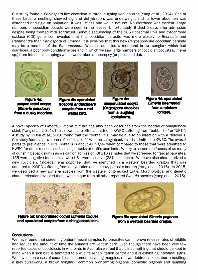
New coccidian species discovered at Kanyana To date we have identified 13 new species of coccidia in avian and reptilian hosts. We have characterized a new species of Isospora, Isospora manorinae from a juvenile yellow-throated miner that came to Kanyana from a mine-site in the Pilbara region of WA (Yang et al., 2016). It was in good body condition and showed no signs of coccidiosis. It was treated for the coccidian infection and was successfully released back at it’s found location 3 weeks later. Phylogenetic analysis revealed that this new species is most closely related to that of Isospora anthochaerae, a new Isospora species we characterized from the red wattlebird, which had an incidence rate of just over 15% (Yang et al., 2014). A grey currawong was admitted to the KWRC with a spinal injury. The bird was emaciated and had diarrhoea. Large numbers of unsporulated coccidian oocysts were seen in the faeces. The bird was euthanased as it had multiple fractures to the keel. A necropsy revealed haemorrhaging and intestinal lesions along the ileum and jejenum (small intestine). Tissue samples were taken for histology and this identified oocysts mostly in the ileum and some in the jejunum region (Yang et al., 2015). Morphological and phylogenetic analysis identified this isolate as a new species which we named Isospora streperae. Recently, we have characterized a new Isospora species from a silvereye. We have named it Isospora butcherae, in honour of KWRC founder and wildlife rehabilitator June Butcher (Yang et al., 2018). Kanyana already has a mammalian coccidian species named after it. In 2006, Bennet et al. described a novel coccidian species in the western barred bandicoot and named it Eimeria kanyana. Eimeria purpureicephali was described from a red-capped parrot that was admitted to KWRC with an injury to its keel. This is the first report of an Eimeria species in a red-capped parrot in Australia (Yang et al., 2016). The incidence rate of Eimeria purpureicephali in red-capped parrots in the Perth area was 4.2%. Eimeria paludosa, which was first described by Leger and Hesse in 1922, from the Eurasian coot and common moorhen, was identified in a dusky moorhen that came into care due to a cat attack. It showed no clinical signs of a coccidian infection. This is the first report of E. paludosa infecting a new host and is the first report of this species on the Australian continent (Yang et al., 2014). We have conducted studies on Eimeria haematodi, often seen in wild rainbow lorikeets that are admitted. Just over 22% of these birds were found to be infected with this butternut-shaped coccidian (Yang et al., 2015).
Page 6 of 8
Our study found a Caryospora-like coccidian in three laughing kookaburras (Yang et al., 2014). One of these birds, a nestling, showed signs of dehydration, was underweight and its lower abdomen was distended and rigid on palpation. It was listless and would not eat. No diarrhoea was evident. Large numbers of coccidian oocysts were seen in the faeces. Unfortunately, it died 5 days after admission despite being treated with Toltrazuril. Genetic sequencing of the 18S ribosomal RNA and cytochrome oxidase (COI) gene loci revealed that this coccidian parasite was more closely to Besnoitia and Hammondia than Caryospora or Eimeria. It is possible that this new Caryospora-like coccidian parasite may be a member of the Eumonospora. We also admitted a moribund brown songlark which had diarrhoea, a poor body condition score and in which we saw large numbers of coccidian oocysts (Eimeria sp.) from intestinal scrapings which were taken at necropsy (unpublished data).
Figure 4a:
unsporulated oocyst (Eimeria paludosa)
from a dusky moorhen.
Figure 4b: sporulated Isospora anthochaera
oocysts from a red wattle bird.
Figure 4c:
unsporulated oocyst (Caryospora daceloe)
from a laughing kookaburra.
Figure 4d: sporulated Eimeria haematodi
from a rainbow lorikeet.
A novel species of Eimeria, Eimeria tiliquae has also been described from the bobtail or shingleback skink (Yang et al., 2014). These lizards are often admitted to KWRC suffering from “bobtail flu” or “URTI”. A study by O’Dea et al., 2016 found that the “bobtail flu” may be due to an infection with a Nidovirus. Our study found a prevalence of coccidia in 21% in the shingleback lizards admitted to KWRC. The overall parasite prevalence in URTI bobtails is about 4X higher when compared to those that were admitted to KWRC for other reasons such as dog attacks or traffic accidents. We try to screen the faeces of as many of our shingleback skinks as we can on admission. Of 216 samples that we screened for faecal parasites, 155 were negative for coccidia whilst 61 were positive (28% incidence). We have also characterized a new coccidian, Choleoeimeria pogonae, that we identified in a western bearded dragon that was admitted to KWRC suffering from dehydration and a heavy parasite burden (Yang et al., 2016). In 2015, we described a new Eimeria species from the western long-necked turtle. Morphological and genetic characterisation revealed that it was unique from all other reported Eimeria species (Yang et al., 2015).
Figure 5a: unsporulated oocyst (Eimeria tilique) and sporulated oocysts from a shingleback skin.
Figure 5b: sporulated Eimeria pogonae
from a western bearded dragon. Conclusions We have found that screening patient faecal samples for parasites can improve release rates of wildlife and reduce the amount of time the animals are kept in care. Even though there have been very few reported cases of coccidiosis in wild birds in Australia we feel that it is something that should be kept in mind when a sick bird is admitted to a wildlife rehabilitation centre and it is exhibiting intestinal signs. We have seen cases of coccidiosis in numerous young magpies, red wattlebirds, a kookaburra nestling, a grey currawong, a brown songlark, common bronzewing pigeons, domestic pigeons and laughing

Page 7 of 8
turtledoves. Treatment for coccidia should be implemented if oocysts are seen in a faecal sample and the animal is showing clinical signs of a coccidian infection.
Acknowledgements The authors gratefully acknowledge Dr Rongchang Yang, Aileen Elliot and Prof. Una Ryan from Murdoch University for their enthusiasm and ongoing support of this project. We also wish to thank June Butcher, Helen Riley and all the volunteers at KWRC for their commitment and dedication in caring for all the animals admitted to the centre. We also thank the other members of the KWRC microscopy team, namely Merryn Pryor and Nathan Jardine. We are also grateful to the veterinarians and staff at the Wattle Grove and Kalamunda Veterinary Hospitals for their expert treatment and care of the wildlife treated at their clinics. References Ball, S., Brown, M., Daszak, P. and Pittilo, R. 1998. Atoxoplasma (Apicomplexa: Eimeriorina: Atoxoplasmatidae) in the Greenfinch (Carduelis chloris). Journal of Parasitology 84, 813-817. Barta, J., Schrenzel, M., Carreno, R., Rideout, B. 2005. The genus Atoxoplasma (Garnham 1950) as a junior objective synonym of the genus Isospora (Schneider 1881) species infecting birds and resurrection of Cystoisopora (Frenkel 1977) as the correct genus for Isospora species infecting mammals. Journal of Parasitology 91: 726-727. Bennet, M., Woolford, L., Ohara, A., Nicholls, P., Warren, K. and Hobbs, R. 2006. A new eimeria species parasitic in western barred bandicoots, Perameles bougainville (Marsupialia: Peramelidae), in western Australia. Journal of Parasitology 92 (6): 1292-1294. Carreno, R. and Barta, J. 1999. An eimeriid origin of isosporoid coccidia with Stieda bodies as shown by phylogenetic analysis of small subunit ribosomal RNA gene sequences. Journal of Parasitology 85: 77–83. Coles, B. 2007. Essentials of avian medicine and surgery, third ed. Blackwell Publishing Ltd. Constable, P. 2016. Overview of Coccidiosis. Merck Veterinary Manual. Dolnik, O., Metzger, B. and Loonen, M. 2011. Keeping the clock set under the midnight sun: diurnal periodicity and synchrony of avian Isospora parasites cycle in the High Artic. Parasitology 138: 1077-1081. Duszynski, D. and Wilber, P. 1997. A guideline for the preparation of species descriptions in the Eimeriidae. Journal of Parasitology 83: 333–336. Duszynski, D.W., Upton, S.J. and Couch, L., 1999. The coccidia of Passeriformes (Isospora spp.). http://biology.unm.edu/biology/coccidia/passer1.html Duszynski, D.W., Couch, L. and Upton, S.J., 2000. Coccidia of the world. Available at: http://biology.unm.edu/biology/coccidia/home.html Fayer, R. 1980. Epidemiology of protozoan infections. Veterinary Parasitology 6: 75-103. Leger, L. and Hesse, E. 1922. Coccidie d’oiseaux palustres, le genre Jarrina n. g. C. R. Hebd. Seances Acad. Sci. 174: 74–77. Levine, N. 1982. The genus Atoxoplasma (Protozoa, Apicomplexa). Journal of Parasitology 68: 719-723. Long, P. 1982. The biology of the coccidia. University Park Press, Baltimore, MD, USA. Mader, D. 2006. Reptile Medicine and Surgery. Saunders Elsevier, Missouri. Martinaud, G., Billaudelle, M. and Moreau, J. 2009. Circadian variation in shedding of the oocysts of Isospora turdi (Apicomplexa) in blackbirds (Turdus merula): An adaptive trait against dessication and ultraviolet radiation. International Journal for Parasitology 39 (6): 735-739. Montali, R., Rose, K., Smith, H. and O'Donoghue, P. 2005. Clinical coccidiosis due to Caryospora (Acicomplexa) in tawny frogmouths, Podargus strigoides (Caprimulgiformes) in Australia. In: Wildlife Disease Association International Conference. Wildlife Disease Association International Conference, Cairns, (Abstract). 26 June - 1 July, 2005. Morrissette, N and Sibley, D. 2002. Cytoskeleton of Apicomplexan parasites. Microbiology Molecular Biology Review. 66(1): 21-38. Munday, B., Mason, R., Wells, R. and Arundel, J. 1971. Further studies on “Limey-disease” of Tasmanian mutton birds (Puffinus tenuirostris). Journal of Wildlife Disease 7: 126. Novilla M., Carpenter, J., Spraker, T and Jeffers, T. 1981. Parenteral development of eimeria coccidia in sandhill and whooping cranes. Journal of Protozoology 28: 248-255.

Page 8 of 8
O’Dea, M., Jackson, B., Jackson, C., Xavier, P. and Warren, K. 2016. Discovery and partial genomic characterisation of a novel Nidovirus associated with respiratory disease in wild shingleback lizards (Tiliqua rugosa). PLOS one. Obendorf, D and McColl, K. 1980. Mortality in little penguins (Eudyptula minor) along the coast of Victoria. Journal of Wildlife Diseases 16: 251-259. Olson, V., Gissing, G., Barta, J. and Middleton, A. 1998. A new Isospora sp. from Carduelis tristis (Aves: Fringillidae) from Ontario, Canada. Journal of Parasitology 84, 153–156. Reece, R. 1989. Hepatic coccidiosis (Eimeria sp.) in a wild magpie-lark (Grallina cyanoleuca). Avian Pathology 18:2, 357-362. Rose, K. 2005. Common diseases of urban wildlife: birds. www.arwh.org Samour J, ed. Avian Medicine. 3rd ed. 2016. Abu Dhabi, United Arab Emirates: Elsevier. Sibley, L. 2004. Intracellular parasite invasion strategies. Science 304: 248-253. Tenter, A., Barta, J., Beveridge, I., Duszynski, D., Mehlhorn, H., Morrison, D., Thompson, A. and Conrad, P. 2002. The conceptual basis for a new classification of the coccidia. International Journal of Parasitology 32: 595–616. Upton, S., Campbell, T., Weigel, M. and McKown, R. 1990. The Eimeria (Apicomplexa) of raptors: review of the literature and description of new species of the genera Caryospora and Eimeria. Canadian Journal of Zoology 68: 1256-1265. Yang, R., Brice, B., Ryan, U., and Bennett, M. 2013. Eimeria tiliquae n. sp. (Apicomplexa: Eimeriidae) from the shingleback skink (Tiliqua rugosa rugosa). Experimental Parasitology 133: 144-149. Yang, R., Brice, B. and Ryan, U. 2014. Isospora anthochaerae n. sp. (Apicomplexa: Eimeriidae) from a Red wattlebird (Anthochaera carunculata) (Passeriformes: Meliphagidae) in Western Australia. Experimental Parasitology 140: 1-7. Yang. R, Brice. B, Elliot. A., Lee. E and Ryan., U. 2014. Morphological and molecular characterisation of Eimeria paludosa coccidian parasite (Apicomplexa: Eimeriidae) in a dusky moorhen (Gallinula tenebrosa, Gould, 1846) in Australia. Experimental Parasitology 147: 16-22. Yang., R, Brice., B and Ryan., U. 2014. A new Caryospora coccidian species (Apicomplexa: Eimeriidae) from the laughing kookaburra (Dacelo novaeguineae). Experimental Parasitology 145: 68–73. Yang, R., Brice, B., Elliot, A., Lee E. and Ryan, U. 2015. Eimeria collieie n. sp. (Apicomplexa: Eimeriidae) from the western long-necked turtle (Chelodina colliei). Experimental Parasitology 154: 75-81. Yang., R, Brice., B and Ryan., U. 2015. Morphological and molecular characterization of Eimeria haematodi, coccidian parasite (Apicomplexa: Eimeriiidae) in a rainbow lorikeet (Trichoglossus haematodus). Experimental Parasitology 153: 123-128. Yang, R., Brice, B., Al Habsi, K., Elliot, A. and Ryan, U. 2015. Isospora streperae n. sp. (Apicomplexa: Eimeriidae) from a grey currawong (Strepera versicolour plumbea) (Passeriformes: Artamidae) in Western Australia. Experimental Parasitology 151-152: 49-55. Yang, R., Brice, B. and Ryan, U. 2016. Morphological and molecular characterization of Choleoeimeria pogonae n. sp. coccidian parasite (Apicomplexa: Eimeriida, 1989, Paperna and Landsberg) in a western bearded dragon (Pogona minor minor). Experimental Parasitology 160: 11-16. Yang, R., Brice, B., and Ryan, U. 2016. Morphological and molecular characterization of Eimeria purpureicephali n. sp. (Apicomplexa: Emeriidae) in a red-capped parrot (Purpureicephalus spurius, Kuhl, 1820) in Western Australia. International Journal for Parasitology: Parasites and Wildlife 5:34-39. Yang, R., Brice, B., Jian, F. and Ryan, U. 2016. Morphological and molecular characterization of Isospora manorinae n. sp. in a yellow-throated miner (Manorina flavigula wayensis) (Gould, 1840). Experimental Parasitology 163:16-23. Yang, R., Brice, B., Oskam, C., Zhang, Y., Brigg, F., Berryman, D. and Ryan, U. 2017. Characterization of two complete Isospora mitochondrial genomes from passerine birds: Isospora serinuse in a domestic canary and Isospora manorinae in a yellow-throated miner. Veterinary Parasitology 237:137-142. Yang, R., Brice, B., and Ryan, U. 2018. Morphological and molecular characterization of Isospora butcherae n. sp. in a silvereye (Zosterops lateralis) (Latham, 1801). Parasitology Research https://doi.org/10.1007/s00436-018-5808-8