cobas egfr mutation test - food and drug · pdf fileclinical outcome data ... the cobas®...
TRANSCRIPT
The Document Revision Information section is located at the end of this document. 06356575001-01EN 1 Doc Rev. 1.0
cobas® EGFR Mutation Test FOR IN VITRO DIAGNOSTIC USE.
cobas® DNA Sample Preparation Kit 24 Tests P/N: 05985536190
cobas® EGFR Mutation Test 24 Tests P/N: 06471463190
NOTICE: The purchase of this product allows the purchaser to use it for amplification and detection of nucleic acid sequences by polymerase chain reaction (PCR) and related processes for human in vitro diagnostics. No general patent or other license of any kind other than this specific right of use from purchase is granted hereby.
TABLE OF CONTENTS
INTENDED USE ..................................................................................................................................................................................................................................................... 2
SUMMARY AND EXPLANATION OF THE TEST ..................................................................................................................................................................................... 2
PRINCIPLES OF THE PROCEDURE ............................................................................................................................................................................................................... 3
Specimen Preparation ..................................................................................................................................................................................................................................... 3
PCR Amplification ............................................................................................................................................................................................................................................. 3
Target Selection ............................................................................................................................................................................................................................................... 3
Target Amplification ....................................................................................................................................................................................................................................... 3
Automated Real-time Mutation Detection ........................................................................................................................................................................................... 3
Selective Amplification .................................................................................................................................................................................................................................. 3
REAGENTS .............................................................................................................................................................................................................................................................. 4
WARNINGS AND PRECAUTIONS ................................................................................................................................................................................................................. 6
STORAGE AND HANDLING REQUIREMENTS ........................................................................................................................................................................................ 6
MATERIALS PROVIDED .................................................................................................................................................................................................................................... 7
MATERIALS REQUIRED BUT NOT PROVIDED ........................................................................................................................................................................................ 7
Instrumentation and Software ..................................................................................................................................................................................................................... 8
SPECIMEN COLLECTION, TRANSPORT, AND STORAGE .................................................................................................................................................................. 8
INSTRUCTIONS FOR USE ................................................................................................................................................................................................................................. 8
Run Size ................................................................................................................................................................................................................................................................. 8
Workflow ............................................................................................................................................................................................................................................................... 8
Reagent Preparation ........................................................................................................................................................................................................................................ 9
Deparaffinization of FFPET Sections Mounted on Slides ................................................................................................................................................................. 9
Deparaffinization of FFPET Sections not Mounted on Slides ......................................................................................................................................................... 9
SPECIMEN PREPARATION ............................................................................................................................................................................................................................. 10
DNA Isolation Procedure ............................................................................................................................................................................................................................. 10
DNA Quantitation: ........................................................................................................................................................................................................................................... 11
Instrument Set-Up: ......................................................................................................................................................................................................................................... 11
Test Order Set-up: ........................................................................................................................................................................................................................................... 11
Dilution Calculation of Specimen DNA Stock: .................................................................................................................................................................................... 11
Specimen Dilution ........................................................................................................................................................................................................................................... 12
Preparation of Working Master Mixes (MMX-1, MMX-2 and MMX-3) .................................................................................................................................. 12
Preparation of Plate ........................................................................................................................................................................................................................................ 13
Starting PCR ....................................................................................................................................................................................................................................................... 14
INTERPRETATION OF RESULTS ................................................................................................................................................................................................................... 14
Retesting of Specimens with Invalid Results ....................................................................................................................................................................................... 14
QUALITY CONTROL .......................................................................................................................................................................................................................................... 14
EGFR
DNA SP
06356575001-02EN 2 Doc Rev. 2.0
Mutant Control ................................................................................................................................................................................................................................................. 14
Negative Control .............................................................................................................................................................................................................................................. 14
PROCEDURAL PRECAUTIONS ..................................................................................................................................................................................................................... 14
PROCEDURAL LIMITATIONS ........................................................................................................................................................................................................................ 14
NON-CLINICAL PERFORMANCE EVALUATION .................................................................................................................................................................................. 15
Analytical Sensitivity – Limit of Blank ..................................................................................................................................................................................................... 15
Analytical Sensitivity Using FFPET Specimen Blends ....................................................................................................................................................................... 15
Minimal Tumor Content ................................................................................................................................................................................................................................ 16
Cross-Reactivity to Other Exon 19 and Exon 21 Mutations ........................................................................................................................................................... 17
Plasmid Samples .............................................................................................................................................................................................................................................. 17
Specificity – Microorganisms and EGFR Homologs ......................................................................................................................................................................... 18
Lung-related Microorganisms ................................................................................................................................................................................................................. 18
Plasmids of EGFR Homologs .................................................................................................................................................................................................................... 18
Interference ........................................................................................................................................................................................................................................................ 18
Necrotic Tissue ................................................................................................................................................................................................................................................. 19
Repeatability ...................................................................................................................................................................................................................................................... 19
Specimen Handling Reproducibility ........................................................................................................................................................................................................ 19
CLINICAL PERFORMANCE EVALUATION .............................................................................................................................................................................................. 19
Clinical Reproducibility .................................................................................................................................................................................................................................. 19
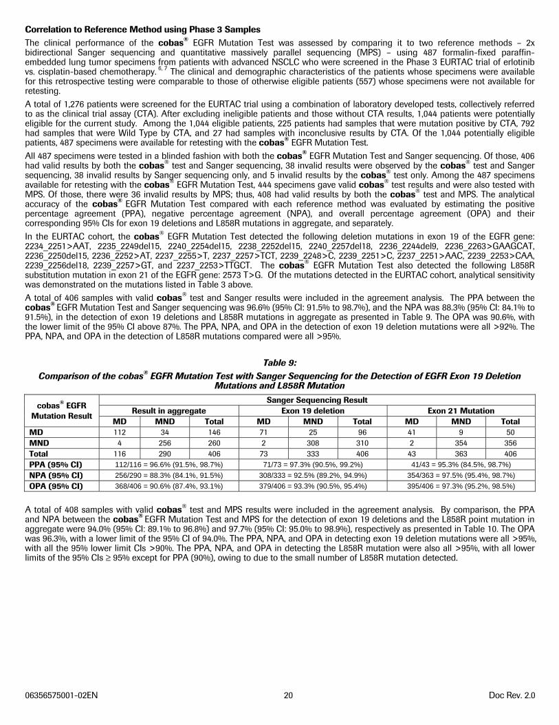
Correlation to Reference Method using Phase 3 Samples ............................................................................................................................................................ 20
Clinical Outcome Data .................................................................................................................................................................................................................................. 21
REFERENCES ........................................................................................................................................................................................................................................................ 22
INTENDED USE The cobas® EGFR Mutation Test is a real-time PCR test for the qualitative detection of exon 19 deletions and exon 21 (L858R) substitution mutations of the epidermal growth factor receptor (EGFR) gene in DNA derived from formalin-fixed paraffin-embedded (FFPET) human non-small cell lung cancer (NSCLC) tumor tissue. The test is intended to be used as an aid in selecting patients with NSCLC for whom Tarceva® (erlotinib), an EGFR tyrosine kinase inhibitor (TKI), is indicated.
Specimens are processed using the cobas® DNA Sample Preparation Kit for manual sample preparation and the cobas z 480 analyzer for automated amplification and detection.
SUMMARY AND EXPLANATION OF THE TEST Activating mutations in the gene encoding EGFR occur primarily in NSCLC, and result in constitutive activation of the kinase activity of the EGFR protein, thereby contributing to the oncogenic process.1 The prevalence of these mutations in unselected cases of NSCLC is approximately 10% - 30%.2, 3 However, these mutations occur more frequently, but not exclusively, in non-smoking/light-smoking female patients of Asian ancestry with adenocarcinoma histologies.4
The most common EGFR mutations in NSCLC include a variety of deletions in exon 19 and the substitution mutation L858R in exon 21; these mutations collectively constitute approximately 85% of EGFR mutations observed in NSCLC.5 The cobas® EGFR Mutation Test is a real-time PCR assay designed to detect deletion mutations in exon 19 and the substitution mutation L858R in exon 21. The cobas® EGFR Mutation Test is used as a companion diagnostic test for Tarceva®, a compound that reversibly inhibits the kinase activity of EGFR, preventing autophosphorylation of tyrosine residues associated with the receptor and thereby inhibiting further downstream signaling that promotes cell survival and proliferation. Erlotinib binding affinity for EGFR exon 19 deletion or exon 21 L858R mutations is higher than its affinity for the wild-type receptor.6 Clinical trials have shown that patients with advanced NSCLC and with exon 19 deletion mutations or L858R substitution mutation in exon 21 that were treated with Tarceva® as first-line treatment, are likely to experience clinical benefit compared to patients treated with chemotherapy.3, 7
The cobas® EGFR Mutation Test detects the following deletion mutations in exon 19 of the EGFR gene: 2235_2249del15, 2236_2250del15, 2238_2252del15, 2239_2248>C, 2240_2254del15, 2240_2257del18, 2237_2253>TTGCT, 2237_2255>T, 2239_2256del18, and 2239_2257>GT, as well as the L858R substitution mutation 2573 T>G in exon 21 of the EGFR gene. All mutations are detected at 5% sensitivity with the exception of the 2240_2257del18 exon 19 deletion mutation, which is detected at a sensitivity of >10% (Table 3).
06356575001-02EN 3 Doc Rev. 2.0
PRINCIPLES OF THE PROCEDURE The cobas® EGFR Mutation Test is based on two major processes: (1) manual specimen preparation to obtain genomic DNA from FFPET; and (2) PCR amplification and detection of target DNA using complementary primer pairs and oligonucleotide probes labeled with fluorescent dyes. The test is designed to detect deletions and complex mutations in exon 19 and L858R in exon 21.
Mutation detection is achieved through PCR analysis with the cobas z 480 analyzer. A mutant control and negative control are included in each run to confirm the validity of the run.
Specimen Preparation FFPET specimens are processed and genomic DNA isolated using the cobas® DNA Sample Preparation Kit, a manual specimen preparation based on nucleic acid binding to glass fibers. A deparaffinized 5 μm section of an FFPET specimen is lysed by incubation at an elevated temperature with a protease and chaotropic lysis/binding buffer that releases nucleic acids and protects the released genomic DNA from DNases. Subsequently, isopropanol is added to the lysis mixture that is then centrifuged through a column with a glass fiber filter insert. During centrifugation, the genomic DNA is bound to the surface of the glass fiber filter. Unbound substances, such as salts, proteins and other cellular impurities, are removed by centrifugation. The adsorbed nucleic acids are washed and then eluted with an aqueous solution. The amount of genomic DNA is spectrophotometrically determined and adjusted to a fixed concentration to be added to the amplification/detection mixture. The target DNA is then amplified and detected on the cobas z 480 analyzer using the amplification and detection reagents provided in the cobas® EGFR Mutation Test kit.
PCR Amplification Target Selection The cobas® EGFR Mutation Test kit uses primers that define specific base-pair sequences for each of the targeted mutations. For the exon 19 deletion mutations, base pair sequences that range from 125 to 141 are targeted and; for the L858R substitution mutation in exon 21, a 138 base pair sequence is targeted; for the internal control in exon 28, a 87 base pair sequence is targeted. Amplification occurs only in the regions of the EGFR gene between the primers; the entire EGFR gene is not amplified.
Target Amplification A derivative of Thermus species Z05-AS1 DNA polymerase is utilized for target amplification. First, the PCR reaction mixture is heated to denature the genomic DNA and expose the primer target sequences. As the mixture cools, the upstream and downstream primers anneal to the target DNA sequences. The Z05 DNA polymerase, in the presence of divalent metal ion and excess dNTP, extends each annealed primer, thus synthesizing a second DNA strand. This completes the first cycle of PCR, yielding a double-stranded DNA copy which includes the targeted base-pair regions of the EGFR gene. This process is repeated for a number of cycles, with each cycle effectively doubling the amount of amplicon DNA.
Automated Real-time Mutation Detection The cobas® EGFR Mutation Test utilizes real-time PCR technology. Each target-specific, oligonucleotide probe in the reaction is labeled with a fluorescent dye that serves as a reporter, and with a quencher molecule that absorbs (quenches) fluorescent emissions from the reporter dye within an intact probe. During each cycle of amplification, probe complementary to the single-stranded DNA sequence in the amplicon binds and is subsequently cleaved by the 5’ to 3’ nuclease activity of the Z05-AS1 DNA Polymerase. Once the reporter dye is separated from the quencher by this nuclease activity, fluorescence of a characteristic wavelength can be measured when the reporter dye is excited by the appropriate spectrum of light. Three different reporter dyes are used to label the mutations targeted by the test. Amplification of the targeted EGFR sequences are detected independently across three reactions by measuring fluorescence at the three characteristic wavelengths in dedicated optical channels.
Selective Amplification Selective amplification of target nucleic acid from the specimen is achieved in the cobas® EGFR Mutation Test by the use of AmpErase (uracil-N-glycosylase) enzyme and deoxyuridine triphosphate (dUTP).8 The AmpErase enzyme recognizes and catalyzes the destruction of DNA strands containing deoxyuridine but not DNA containing thymidine. Deoxyuridine is not present in naturally occurring DNA but is always present in amplicon due to the use of dUTP in place of deoxythymidine triphosphate as one of the nucleotide triphosphates in the Master Mix reagents; therefore, only amplicon contains deoxyuridine. Deoxyuridine renders contaminating amplicon susceptible to destruction by AmpErase enzyme prior to amplification of the target DNA. The AmpErase enzyme, which is included in the Master Mix reagents, catalyzes the cleavage of deoxyuridine-containing DNA at the deoxyuridine residues by opening the deoxyribose chain at the C1-position. When heated in the first thermal cycling step at alkaline pH, the amplicon DNA chain breaks at the position of the deoxyuridine, thereby rendering the DNA non-amplifiable. The AmpErase enzyme is inactive at temperatures above 55ºC, i.e., throughout the thermal cycling steps, and therefore does not destroy target amplicon.
06356575001-02EN 4 Doc Rev. 2.0
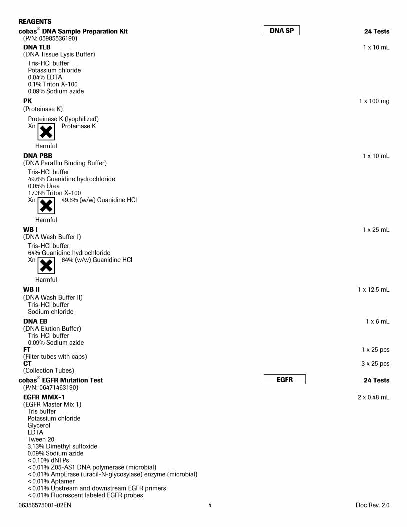
REAGENTS cobas® DNA Sample Preparation Kit 24 Tests
(P/N: 05985536190) DNA TLB 1 x 10 mL (DNA Tissue Lysis Buffer)
Tris-HCl buffer Potassium chloride 0.04% EDTA 0.1% Triton X-100 0.09% Sodium azide
PK 1 x 100 mg (Proteinase K)
Proteinase K (lyophilized) Xn Proteinase K
Harmful
DNA PBB 1 x 10 mL (DNA Paraffin Binding Buffer)
Tris-HCl buffer 49.6% Guanidine hydrochloride 0.05% Urea 17.3% Triton X-100 Xn 49.6% (w/w) Guanidine HCI
Harmful
WB I 1 x 25 mL (DNA Wash Buffer I)
Tris-HCl buffer 64% Guanidine hydrochloride Xn 64% (w/w) Guanidine HCI
Harmful
WB II 1 x 12.5 mL (DNA Wash Buffer II)
Tris-HCl buffer Sodium chloride
DNA EB 1 x 6 mL (DNA Elution Buffer)
Tris-HCl buffer 0.09% Sodium azide
FT 1 x 25 pcs (Filter tubes with caps) CT 3 x 25 pcs (Collection Tubes)
cobas® EGFR Mutation Test 24 Tests (P/N: 06471463190) EGFR MMX-1 2 x 0.48 mL (EGFR Master Mix 1)
Tris buffer Potassium chloride Glycerol EDTA Tween 20 3.13% Dimethyl sulfoxide 0.09% Sodium azide <0.10% dNTPs <0.01% Z05-AS1 DNA polymerase (microbial) <0.01% AmpErase (uracil-N-glycosylase) enzyme (microbial) <0.01% Aptamer <0.01% Upstream and downstream EGFR primers <0.01% Fluorescent labeled EGFR probes
DNA SP
EGFR
DNA SP
EGFR
06356575001-02EN 5 Doc Rev. 2.0
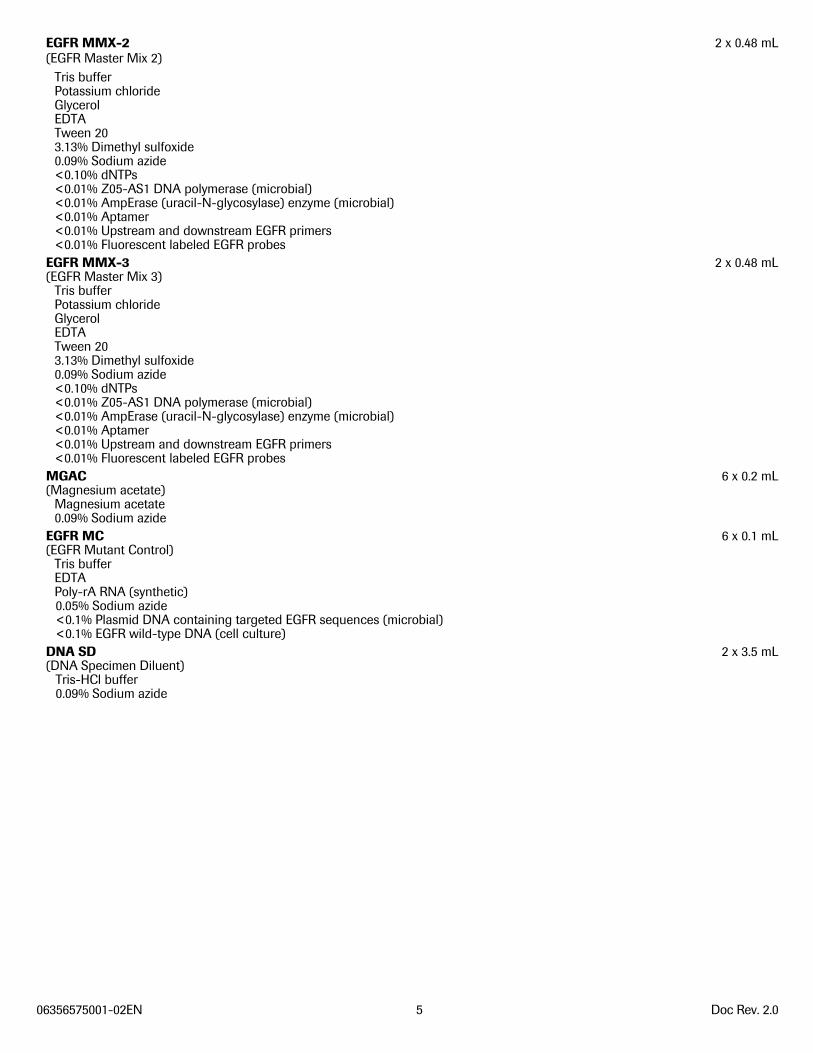
EGFR MMX-2 2 x 0.48 mL (EGFR Master Mix 2)
Tris buffer Potassium chloride Glycerol EDTA Tween 20 3.13% Dimethyl sulfoxide 0.09% Sodium azide <0.10% dNTPs <0.01% Z05-AS1 DNA polymerase (microbial) <0.01% AmpErase (uracil-N-glycosylase) enzyme (microbial) <0.01% Aptamer <0.01% Upstream and downstream EGFR primers <0.01% Fluorescent labeled EGFR probes
EGFR MMX-3 2 x 0.48 mL (EGFR Master Mix 3)
Tris buffer Potassium chloride Glycerol EDTA Tween 20 3.13% Dimethyl sulfoxide 0.09% Sodium azide <0.10% dNTPs <0.01% Z05-AS1 DNA polymerase (microbial) <0.01% AmpErase (uracil-N-glycosylase) enzyme (microbial) <0.01% Aptamer <0.01% Upstream and downstream EGFR primers <0.01% Fluorescent labeled EGFR probes
MGAC 6 x 0.2 mL (Magnesium acetate)
Magnesium acetate 0.09% Sodium azide
EGFR MC 6 x 0.1 mL (EGFR Mutant Control)
Tris buffer EDTA Poly-rA RNA (synthetic) 0.05% Sodium azide <0.1% Plasmid DNA containing targeted EGFR sequences (microbial) <0.1% EGFR wild-type DNA (cell culture)
DNA SD 2 x 3.5 mL (DNA Specimen Diluent)
Tris-HCl buffer 0.09% Sodium azide
06356575001-02EN 6 Doc Rev. 2.0
WARNINGS AND PRECAUTIONS
A. FOR IN VITRO DIAGNOSTIC USE.
B. This test is for use with FFPET NSCLC specimens.
C. Do not pipette by mouth.
D. Do not eat, drink or smoke in laboratory work areas.
E. Avoid microbial and DNA contamination of reagents.
F. Dispose of unused reagents and waste in accordance with country, federal, state and local regulations.
G. Do not use kits after their expiration dates.
H. Do not pool reagents from different kits or lots.
I. Gloves must be worn and must be changed between handling specimens and reagents to prevent contamination.
J. To avoid contamination of the working Master Mix (working MMX) with DNA specimens, amplification and detection should be performed in an area separated from DNA Isolation. The amplification and detection work area should be thoroughly cleaned before working MMX preparation. For proper cleaning, all surfaces including racks and pipettors should be thoroughly wiped with 0.5% sodium hypochlorite solution followed by wiping with a 70% ethanol solution.
K. DNA PBB and WB I contain guanidine hydrochloride. If liquid containing this buffer is spilled, clean with suitable laboratory detergent and water. If a spill occurs with potentially infectious agents, clean the affected area first with laboratory detergent and water, and then with 0.5% sodium hypochlorite*. If spills occur on the cobas z 480 analyzer, follow the instructions in the cobas z 480 analyzer Instrument Manual.
*NOTE: Commercial liquid household bleach typically contains sodium hypochlorite at a concentration of 5.25%. A 1:10 dilution of household bleach will produce a 0.5% sodium hypochlorite solution.
L. Specimens should be handled as infectious using safe laboratory procedures such as those outlined in Biosafety in Microbiological and Biomedical Laboratories9 and in the CLSI Document M29-A3.10
M. DNA PBB contains Triton X-100, an irritant to mucous membranes. Avoid contact with eyes, skin, and mucous membranes.
N. DNA TLB, DNA EB, MGAC, EGFR MMX-1, EGFR MMX-2, EGFR MMX-3, EGFR MC, and DNA SD contain sodium azide. Sodium azide may react with lead and copper plumbing to form highly explosive metal azides. While disposing of sodium azide containing solutions down laboratory sinks, flush the drains with a large volume of cold water to prevent azide buildup.
O. Xylene is a hazardous chemical and should be used in a chemical hood. Discard into chemical waste in accordance with local, state, and federal regulations.
P. Wear eye protection, laboratory coats, and disposable gloves when handling any reagents. Avoid contact of these materials with the skin, eyes, or mucous membranes. If contact does occur, immediately wash with large amounts of water. Burns can occur if left untreated. If spills occur, dilute with water before wiping dry.
Q. All disposable items are for one time use. Do not reuse.
R. Do not use disposable items beyond their expiration date.
S. Do not use sodium hypochlorite solution (bleach) for cleaning the cobas z 480 analyzer. Clean the cobas z 480 analyzer according to procedures described in the cobas z 480 analyzer Instrument Manual.
T. For additional warnings, precautions and procedures to reduce the risk of contamination for the cobas z 480 analyzer, consult the cobas z 480 analyzer Instrument Manual.
U. The use of sterile disposable pipettes and DNase-free pipettor tips is recommended.
STORAGE AND HANDLING REQUIREMENTS A. With the exception of the PK reagent, do not freeze reagents.
B. Store DNA TLB, DNA PBB, WB I, WB II, DNA EB, PK, FT, and CT at 15ºC to 30ºC. Once opened, DNA TLB, DNA PBB, WB I, WB II, DNA EB, and PK are stable for up to 8 uses over 90 days or until the expiration date, whichever comes first.
C. After addition of sterile, nuclease free water to PK, store unused reconstituted PK in 450 μL aliquots at -20ºC. Once reconstituted, PK must be used within 90 days or until the expiration date, whichever comes first.
D. After addition of absolute ethanol, store WB I and WB II at 15ºC to 30ºC. These working solutions are stable for 90 days or until the expiration date, whichever comes first.
E. Store MGAC, EGFR MMX-1, EGFR MMX-2, EGFR MMX-3, EGFR MC, and DNA SD at 2ºC to 8°C. Once opened, these reagents are stable for 4 uses over 90 days or until the expiration date, whichever comes first.
F. EGFR MMX-1, EGFR MMX-2, EGFR MMX-3, and working MMX (prepared by the addition of MGAC to EGFR MMX-1 or EGFR MMX-2 or EGFR MMX-3) should be protected from prolonged exposure to light.
G. Working MMX must be stored at 2ºC to 8ºC in the dark. The prepared specimens and controls must be added within 1 hour of preparation of the working MMX.
06356575001-02EN 7 Doc Rev. 2.0
H. Processed specimens (extracted DNA) are stable for up to 24 hours at 15ºC to 30°C or up to 14 days at 2ºC to 8°C or up to 60 days at -15ºC to -25°C or after undergoing 3 freeze thaws when stored at -15ºC to -25°C. Extracted DNA should be amplified within the recommended storage periods or before the expiration date of the cobas® DNA Sample Preparation Kit used to extract the DNA, whichever comes first.
I. Amplification must be started within 1 hour from the time that the processed specimens and controls are added to the working MMX (prepared by the addition of MGAC to EGFR MMX-1 or EGFR MMX-2 or EGFR MMX-3).
MATERIALS PROVIDED
A. cobas® DNA Sample Preparation Kit 24 Tests (P/N: 05985536190)
DNA TLB (DNA Tissue Lysis Buffer)
PK (Proteinase K)
DNA PBB (DNA Paraffin Binding Buffer)
WB I (DNA Wash Buffer I)
WB II (DNA Wash Buffer II)
DNA EB (DNA Elution Buffer)
FT (Filter tubes with caps)
CT (Collection Tubes)
B. cobas® EGFR Mutation Test 24 Tests (P/N: 06471463190)
MGAC (Magnesium acetate) (Cap with Yellow Button)
EGFR MMX-1 (EGFR Master Mix 1) (Cap with White Button)
EGFR MMX-2 (EGFR Master Mix 2) (Cap with Gold Button)
EGFR MMX-3 (EGFR Master Mix 3) (Cap with Teal Button)
EGFR MC (EGFR Mutant Control) (Cap with Red Button)
DNA SD (DNA Specimen Diluent)
MATERIALS REQUIRED BUT NOT PROVIDED • Xylene (ACS, ≥ 98.5% xylenes)
• Absolute ethanol (for Molecular Biology)
• Isopropanol (ACS, ≥ 99.5%)
• Sterile, nuclease-free water (For Molecular Biology)
• Sterile disposable, serological pipettes: 5 and 25 mL
• cobas® 4800 System Microwell Plate (AD-Plate) and Sealing Foil (Roche P/N 05232724001)
• cobas® 4800 Sealing Foil Applicator (Roche P/N 04900383001)
• Adjustable Pipettors* (capacity 10 μL, 20 μL, 200 μL, and 1000 μL) with aerosol barrier or positive displacement DNase-free tips
• Pipette aid (Drummond P/N: 4-000-100 or equivalent)
• Bench top microcentrifuge capable of 20,000 x g**
DNA SP
EGFR
DNA SP
EGFR
06356575001-02EN 8 Doc Rev. 2.0
• Two (2) dry heat blocks capable of heating microcentrifuge tubes to 56ºC and 90ºC**
• 1.5 mL Safe-Lock microcentrifuge tubes, sterile, RNase/DNase free, PCR grade (Eppendorf, Cat# 022363212)
• Spectrophotometer for measuring DNA concentration **
• Vortex mixer**
• Microcentrifuge tube racks
• Disposable gloves, powder-free
• Calibrated thermometers for dry heat block**
• Waterbath** capable of maintaining 37ºC
• Single edged blade or similar
* Pipettors should be maintained according to the manufacturer’s instructions and accurate within 3% of stated volume. Aerosol barrier or positive displacement DNase-free tips must be used where specified to prevent specimen degradation and cross-contamination.
** All equipment should be properly maintained according to manufacturer’s instructions.
Instrumentation and Software • cobas z 480 analyzer
• cobas® 4800 SR2 System Control Unit with OSXP image
• cobas® 4800 SR2 System Software version 2.0 or higher configured with the EGFR Analysis Package
• Barcode Reader ext USB
• Printer
SPECIMEN COLLECTION, TRANSPORT, AND STORAGE NOTE: Handle all specimens as if they are capable of transmitting infectious agents.
A. Specimen Collection NSCLC FFPET specimens have been validated for use with the cobas® EGFR Mutation Test.
B. Specimen Transport NSCLC FFPET specimens can be transported at 15ºC to 30ºC. Transportation of FFPET specimens must comply with country, federal, state, and local regulations for the transport of etiologic agents.12
C. Specimen Storage Stability of FFPET specimens stored at 15-30°C for up to 12 months after the date of collection has been confirmed. 5 micron sections mounted on slides may be stored at 15-30°C for up to 60 days.
INSTRUCTIONS FOR USE NOTE: Only NSCLC FFPET sections of 5 μm thickness containing at least 10% tumor content by area are to be used in
the cobas® EGFR Mutation Test. Any specimen containing less that 10% tumor content by area should be macro-dissected after deparaffinization.
NOTE: Refer to the cobas z 480 analyzer Instrument Manual for detailed operating instructions for the cobas z 480 analyzer.
NOTE: Dry heat blocks capable of heating microcentrifuge tubes should be turned on and set at 56°C and 90°C.
Run Size A single run can include from 1 to 30 specimens (plus controls) per 96 well Microwell plate. When running more than 24 specimens, multiple cobas® EGFR Mutation Test kits will be required.
The cobas® EGFR Mutation Test contains sufficient reagents for 8 runs of 3 specimens (plus controls) for a maximum of 24 specimens per kit.
Workflow The cobas® EGFR Mutation Test consists of manual specimen preparation using the cobas® DNA Sample Preparation Kit followed by amplification/detection on the cobas z 480 analyzer using the cobas® EGFR Mutation Test kit.
06356575001-02EN 9 Doc Rev. 2.0
Reagent Preparation A. Reconstitute Proteinase K (PK) by adding 4.5 mL of sterile, nuclease-free (PCR grade) water to the vial using a sterile,
disposable 5-mL serological pipette. Mix by inverting the vial 5 to 10 times. Aliquot 450 μL of reconstituted PK into 1.5 mL Safe-Lock microcentrifuge tubes and store at -20ºC. If the Proteinase K has already been reconstituted and frozen, thaw sufficient number of aliquots to process the number of specimens to be run prior to deparaffinization (70 μL of reconstituted PK is required for each specimen).
B. All solutions stored at 15-30°C should be clear. If precipitate is present in any reagent, warm the solution in a 37°C water bath until the precipitate dissolves. Do not use until all precipitate has been dissolved.
C. Prepare working DNA Wash Buffer I (WB I) by adding 15 mL of absolute ethanol to the bottle of WB I. Mix by inverting the bottle 5 to 10 times. Make a note on the bottle that ethanol has been added and the date. Store working WB I at 15ºC to 30ºC.
D. Prepare working DNA Wash Buffer II (WB II) by adding 50 mL of absolute ethanol to the bottle of WB II. Mix by inverting the bottle 5 to 10 times. Make a note on the bottle that ethanol has been added and the date. Store working WB II at 15ºC to 30ºC.
Deparaffinization of FFPET Sections Mounted on Slides NOTE: Xylene is a hazardous chemical. All steps for deparaffinization should be performed under a chemical hood. See
Warnings and Precautions.
NOTE: If the specimen contains less than 10% tumor content by area, the section must be macro-dissected
A. Add a slide with a mounted 5 μm FFPET section to a container with sufficient xylene to cover the tissue; soak for 5 minutes.
B. Transfer the slide to a container with sufficient absolute ethanol to cover the tissue; soak for 5 minutes.
C. Remove the slide from the ethanol and allow the section to air dry completely (5 to 10 minutes).
D. Perform macro-dissection if the specimen contains less than 10% tumor content by area.
E. Label one 1.5 mL Safe-Lock microcentrifuge tube for each specimen with the specimen identification information.
F. Add 180 μL DNA TLB to the 1.5-mL Safe-Lock microcentrifuge tube.
G. Add 70 μL of reconstituted PK to the Safe-Lock tube containing DNA TLB.
H. Scrape the tissue off the slide and into the Safe-Lock tube. Immerse the tissue in the DNA TLB/PK mixture.
I. Continue with Step A of the DNA Isolation procedure.
Deparaffinization of FFPET Sections not Mounted on Slides NOTE: Xylene is a hazardous chemical. All steps for deparaffinization should be performed under a chemical hood. See
Warnings and Precautions.
NOTE: If the specimen contains less than 10% tumor content by area, the section must be mounted on a slide for macro-dissection and the procedure detailed in ‘Deparaffinization of FFPET Sections Mounted on Slides’ must be followed.
A. Place one 5-micron FFPET section into a 1.5 mL Safe-Lock microcentrifuge tube labeled with the specimen identification information for each specimen.
B. Add 500 μL Xylene to the Safe-Lock tube containing the FFPET section.
C. Mix well by vortexing for 10 seconds.
D. Let the tube stand for 5 minutes at 15°C to 30°C.
E. Add 500 μL absolute ethanol and mix by vortexing for 10 seconds.
F. Let the tube stand for 5 minutes at 15°C to 30°C.
G. Centrifuge at 16,000 x g to 20,000 x g for 2 minutes. Remove the supernatant without disturbing the pellet. Discard the supernatant into chemical waste.
H. Add 1 mL absolute ethanol and vortex for 10 seconds.
I. Centrifuge at 16,000 x g to 20,000 x g for 2 minutes. Remove the supernatant without disturbing the pellet. Discard the supernatant into chemical waste.
NOTE: If the pellet is floating in the remaining supernatant, spin again for 1 minute at 16,000 x g to 20,000 x g. Remove any remaining supernatant.
J. Dry the tissue pellet for 10 minutes at 56°C in a heating block with the tube open.
NOTE: Make sure the ethanol is completely evaporated and the pellet is dry before proceeding to the next step.
NOTE: If needed, dry pellets can be stored up to 24 hours at 2°C to 8°C.
06356575001-02EN 10 Doc Rev. 2.0
K. Resuspend the tissue pellet in 180 μL DNA Tissue Lysis Buffer (DNA TLB).
L. Add 70 μL of reconstituted PK.
M. Continue with Step A of the DNA Isolation procedure.
SPECIMEN PREPARATION DNA Isolation Procedure NOTE: Process a negative control concurrently with the specimen(s). Prepare the negative control by combining 180 μL
DNA Tissue Lysis Buffer (DNA TLB) and 70 μL PK solution in a 1.5 mL Safe-Lock microcentrifuge tube labeled as NEG CT. The negative control should be processed following the same procedure as the specimens.
A. Vortex the tubes containing the specimen/DNA TLB/PK mixture and the negative control mixture (NEG CT) for 30 seconds.
NOTE: The tissue must be fully immersed in the DNA TLB/PK mixture.
B. Place tubes in the 56ºC dry heat block and incubate for 60 minutes.
C. Vortex the tubes for 10 seconds.
NOTE: The tissue must be fully immersed in the DNA TLB/PK mixture.
D. Place tubes in the 90ºC dry heat block and incubate for 60 minutes.
NOTE: During the incubation, prepare the required number of filter tubes (FTs) with hinged caps by placing the FT onto a collection tube (CT) and labeling each FT cap with the proper specimen or control identification.
NOTE: Each specimen will need 1 FT, 3 CTs and 1 elution tube (1.5 mL microcentrifuge tube).
NOTE: During the incubation, label the required number of elution tubes (1.5 mL microcentrifuge tube) with the proper specimen or control identification information.
E. Allow the tubes to cool to 15°C to 30°C. After cooling, pulse-centrifuge the tubes to collect liquid from the caps.
F. Add 200 μL DNA PBB to each tube; mix by pipetting up and down 3 times.
G. Incubate the tubes at 15°C to 30°C for 10 minutes.
H. Add 100 μL isopropanol to each tube; mix lysate by pipetting up and down 3 times.
I. Transfer each lysate into the appropriately labeled FT/CT unit.
J. Centrifuge the FT/CT units at 8,000 x g for 1 minute.
K. Place each FT onto a new CT. Discard the flow-through from the old CT into chemical waste, and properly dispose of the used CT.
L. Add 500 μL working WB I to each FT.
NOTE: Preparation of working WB I is described in the Reagent Preparation section.
M. Centrifuge the FT/CT units at 8,000 x g for 1 minute.
N. Discard the flow-through in each CT into chemical waste. Place the FT back into the same CT.
O. Add 500 μL working WB II to each FT.
NOTE: Preparation of working WB II is described in the Reagent Preparation section.
P. Centrifuge the FT/CT units at 8,000 x g for 1 minute.
Q. Place each FT onto a new CT. Discard the flow-through from the old CT into chemical waste, and properly dispose of the used CT.
R. Centrifuge the FT/CT units at 16,000 to 20,000 x g for 1 minute to dry the filter membranes.
S. Place each FT into an elution tube (1.5 mL microcentrifuge tube) pre-labeled with specimen or control identification. Discard the flow-through from the used CT into chemical waste, and properly dispose of the used CT.
T. Add 100 μL DNA EB to the center of each FT membrane without touching the FT membrane.
U. Incubate the FT with elution tube at 15°C to 30°C for 5 minutes.
V. Centrifuge the FT with elution tube at 8,000 x g for 1 minute to collect eluate into the elution tube. Properly dispose of the used FT.
W. Close the cap on the elution tube. The elution tube contains the DNA Stock. Proceed to Step A in the DNA Quantitation section.
NOTE: Measurement of DNA concentration should be performed immediately after the DNA Isolation procedure and prior to storage.
06356575001-02EN 11 Doc Rev. 2.0
DNA Quantitation:
A. Mix each DNA Stock by vortexing for 5 seconds.
B. Quantify DNA using a spectrophotometer according to the manufacturer’s protocol. Use DNA EB as the blank for the instrument. An average of two consistent readings is necessary. The two measurements should be within ±10% of each other when the DNA concentration readings are ≥ 20.0 ng/μL. For DNA concentration readings < 20.0 ng/μL, the two measurements should be within ± 2 ng/μL. If the two measurements are not within +/- 10% of each other when the DNA concentration readings are ≥20.0 ng/μL or within +/- 2 ng/μL when the DNA concentration readings are < 20.0 ng/μL, an additional 2 readings must be taken until the requirements are met. The average of these two new measurements should then be calculated.
NOTE: The DNA Stock from the processed negative control (NEG CT) does not need to be measured.
C. The DNA Stock concentration from the specimens must be ≥2 ng/μL to perform the cobas® EGFR Mutation Test. Three amplification/detections are run per specimen, using 25 uL of a 2 ng/μL dilution of DNA Stock (total of 50 ng DNA) for each amplification/detection.
NOTE: Each DNA Stock must have a minimum concentration of 2 ng/μL to perform the cobas® EGFR Mutation Test. If the concentration of a DNA Stock is <2 ng/μL, repeat the deparaffinization, DNA Isolation, and DNA Quantitation procedures for that specimen using two 5 μm FFPET sections. For mounted specimens, after deparaffinization, combine the tissue from both sections into one tube, immerse the tissue in DNA TLB + PK, and perform DNA Isolation and Quantitation as described above. For unmounted specimens, combine two sections into one tube and immerse the tissue in DNA TLB + PK, and perform DNA Isolation and Quantitation as described above. If the DNA Stock is still <2 ng/μL, request another FFPET specimen section from the referring clinical site.
NOTE: Processed specimens (extracted DNA) are stable for up to 24 hours at 15ºC to 30°C or up to 14 days at 2ºC to 8°C or up to 60 days at -15ºC to -25°C or after undergoing 3 freeze thaws when stored at -15ºC to -25°C. Extracted DNA should be amplified within the recommended storage periods or before the expiration date of the cobas® DNA Sample Preparation Kit used to extract the DNA, whichever comes first.
AMPLIFICATION AND DETECTION NOTE: To avoid contamination of working MMX with DNA specimens, amplification and detection should be performed
in an area separated from DNA Isolation. The amplification and detection work area should be thoroughly cleaned before working MMX preparation. For proper cleaning, all surfaces including racks and pipettors should be thoroughly wiped with 0.5% sodium hypochlorite solution followed by wiping with a 70% ethanol solution. Commercial liquid household bleach typically contains sodium hypochlorite at a concentration of 5.25%. A 1:10 dilution of household bleach will produce a 0.5% sodium hypochlorite solution.
Instrument Set-Up: Refer to the cobas z 480 analyzer Instrument Manual for detailed instruction for the cobas z 480 set up.
Test Order Set-up: Refer to the cobas® 4800 system Operator’s Manual Software Version 2.0 for cobas® EGFR Mutation Test (cobas® 4800 EGFR Operator’s Manual) for detailed instructions on the EGFR workflow steps.
Dilution Calculation of Specimen DNA Stock: Dilution Calculation for DNA Stock Concentrations from 2 ng/μL to 36 ng/μL
NOTE: DNA stocks from specimens should be diluted immediately prior to amplification and detection.
NOTE: Three (3) amplification/detections are run for each specimen requiring a total volume of 75 μL (25 μL for each of three reactions) of a 2 ng/μL dilution of DNA Stock (total of 150 ng DNA).
A. For each specimen, calculate the volume (μL) of DNA stock needed:
μL of DNA stock = (90 μL x 2 ng/μL) ÷ DNA Stock concentration [ng/μL]
B. For each specimen, calculate the volume (μL) of DNA Specimen Diluent (DNA SD) needed:
μL of DNA SD = 90 μL – μL of DNA Stock
Example:
DNA stock concentration = 6.5 ng/μL
A. μL of DNA Stock = (90 μL x 2 ng/μL) ÷ 6.5 ng/μL = 27.7 μL
B. μL of DNA SD = (90 μL – 27.7 μL) = 62.3 μL
Dilution Calculation for DNA Stock Concentrations >36 ng/μL
NOTE: DNA Stocks from specimens should be diluted immediately prior to amplification and detection.
NOTE: Three (3) amplification/detections are run for each specimen requiring a total volume of 75 μL (25 μL for each of three reactions) of a 2 ng/μL dilution of DNA stock (total of 150 ng DNA).
06356575001-02EN 12 Doc Rev. 2.0
A. At DNA Stock concentrations > 36 ng/μL, use the following formula to calculate the amount of DNA Specimen Diluent (DNA SD) required to prepare at least 90 μL of diluted DNA stock. This is to ensure that each specimen uses a minimum of 5 μL of DNA stock.
B. For each specimen, calculate the volume (μL) of DNA SD needed to dilute 5 μL of DNA Stock to 2 ng/μL: Vol. of DNA SD required in μL = [(5 μL of DNA stock x DNA stock concentration in ng/μL) / 2 ng/μL] – 5 μL
Example:
DNA stock concentration = 100 ng/μL
A. Vol. of DNA SD required in μL = [(5 μL x 100 ng/μL) / 2 ng/μL] – 5 μL = 245 μL
B. Use the calculated volume of DNA SD to dilute 5 μL of DNA stock.
Specimen Dilution A. Prepare the appropriate number of 1.5 mL Safe-Lock microcentrifuge tubes for DNA Dilutions by labeling them with the proper
specimen identification.
B. Using a pipettor with an aerosol-resistant tip, pipette the calculated volumes of DNA SD into the respectively labeled tubes. Pipette 45 μL of DNA SD into a Safe-Lock tube labeled as NEG CT.
C. Vortex each DNA stock and the negative control for 5 to 10 seconds.
D. Using a pipettor with an aerosol-resistant pipette tip (new tip for each pipetting), gently pipette the calculated volume of each DNA stock into the respective tube containing DNA SD. Pipette 45 μL of negative control (extracted eluate) into the NEG CT tube.
E. Cap the tubes and vortex each for 5 to 10 seconds.
F. Change gloves.
Preparation of Working Master Mixes (MMX-1, MMX-2 and MMX-3) NOTE: EGFR MMX-1, EGFR MMX-2, EGFR MMX-3, and working MMX are light-sensitive and must be protected from
prolonged exposure to light.
NOTE: Due to the viscosity of the EGFR MIXES and working MMX, pipette slowly to ensure all mix is completely dispensed from the tip.
NOTE: The EGFR MMX-1, EGFR MMX-2, and EGFR MMX-3 may appear light blue/purplish. This does not affect the performance of the reagent.
Prepare three bulk working MMX, one containing EGFR MMX-1, one containing EGFR MMX-2, and the other containing EGFR MMX-3 in separate 1.5 mL Safe-Lock microcentrifuge tubes.
A. Calculate the volume of EGFR MMX-1 or EGFR MMX-2 or EGFR MMX-3 required for each working MMX using the following formula:
Volume of EGFR MMX-1 or EGFR MMX-2 or EGFR MMX-3 required = (Number of Specimens + 2 Controls +1) x 20 μL
B. Calculate the volume of MGAC required for each working MMX using the following formula: Volume of MGAC required = (Number of Specimens + 2 Controls +1) x 5 μL
Use Table 1 to determine the volume of each reagent needed for the preparation of working MMX based on the number of specimens included in the run.
Table 1: Volumes of Reagents Needed for Working MMX-1, Working MMX-2 and Working MMX-3
# of Specimens* 1 2 3 4 5 6 7 8 9 10
MMX 20 μL 80 100 120 140 160 180 200 220 240 260
MGAC 5 μL 20 25 30 35 40 45 50 55 60 65
Total Vol. for Each Working MMX (μL) 100 125 150 175 200 225 250 275 300 325
* Volumes for # of Specimens is based on the sum of the # Specimens + 2 Controls + 1
C. Remove the appropriate number of EGFR MMX-1, EGFR MMX-2, EGFR MMX-3, and MGAC vials from 2°C to 8°C storage. Vortex each reagent for 5 seconds and collect liquid at the bottom of the tube before use. Label a sterile microcentrifuge tube for working MMX-1, working MMX-2, and working MMX-3.
D. Add the calculated volume of EGFR MMX-1 or EGFR MMX-2 or EGFR MMX-3 to their respective working MMX tube.
E. Add the calculated volume of MGAC to the working MMX tubes.
F. Vortex the tubes for 3 to 5 seconds to ensure adequate mixing.
06356575001-02EN 13 Doc Rev. 2.0
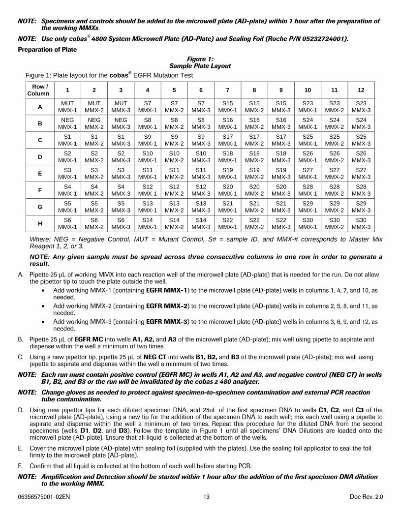
NOTE: Specimens and controls should be added to the microwell plate (AD-plate) within 1 hour after the preparation of the working MMXs.
NOTE: Use only cobas® 4800 System Microwell Plate (AD-Plate) and Sealing Foil (Roche P/N 05232724001).
Preparation of Plate Figure 1:
Sample Plate Layout Figure 1: Plate layout for the cobas® EGFR Mutation Test
Row / Column
1 2 3 4 5 6 7 8 9 10 11 12
A MUT
MMX-1 MUT
MMX-2 MUT
MMX-3 S7
MMX-1 S7
MMX-2 S7
MMX-3 S15
MMX-1 S15
MMX-2 S15
MMX-3 S23
MMX-1 S23
MMX-2 S23
MMX-3
B NEG
MMX-1 NEG
MMX-2 NEG
MMX-3 S8
MMX-1 S8
MMX-2 S8
MMX-3 S16
MMX-1 S16
MMX-2 S16
MMX-3 S24
MMX-1 S24
MMX-2 S24
MMX-3
C S1
MMX-1 S1
MMX-2 S1
MMX-3 S9
MMX-1 S9
MMX-2 S9
MMX-3 S17
MMX-1 S17
MMX-2 S17
MMX-3 S25
MMX-1 S25
MMX-2 S25
MMX-3
D S2
MMX-1 S2
MMX-2 S2
MMX-3 S10
MMX-1 S10
MMX-2 S10
MMX-3 S18
MMX-1 S18
MMX-2 S18
MMX-3 S26
MMX-1 S26
MMX-2 S26
MMX-3
E S3
MMX-1 S3
MMX-2 S3
MMX-3 S11
MMX-1 S11
MMX-2 S11
MMX-3 S19
MMX-1 S19
MMX-2 S19
MMX-3 S27
MMX-1 S27
MMX-2 S27
MMX-3
F S4
MMX-1 S4
MMX-2 S4
MMX-3 S12
MMX-1 S12
MMX-2 S12
MMX-3 S20
MMX-1 S20
MMX-2 S20
MMX-3 S28
MMX-1 S28
MMX-2 S28
MMX-3
G S5
MMX-1 S5
MMX-2 S5
MMX-3 S13
MMX-1 S13
MMX-2 S13
MMX-3 S21
MMX-1 S21
MMX-2 S21
MMX-3 S29
MMX-1 S29
MMX-2 S29
MMX-3
H S6
MMX-1 S6
MMX-2 S6
MMX-3 S14
MMX-1 S14
MMX-2 S14
MMX-3 S22
MMX-1 S22
MMX-2 S22
MMX-3 S30
MMX-1 S30
MMX-2 S30
MMX-3
Where: NEG = Negative Control, MUT = Mutant Control, S# = sample ID, and MMX-# corresponds to Master Mix Reagent 1, 2, or 3.
NOTE: Any given sample must be spread across three consecutive columns in one row in order to generate a result.
A. Pipette 25 μL of working MMX into each reaction well of the microwell plate (AD-plate) that is needed for the run. Do not allow the pipettor tip to touch the plate outside the well.
• Add working MMX-1 (containing EGFR MMX-1) to the microwell plate (AD-plate) wells in columns 1, 4, 7, and 10, as needed.
• Add working MMX-2 (containing EGFR MMX-2) to the microwell plate (AD-plate) wells in columns 2, 5, 8, and 11, as needed.
• Add working MMX-3 (containing EGFR MMX-3) to the microwell plate (AD-plate) wells in columns 3, 6, 9, and 12, as needed.
B. Pipette 25 μL of EGFR MC into wells A1, A2, and A3 of the microwell plate (AD-plate); mix well using pipette to aspirate and dispense within the well a minimum of two times.
C. Using a new pipettor tip, pipette 25 μL of NEG CT into wells B1, B2, and B3 of the microwell plate (AD-plate); mix well using pipette to aspirate and dispense within the well a minimum of two times.
NOTE: Each run must contain positive control (EGFR MC) in wells A1, A2 and A3, and negative control (NEG CT) in wells B1, B2, and B3 or the run will be invalidated by the cobas z 480 analyzer.
NOTE: Change gloves as needed to protect against specimen-to-specimen contamination and external PCR reaction tube contamination.
D. Using new pipettor tips for each diluted specimen DNA, add 25uL of the first specimen DNA to wells C1, C2, and C3 of the microwell plate (AD-plate), using a new tip for the addition of the specimen DNA to each well; mix each well using a pipette to aspirate and dispense within the well a minimum of two times. Repeat this procedure for the diluted DNA from the second specimens (wells D1, D2, and D3). Follow the template in Figure 1 until all specimens’ DNA Dilutions are loaded onto the microwell plate (AD-plate). Ensure that all liquid is collected at the bottom of the wells.
E. Cover the microwell plate (AD-plate) with sealing foil (supplied with the plates). Use the sealing foil applicator to seal the foil firmly to the microwell plate (AD-plate).
F. Confirm that all liquid is collected at the bottom of each well before starting PCR.
NOTE: Amplification and Detection should be started within 1 hour after the addition of the first specimen DNA dilution to the working MMX.
06356575001-02EN 14 Doc Rev. 2.0
Starting PCR Refer to the cobas® EGFR Operator’s Manual for detailed instructions on the EGFR workflow steps.
INTERPRETATION OF RESULTS NOTE: All run and specimen validation is performed by the cobas®4800 software.
NOTE: A valid test run may include both valid and invalid sample results.
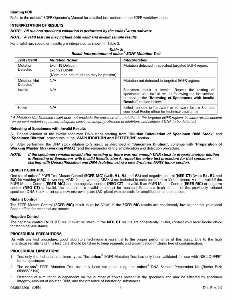
For a valid run, specimen results are interpreted as shown in Table 2.
Table 2: Result Interpretation of cobas® EGFR Mutation Test
Test Result Mutation Result Interpretation Mutation Detected
Exon 19 Deletion Exon 21 L858R (More than one mutation may be present)
Mutation detected in specified targeted EGFR region.
Mutation Not Detected*
N/A Mutation not detected in targeted EGFR regions
Invalid N/A Specimen result is invalid. Repeat the testing of specimens with invalid results following the instructions outlined in the “Retesting of Specimens with Invalid Results” section below.
Failed N/A Failed run due to hardware or software failure. Contact your local Roche office for technical assistance
* A Mutation Not Detected result does not preclude the presence of a mutation in the targeted EGFR regions because results depend on percent mutant sequences, adequate specimen integrity, absence of inhibitors, and sufficient DNA to be detected.
Retesting of Specimens with Invalid Results A. Repeat dilution of the invalid specimen DNA stock starting from “Dilution Calculation of Specimen DNA Stock” and “Specimen Dilution” procedures in the “AMPLIFICATION and DETECTION” section.
B. After performing the DNA stock dilution to 2 ng/μL as described in “Specimen Dilution”, continue with “Preparation of Working Master Mix (working MMX)” and the remainder of the amplification and detection procedure.
NOTE: If the specimen remains invalid after retesting or there was not enough DNA stock to prepare another dilution in Retesting of Specimens with Invalid Results, step A, repeat the entire test procedure for that specimen, starting with Deparaffinization and DNA Isolation using a new 5-micron FFPET tumor section.
QUALITY CONTROL One set of cobas® EGFR Test Mutant Control (EGFR MC) (wells A1, A2 and A3) and negative control (NEG CT) (wells B1, B2 and B3) for working MMX-1, working MMX-2, and working MMX-3 are included in each run of up to 30 specimens. A run is valid if the EGFR Mutant Control (EGFR MC) and the negative control (NEG CT) are valid. If an EGFR Mutant Control (EGFR MC) or negative control (NEG CT) is invalid, the entire run is invalid and must be repeated. Prepare a fresh dilution of the previously isolated specimen DNA Stock to set up a new microwell plate (AD-plate) with controls for amplification and detection.
Mutant Control The EGFR Mutant Control (EGFR MC) result must be ‘Valid’. If the EGFR MC results are consistently invalid, contact your local Roche office for technical assistance.
Negative Control The negative control (NEG CT) result must be ‘Valid’. If the NEG CT results are consistently invalid, contact your local Roche office for technical assistance.
PROCEDURAL PRECAUTIONS As with any test procedure, good laboratory technique is essential to the proper performance of this assay. Due to the high analytical sensitivity of this test, care should be taken to keep reagents and amplification mixtures free of contamination.
PROCEDURAL LIMITATIONS 1. Test only the indicated specimen types. The cobas® EGFR Mutation Test has only been validated for use with NSCLC FFPET
tumor specimens.
2. The cobas® EGFR Mutation Test has only been validated using the cobas® DNA Sample Preparation Kit (Roche P/N: 05985536190).
3. Detection of a mutation is dependent on the number of copies present in the specimen and may be affected by specimen integrity, amount of isolated DNA, and the presence of interfering substances.
06356575001-02EN 15 Doc Rev. 2.0
4. Reliable results are dependent on adequate specimen fixation, transport, storage and processing. Follow the procedures in this Package Insert and in the cobas® EGFR Operator’s Manual.
5. The effects of other potential variables such as specimen fixation variables have not been evaluated.
6. The addition of AmpErase enzyme into the cobas® EGFR Mutation Test Master Mix enables selective amplification of target DNA; however, good laboratory practices and careful adherence to the procedures specified in this Package Insert are necessary to avoid contamination of reagents.
7. Use of this product must be limited to personnel trained in the techniques of PCR and the use of the cobas® 4800 system.
8. Only the cobas z 480 analyzer has been validated for use with this product. No other thermal cycler with real-time optical detection can be used with this product.
9. The presence of PCR inhibitors may cause false negative or invalid results.
10. Though rare, mutations within the genomic DNA regions of the EGFR gene covered by the primers or probes used in the cobas® EGFR Mutation Test may result in failure to detect presence of a mutation in exons 19 and 21 (results of “Mutation Not Detected”).
11. The cobas® EGFR Mutation Test shows cross-reactivity (results of “Mutation Detected”) to the exon 19 L747S mutation, a rare acquired mutation that may confer resistance to TKI treatment.12
12. The cobas® EGFR Mutation Test shows cross-reactivity (results of “Mutation Detected”) to additional rare exon 19 deletion mutations, and exon 21 L858R substitution mutation. Refer to Table 6 and 7 in the Non-clinical Performance Evaluation section below for more details.
13. The cobas® EGFR Mutation Test is validated for use with 50 ng of DNA per reaction well. DNA input amounts lower than 50 ng per reaction well are not recommended.
14. The cobas® EGFR Mutation Test is a qualitative test. The test is not for quantitative measurements of percent mutation.
15. NSCLC FFPET specimens containing degraded DNA may affect the ability of the test to detect the EGFR mutations.
16. Samples with results reported as “Mutation Not Detected” may harbor EGFR mutations not detected by the assay.
17. The cobas® EGFR Mutation Test detects EGFR mutations in NSCLC patients whose tumors have the exon 19 deletions or exon 21 L858R substitution mutations, but not any other EGFR mutations.
NON-CLINICAL PERFORMANCE EVALUATION For the non-clinical studies described below, percentage of tumor was assessed by pathology review. Bi-directional Sanger sequencing and massively parallel sequencing (MPS) were used to select the specimens for testing. Percentage of mutation of NSCLC FFPET specimen was determined using a MPS method.
Analytical Sensitivity – Limit of Blank To assess performance of the cobas® EGFR Mutation Test in the absence of template and to ensure that a blank sample does not generate an analytical signal that might indicate a low concentration of mutation, samples with no template and NSCLC FFPET EGFR wild-type specimens were evaluated. No detectable Ct results were identified in the mutant channel in the presence of EGFR wild-type DNA or in either channel following a no template sample.
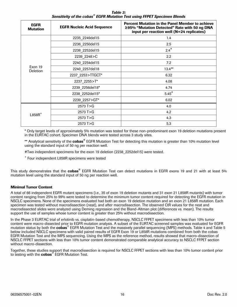
Analytical Sensitivity Using FFPET Specimen Blends Eleven FFPET specimen DNA extracts for the exon 19 Deletion mutations and four FFPET specimen DNA extracts for the L858R mutation were blended with EGFR wild-type FFPET specimen extracts to achieve blends with samples targeting 10, 5.0, 2.5 and 1.25% mutation level as determined by a massively parallel sequencing method (MPS), that was validated for the use for detecting the EGFR exon 19 deletions and exon 21 L858R mutation. Serial dilutions of each specimen blend were prepared and eight (8) replicates of each panel member were run using each of 3 cobas® EGFR Mutation Test kit lots (n=24/panel member). The sensitivity of each sample was determined by the lowest amount of DNA that gave an EGFR “Mutation Detected” rate of at least 95% for the targeted mutation, shown in Table 3.
06356575001-02EN 16 Doc Rev. 2.0
Table 3: Sensitivity of the cobas® EGFR Mutation Test using FFPET Specimen Blends
EGFR Mutation EGFR Nucleic Acid Sequence
Percent Mutation in the Panel Member to achieve ≥95% “Mutation Detected” Rate with 50 ng DNA
input per reaction well (N=24 replicates)
Exon 19 Deletion
2235_2249del15 1.4
2236_2250del15 2.5
2238_2252del15 2.4#
2239_2248>C 2.2
2240_2254del15 7.2
2240_2257del18 13.4**
2237_2253>TTGCT* 6.32
2237_2255>T* 4.08
2239_2256del18* 4.74
2238_2252del15* 5.45#
2239_2257>GT* 6.02
L858R+
2573 T>G 4.0
2573 T>G 4.2
2573 T>G 4.3
2573 T>G 5.3
* Only target levels of approximately 5% mutation was tested for these non-predominant exon 19 deletion mutations present in the EURTAC cohort. Specimen DNA blends were tested across 3 study sites.
** Analytical sensitivity of the cobas® EGFR Mutation Test for detecting this mutation is greater than 10% mutation level using the standard input of 50 ng per reaction well.
#Two independent specimens for the exon 19 deletion (2238_2252del15) were tested. + Four independent L858R specimens were tested
This study demonstrates that the cobas® EGFR Mutation Test can detect mutations in EGFR exons 19 and 21 with at least 5% mutation level using the standard input of 50 ng per reaction well.
Minimal Tumor Content A total of 66 independent EGFR mutant specimens (i.e., 35 of exon 19 deletion mutants and 31 exon 21 L858R mutants) with tumor content ranging from 25% to 99% were tested to determine the minimum tumor content required for detecting the EGFR mutation in NSCLC specimens. None of the specimens evaluated had both an exon 19 deletion mutation and an exon 21 L858R mutation. Each specimen was tested without macrodissection (neat), and after macrodissection. The observed CtR values for the neat and macrodissected slides were analyzed using Deming regression and the Bland-Altman plot (differences vs. mean). The results support the use of samples whose tumor content is greater than 25% without macrodissection.
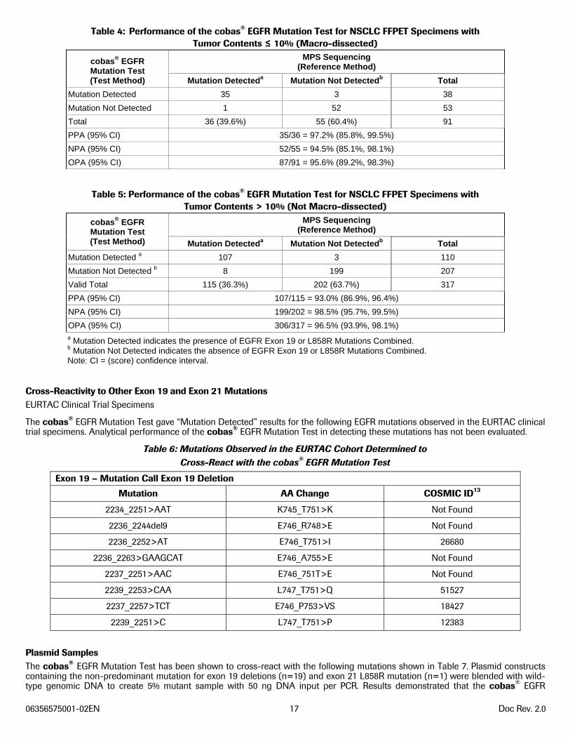
In the Phase 3 EURTAC trial of erlotinib vs. cisplatin-based chemotherapy, NSCLC FFPET specimens with less than 10% tumor content were macro-dissected prior to EGFR mutation analysis. A subset of the EURTAC screened samples was evaluated for EGFR mutation status by both the cobas® EGFR Mutation Test and the massively parallel sequencing (MPS) methods. Table 4 and Table 5 below included NSCLC specimens with valid paired results of EGFR Exon 19 or L858R mutations combined from both the cobas EGFR Mutation Test and the MPS sequencing. Using the MPS as the reference method, results showed that macro-dissection of NSCLC FFPET sections with less than 10% tumor content demonstrated comparable analytical accuracy to NSCLC FFPET section without macro-dissection.
Together, these studies support that macrodissection is required for NSCLC FFPET sections with less than 10% tumor content prior to testing with the cobas® EGFR Mutation Test.
06356575001-02EN 17 Doc Rev. 2.0
Table 4: Performance of the cobas® EGFR Mutation Test for NSCLC FFPET Specimens with Tumor Contents ≤ 10% (Macro-dissected)
cobas® EGFR Mutation Test (Test Method)
MPS Sequencing (Reference Method)
Mutation Detecteda Mutation Not Detectedb Total
Mutation Detected 35 3 38
Mutation Not Detected 1 52 53
Total 36 (39.6%) 55 (60.4%) 91
PPA (95% CI) 35/36 = 97.2% (85.8%, 99.5%)
NPA (95% CI) 52/55 = 94.5% (85.1%, 98.1%)
OPA (95% CI) 87/91 = 95.6% (89.2%, 98.3%)
Table 5: Performance of the cobas® EGFR Mutation Test for NSCLC FFPET Specimens with Tumor Contents > 10% (Not Macro-dissected)
cobas® EGFR Mutation Test (Test Method)
MPS Sequencing (Reference Method)
Mutation Detecteda Mutation Not Detectedb Total
Mutation Detected a 107 3 110
Mutation Not Detected b 8 199 207
Valid Total 115 (36.3%) 202 (63.7%) 317
PPA (95% CI) 107/115 = 93.0% (86.9%, 96.4%)
NPA (95% CI) 199/202 = 98.5% (95.7%, 99.5%)
OPA (95% CI) 306/317 = 96.5% (93.9%, 98.1%) a Mutation Detected indicates the presence of EGFR Exon 19 or L858R Mutations Combined. b Mutation Not Detected indicates the absence of EGFR Exon 19 or L858R Mutations Combined. Note: CI = (score) confidence interval.
Cross-Reactivity to Other Exon 19 and Exon 21 Mutations EURTAC Clinical Trial Specimens
The cobas® EGFR Mutation Test gave “Mutation Detected” results for the following EGFR mutations observed in the EURTAC clinical trial specimens. Analytical performance of the cobas® EGFR Mutation Test in detecting these mutations has not been evaluated.
Table 6: Mutations Observed in the EURTAC Cohort Determined to Cross-React with the cobas® EGFR Mutation Test
Exon 19 – Mutation Call Exon 19 Deletion
Mutation AA Change COSMIC ID13
2234_2251>AAT K745_T751>K Not Found
2236_2244del9 E746_R748>E Not Found
2236_2252>AT E746_T751>I 26680
2236_2263>GAAGCAT E746_A755>E Not Found
2237_2251>AAC E746_751T>E Not Found
2239_2253>CAA L747_T751>Q 51527
2237_2257>TCT E746_P753>VS 18427
2239_2251>C L747_T751>P 12383
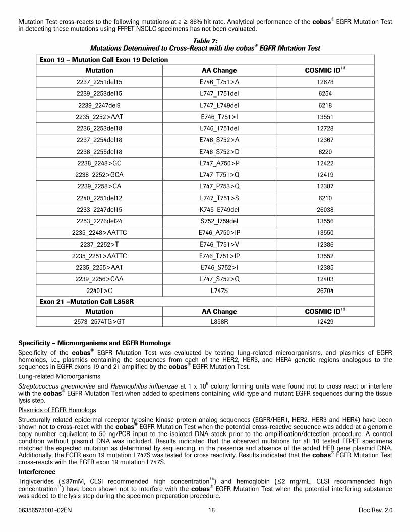
Plasmid Samples The cobas® EGFR Mutation Test has been shown to cross-react with the following mutations shown in Table 7. Plasmid constructs containing the non-predominant mutation for exon 19 deletions (n=19) and exon 21 L858R mutation (n=1) were blended with wild-type genomic DNA to create 5% mutant sample with 50 ng DNA input per PCR. Results demonstrated that the cobas® EGFR
06356575001-02EN 18 Doc Rev. 2.0
Mutation Test cross-reacts to the following mutations at a ≥ 86% hit rate. Analytical performance of the cobas® EGFR Mutation Test in detecting these mutations using FFPET NSCLC specimens has not been evaluated.
Table 7: Mutations Determined to Cross-React with the cobas® EGFR Mutation Test
Exon 19 – Mutation Call Exon 19 Deletion
Mutation AA Change COSMIC ID13
2237_2251del15 E746_T751>A 12678
2239_2253del15 L747_T751del 6254
2239_2247del9 L747_E749del 6218
2235_2252>AAT E746_T751>I 13551
2236_2253del18 E746_T751del 12728
2237_2254del18 E746_S752>A 12367
2238_2255del18 E746_S752>D 6220
2238_2248>GC L747_A750>P 12422
2238_2252>GCA L747_T751>Q 12419
2239_2258>CA L747_P753>Q 12387
2240_2251del12 L747_T751>S 6210
2233_2247del15 K745_E749del 26038
2253_2276del24 S752_I759del 13556
2235_2248>AATTC E746_A750>IP 13550
2237_2252>T E746_T751>V 12386
2235_2251>AATTC E746_T751>IP 13552
2235_2255>AAT E746_S752>I 12385
2239_2256>CAA L747_S752>Q 12403
2240T>C L747S 26704
Exon 21 –Mutation Call L858R Mutation AA Change COSMIC ID13
2573_2574TG>GT L858R 12429
Specificity – Microorganisms and EGFR Homologs Specificity of the cobas® EGFR Mutation Test was evaluated by testing lung-related microorganisms, and plasmids of EGFR homologs, i.e., plasmids containing the sequences from each of the HER2, HER3, and HER4 genetic regions analogous to the sequences in EGFR exons 19 and 21 amplified by the cobas® EGFR Mutation Test. Lung-related Microorganisms Streptococcus pneumoniae and Haemophilus influenzae at 1 x 10⁶ colony forming units were found not to cross react or interfere with the cobas® EGFR Mutation Test when added to specimens containing wild-type and mutant EGFR sequences during the tissue lysis step. Plasmids of EGFR Homologs Structurally related epidermal receptor tyrosine kinase protein analog sequences (EGFR/HER1, HER2, HER3 and HER4) have been shown not to cross-react with the cobas® EGFR Mutation Test when the potential cross-reactive sequence was added at a genomic copy number equivalent to 50 ng/PCR input to the isolated DNA stock prior to the amplification/detection procedure. A control condition without plasmid DNA was included. Results indicated that the observed mutations for all 10 tested FFPET specimens matched the expected mutation as determined by sequencing, in the presence and absence of the added HER gene plasmid DNA. Additionally, the EGFR exon 19 mutation L747S was tested for cross reactivity. Results indicated that the cobas® EGFR Mutation Test cross-reacts with the EGFR exon 19 mutation L747S. Interference Triglycerides (≤37mM, CLSI recommended high concentration14) and hemoglobin (≤2 mg/mL, CLSI recommended high concentration14) have been shown not to interfere with the cobas® EGFR Mutation Test when the potential interfering substance was added to the lysis step during the specimen preparation procedure.
06356575001-02EN 19 Doc Rev. 2.0
Necrotic Tissue NSCLC FFPET specimens with necrotic tissue content up to 60% for EGFR mutant and 85% in wild-type specimens have been shown not to interfere with the call results using the cobas® EGFR Mutation Test. Repeatability Repeatability of the cobas® EGFR Mutation Test was assessed using six FFPET specimens, including: 4 wild-type specimens; 2 mutant specimens with exon 19 deletion and L858R mutations respectively. These specimens were tested in duplicate by two operators, using two different reagent lots and two cobas z 480 analyzers over 4 days. A total of 32 replicates were evaluated per sample. The cobas® EGFR Mutation Test had a correct call rate of 99% (190/192). Specimen Handling Reproducibility The reproducibility of the DNA Sample Preparation Kit was examined using sections taken from three FFPET specimen blocks, one containing an exon 19 deletion mutation, one containing an L858R mutation, and one that is wild-type. Each specimen was tested in duplicate at each site on each day. The specimen sections for a given specimen were randomized and tested over a six day period across three sites using one operator at each site, one cobas z 480 analyzer at each site, three cobas® DNA Sample Preparation Kit lots, and one cobas EGFR Mutation Test kit lots. On each test day, each operator isolated and tested the DNA from two NSCLC FFPET curl sections for each specimen using the cobas® EGFR Mutation Test. All specimens reported valid and correct results through-out the six days of testing. For all specimens and operators combined, the cobas® EGFR Mutation Test had a correct call rate of 100% (108/108).
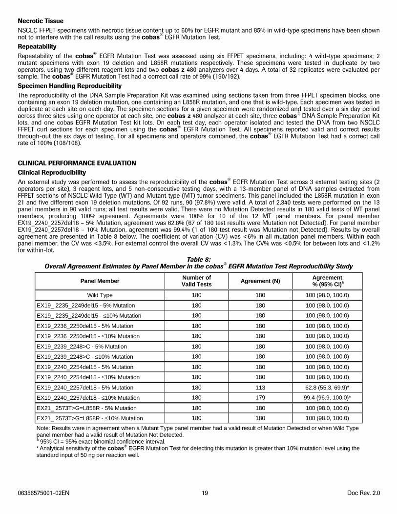
CLINICAL PERFORMANCE EVALUATION Clinical Reproducibility An external study was performed to assess the reproducibility of the cobas® EGFR Mutation Test across 3 external testing sites (2 operators per site), 3 reagent lots, and 5 non-consecutive testing days, with a 13-member panel of DNA samples extracted from FFPET sections of NSCLC Wild Type (WT) and Mutant type (MT) tumor specimens. This panel included the L858R mutation in exon 21 and five different exon 19 deletion mutations. Of 92 runs, 90 (97.8%) were valid. A total of 2,340 tests were performed on the 13 panel members in 90 valid runs; all test results were valid. There were no Mutation Detected results in 180 valid tests of WT panel members, producing 100% agreement. Agreements were 100% for 10 of the 12 MT panel members. For panel member EX19_2240_2257del18 – 5% Mutation, agreement was 62.8% (67 of 180 test results were Mutation not Detected). For panel member EX19_2240_2257del18 – 10% Mutation, agreement was 99.4% (1 of 180 test result was Mutation not Detected). Results by overall agreement are presented in Table 8 below. The coefficient of variation (CV) was <6% in all mutation panel members. Within each panel member, the CV was <3.5%. For external control the overall CV was <1.3%. The CV% was <0.5% for between lots and <1.2% for within-lot.
Table 8: Overall Agreement Estimates by Panel Member in the cobas® EGFR Mutation Test Reproducibility Study
Panel Member Number of Valid Tests
Agreement (N) Agreement % (95% CI)a
Wild Type 180 180 100 (98.0, 100.0)
EX19_ 2235_2249del15 - 5% Mutation 180 180 100 (98.0, 100.0)
EX19_ 2235_2249del15 - ≤10% Mutation 180 180 100 (98.0, 100.0)
EX19_2236_2250del15 - 5% Mutation 180 180 100 (98.0, 100.0)
EX19_2236_2250del15 - ≤10% Mutation 180 180 100 (98.0, 100.0)
EX19_2239_2248>C - 5% Mutation 180 180 100 (98.0, 100.0)
EX19_2239_2248>C - ≤10% Mutation 180 180 100 (98.0, 100.0)
EX19_2240_2254del15 - 5% Mutation 180 180 100 (98.0, 100.0)
EX19_2240_2254del15 - ≤10% Mutation 180 180 100 (98.0, 100.0)
EX19_2240_2257del18 - 5% Mutation 180 113 62.8 (55.3, 69.9)*
EX19_2240_2257del18 - ≤10% Mutation 180 179 99.4 (96.9, 100.0)*
EX21_ 2573T>G=L858R - 5% Mutation 180 180 100 (98.0, 100.0)
EX21_ 2573T>G=L858R - ≤10% Mutation 180 180 100 (98.0, 100.0)
Note: Results were in agreement when a Mutant Type panel member had a valid result of Mutation Detected or when Wild Type panel member had a valid result of Mutation Not Detected. a 95% CI = 95% exact binomial confidence interval. * Analytical sensitivity of the cobas® EGFR Mutation Test for detecting this mutation is greater than 10% mutation level using the standard input of 50 ng per reaction well.
06356575001-02EN 20 Doc Rev. 2.0
Correlation to Reference Method using Phase 3 Samples The clinical performance of the cobas® EGFR Mutation Test was assessed by comparing it to two reference methods – 2x bidirectional Sanger sequencing and quantitative massively parallel sequencing (MPS) – using 487 formalin-fixed paraffin-embedded lung tumor specimens from patients with advanced NSCLC who were screened in the Phase 3 EURTAC trial of erlotinib vs. cisplatin-based chemotherapy. 6, 7 The clinical and demographic characteristics of the patients whose specimens were available for this retrospective testing were comparable to those of otherwise eligible patients (557) whose specimens were not available for retesting. A total of 1,276 patients were screened for the EURTAC trial using a combination of laboratory developed tests, collectively referred to as the clinical trial assay (CTA). After excluding ineligible patients and those without CTA results, 1,044 patients were potentially eligible for the current study. Among the 1,044 eligible patients, 225 patients had samples that were mutation positive by CTA, 792 had samples that were Wild Type by CTA, and 27 had samples with inconclusive results by CTA. Of the 1,044 potentially eligible patients, 487 specimens were available for retesting with the cobas® EGFR Mutation Test. All 487 specimens were tested in a blinded fashion with both the cobas® EGFR Mutation Test and Sanger sequencing. Of those, 406 had valid results by both the cobas® test and Sanger sequencing, 38 invalid results were observed by the cobas® test and Sanger sequencing, 38 invalid results by Sanger sequencing only, and 5 invalid results by the cobas® test only. Among the 487 specimens available for retesting with the cobas® EGFR Mutation Test, 444 specimens gave valid cobas® test results and were also tested with MPS. Of those, there were 36 invalid results by MPS; thus, 408 had valid results by both the cobas® test and MPS. The analytical accuracy of the cobas® EGFR Mutation Test compared with each reference method was evaluated by estimating the positive percentage agreement (PPA), negative percentage agreement (NPA), and overall percentage agreement (OPA) and their corresponding 95% CIs for exon 19 deletions and L858R mutations in aggregate, and separately. In the EURTAC cohort, the cobas® EGFR Mutation Test detected the following deletion mutations in exon 19 of the EGFR gene: 2234_2251>AAT, 2235_2249del15, 2240_2254del15, 2238_2252del15, 2240_2257del18, 2236_2244del9, 2236_2263>GAAGCAT, 2236_2250del15, 2236_2252>AT, 2237_2255>T, 2237_2257>TCT, 2239_2248>C, 2239_2251>C, 2237_2251>AAC, 2239_2253>CAA, 2239_2256del18, 2239_2257>GT, and 2237_2253>TTGCT. The cobas® EGFR Mutation Test also detected the following L858R substitution mutation in exon 21 of the EGFR gene: 2573 T>G. Of the mutations detected in the EURTAC cohort, analytical sensitivity was demonstrated on the mutations listed in Table 3 above. A total of 406 samples with valid cobas® test and Sanger results were included in the agreement analysis. The PPA between the cobas® EGFR Mutation Test and Sanger sequencing was 96.6% (95% CI: 91.5% to 98.7%), and the NPA was 88.3% (95% CI: 84.1% to 91.5%), in the detection of exon 19 deletions and L858R mutations in aggregate as presented in Table 9. The OPA was 90.6%, with the lower limit of the 95% CI above 87%. The PPA, NPA, and OPA in the detection of exon 19 deletion mutations were all >92%. The PPA, NPA, and OPA in the detection of L858R mutations compared were all >95%.
Table 9: Comparison of the cobas® EGFR Mutation Test with Sanger Sequencing for the Detection of EGFR Exon 19 Deletion
Mutations and L858R Mutation
cobas® EGFR Mutation Result
Sanger Sequencing ResultResult in aggregate Exon 19 deletion Exon 21 Mutation
MD MND Total MD MND Total MD MND TotalMD 112 34 146 71 25 96 41 9 50MND 4 256 260 2 308 310 2 354 356Total 116 290 406 73 333 406 43 363 406PPA (95% CI) 112/116 = 96.6% (91.5%, 98.7%) 71/73 = 97.3% (90.5%, 99.2%) 41/43 = 95.3% (84.5%, 98.7%)
NPA (95% CI) 256/290 = 88.3% (84.1%, 91.5%) 308/333 = 92.5% (89.2%, 94.9%) 354/363 = 97.5% (95.4%, 98.7%)
OPA (95% CI) 368/406 = 90.6% (87.4%, 93.1%) 379/406 = 93.3% (90.5%, 95.4%) 395/406 = 97.3% (95.2%, 98.5%)
A total of 408 samples with valid cobas® test and MPS results were included in the agreement analysis. By comparison, the PPA and NPA between the cobas® EGFR Mutation Test and MPS for the detection of exon 19 deletions and the L858R point mutation in aggregate were 94.0% (95% CI: 89.1% to 96.8%) and 97.7% (95% CI: 95.0% to 98.9%), respectively as presented in Table 10. The OPA was 96.3%, with a lower limit of the 95% CI of 94.0%. The PPA, NPA, and OPA in detecting exon 19 deletion mutations were all >95%, with all the 95% lower limit CIs >90%. The PPA, NPA, and OPA in detecting the L858R mutation were also all >95%, with all lower limits of the 95% CIs ≥ 95% except for PPA (90%), owing to due to the small number of L858R mutation detected.
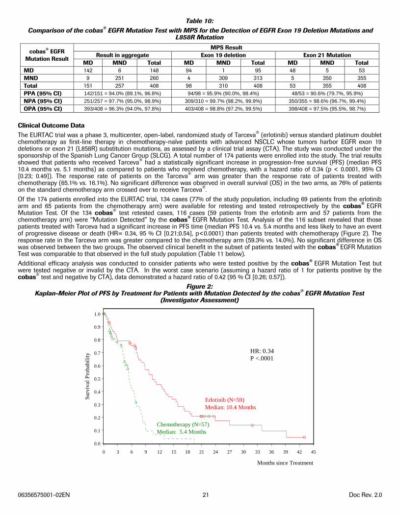
06356575001-02EN 21 Doc Rev. 2.0
Table 10: Comparison of the cobas® EGFR Mutation Test with MPS for the Detection of EGFR Exon 19 Deletion Mutations and
L858R Mutation
cobas® EGFR Mutation Result
MPS ResultResult in aggregate Exon 19 deletion Exon 21 Mutation
MD MND Total MD MND Total MD MND TotalMD 142 6 148 94 1 95 48 5 53MND 9 251 260 4 309 313 5 350 355Total 151 257 408 98 310 408 53 355 408PPA (95% CI) 142/151 = 94.0% (89.1%, 96.8%) 94/98 = 95.9% (90.0%, 98.4%) 48/53 = 90.6% (79.7%, 95.9%)
NPA (95% CI) 251/257 = 97.7% (95.0%, 98.9%) 309/310 = 99.7% (98.2%, 99.9%) 350/355 = 98.6% (96.7%, 99.4%)
OPA (95% CI) 393/408 = 96.3% (94.0%, 97.8%) 403/408 = 98.8% (97.2%, 99.5%) 398/408 = 97.5% (95.5%, 98.7%)
Clinical Outcome Data The EURTAC trial was a phase 3, multicenter, open-label, randomized study of Tarceva® (erlotinib) versus standard platinum doublet chemotherapy as first-line therapy in chemotherapy-naïve patients with advanced NSCLC whose tumors harbor EGFR exon 19 deletions or exon 21 (L858R) substitution mutations, as assessed by a clinical trial assay (CTA). The study was conducted under the sponsorship of the Spanish Lung Cancer Group (SLCG). A total number of 174 patients were enrolled into the study. The trial results showed that patients who received Tarceva® had a statistically significant increase in progression-free survival (PFS) (median PFS 10.4 months vs. 5.1 months) as compared to patients who received chemotherapy, with a hazard ratio of 0.34 (p < 0.0001, 95% CI [0.23; 0.49]). The response rate of patients on the Tarceva® arm was greater than the response rate of patients treated with chemotherapy (65.1% vs. 16.1%). No significant difference was observed in overall survival (OS) in the two arms, as 76% of patients on the standard chemotherapy arm crossed over to receive Tarceva®. Of the 174 patients enrolled into the EURTAC trial, 134 cases (77% of the study population, including 69 patients from the erlotinib arm and 65 patients from the chemotherapy arm) were available for retesting and tested retrospectively by the cobas® EGFR Mutation Test. Of the 134 cobas® test retested cases, 116 cases (59 patients from the erlotinib arm and 57 patients from the chemotherapy arm) were “Mutation Detected” by the cobas® EGFR Mutation Test. Analysis of the 116 subset revealed that those patients treated with Tarceva had a significant increase in PFS time (median PFS 10.4 vs. 5.4 months and less likely to have an event of progressive disease or death (HR= 0.34, 95 % CI [0.21;0.54], p<0.0001) than patients treated with chemotherapy (Figure 2). The response rate in the Tarceva arm was greater compared to the chemotherapy arm (59.3% vs. 14.0%). No significant difference in OS was observed between the two groups. The observed clinical benefit in the subset of patients tested with the cobas® EGFR Mutation Test was comparable to that observed in the full study population (Table 11 below). Additional efficacy analysis was conducted to consider patients who were tested positive by the cobas® EGFR Mutation Test but were tested negative or invalid by the CTA. In the worst case scenario (assuming a hazard ratio of 1 for patients positive by the cobas® test and negative by CTA), data demonstrated a hazard ratio of 0.42 (95 % CI [0.26; 0.57]).
Figure 2: Kaplan-Meier Plot of PFS by Treatment for Patients with Mutation Detected by the cobas® EGFR Mutation Test
(Investigator Assessment)
Surv
ival
Pro
babi
lity
0.0
0.1
0.2
0.3
0.4
0.5
0.6
0.7
0.8
0.9
1.0
Months since Treatment
0 3 6 9 12 15 18 21 24 27 30 33 36 39 42 45
HR: 0.34P <.0001
Erlotinib (N=59)Median: 10.4 Months
Chemotherapy (N=57)Median: 5.4 Months
06356575001-02EN 22 Doc Rev. 2.0
Table 11: Clinical Benefit of Patients Tested With The cobas® EGFR Mutation Test is Comparable to That Observed in the EURTAC Population
Parameter
cobas®-positive Populationn = 116
EURTAC n = 173*
Chemotherapyn = 57
Erlotinibn = 59
Chemotherapy n = 87
Erlotinibn = 86
PFS Median (Months) 5.4 10.4 5.1 10.4Hazard Ratio 0.34 0.34 Hazard Ratio 95% CI [0.21; 0.54] [0.23; 0.49] P-Value (log-rank test) <0.0001 <0.0001 *Note: One patient withdrew consent after completion of the EURTAC study, which resulted in a dataset of n = 173
REFERENCES
1. Sharma SV, Bell DW, Settleman J, Haber DA. Epidermal growth factor receptor mutations in lung cancer. Nature Reviews Cancer.
2007;7:169-181.
2. Pao W, Chmielecki J. Rational, biologically based treatment of EGFR-mutant non-small-cell lung cancer. Nat Rev Cancer.
2010;10:760-774.
3. Zhou C, Wu Y-L, Chen G, et al. Erlotinib versus chemotherapy as first-line treatment for patients with advanced EGFR mutation-
positive non-small-cell lung cancer (OPTIMAL, CTONG-0802): a multicentre, open-label, randomised, phase 3 study. Lancet Onc.
2011; 12:735-742
4. Paz-Ares L, Soulières D, Melezínek I, et al. Clinical outcomes in non-small-cell lung cancer patients with EGFR mutations: pooled
analysis. J Cell Mol Med 2010;14:51-69.
5. American Cancer Society. Cancer Facts and Figures, 2012.
6. Tarceva® (erlotinib) Package Insert
7. Rosell, R., et al. Erlotinib versus standard chemotherapy as first-line treatment for European patients with advanced EGFR mutation-
positive non-small-cell lung cancer (EURTAC): a multicentre, open-label, randomised phase 3 trial. The Lancet Oncology. 2012;13
v3:239-246.
8. Longo, M.C., Berninger, M.S. and Hartley, J.L. 1990. Use of uracil DNA glycosylase to control carry-over contamination in
polymerase chain reactions. Gene. 93:125-128.
9. Chosewood, L.C. and Wilson, D.E. Biosafety and Microbiological and Biomedical Laboratories. HHS Publication Fifth # edition.(CDC)
21-1112. 2009.
10. Clinical and Laboratory Standards Institute (CLSI). Protection of Laboratory Workers from Occupationally Acquired Infections.
Approved Guideline-Third Edition. CLSI Document M29-A3 Villanova, PA:CLSI, 2005.
11. International Air Transport Association. Dangerous Goods Regulations, 52nd Edition. 2011.
12. Costa DB, Nguyen KS, Cho BC, Sequist LV, Jackman DM, Riely GJ, Yeap BY, Halmos B, Kim JH, Jänne PA, Huberman MS, Pao W,
Tenen DG, Kobayashi S. Effects of erlotinib in EGFR mutated non-small cell lung cancers with resistance to gefitinib. Clin Cancer
Res. 2008 Nov 1;14(21):7060-7
13. Catalogue of Somatic Mutations in Cancer (COSMIC), 2011, v.51, http://www.sanger.ac.uk/genetics/CGP/cosmic.
14. Clinical and Laboratory Standards Institute (CLSI) EP7-A2, Interference Testing in Clinical Chemistry; Approved Guidelines – Second
Edition, Appendix D 2005.
06356575001-02EN 23 Doc Rev. 2.0
Document Revision Information
Doc Rev. 1.0 4/2013
First Publishing.
Roche Molecular Systems, Inc. 1080 US Highway 202 South Branchburg, NJ 08876 USA A Member of the Roche Group
Distributed by
Roche Diagnostics 9115 Hague Road Indianapolis, IN 46250-0457 USA(For Technical Assistance call the Roche Response Center toll free: 1-800-526-1247)
COBAS, COBAS Z, and AMPERASE are trademarks of Roche.
Carryover prevention technology in the AmpErase enzyme is covered by U.S. Patent 5,035,996 and foreign counterparts owned by Invitrogen Corporation and licensed to Roche Molecular Systems, Inc.
The cyanine dyes in this product are subject to patent rights of GE Healthcare Bio-Sciences Corp. and Carnegie Mellon University and are licensed to Roche solely for incorporation into components of research and in-vitro diagnostic kits. Any use of such kits for purposes other than research or in-vitro diagnostics requires sublicenses from GE Healthcare Bio-Sciences Corp., Piscataway, New Jersey, U.S.A. and Carnegie Mellon University, Pittsburgh, Pennsylvania, U.S.A.
Certain EGFR sequences in this product are covered by one of more patents of Genzyme Corp. and Dana Farber Cancer Institute and The General Hospital Corporation and licensed to Roche Molecular Systems, Inc. under U.S. Patent No. 7,964,349 and other U.S. and foreign patents pending.
EPPENDORF is a trademark of Eppendorf AG.
PIPET-AID is a trademark of Drummond Scientific.
NANODROP is a trademark of Thermo Scientific.
Copyright 2013 Roche Molecular Systems, Inc. 07/2013 06356575001-02 Doc Rev. 2.0
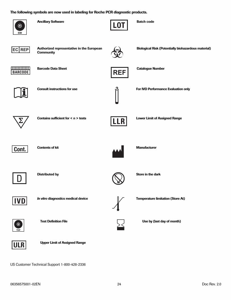
06356575001-02EN 24 Doc Rev. 2.0
The following symbols are now used in labeling for Roche PCR diagnostic products. Ancillary Software
Batch code
Authorized representative in the EuropeanCommunity
Biological Risk (Potentially biohazardous material)
Barcode Data Sheet
Catalogue Number
Consult instructions for use
For IVD Performance Evaluation only
Contains sufficient for < n > tests
Lower Limit of Assigned Range
Contents of kit
Manufacturer
Distributed by
Store in the dark
In vitro diagnostics medical device
Temperature limitation (Store At)
Test Definition File
Use by (last day of month)
Upper Limit of Assigned Range
US Customer Technical Support 1-800-428-2336
TDF
SW