co-regulation of iron metabolism and virulence associated
TRANSCRIPT

RESEARCH ARTICLE
Co-regulation of Iron Metabolism and
Virulence Associated Functions by Iron and
XibR, a Novel Iron Binding Transcription
Factor, in the Plant Pathogen Xanthomonas
Sheo Shankar Pandey1,2, Pradeep Kumar Patnana1, Santosh Kumar Lomada1,
Archana Tomar1, Subhadeep Chatterjee1*
1 Centre for DNA Fingerprinting and Diagnostics, Nampally, India, 2 Graduate studies, Manipal University,
Manipal, India
Abstract
Abilities of bacterial pathogens to adapt to the iron limitation present in hosts is critical to
their virulence. Bacterial pathogens have evolved diverse strategies to coordinately regulate
iron metabolism and virulence associated functions to maintain iron homeostasis in
response to changing iron availability in the environment. In many bacteria the ferric uptake
regulator (Fur) functions as transcription factor that utilize ferrous form of iron as cofactor to
regulate transcription of iron metabolism and many cellular functions. However, mecha-
nisms of fine-tuning and coordinated regulation of virulence associated function beyond iron
and Fur-Fe2+ remain undefined. In this study, we show that a novel transcriptional regulator
XibR (named Xanthomonas iron binding regulator) of the NtrC family, is required for fine-
tuning and co-coordinately regulating the expression of several iron regulated genes and vir-
ulence associated functions in phytopathogen Xanthomonas campestris pv. campestris
(Xcc). Genome wide expression analysis of iron-starvation stimulon and XibR regulon,
GUS assays, genetic and functional studies of xibR mutant revealed that XibR positively
regulates functions involved in iron storage and uptake, chemotaxis, motility and negatively
regulates siderophore production, in response to iron. Furthermore, chromatin immunopre-
cipitation followed by quantitative real-time PCR indicated that iron promoted binding of the
XibR to the upstream regulatory sequence of operon’s involved in chemotaxis and motility.
Circular dichroism spectroscopy showed that purified XibR bound ferric form of iron. Electro-
phoretic mobility shift assay revealed that iron positively affected the binding of XibR to the
upstream regulatory sequences of the target virulence genes, an effect that was reversed
by ferric iron chelator deferoxamine. Taken together, these data revealed that how XibR
coordinately regulates virulence associated and iron metabolism functions in Xanthomo-
nads in response to iron availability. Our results provide insight of the complex regulatory
mechanism of fine-tuning of virulence associated functions with iron availability in this impor-
tant group of phytopathogen.
PLOS Pathogens | DOI:10.1371/journal.ppat.1006019 November 30, 2016 1 / 39
a11111
OPENACCESS
Citation: Pandey SS, Patnana PK, Lomada SK,
Tomar A, Chatterjee S (2016) Co-regulation of Iron
Metabolism and Virulence Associated Functions by
Iron and XibR, a Novel Iron Binding Transcription
Factor, in the Plant Pathogen Xanthomonas. PLoS
Pathog 12(11): e1006019. doi:10.1371/journal.
ppat.1006019
Editor: Wenbo Ma, University of California
Riverside, UNITED STATES
Received: August 1, 2016
Accepted: October 21, 2016
Published: November 30, 2016
Copyright: © 2016 Pandey et al. This is an open
access article distributed under the terms of the
Creative Commons Attribution License, which
permits unrestricted use, distribution, and
reproduction in any medium, provided the original
author and source are credited.
Data Availability Statement: Microarray raw data
is available via the NCBI GEO repository (accession
number GSE89267). All other relevant data are
within the paper and its Supporting Information
files.
Funding: This study was supported by funding to
SC from Department of Biotechnology (DBT),
CSIR-HRDG, DST-SERB, Government of India and
core funding from CDFD. SSP was recipient of
Junior and Senior Research Fellowships from the
Council of Scientific and Industrial Research

Author Summary
Pathogenic bacteria exhibit tight regulation of iron homeostasis in order to meet iron
requirements of living in different environmental conditions, including in the host. The
ferric uptake regulator (Fur) regulates the expression of genes involved in iron metabolism
in response to change in iron availability in several bacteria. However, beyond Fur, mech-
anisms of fine-tuning expression of iron regulated genes and virulence associated func-
tions in response to iron availability is largely undefined. Here, we show that a novel ferric
iron binding transcription factor, XibR, is required for optimum virulence in phytopatho-
gen Xanthomonas campestris pv. campestris (Xcc) by coordinately regulating expression of
genes involved in iron metabolism and several virulence associated function such as che-
motaxis and motility. XibR directly binds to the upstream regulatory sequence of chemo-
taxis, and flagellar cluster in the presence of ferric form of iron. Furthermore, the xibRmutant exhibited reduced growth and intracellular iron content under low-iron condi-
tion, which were phenocopied by mutants in the iron storage and uptake genes. This
study provides for the mechanistic insight for the first time into the involvement of a ferric
binding transcription factor in the coordinated regulation of iron metabolism and viru-
lence associated functions.
Introduction
Iron homeostasis is vital for survival and cellular metabolism in many organisms. Bacteria
maintain cellular iron homeostasis by coordinately regulating iron uptake, metabolism and
storage, to achieve sufficient iron under iron-replete condition, and to store intracellular iron
surplus for utilization under condition of iron limitation [1]. Iron is required for virulence of
several animal and plant pathogenic bacteria [1–3]. The availability of iron within the host
plays a critical role in the growth and survival of the pathogens. In animal hosts, iron-with-
holding strategies are employed to limit iron availability to infecting pathogens [1]. Similarly,
in plants, several studies have shown that iron availability is likely to be a limiting factor for
pathogen growth within host [2,3].
Bacteria employ a variety of strategies to sequester iron from the environment for survival.
These include secretion and uptake of low molecular weight iron chelators called siderophores,
transport of the ferrous form of iron by the ferrous iron transporter (Feo), several metal-type
ABC transporters [1,4]. Certain pathogenic bacteria are also able to utilize host-iron complexes
such as transferrin, lactoferrin and heme, when exogenous iron sources are restricted [1,5].
However, excess of free iron is toxic to the cell as it causes the production of Reactive Oxygen
Species (ROS) by the Fenton reaction [4]. Hence, bacteria tightly coordinate the expression of
the iron homeostasis machinery which includes iron uptake, storage and distribution in
response to iron availability to ensure proper iron homeostasis. In addition, it has been shown
that pathogenic bacteria utilizes iron as regulatory signal to coordinately regulate the expres-
sion of virulence genes such as toxins, hemolysins, and hydrolyzing enzymes, as low-iron con-
ditions triggers the expression of iron uptake systems as well virulence associated factors,
mimicking limited iron availability inside the host environment [6,7].
In many bacteria Ferric-uptake regulator (Fur) is involved in the coordinated regulation of
gene expression in response to iron availability. Fur utilizes Fe2+ as a cofactor and represses
the expression of iron uptake and metabolism genes under iron sufficiency, and causes de-
repression in the absence of Fe2+ under conditions of iron restriction. Fur-Fe2+ also has been
Xanthomonas Iron Binding Regulator and Iron, Coordinate Virulence Associated Function
PLOS Pathogens | DOI:10.1371/journal.ppat.1006019 November 30, 2016 2 / 39
(CSIR), India. The funders had no role in study
design, data collection and analysis, decision to
publish, or preparation of the manuscript.
Competing Interests: The authors have declared
that no competing interests exist.

reported to be involved in the positive regulation of expression of genes involved in iron stor-
age proteins, superoxide dismutase, and catalase. In addition to regulating genes involved in
iron uptake and metabolism, Fur has been shown to regulate diverse cellular process such as
respiration, TCA cycle, glycolysis, oxidative stress [7–9]. However, mechanism of fine-tuning
iron metabolism and virulence associated functions beyond ferrous responsive Fur-like tran-
scription factor (TF) remains undefined in pathogenic bacteria.
Bacteria belonging to the genus Xanthomonas causes diseases in several economically
important plants [10,11]. Xanthomonads encodes an xss (Xanthomonas siderophore synthesis)
operon which is required for the production of siderophore vibrioferrin under iron-restricted
conditions, and iron metabolism plays a critical role in their virulence [12–14]. These phyto-
pathogen use cell-cell signaling mediated by diffusible quorum sensing signal molecule to reg-
ulate the expression of iron uptake and metabolism functions contributing to virulence and
growth within host [13,15]. In Xanthomonas campestris pv. campestris (Xcc) and Xanthomonasoryzae pv. oryzae (Xoo), which are important models to study bacterial phytopathogenesis, it
has been shown that Fur is involved in the suppression of siderophore production and furmutants are deficient in virulence and hypersensitive to oxidative stress [16]. However, little is
known about mechanisms of fine-tuning expression of iron regulated genes, beyond iron regu-
lation mediated via cell-cell signaling and Fur in this important group of phytopathogens.
Xcc produces moderate amount of siderophore only in iron-limiting conditions. In order
to gain insight into iron metabolism and regulatory functions involved in iron metabolism, we
performed a genetic screen to identify mutants overproducing siderophores. Three mutants
were identified that had transposon insertions in an ntrC family of transcription factor
(XC_3760; named xibR; Xanthomonas iron binding regulator), that significantly overproduce
siderophore compared to the parental wild-type strain (Xcc 8004).
NtrC family of transcription factors has been shown to be involved in the regulation of
diverse physiological process such as extracellular polysaccharide production, nitrogen metab-
olism, biofilm formation in diverse bacteria [17–20]. However, role of NtrC family of tran-
scription factor in regulation of iron metabolism and sensing has not been identified.
In this study, we show that the Xcc XibR (an NtrC family of response regulator) is involved
in global regulation of functions involved in iron uptake, storage and virulence in response to
changes in iron availability. XibR positively regulates motility and chemotaxis in response to
iron starvation and also contributes to biofilm formation. Genome wide transcriptional analy-
sis of iron starvation stimulon and XibR regulon indicated that in Xcc, XibR regulates the pro-
duction of virulence associated function in response to iron availability. Our results provide
insight of the mechanism of fine-tuning of virulence associated functions with iron availability
in this important phytopathogen.
Results
xibR regulates siderophores production in Xanthomonas campestris pv.
campestris
A genetic screen was performed by genome-wide Tn5-transposon mediated random mutagen-
esis to identify mutants altered in siderophore production in Xcc (See Supporting Materials
and Methods; S1 Table; S1 Fig). We isolated three siderophore overproducing mutants
(xibRM2, xibRM1 and xibRB1) on petone–sucrose agar-chrome azurol sulphonate (PSA-CAS)
plates containing 2,20-dipyridyl (DP) that carried three independent transposon insertions in
xibR (a ntrC family of response regulator; XC_3760) (S1B and S1C Fig; S1 Table and S2 Table).
In addition, we also made an in-frame deletion in the xibR gene, ΔxibR, which also exhibited
siderophore overproduction, similar to the transposon induced mutant strains (Fig 1A and 1B;
Xanthomonas Iron Binding Regulator and Iron, Coordinate Virulence Associated Function
PLOS Pathogens | DOI:10.1371/journal.ppat.1006019 November 30, 2016 3 / 39

Xanthomonas Iron Binding Regulator and Iron, Coordinate Virulence Associated Function
PLOS Pathogens | DOI:10.1371/journal.ppat.1006019 November 30, 2016 4 / 39

S1D Fig; S2 Table;). Quantification of vibrioferrin siderophore isolated form the cell free cul-
ture supernatant of different strains of Xcc by Amberlite XAD-16 column chromatography
and high performance liquid chromatography (HPLC) indicated that the ΔxibR mutant pro-
duced at least 4-fold more vibrioferrin than that produced by the wild-type Xcc 8004 strain
(Fig 1B; S2 Fig). Complementation of the xibR mutants ΔxibR, xibRM2, xibRM1 and xibRB1by in trans expression of the plasmid borne wild-type xibR allele (pSSP30) restored the wild-
type levels of siderophore (Fig 1A and 1B; S1C and S1D Fig). As a control, the ΔxibRΔxssAdouble deletion mutant strain [xibR and xssA (Xanthomonas siderophore synthesis A)] failed
to produce any detectable level of siderophore (Fig 1B and S1D and S1E Fig).
It has been reported that the aspartate at 54th or 55th residue of NtrC receiver domain posi-
tion gets phosphorylated by the cognate sensor kinase (SK), which is required for the regula-
tion of transcription of downstream genes [21–24]. Interestingly, in trans expression of a point
mutant of xibR in the putative conserved aspartate residue phosphorylation site (D55AXibR;
pSSP39) failed to rescue the siderophore overproduction phenotype of ΔxibR mutant (Fig 1B
and S1D Fig).
In an attempt to understand the role of iron and XibR mediated regulation of siderophore
biosynthesis, we performed expression analysis of xssA by real-time qRT-PCR and chromo-
somal reporter fusion PxssA::gusA in the wild-type Xcc 8004, ΔxibR and ΔxibR mutant harbor-
ing the complementing plasmid (ΔxibR/pSSP30) strains grown in PS, low-iron (PS + DP) and
low-iron medium supplemented with FeSO4 (Fig 1C and 1D; S3 Fig). Expression of xssA was
approximately 3-fold higher in the ΔxibR mutant compared to the wild-type Xcc 8004 strain
grown under low-iron condition. Complementation of the ΔxibR mutant by in trans expres-
sion of wild-type xibR or exogenous supplementation of FeSO4 suppressed the expression of
xssA (Fig 1C and 1D; S3 Fig). Notably, in trans expression of the multicopy plasmid borne
wild-type xibR allele suppressed the expression and production of siderophore in the wild-type
strain compared to the empty vector pHM1 which had no effect on siderophore production
(Fig 1B and 1E; S1D Fig).
Interestingly, expression analysis of xibR by real-time qRT-PCR and chromosomal reporter
fusion PxibR::gusA indicated that the xibR is induced under iron-replete conditions in the
wild-type Xcc 8004 (S4 Fig).
Fig 1. xibR suppresses siderophore production in Xcc. (A) Upper lane; siderophore production by Xcc strains on PSA-CAS plates containing 75 μM
ferrous iron chelator 2,20-Dipyridyl (DP) (PSA-CAS + DP). Lower lane; siderophore isolated from cell free culture supernatant of different Xcc strains grown
under low-iron condition (PS + 100 μM DP using Amberlite XAD-16 resin column chromatography. Cell normalized siderophore fractions were loaded on
PSA-CAS plate wells. Strains: Xcc 8004 (wild-type strain), ΔxibR (xibR deletion mutant), ΔxibRΔxssA [xibR and xssA (Xanthomonas siderophore synthesis
A) double mutant], and strains harboring the plasmid containing either the wild-type xibR allele (pSSP30) or a point mutant of xibR in the putative conserved
aspartate residue phosphorylation site (D55AXibR; pSSP39), xssA (pAP15; wild-type xssA allele) and pHM1. Here onward all the experiments repeated three
times until not mentioned. (B) Quantification of purified vibrioferrin using high-performance liquid chromatography (HPLC). Different Xcc strains were grown
up to 1.0 X 10⁹cells/ml under low-iron condition (PS + 100 μM DP). Siderophore was purified from cell free culture supernatants using Amberlite XAD-16 resin
columns. Cell normalized active fraction of siderophore was analyzed by HPLC. The vibrioferrin was detected at 300 nm and the concentration was
determined while comparing the peak area (mUA ×min) with standard curves generated from a known concentration of pure standard vibrioferrin. * indicate
P < 0.05 in student’s t test (T-test). Error bars represent SD of the mean (n = 3). (C) Relative quantification of expression of the siderophore biosynthesis gene
(xssA) of Xcc by real-time qRT-PCR. Different strains of Xcc; Xcc 8004, ΔxibR and ΔxibR harboring the plasmid containing the wild-type xibR allele (pSSP30),
were grown to OD600 1.2 in PS, PS + 100 μM DP (low–iron condition), and PS + 100 μM DP + 100 μM FeSO4. 16S ribosomal RNA was used as an
endogenous control to normalize the RNA for cellular abundance. * indicates P < 0.01 in Student’s t test. Standard errors were calculated based on at least
three independent experiments. (D) Transcriptional analysis of xssA gene in the Xanthomonas siderophore synthesis cluster. Expression analysis of xssA
gene in the Xanthomonas siderophore synthesis cluster in the wild-type (Xcc 8004 PxssA::gusA), ΔxibR (ΔxibR PxssA::gusA), and ΔxibR PxssA::gusA
mutant harboring the complementing plasmid pSSP30 grown in PS medium containing 100 μM DP (low-iron condition). Error bars represent SD of the mean
(n = 3) cell normalized Glucuronidase (GUS) activity represented as nanomoles of 4-methyl-umbelliferone (4-MU) produced per minute. * indicates P < 0.05
in Student’s t test, significant difference between the data obtained for the ΔxibR PxssA::gusA strain compared to those obtained from the parental strain Xcc
8004 PxssA::gusA and the complemented strain (ΔxibR/pSSP30 PxssA::gusA). (E) Expression analysis of PxssA::gusA in the wild-type strain (Xcc 8004
PxssA::gusA) harboring either the plasmid containing the wild-type xibR allele (pSSP30) or the empty vector (pHM1). Error bars represent SD of the mean
(n = 3) cell normalized Glucuronidase (GUS) activity. * indicates P < 0.01 in Student’s t test, significant difference between the data obtained for the Xcc 8004
PxssA::gusA/pSSP30 strain compared to those obtained from the Xcc 8004 PxssA::gusA/pHM1 (vector control).
doi:10.1371/journal.ppat.1006019.g001
Xanthomonas Iron Binding Regulator and Iron, Coordinate Virulence Associated Function
PLOS Pathogens | DOI:10.1371/journal.ppat.1006019 November 30, 2016 5 / 39

The Xcc XibR and NtrC are two functionally distinct members of the NtrC
family proteins
The XibR shares 32% identity and 47% similarity with NtrC from Xcc (ntrC; XC_0198) (Fig
2A). In Xcc the ntrC (XC_0198), also known as glnG, is present in the gln cluster along with
the cognate sensor kinase NtrB or GlnL which are involved in the transcriptional regulation of
genes involved in nitrogen metabolism [17]. In contrast, the XibR is an orphan regulator as it
is not linked with a sensor protein. XibR and GlnG are members of the NtrC family proteins
with both containing three characteristic functional domains; the N-terminal receiver (Rec)
domain, the central σ54 interacting domain or AAA+ domain and the C-terminal DNA bind-
ing domain or HTH domain (Fig 2A and 2B). Sequence analysis and homology modeling of
XibR of Xcc indicated that the Rec domain, σ54 interacting domain, and DNA binding
domain exhibited 32%, 55% and 35% identities with the GlnG respectively. In addition, the
aspartate at 15th, 16th, 17th and 55th positions in the Rec domain; GETGTGK and GAFTGA
motifs of the σ54 interacting domain are well conserved between GlnG and XibR (Fig 2A and
2B). In order to address whether XibR and GlnG exhibit functional similarity, we made an in-
frame deletion mutant of the Xcc glnG gene (ΔglnG) (S2 Table). GlnG act as a positive regula-
tor of glutamine synthetase which enables bacteria to survive in the medium with arginine as a
sole nitrogen source [25,26]. The ΔglnG mutant of Xcc did not over produce siderophore,
however, exhibited growth deficiency in minimal medium containing arginine as a sole nitro-
gen source, a phenotype, that can be rescued by in trans expression of the wild-type glnG allele
(Fig 2D; S5 Fig). In contrast, the ΔxibR mutant did not exhibited growth deficiency on mini-
mal medium containing arginine as a sole nitrogen source (S5B Fig). Further to investigate the
functional overlap between the conserved domains in XibR and NtrC, we made domain
swapped variants of XibR and NtrC (GlnG) (Fig 2C). All three strains of ΔxibR harboring the
swapped domain from GlnG failed to complement for siderophore overproduction (Fig 2D
and S5A Fig). Similarly, all three strains of ΔglnG harboring the swapped domain from XibR
failed to complement the growth deficiency in minimal media having arginine as sole nitrogen
source (S5B Fig). In trans expression of domain swapped variants of XibR and NtrC allele
failed to rescue the ΔxibR and ΔglnG mutant phenotypes (Fig 2D; S5 Fig).
Furthermore, phylogenetic analysis at NCBI database using the UPGMA method after
amino acid sequence alignment of XibR homologs with ClustalW and phylip 3.67 (mobyle.
pasteur.fr/cgi-bin/portal) suggested that XibR is conserved among several members of the
Xanthomonas group of phytopathogens. In addition, homologs of XibR are present in the
other bacteria such as Pseudoxanthomonas dokdonensis (Psedo; KRG68042), Lysobacter sp.
URHA0019 (Lyso; WP_027082117); Bordetella bronchiseptica (Bor; WP_003811339) (S6 Fig).
Genome-wide expression analysis of the iron-starvation stimulon and
XibR regulon in Xcc
To gain insight into the role of iron and/or XibR in regulating global gene expression in Xcc,
we performed microarray-based gene expression analysis using the Agilent 8x15k array based
on the sequenced strain of Xcc 8004 (See Materials and Methods). Genes regulated by iron-
starvation and/or XibR were identified by comparing the gene expression in the wild-type Xcc
8004 strain grown under iron-replete and low-iron condition and comparing the ΔxibRmutant with the parental strain. Differentially expressed genes showing a log2 fold change of
1.5 in the two biological replicates were considered for further analysis. T-test p-value was cal-
culated using volcano Plot (See Materials and Methods)).
Analysis of transcriptional changes in response to low-iron condition in the wild-type Xcc
8004 and in the ΔxibR mutant strain indicated that both low-iron condition and/or xibR
Xanthomonas Iron Binding Regulator and Iron, Coordinate Virulence Associated Function
PLOS Pathogens | DOI:10.1371/journal.ppat.1006019 November 30, 2016 6 / 39

Xanthomonas Iron Binding Regulator and Iron, Coordinate Virulence Associated Function
PLOS Pathogens | DOI:10.1371/journal.ppat.1006019 November 30, 2016 7 / 39
mutation had significant effect on genes expression in Xcc (Fig 3; S7 Fig). Differentially regu-
lated genes were grouped under 19 major functional categories (see Materials and Methods;
Fig 3B; S3–S10 Tables). The putative operons regulated by low-iron condition and/or XibR
were predicted based on the Xcc 8004 genome sequence [27] and expression patterns seen in
microarray analysis (S8 Fig).
Transcriptional response to low-iron condition indicated that iron starvation affected the
expression of broad spectrum of genes in Xcc with a variety of functions [505 genes; 217 upre-
gulated and 288 downregulated under low -iron condition; (Fig 3B)]. We identified 73 genes
which were positively regulated by both XibR and low-iron condition (Fig 3B; S9 Table). Inter-
estingly, many of these differentially regulated genes belonging to this group encode virulence
associated functions such as flagellar biogenesis and regulation (23 genes), secretion compo-
nents and effectors (4 genes), and chemotaxis (5 genes) (S9 Table). We also identified 12 genes
which were repressed by both XibR and low-iron (Fig 3B; S10 Table). 132 genes were induced
under low-iron condition but were not affected by the xibR mutation (Fig 3B; S5 Table). This
group of genes included 15 iron uptake and metabolism related genes, 7 pathogenicity associ-
ated genes, 28 secretion components and effectors, and 7 two component systems associated
genes (Fig 3B; S5 Table). Similarly, 203 genes, which included several bacterioferritins (iron
storage proteins), were repressed under low-iron condition but not in the ΔxibR mutant (Fig
3B; S6 Table). Notably, 74 genes, which includes iron storage (bacterioferritins like proteins)
and iron receptor encoding genes were positively regulated by XibR but repressed under low-
iron condition (Fig 3B. S7 Table). Similarly, 11 genes were negatively regulated by XibR but
activated by low-iron condition (Fig 3B. S8 Table). 402 genes were positively regulated by
XibR but were not affected by low-iron condition (Fig 3B; S3 Table). Several of these genes
which were positively regulated by XibR are involved in iron uptake/metabolism and virulence
associated functions. These included 13 iron uptake and metabolism related genes, 4 pathoge-
nicity associated genes, 14 secretion components and effectors, 14 flagellar and motility related
genes, 4 attachment associated genes, 10 chemotaxis related genes and 18 two component sys-
tems associated genes, respectively (Fig 3B; S3 Table). Similarly, we also identified 164 genes
which were repressed by XibR but not influenced by low-iron condition (Fig 3B; S4 Table).
We also performed quantitative real-time Reverse Transcriptase-Polymerase Chain Reac-
tion (qRT-PCR) to confirm the pattern of expression of low-iron condition and/or XibR regu-
lated genes, representing the set of genes which are affected by xibR only (Class I), influenced
by both xibR and iron limitation (Class II) and genes affected by iron limitation only (Class
III) performed real-time qRT-PCR for the low-iron condition and/or XibR regulated virulence
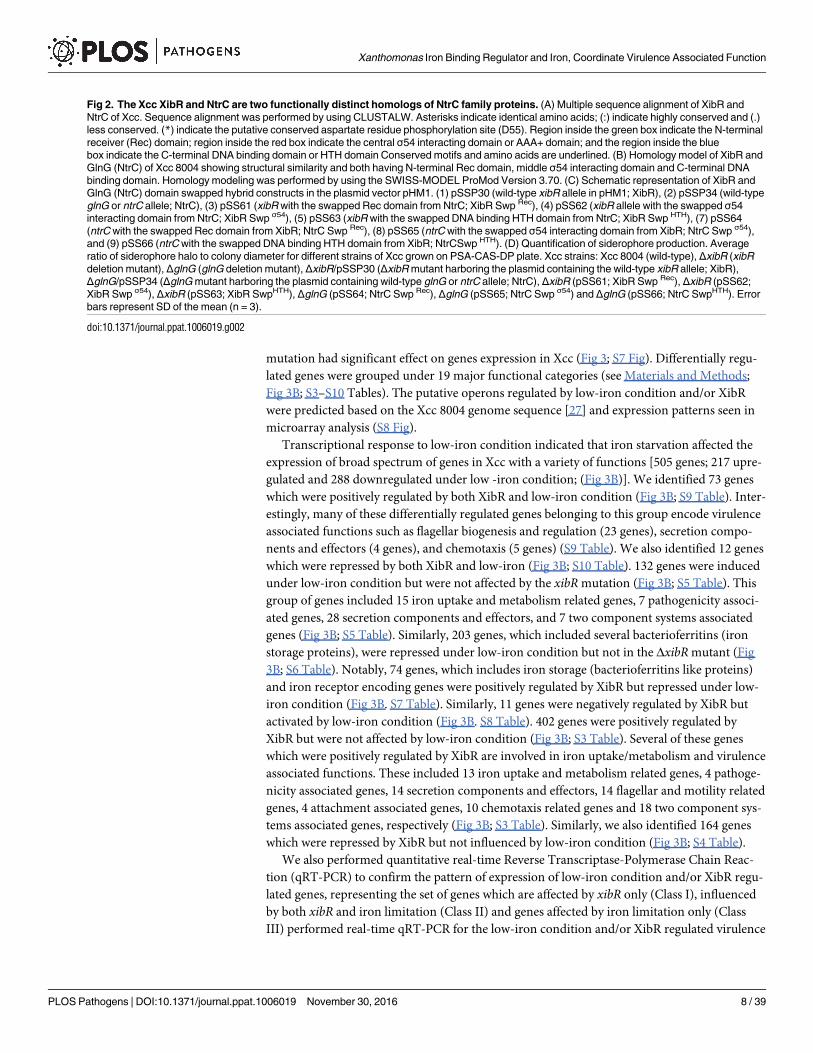
Fig 2. The Xcc XibR and NtrC are two functionally distinct homologs of NtrC family proteins. (A) Multiple sequence alignment of XibR and
NtrC of Xcc. Sequence alignment was performed by using CLUSTALW. Asterisks indicate identical amino acids; (:) indicate highly conserved and (.)
less conserved. (*) indicate the putative conserved aspartate residue phosphorylation site (D55). Region inside the green box indicate the N-terminal
receiver (Rec) domain; region inside the red box indicate the central σ54 interacting domain or AAA+ domain; and the region inside the blue
box indicate the C-terminal DNA binding domain or HTH domain Conserved motifs and amino acids are underlined. (B) Homology model of XibR and
GlnG (NtrC) of Xcc 8004 showing structural similarity and both having N-terminal Rec domain, middle σ54 interacting domain and C-terminal DNA
binding domain. Homology modeling was performed by using the SWISS-MODEL ProMod Version 3.70. (C) Schematic representation of XibR and
GlnG (NtrC) domain swapped hybrid constructs in the plasmid vector pHM1. (1) pSSP30 (wild-type xibR allele in pHM1; XibR), (2) pSSP34 (wild-type
glnG or ntrC allele; NtrC), (3) pSS61 (xibR with the swapped Rec domain from NtrC; XibR Swp Rec), (4) pSS62 (xibR allele with the swapped σ54
interacting domain from NtrC; XibR Swp σ54), (5) pSS63 (xibR with the swapped DNA binding HTH domain from NtrC; XibR Swp HTH), (7) pSS64
(ntrC with the swapped Rec domain from XibR; NtrC Swp Rec), (8) pSS65 (ntrC with the swapped σ54 interacting domain from XibR; NtrC Swp σ54),
and (9) pSS66 (ntrC with the swapped DNA binding HTH domain from XibR; NtrCSwp HTH). (D) Quantification of siderophore production. Average
ratio of siderophore halo to colony diameter for different strains of Xcc grown on PSA-CAS-DP plate. Xcc strains: Xcc 8004 (wild-type), ΔxibR (xibR
deletion mutant), ΔglnG (glnG deletion mutant), ΔxibR/pSSP30 (ΔxibR mutant harboring the plasmid containing the wild-type xibR allele; XibR),
ΔglnG/pSSP34 (ΔglnG mutant harboring the plasmid containing wild-type glnG or ntrC allele; NtrC), ΔxibR (pSS61; XibR Swp Rec), ΔxibR (pSS62;
XibR Swp σ54), ΔxibR (pSS63; XibR SwpHTH), ΔglnG (pSS64; NtrC Swp Rec), ΔglnG (pSS65; NtrC Swp σ54) and ΔglnG (pSS66; NtrC SwpHTH). Error
bars represent SD of the mean (n = 3).
doi:10.1371/journal.ppat.1006019.g002
Xanthomonas Iron Binding Regulator and Iron, Coordinate Virulence Associated Function
PLOS Pathogens | DOI:10.1371/journal.ppat.1006019 November 30, 2016 8 / 39

Fig 3. Genome-wide expression analysis of the iron-starvation and/or XibR regulon in Xcc. (A) The map of differentially expressed genes in response
to iron limitation and/or xibR mutation in Xcc represented by using circos plot. From the outer to the inner circle, Track 1 shows circular genome of Xcc 8004
Xanthomonas Iron Binding Regulator and Iron, Coordinate Virulence Associated Function
PLOS Pathogens | DOI:10.1371/journal.ppat.1006019 November 30, 2016 9 / 39

associated factors to validate the microarray results. Real-time qRT-PCR of differentially
expressed class I-III genes corroborated the microarray results (Fig 3C).
ΔxibR mutant of Xcc exhibit altered ferric iron uptake and storage
Analysis of our microarray data indicated that several genes related to iron storage (ferritin-
like proteins), putative outer membrane receptor for ferric iron uptake (fhuE) and putative
TonB dependent receptors were positively regulated by XibR (S3 Table; S9 Table). To under-
stand the role of xibR in iron uptake and storage, we performed in vitro 55Fe3+, 55Fe2+ and55Fe3+-vibrioferrin complex uptake assays with wild-type Xcc 8004, the ΔxibR mutant, ΔxibRmutant harboring the plasmid-borne wild-type xibR allele (ΔxibR/pSSP30), and the ΔxibR har-
boring the plasmid-borne point mutant D55AXibR allele (ΔxibR/pSSP39) as described previ-
ously [13,28,29] with few modifications (see Materials and Methods). The total amount of55Fe3+ incorporated into the ΔxibR mutant or ΔxibR/pSSP39 was significantly less than that
incorporated into the wild-type Xcc 8004 and the ΔxibR/pSSP30 over the 10 min time-course
of the experiment (Fig 4A). Interestingly, uptake assay in the presence of vibrioferrin (1:1 ratio
of 55Fe3+ and vibrioferrin) indicated that the total amount of 55Fe3+ incorporated was signifi-
cantly higher in the ΔxibR or ΔxibR/pSSP39 strains compared to that incorporated into either
the wild-type Xcc 8004 or ΔxibR/pSSP30 (Fig 4B). In contrast, there was no significant differ-
ence in the amount of radiolabelled Fe2+ incorporated into four of these strains (S9A Fig).
Reduced incorporation of radiolabelled ferric iron in the ΔxibR mutant compared to the
wild-type strain indicated that XibR may be involved in the regulation of expression of low-
affinity or non-vibrioferrin mediated ferric iron uptake or storage system/s under iron-deplete
condition. Our microarray based expression analysis indicated that an outer membrane recep-
tor for ferric iron uptake (fhuE; XC_0924), was positively regulated by low-iron and XibR (S9
Table). XC_0924 appears to be arranged in an operon with XC_0925, an outer membrane
receptor for ferric iron uptake), which exhibit 49% identity and 67% similarity with previously
reported FauA of human pathogenic bacteria Bordetella pertussis [30] and 47% identity and
68% similarity with FpvA of Pseudomonas aeruginosa [31], respectively. Real-time qRT-PCR
analysis indicated that XC_0925 is also induced under low-iron condition and ΔxibR mutant
exhibited reduced expression compared to the Xcc 8004 and ΔxibR/pSSP30 under iron-deplete
condition (Fig 4C). In contrast, the expression of xsuA (Xanthomonas siderophore uptake)
was approximately 6-fold higher in the ΔxibR mutant compared to the wild-type Xcc 8004 and
ΔxibR/pSSP30 strains grown either in iron-replete or under low-iron conditions, respectively
(Fig 4D). We did not observe any significant difference in the expression of feoB (ferrous iron
transporter) and ferrous uptake regulator (fur) in the ΔxibR mutant compared to the wild-type
Xcc 8004 and ΔxibR/pSSP30 strains by real-time qRT-PCR (S9B and S9C Fig).
(~5.15Mb) with scale in Mb; Track 2, loci presentation of Xcc 8004 circular genome; Track 3, differentially expressed genes in ΔxibR mutant versus wild-type
Xcc 8004 strain grown under iron-replete condition (PS medium); Track 4, differentially expressed genes in ΔxibR mutant grown under low-iron condition (PS
+ DP) versus iron-replete condition; Track 5, differentially expressed genes in ΔxibR mutant versus wild-type Xcc 8004 strain, both grown under low-iron
condition; and Track 6, differentially expressed genes in the wild-type strain grown under low-iron condition versus iron-replete condition. Color scale
indicates log2 –fold change of expression (from green for downregulated to red for upregulated). Low-iron was made by addition of 100 μM DP to PS
medium. (B) Venn diagram showing the overlap and unique subset of genes belonging to different functional groups of Xcc whose expression is upregulated
or downregulated under low-iron condition and/or XibR. For detail list of genes please see S3 to S10 Tables. (C) Expression analysis by microarray and real-
time qRT-PCR indicating xibR and/or iron limitation regulated genes involved in flagellar biogenesis and regulation, metabolism, chemotaxis, and virulence.
The y-axis represents log2-fold change in expression. For RT-PCR, data were normalized to an internal 16S rRNA control, and the relative changes in the
transcriptional level were calculated as a ratio of transcript levels of ΔxibR versus wild-type Xcc 8004 strain grown in PS medium (iron-replete condition), and
Xcc 8004 grown under low-iron condition (PS + DP) versus that grown in PS medium (iron-replete condition) using log2 of fold difference method. I, II and III
represent set of genes which are affected by xibR only (I), influenced by both xibR and iron limitation (II) and genes affected by iron limitation only (III). Data
represents the means ± S.E. (n = 3).
doi:10.1371/journal.ppat.1006019.g003
Xanthomonas Iron Binding Regulator and Iron, Coordinate Virulence Associated Function
PLOS Pathogens | DOI:10.1371/journal.ppat.1006019 November 30, 2016 10 / 39

Xanthomonas Iron Binding Regulator and Iron, Coordinate Virulence Associated Function
PLOS Pathogens | DOI:10.1371/journal.ppat.1006019 November 30, 2016 11 / 39

We next performed streptonigrin sensitivity assay, which depends on the intracellular iron
levels, to assess intracellular iron content in different strains of Xcc [13,32]. Streptonigrin sen-
sitivity assay indicated that the wild-type Xcc 8004 and ΔxibR/pSSP30 strains were hypersensi-
tive to streptonigrin compared to the ΔxibR and ΔxibR/pSSP39 (Fig 4E; S9D Fig), indicative of
low intracellular iron level in the ΔxibR mutant. In addition, measurement of intracellular iron
content in different strains of Xcc by Inductively Coupled Plasma-Optical Emission Spectrom-
etry (ICP-OES) indicated that the ΔxibR and ΔxibR/pSSP39 strains contained less intracellular
iron (approximately 26%) compared to either the wild-type Xcc 8004 or ΔxibR/pSSP30, respec-
tively (Fig 4F). Expression analysis by real-time qRT-PCR indicated that iron storage-related
putative ferritin genes XC_2164, XC_2190 and XC_3752 were down regulated in the ΔxibRmutant compared to the in wild-type Xcc 8004 and ΔxibR/pSSP30 under low-iron condition
(Fig 4G and 4H; S9H Fig). Growth assays under low-iron conditions indicated that the ΔxibRand ΔxibR/pSSP39 strains exhibited reduced growth compared to either the wild-type Xcc
8004 or ΔxibR/pSSP30, which could be rescued by exogenous iron supplementation (S9E–S9G
Fig; S11 Table).
Since ΔxibR mutant exhibited lower level of intracellular iron content and growth defi-
ciency under low-iron condition, it follows that some of the genes identified in our expression
analysis by microarray and real-time qRT-PCR may have a role in iron metabolism. In order
to examine this, we made three deletion strains; 1. ΔyciE ΔyciF ΔXC_3754, triple deletion
mutant strain with the deletion of entire cluster of bacterioferritin-related protein encoding
genes XC_3752, XC_3753 and XC_3754); 2. ΔfhuEΔXC_0925, double deletion mutant in the
receptors for non-vibrioferrin mediated ferric iron uptake encoding genes (XC_0924 and
XC_0925); and 3. ΔfecR (XC_0057) encoding the periplasmic iron dicitrate sensor [33]. The
bacterioferritin-related protein encoding genes (XC_3752, XC_3753 and XC_3754) and the
genes encoding the ferric iron uptake proteins (XC_0924 and XC_0925) are regulated by both
xibR and low-iron condition, whereas, the expression of fecR is only influence by low-iron con-
dition (S5 Table. S9I Fig). Streptonigrin sensitivity and growth assays under low-iron condi-
tion indicated that the triple deletion strain ΔyciE ΔyciF ΔXC_3754 and the ΔfecR mutant
exhibited lower intracellular iron levels compared to the wild-type strain, whereas there was
not much significant difference in the streptonigrin sensitivity and growth under low-iron
condition in the ΔfhuE ΔXC_0925 double mutant compared to the wild-type Xcc 8004 strain
(S9J–S9N Fig; S11 Table)
XibR and low-iron condition induces chemotaxis and motility in Xcc
Expression analysis by microarray and real-time qRT-PCR indicated that several chemotaxis
and motility-related genes are positively regulated by XibR and low-iron condition (Fig 3;
Fig 4. ΔxibR mutant of Xcc exhibit altered ferric iron uptake and defect in iron storage. (A) ΔxibR mutant exhibits defect in ferric iron uptake. Transport
was initiated by addition of 0.5 μM 55FeCl3 to cell suspensions of Xcc 8004, ΔxibR, ΔxibR/pSSP30 and ΔxibR/pSSP39 grown under low-iron condition. Low-
iron was made by addition of 150 μM DP to PS medium. Incorporation of radiolabelled Fe3+ was detected by scintillation counter. (B) ΔxibR mutant exhibits
enhanced uptake of ferric iron-vibrioferrin complex. Transport was initiated by the addition of 0.5 μM 1:1 ratios of 55FeCl3 and vibrioferrin to the cell
suspensions of different Xcc strains grown under low-iron condition. (C and D) Relative quantification of the expression of Xanthomonas siderophore uptake
gene (xsuA) and outer membrane receptor for ferric iron (XC_0925) of Xcc grown under PS, PS + 100 μM DP, and PS + 100 μM DP + 100 μM FeSO4 by real-
time qRT-PCR. 16S ribosomal RNA was used as an endogenous control to normalize the RNA for cellular abundance. (E) Streptonigrin (SNG) sensitivity
plate assay. Different Xcc strains were grown in PS media at a density of 1 × 10⁹ cells/ml. 4 μL of cultures from each serial dilution was spotted on PSA plates
containing 1μg/ml SNG and 0.01 M sodium citrate. Plates were incubated for 72 h at 28˚C to observe bacterial growth. (F) Intracellular iron content
quantification determined by atomic absorption spectrophotometry. Different Xcc strains were grown at a density of 1.2 OD600 in PS medium. Cells were
harvested, freeze dried, and determined the iron content by Inductively Coupled Plasma-Optical Emission Spectrometry (ICP-OES). (G and H) Relative
quantification of the expression of Xanthomonas putative ferritin-like protein (XC_2164) and putative ferritin related protein (XC_2190) of Xcc grown under
PS, PS + 100 μM DP, and PS + 100 μM DP + 100 μM FeSO4 by real-time qRT-PCR. Data shown in the graphs are mean ± S.E. (n = 3). * Indicating p-
value < 0.05, **indicating p-value < 0.01 and *** indicating p-value < 0.001 statistically significance by paired student t-test.
doi:10.1371/journal.ppat.1006019.g004
Xanthomonas Iron Binding Regulator and Iron, Coordinate Virulence Associated Function
PLOS Pathogens | DOI:10.1371/journal.ppat.1006019 November 30, 2016 12 / 39
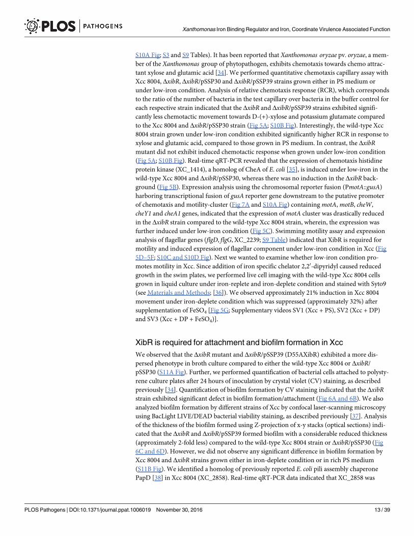
S10A Fig; S3 and S9 Tables). It has been reported that Xanthomonas oryzae pv. oryzae, a mem-
ber of the Xanthomonas group of phytopathogen, exhibits chemotaxis towards chemo attrac-
tant xylose and glutamic acid [34]. We performed quantitative chemotaxis capillary assay with
Xcc 8004, ΔxibR, ΔxibR/pSSP30 and ΔxibR/pSSP39 strains grown either in PS medium or
under low-iron condition. Analysis of relative chemotaxis response (RCR), which corresponds
to the ratio of the number of bacteria in the test capillary over bacteria in the buffer control for
each respective strain indicated that the ΔxibR and ΔxibR/pSSP39 strains exhibited signifi-
cantly less chemotactic movement towards D-(+)-xylose and potassium glutamate compared
to the Xcc 8004 and ΔxibR/pSSP30 strain (Fig 5A; S10B Fig). Interestingly, the wild-type Xcc
8004 strain grown under low-iron condition exhibited significantly higher RCR in response to
xylose and glutamic acid, compared to those grown in PS medium. In contrast, the ΔxibRmutant did not exhibit induced chemotactic response when grown under low-iron condition
(Fig 5A; S10B Fig). Real-time qRT-PCR revealed that the expression of chemotaxis histidine
protein kinase (XC_1414), a homolog of CheA of E. coli [35], is induced under low-iron in the
wild-type Xcc 8004 and ΔxibR/pSSP30, whereas there was no induction in the ΔxibR back-
ground (Fig 5B). Expression analysis using the chromosomal reporter fusion (PmotA::gusA)
harboring transcriptional fusion of gusA reporter gene downstream to the putative promoter
of chemotaxis and motility-cluster (Fig 7A and S10A Fig) containing motA, motB, cheW,
cheY1 and cheA1 genes, indicated that the expression of motA cluster was drastically reduced
in the ΔxibR strain compared to the wild-type Xcc 8004 strain, wherein, the expression was
further induced under low-iron condition (Fig 5C). Swimming motility assay and expression
analysis of flagellar genes (flgD, flgG, XC_2239; S9 Table) indicated that XibR is required for
motility and induced expression of flagellar component under low-iron condition in Xcc (Fig
5D–5F; S10C and S10D Fig). Next we wanted to examine whether low-iron condition pro-
motes motility in Xcc. Since addition of iron specific chelator 2,20-dipyridyl caused reduced
growth in the swim plates, we performed live cell imaging with the wild-type Xcc 8004 cells
grown in liquid culture under iron-replete and iron-deplete condition and stained with Syto9
(see Materials and Methods; [36]). We observed approximately 21% induction in Xcc 8004
movement under iron-deplete condition which was suppressed (approximately 32%) after
supplementation of FeSO4 [Fig 5G; Supplementary videos SV1 (Xcc + PS), SV2 (Xcc + DP)
and SV3 (Xcc + DP + FeSO4)].
XibR is required for attachment and biofilm formation in Xcc
We observed that the ΔxibR mutant and ΔxibR/pSSP39 (D55AXibR) exhibited a more dis-
persed phenotype in broth culture compared to either the wild-type Xcc 8004 or ΔxibR/
pSSP30 (S11A Fig). Further, we performed quantification of bacterial cells attached to polysty-
rene culture plates after 24 hours of inoculation by crystal violet (CV) staining, as described
previously [34]. Quantification of biofilm formation by CV staining indicated that the ΔxibRstrain exhibited significant defect in biofilm formation/attachment (Fig 6A and 6B). We also
analyzed biofilm formation by different strains of Xcc by confocal laser-scanning microscopy
using BacLight LIVE/DEAD bacterial viability staining, as described previously [37]. Analysis
of the thickness of the biofilm formed using Z-projection of x-y stacks (optical sections) indi-
cated that the ΔxibR and ΔxibR/pSSP39 formed biofilm with a considerable reduced thickness
(approximately 2-fold less) compared to the wild-type Xcc 8004 strain or ΔxibR/pSSP30 (Fig
6C and 6D). However, we did not observe any significant difference in biofilm formation by
Xcc 8004 and ΔxibR strains grown either in iron-deplete condition or in rich PS medium
(S11B Fig). We identified a homolog of previously reported E. coli pili assembly chaperone
PapD [38] in Xcc 8004 (XC_2858). Real-time qRT-PCR data indicated that XC_2858 was
Xanthomonas Iron Binding Regulator and Iron, Coordinate Virulence Associated Function
PLOS Pathogens | DOI:10.1371/journal.ppat.1006019 November 30, 2016 13 / 39

Fig 5. Chemotaxis and motility are regulated by xibR and induced under iron limitation. (A) Quantitative chemotaxis capillary assay in response to D-
(+)-Xylose with different Xcc strains grown under PS, PS + 100 μM DP and PS + 100 μM DP + 100 μM FeSO4. Cells were incubated at 28˚C with capillaries
containing D-(+)-Xylose (1.2 mM) and PBS. Relative chemotaxis response was determined by migrated bacterial cells in capillary containing D-(+)-Xylose
over the migrated bacterial cells in capillary containing PBS. Data are shown as mean ± S.E. (n = 3). The experiment was repeated two times. (B) Relative
quantification of the expression of chemotaxis histidine protein kinase (cheA3) of Xcc grown under PS, PS + 100 μM DP, and PS + 100 μM DP + 100 μM
FeSO4 by real-time qRT-PCR. 16S ribosomal RNA was used as an endogenous control to normalize the RNA for cellular abundance. (C) Expression
analysis of motA operon in wild-type Xcc 8004 and ΔxibR mutant grown under PS, PS + 100 μM DP, and PS + 100 μM DP + 100 μM FeSO4 by monitoring
the β-glucuronidase (GUS) activity. (D) Swim plate motility assay for different Xcc strains; Xcc 8004, ΔxibR, ΔxibR/pSSP30 and ΔxibR/pSSP39. (E) Motility
zone diameter quantification from semisolid swim plate motility assay. (F) Relative quantification of the expression of flagellar biosynthesis gene for a cell-
distal portion of basal-body rod (XC_2239) of Xcc grown under PS, PS + 100 μM DP, and PS + 100 μM DP + 100 μM FeSO4 by real-time qRT-PCR. (G)
Xanthomonas Iron Binding Regulator and Iron, Coordinate Virulence Associated Function
PLOS Pathogens | DOI:10.1371/journal.ppat.1006019 November 30, 2016 14 / 39

down-regulated in ΔxibR mutant compared to the wild-type Xcc 8004 and complemented
strain ΔxibR/pSSP30. However, there was no significant difference under low-iron condition
compared to the PS medium (Fig 6E).
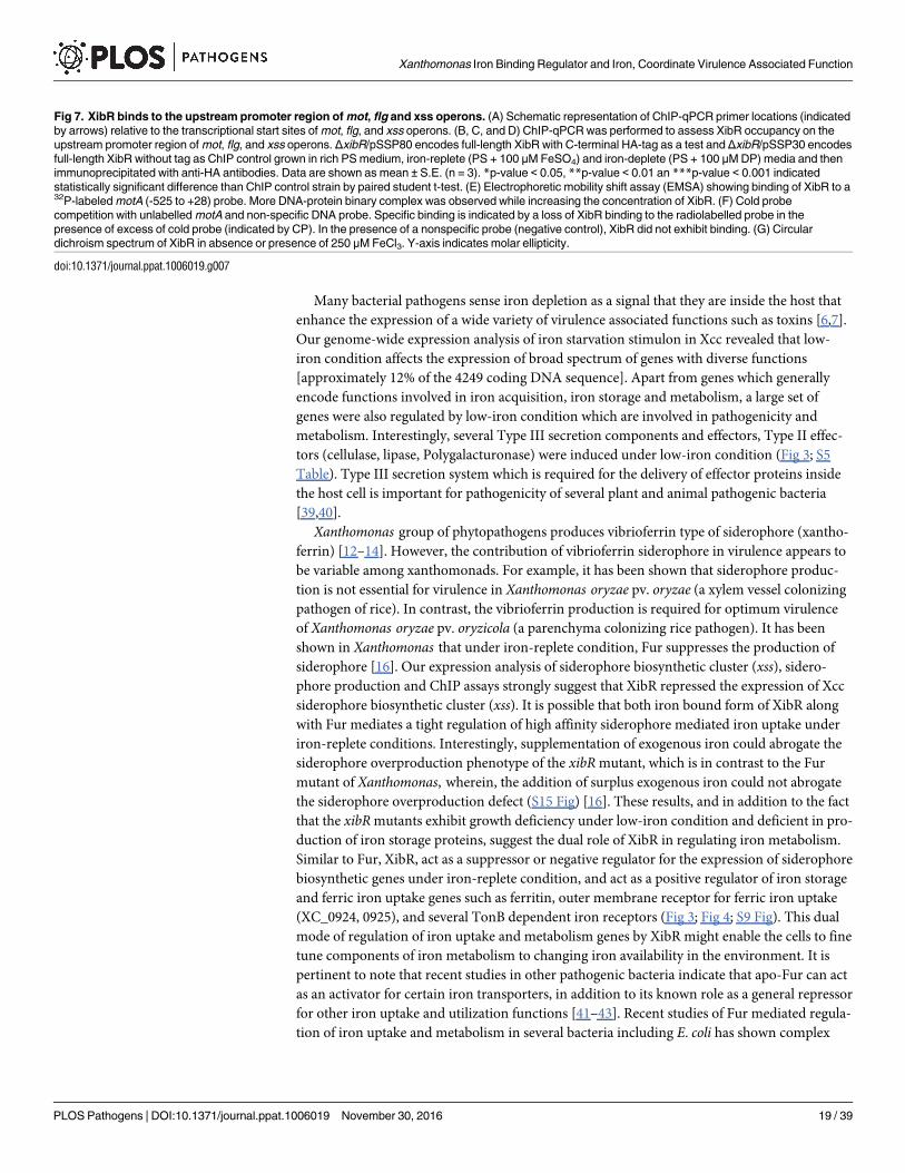
XibR regulates transcription of mot, flg and xsuA operons by binding to
the promoters
Our expression analysis indicated that XibR is involved in the regulation of mot, flg, and xssoperons. To determine whether XibR is capable of binding to the upstream region of mot, flg,
and xss operons (Fig 7A), we performed chromatin immunoprecipitation (ChIP) using epi-
tope-tagged XibR expressed in the ΔxibR strain and tagless XibR as ChIP control. Complemen-
tation experiment established that the HA-tag did not affect the function of the XibR protein
(S12A Fig). The ΔxibR/pSSP80 (HA-tagged XibR) and ΔxibR/pSSP30 (tagless-XibR) strains
were grown under iron-replete and iron-deplete conditions. DNA-protein cross-linking was
done by formaldehyde treatment to bacterial cells followed by immunoprecipitation of XibR
from lysates using anti-HA antibodies by a sandwich technique (see Materials and Methods).
ChIP followed by quantitative real-time PCR (ChIP-qPCR) of captured DNA fragments indi-
cated binding of XibR to the motA and flg upstream sequence (Fig 7B and 7C). Interestingly,
XibR exhibited significant binding to xss promoter only under iron-replete condition (Fig 7D).
We also performed electrophoretic mobility shift assay (EMSA) to study the in vitro binding
of XibR to motA promoter using the purified C-terminal His-tagged XibR (S2 Table; S12B–
S12D Fig) and 553 bp upstream region of motA comparising the sequence from -525 to +28
(see Materials and Methods). EMSA analysis indicated that XibR specifically bind the radiola-
belled motA fragment (Fig 7E and 7F). We also observed binding of XibR to the 393 bp
upstream region of xss cluster comprising the sequence from -188 to +205 (S13A Fig). To rule
out the possibility of probe shift due to DNA-protein aggregation, we performed EMSA with a
non-specific 32P-labelled probe, which did not exhibit any shift (Fig 7F). Interestingly, we
observed that the presence of ferric form of iron in the EMSA binding buffer promoted invitro binding of XibR to DNA (S13B Fig). However, except for ferrous (Fe2+), presence of
other metal ions did not affect the binding of XibR to the target DNA (S13C and S13D Fig).
Since under aerobic conditions, ferrous iron is oxidized to ferric form of iron, and to rule out
the possibility that the increased binding of XibR exhibited in the presence of ferrous iron may
be due to conversion to ferric form, we added ferric specific chelator deferoxamine mesylate in
the binding buffer. Addition of deferoxamine mesylate with ferrous form of iron drastically
reduced the binding of XibR to the target DNA (S13C Fig).
In an attempt to identify putative consensus motif in the xibR regulated upstream regula-
tory sequence of mot, flg and xss, we performed consensus sequences search using the MEME
(Multiple Em for Motif Elicitation; at http://meme-suite.org/tools/meme), which predicted five
consensus sequence (S14 Fig; S12 Table). Sequence analysis of 200 bp upstream region of all the
XibR regulated genes identified in the microarray experiment indicated that 19.29% of them
harbor either of the consensus motif (motif 1 = 1.09%; 2 = 1.29%; 3 = 3.96%; 4 = 2.73%; and
5 = 10.23%) (S12 Table). It is possible that the consensus motif 5 (CAGAACGACAAC), which
constitute 10.23% of the XibR regulated genes, could be the potential direct target of XibR.
Bacterial velocity measurement from the live cell imaging of bacterial movement at single cell level by using manual tracking and chemotaxis tools with
ImageJ software. Cells were grown in PS, low-iron (PS + 100 μM 2,20-dipyridyl) and PS + 100 μM 2,20-dipyridyl + 100 μM FeSO4 at 28˚C up to mid-
exponential phase, stained with Syto9 and incubated for 10 min at 28˚C. The stained cells were loaded into a chamber of sterile glass bottom plates
containing PS medium with 0.3% agar and visualized on epifluorescence microscope. Values are mean of at least 25 bacteria up to 20 frames. The
experiment was repeated three times. Error bars are SEM. Data shown in the graphs are mean ± S.E. (n = 3).* Indicating p-value < 0.05, **indicating p-
value < 0.01 and *** indicating p-value < 0.001 statistically significance by paired student t-test.
doi:10.1371/journal.ppat.1006019.g005
Xanthomonas Iron Binding Regulator and Iron, Coordinate Virulence Associated Function
PLOS Pathogens | DOI:10.1371/journal.ppat.1006019 November 30, 2016 15 / 39

Xanthomonas Iron Binding Regulator and Iron, Coordinate Virulence Associated Function
PLOS Pathogens | DOI:10.1371/journal.ppat.1006019 November 30, 2016 16 / 39
Furthermore, we performed circular dichroism (CD) spectroscopy to detect possible struc-
tural changes in XibR in the presence of ferric iron. CD spectroscopy measurements suggested
that iron elicited conformational changes upon binding to XibR that result in reduced content
of α-helix in the protein (Fig 7G).
xibR is required for optimum virulence
To understand the role of xibR in the virulence of Xcc, we performed infection studies with
wild-type Xcc 8004, ΔxibR, ΔxibR/pSSP30 and ΔxibR/pSSP39 strains on cabbage plant. For
infection studies, 30-day old cabbage leaves were inoculated with bacterial cell suspension by
leaf clip method and monitored the lesion development, bacterial growth and migration inside
the leaves (Fig 8). Infection studies suggested that the ΔxibR and ΔxibR/pSSP39 strains exhib-
ited significant reduction in lesion development, growth and migration inside the leaves com-
pared to either the wild-type Xcc 8004 or ΔxibR/pSSP30 (Fig 8A–8D) strain. Expression
analysis by real-time qRT-PCR indicated that virulence associated functions such as Type III
secretion protein ATPase encoding gene (XC_3006), hrpB1, hrcV, hrpG and hrpX, were down-
regulated in the ΔxibR mutant compared to either the wild-type Xcc 8004 or the ΔxibR/
pSSP30, respectively (Fig 8E).
Discussion
Ability of the bacterial pathogen to respond and adapt to iron limiting condition inside host is
essential to their virulence. Pathogenic bacteria utilize diverse and efficient iron uptake systems
that enable them to scavenge various forms of iron from the environment under iron-
restricted conditions. Due to essentiality of iron in bacterial growth and survival particularly in
iron limiting environment and also the potential toxic effect due to iron overload, bacteria
tightly regulate iron uptake, metabolism and distribution in response to environmental cues
mediated by iron dependent regulators such as Fur and DtxR [4,7,9].
Fur and Fur-like homologs play an important role in regulating iron metabolism and viru-
lence in many Gram-negative bacteria. Fur binds to ferrous (Fe2+) form of iron which acts as a
co-repressor, and suppresses the expression of high affinity iron uptake system under iron-
replete condition [7,9]. Fur also has been shown to act as an activator of virulence associated
factors in bacterial pathogens such as Neisseria meningitidis, Salmonella typhimurium and Heli-cobacter pylori [9]. However, apart from Fur and DtxR type ferrous binding regulators, little is
known about how iron regulated genes and virulence associated functions are fine-tuned and
coordinately regulated by other iron responsive regulatory proteins.
In this study we showed that a novel NtrC family of response regulator, XibR binds to ferric
form of iron and regulates the expression of several iron metabolism and virulence associated
functions in important phytopathogen Xanthomonas campestris pv. campestris (Xcc). To our
knowledge iron-responsive ferric binding regulator has not been reported in any bacteria.
Fig 6. xibR promotes Biofilm formation. (A) Biofilm formation by Xcc 8004, ΔxibR, ΔxibR/pSSP30 and ΔxibR/pSSP39 strains in the static biofilm after
24 hrs of growth and staining with 0.1% Crystal Violet. (B) Quantification of attached cells of different Xcc strains in the static biofilm after 24 hours of
growth. Attached cells were stained with Crystal Violet (CV), dissolved in ethanol and quantified by measuring absorbance at 570 nm. Data are shown as
mean ± S.E. (n = 3). (C) Representative confocal laser-scanning microscopy (CLSM) images of biofilms formed on glass slides at the air–media interface
by different Xcc strains grown in PS medium for 24h, and stained with BacLight LIVE/DEAD stain. Each 3D image represents the layer in the Z-stack. (D)
Average biofilm thickness of different strains of Xcc formed on the glass slide at the air-media interphase. For quantification of the thickness, five
independent biofilms were scanned with CLSM at ten randomly selected positions and thickness was determined through height of the biofilm. Data are
shown as mean ± S.E. (n = 3). ** Indicating p-value < 0.01 and *** indicating p-value < 0.001 statistically significance by paired student t-test. (E) Relative
quantification of the expression of pili assembly chaperone of Xcc grown under PS, PS + 100 μM DP, and PS + 100 μM DP + 100 μM FeSO4 by real-time
qRT-PCR. 16S ribosomal RNA was used as an endogenous control to normalize the RNA for cellular abundance. Data are shown as mean ± S.E. (n = 3).
doi:10.1371/journal.ppat.1006019.g006
Xanthomonas Iron Binding Regulator and Iron, Coordinate Virulence Associated Function
PLOS Pathogens | DOI:10.1371/journal.ppat.1006019 November 30, 2016 17 / 39

Xanthomonas Iron Binding Regulator and Iron, Coordinate Virulence Associated Function
PLOS Pathogens | DOI:10.1371/journal.ppat.1006019 November 30, 2016 18 / 39
Many bacterial pathogens sense iron depletion as a signal that they are inside the host that
enhance the expression of a wide variety of virulence associated functions such as toxins [6,7].
Our genome-wide expression analysis of iron starvation stimulon in Xcc revealed that low-
iron condition affects the expression of broad spectrum of genes with diverse functions
[approximately 12% of the 4249 coding DNA sequence]. Apart from genes which generally
encode functions involved in iron acquisition, iron storage and metabolism, a large set of
genes were also regulated by low-iron condition which are involved in pathogenicity and
metabolism. Interestingly, several Type III secretion components and effectors, Type II effec-
tors (cellulase, lipase, Polygalacturonase) were induced under low-iron condition (Fig 3; S5
Table). Type III secretion system which is required for the delivery of effector proteins inside
the host cell is important for pathogenicity of several plant and animal pathogenic bacteria
[39,40].
Xanthomonas group of phytopathogens produces vibrioferrin type of siderophore (xantho-
ferrin) [12–14]. However, the contribution of vibrioferrin siderophore in virulence appears to
be variable among xanthomonads. For example, it has been shown that siderophore produc-
tion is not essential for virulence in Xanthomonas oryzae pv. oryzae (a xylem vessel colonizing
pathogen of rice). In contrast, the vibrioferrin production is required for optimum virulence
of Xanthomonas oryzae pv. oryzicola (a parenchyma colonizing rice pathogen). It has been
shown in Xanthomonas that under iron-replete condition, Fur suppresses the production of
siderophore [16]. Our expression analysis of siderophore biosynthetic cluster (xss), sidero-
phore production and ChIP assays strongly suggest that XibR repressed the expression of Xcc
siderophore biosynthetic cluster (xss). It is possible that both iron bound form of XibR along
with Fur mediates a tight regulation of high affinity siderophore mediated iron uptake under
iron-replete conditions. Interestingly, supplementation of exogenous iron could abrogate the
siderophore overproduction phenotype of the xibR mutant, which is in contrast to the Fur
mutant of Xanthomonas, wherein, the addition of surplus exogenous iron could not abrogate
the siderophore overproduction defect (S15 Fig) [16]. These results, and in addition to the fact
that the xibR mutants exhibit growth deficiency under low-iron condition and deficient in pro-
duction of iron storage proteins, suggest the dual role of XibR in regulating iron metabolism.
Similar to Fur, XibR, act as a suppressor or negative regulator for the expression of siderophore
biosynthetic genes under iron-replete condition, and act as a positive regulator of iron storage
and ferric iron uptake genes such as ferritin, outer membrane receptor for ferric iron uptake
(XC_0924, 0925), and several TonB dependent iron receptors (Fig 3; Fig 4; S9 Fig). This dual
mode of regulation of iron uptake and metabolism genes by XibR might enable the cells to fine
tune components of iron metabolism to changing iron availability in the environment. It is
pertinent to note that recent studies in other pathogenic bacteria indicate that apo-Fur can act
as an activator for certain iron transporters, in addition to its known role as a general repressor
for other iron uptake and utilization functions [41–43]. Recent studies of Fur mediated regula-
tion of iron uptake and metabolism in several bacteria including E. coli has shown complex
Fig 7. XibR binds to the upstream promoter region of mot, flg and xss operons. (A) Schematic representation of ChIP-qPCR primer locations (indicated
by arrows) relative to the transcriptional start sites of mot, flg, and xss operons. (B, C, and D) ChIP-qPCR was performed to assess XibR occupancy on the
upstream promoter region of mot, flg, and xss operons. ΔxibR/pSSP80 encodes full-length XibR with C-terminal HA-tag as a test and ΔxibR/pSSP30 encodes
full-length XibR without tag as ChIP control grown in rich PS medium, iron-replete (PS + 100 μM FeSO4) and iron-deplete (PS + 100 μM DP) media and then
immunoprecipitated with anti-HA antibodies. Data are shown as mean ± S.E. (n = 3). *p-value < 0.05, **p-value < 0.01 an ***p-value < 0.001 indicated
statistically significant difference than ChIP control strain by paired student t-test. (E) Electrophoretic mobility shift assay (EMSA) showing binding of XibR to a32P-labeled motA (-525 to +28) probe. More DNA-protein binary complex was observed while increasing the concentration of XibR. (F) Cold probe
competition with unlabelled motA and non-specific DNA probe. Specific binding is indicated by a loss of XibR binding to the radiolabelled probe in the
presence of excess of cold probe (indicated by CP). In the presence of a nonspecific probe (negative control), XibR did not exhibit binding. (G) Circular
dichroism spectrum of XibR in absence or presence of 250 μM FeCl3. Y-axis indicates molar ellipticity.
doi:10.1371/journal.ppat.1006019.g007
Xanthomonas Iron Binding Regulator and Iron, Coordinate Virulence Associated Function
PLOS Pathogens | DOI:10.1371/journal.ppat.1006019 November 30, 2016 19 / 39

Fig 8. xibR is required for optimal virulence. (A) Infected cabbage leaves with different Xcc strain showing symptoms as a lesion at 15 days
postinoculation. 30 days old plants were inoculated with bacterial cultures (1 X 10⁹ cells/ml suspension) of different Xcc strains by clip method. (B)
In planta growth assays of Xcc 8004, ΔxibR, ΔxibR/pSSP30 and ΔxibR/pSSP39 strains. Bacterial populations were measured by crushing the
leaves of 1cm2 areas for each and serial dilution plating at the indicated post inoculation days. Data are shown as mean ± S.E. (n = 3). (C) In
planta bacterial migration assay was performed by inoculating 1cm pieces of infected leave, cut from base to tip with sterile scissors on rich PS
medium with respective antibiotics. Migration was estimated by observing colonies formed after 1 to 3 days by the bacterial ooze from the cut
ends of cabbage leaf pieces. (D) Quantification of lesion length at 15 days post inoculation. Data shown as mean ± S.E. (n = 25). (E) Relative
quantification of the expression of different Type III secretion system hrp genes of Xcc 8004, ΔxibR, and ΔxibR/pSSP30 strains by real-time
qRT-PCR. * Indicating p-value < 0.05, **indicating p-value < 0.01 and *** indicating p-value < 0.001 statistically significance by paired student t-
test.
doi:10.1371/journal.ppat.1006019.g008
Xanthomonas Iron Binding Regulator and Iron, Coordinate Virulence Associated Function
PLOS Pathogens | DOI:10.1371/journal.ppat.1006019 November 30, 2016 20 / 39

regulatory roles beyond iron metabolism to coordinate complex cellular process. Fur bound
with or without iron can act as a repressor as well as activator for different set of genes, which
is in contrast to the classical repressor role reported for iron uptake and metabolism [9,44]. In
our study, we have shown that low-iron condition induces motility and chemotaxis functions
in Xcc, which are positively regulated by XibR (Fig 5; S10 Fig). It has been shown that motility
and chemotaxis plays an important role in the host-pathogen interaction in several animal and
plant pathogenic bacteria, and in many pathogens, motility is essential in some phases of their
life style and that virulence and motility are often closely linked by complex regulatory net-
works [45–49]. In Xanthomonas group of phytopathogens, chemotaxis driven motility plays
an important role in the virulence and has been implicated for the entry of the pathogen inside
the host through openings known as hydathodes [34,50]. Furthermore, in Xanthomonas cam-pestris pv. campestris, it has been shown that genes involved in flagellar biogenesis, chemotaxis
and iron uptake and metabolism (ferric iron uptake) are required for optimum virulence
[14,50]. Interestingly, (XC_2234) flgB, XC_2298 (motD), XC_2241 (flgI), XC_2260 (fliF) genes,
which are regulated by XibR (S3 Table; S9 Table), has been shown to be required for the viru-
lence of Xcc [50].
Notwithstanding to our in vitro electrophoretic mobility shift assay, which indicated
requirement of ferric form of iron for binding of XibR to the motA upstream regulatory
sequence, the in vivo ChIP experiments indicated binding of XibR to the motA and flgGupstream regulatory sequence either under iron-replete or iron-deplete condition. It may be
possible that under in vivo condition, XibR bound with or without iron (apo or the holo form
of XibR) can act as an activator of motA and flgG cluster. Under iron starvation condition,
wherein, the apo-XibR may be the predominant form in the cell, it may act as a strong activa-
tor of motility and chemotaxis genes (Fig 9).
In this study, we have shown that iron starvation induces the expression of several genes
involved in motility, chemotaxis and functions involved in iron metabolism. Importantly,
XibR is involved in the regulation of several of these iron responsive genes. Furthermore, we
have shown by EMSA and in vivo ChIP experiments that binding of XibR to the regulatory
sequences of these virulence associated locus is affected by iron availability. These results
strongly suggest the co-regulatory role of both iron and XibR in regulating virulence associated
functions. XibR suppresses expression of siderophore biosynthesis and uptake genes under
iron-replete condition and positively regulate several iron storage and putative low-affinity
iron uptake genes under iron-deplete condition (Fig 1; Fig 4; S9 Fig). This indicates that XibR
is involved in the fine-tuning the expression of components of iron metabolism in response to
exogenous iron availability. We have proposed a model which describes the role of XibR in the
regulation of iron metabolism and virulence associated function in Xcc (Fig 9). Under iron-
replete condition, holo-XibR (XibR-Fe3+) represses expression of Xanthomonas siderophore
synthesis (xss) cluster along with Fur-Fe2+. XibR positively regulates chemotaxis and motility
in Xcc. Under iron-deplete condition, wherein, the apo-XibR may be the predominant form in
the cell, may act as a strong activator of motility and chemotaxis genes. The apo-XibR posi-
tively regulates expression of outer membrane receptors for ferric iron uptake, iron storage
proteins (bacterioferritin). XibR regulates the expression of several cellular functions such as
biofilm formation and production of virulence associated functions (Type III effectors and
regulators).
Recent studies of Fur regulon in E. coli and other pathogenic bacteria has indicated complex
regulatory mode of Fur and iron in the regulation of diverse cellular functions, wherein, Fur
can serve as a dual role of activator and repressor either in the presence or absence of iron
[9,44]. In addition, Fur has been shown to have a dual regulatory role in the expression of com-
mon target gene. For example, in E. coli Fur has been shown to indirectly regulate the
Xanthomonas Iron Binding Regulator and Iron, Coordinate Virulence Associated Function
PLOS Pathogens | DOI:10.1371/journal.ppat.1006019 November 30, 2016 21 / 39

expression of aconitase (acnA) by suppressing the expression of small RNA RhyB which can
lead to degradation of acnA under iron-replete condition in addition to its direct activation
under iron-replete condition.
Comparison of iron starvation stimulon and XibR regulon indicated that although several
functions are coordinately regulated by both XibR and low-iron condition, however, many of
the functions are also uniquely regulated by both these factors. For example only 23% (170 of
736 genes) of the XibR-dependent genes are also influenced by iron starvation condition. Simi-
larly, 33% of differentially expressed genes under low-iron condition are also regulated by
XibR (Fig 3). This indicate that apart from iron and XibR, other environmental signals, or
Fig 9. A proposed model for the role XibR in the regulation of iron homeostasis, chemotaxis, motility, biofilm formation, and virulence in Xcc. XibR
is phosphorylated by a yet-unknown sensor kinase in response to change in environmental condition such as iron availability or host environment. Under iron-
replete condition, holo-XibR (XibR-Fe3+) represses expression of Xanthomonas siderophore synthesis (xss) cluster along with Fur-Fe2+. XibR positively
regulates chemotaxis and motility in Xcc. Under iron-deplete condition, wherein, the apo-XibR may be the predominant form in the cell, may act as a strong
activator of motility and chemotaxis genes. The apo-XibR positively regulates expression of outer membrane receptors for ferric iron uptake, iron storage
proteins (ferritin). XibR regulates the expression of several cellular functions such as biofilm formation and production of virulence associated functions (Type
III effectors and regulators).
doi:10.1371/journal.ppat.1006019.g009
Xanthomonas Iron Binding Regulator and Iron, Coordinate Virulence Associated Function
PLOS Pathogens | DOI:10.1371/journal.ppat.1006019 November 30, 2016 22 / 39

other iron-responsive transcription factors or additional transcription factors may play a role
in the regulation of XibR and low-iron condition-dependent genes. Interestingly, several tran-
scription factors (TFs) were differentially regulated by both XibR and under low-iron condi-
tion such as several LysR, AraC and TetR family of TFs. It is possible that these TF may be also
involved in the regulation of expression of XibR and low-iron condition dependent genes to
provide additional fine tuning of coordinated regulation of various cellular processes.
Interestingly, it has been reported that majority of the Fur-dependent genes were not
directly regulated by Fur and has been proposed to be targets of indirect fur regulation, or
other transcription factors responsive to variety of environmental condition including iron, as
variation in iron availability can lead to diverse physiological changes [9,44]. This indicates
that regulation of iron metabolism and coordination of different cellular functions associated
with iron availability is mediated by complex regulatory network, which varies substantially in
different bacterial species and may serve as an adaptation to suite different lifestyle. For exam-
ple, it has been shown that under low-iron condition, apo-Fur and apo-IscR suppresses biofilm
formation in E. coli [44,51]. In contrast, biofilm formation assay and expression analysis of pili
assembly chaperon indicated that XibR positively regulates biofilm formation in Xcc (Fig 6).
Our in vitro EMSA assay and circular dichroism (CD) spectroscopy analysis suggest that
XibR binds with ferric form of iron. Although ferrous form of iron is the predominant form
which is involved in cellular metabolism, iron is mostly stored in the ferric form in storage
protein such as ferritin, bacterioferritin [7]. It has been proposed that Fur, in addition to its
role as an iron-responsive transcriptional regulator can have an additional function as ferrous
iron buffer-storage protein [7]. It is possible that XibR which binds to ferric form of iron has
an additional role of ferric iron storage, in addition to other ferric iron storage proteins and
serve as an iron source under iron-replete condition.
In summary, we have identified a novel ferric iron binding transcription factor XibR, which
belong to the NtrC family of protein in Xcc. XibR plays a dual role in iron metabolism, sup-
pression of siderophore expression under iron-replete condition and positively regulates the
expression of outer membrane receptors for iron/iron complex uptake. XibR and low-iron
condition coordinately regulates several virulence associated functions such as motility, che-
motaxis. We have shown that XibR binds to ferric form of iron which is in contrast to other
iron responsive transcription factor which binds to ferrous form of iron. Our results reveal
complex regulatory roles of iron and XibR beyond iron metabolism to coordinate complex cel-
lular process.
Materials and Methods
Bacterial strains, plasmids and culture conditions
The bacterial strains and plasmids used in this study are listed in S2 Table. Xanthomonas cam-pestris pv. campestris 8004 and Xanthomonas oryzae pv. oryzae strains were grown in 28˚C in
PS medium [52] and 200 rpm (New Brunswick Scientific, Innova 43, Edison, NJ, USA). The E.
coli strains were grown in Luria-Bertani medium [53] at 37˚C and 200 rpm. The concentra-
tions of antibiotics were used were rifampicin (Rif; 50 μg/ml), spectinomycin (Spec; 50 μg/ml),
kanamycin (Kan; 50 μg/ml), gentamycin (Gent; 5 μg/ml), tetracycline (Tet; 5 μg/ml) and nali-
dixic acid (Nal; 50 μg/ml). 2, 20-dipyridyl (Fluka Analytical, Steinheim, Westphalia, Germany)
was used as an iron chelator in low-iron medium.
Molecular biology and microbiology techniques
Standard molecular biology and genetics related techniques including genomic DNA isolation,
plasmid isolation and gel extraction were done as described previously [54] or by using kits
Xanthomonas Iron Binding Regulator and Iron, Coordinate Virulence Associated Function
PLOS Pathogens | DOI:10.1371/journal.ppat.1006019 November 30, 2016 23 / 39

provided by Qiagen (QIAGEN Inc., Valencia, CA, USA). PCR amplifications were performed
with high-fidelity accutaq polymerase (Sigma-Aldrich, St. Louis, MO, USA) and Taq polymerase
(Thermo Fisher Scientific, Waltham, MA, USA) as per manufacturer’s instructions. Restriction
digestions and ligations were carried out with enzymes provided by New England Biolabs (Ips-
wich, MA, USA) as per manufacturer’s instructions. Transformations were done by conjugation,
electroporation or heat shock method. The oligos used in this study are listed in S13 Table.
Generation of deletion strains
In frame deletion constructs were made as described previously [55] with few modifications.
The 5’ and 3’ flanking regions of the gene to be deleted (approximately 300bp) were amplified,
digested with one common inward restriction enzyme, ligated and cloned in suicidal vector
pK18mobsacB. Subsequently, the deletion was accomplished by allelic exchange and homolo-
gous recombination while utilizing the suicide vector pK18mobsacB harbouring deletion con-
struct of the gene of interest (see Supporting Materials and Methods).
CAS plate assays for siderophore production
The siderophore production assays were done on CAS agar plates [56] with certain modifica-
tions [15]. Individual colonies of different Xanthomonas strains were spotted on the PSA-CAS
plates, and were incubated at 28˚C. CAS production assays under the low-iron condition was
done by adding the ferrous iron chelator 75 μM 2, 20-bipyridyl in PSA-CAS plates.
Screen for isolating siderophore over-production mutants of Xcc
For screening siderophore overproducing mutants, 12,000 colonies from a transposon (Tn5;
[57]) induced mutant library were grown on peptone sucrose agar (PSA)-chrome azurol sulfo-
nate (CAS) siderophore indicator plates [56] containing 75 μM 2,20-dipyridyl. Plates were incu-
bated at 28˚C for 24 h. Appearance of orange halo indicative of secreted siderophore production
was scored by measuring the halo diameter (See supporting Materials and Methods for detail).
Siderophore estimation from the cell-free culture supernatant of different
strains of Xcc by HPLC
Siderophores estimation of different Xcc strains was done by the HPLC quantification of the
purified vibrioferrin from cell-free culture supernatant as described previously [13,14] with
few modifications. Briefly, different Xcc strains were grown up to the density of 1.0 X 10⁹cells/
ml in rich PS medium with respective antibiotics. 0.2% of inoculum transferred to fresh PS
medium (1 litre) supplemented with 100 μM 2, 2’-dipyridyl, grown to the density of 1.0 X
109cells/ml. The bacterial cell cultures were centrifuged at 17000g for 50 min. Siderophore
from the cell-free supernatants from different cultures were isolated by using Amberlite XAD-
16 resin columns as described previously [58,59] with few modifications. Acidified supernatant
with HCl up to pH 2 was allowed to pass through the prepared column (2.4 X 30 cm) and
eluted with 200 ml of methanol. Flow-through was collected into 80 fractions and each fraction
was tested for siderophores using CAS plate assay. Estimation of siderophores from concen-
trated positive fractions were carried out as described previously [59] with few modifications.
We passed the samples through Agilent 1100 series HPLC system (Agilent, Santa Clara, CA,
USA) and further data were recorded and processed by using chemstation software (Agilent
1100). The siderophore concentration was determined while comparing the peak area with the
standard curves generated using a known concentration of a pure standard vibrioferrin (a
kind gift provided by Masaki J Fujita, Hokkaido University, Japan).
Xanthomonas Iron Binding Regulator and Iron, Coordinate Virulence Associated Function
PLOS Pathogens | DOI:10.1371/journal.ppat.1006019 November 30, 2016 24 / 39

Gene expression profiling
Agilent 8x15K gene expression microarray was designed with probes having 60-mer oligonu-
cleotides from NCBI transcript sequences of Xanthomonas campestris pv. campestris str. 8004
which comprised of a single circular chromosome comprising 5,148,708 bp nucleotides with
4332 predicted protein coding gene [27]. The 15K array comprised of a total number of 15,744
features including 15,208 probes, 536 Agilent controls. All the oligonucleotides were designed
and synthesized in situ as per the standard algorithms and methodologies used by Agilent
Technologies for 60-mer in situ oligonucleotide DNA microarray. Out of 4332 number of
transcripts, 4234 probes were designed from NCBI Transcript sequences with an average of 1
probe per sequence. For 150 transcripts, additionally 2 probes were designed. The blast search
analysis was performed against the respective sequence databases to check the specificity of the
probes. Finally, 4560 probes were designed for NCBI Transcript sequences and specific probes
were replicated to fill the blank spots. Total RNA was extracted from Xcc 8004 wild-type and
ΔxibR mutant cultures at mid-exponential phase grown in either rich PS or low-iron media.
Labelling, microarray hybridization, scanning and data analysis were performed as per manu-
facturer’s instruction (See supporting Materials and Methods for detail). Data extraction from
Images was done using Agilent Feature Extraction software. Feature extracted data was ana-
lyzed using GeneSpring GX version 11software from Agilent. Normalization of the data was
done in GeneSpring GX using the 75th percentile shift and Normalization to Specific Samples.
Samples were grouped based on the replicates. Genes that were significantly up regulated by
0.6 or more or down regulated by -0.6 or less fold (log2–fold change) were identified.T-test p-
value was calculated using volcano Plot. Differentially regulated genes were clustered using
hierarchical clustering to identify significant gene expression patterns (see supporting Materi-
als and Methods). Differentially regulated genes were grouped under following 19 major func-
tional categories: (i) iron metabolism, (ii) nitrogen metabolism, (iii) pathogenesis-related
genes, (iv) secretion components, (v) flagella biogenesis and regulation, (vi) fimbrial and non-
fimbrial adhesions, (vii) extracellular polysaccharides, (viii) chemotaxis, (ix) two-component
system, (x) transcriptional regulators, (xi) small nucleotide binding protein, (xii) membrane
protein transporters and efflux proteins, (xiii) energy and metabolism, (xiv) stress response,
(xv) replication and maintenance, (xvi) cell wall biogenesis (xvii) phage-related genes (xviii)
hypothetical proteins and (xix) others (Fig 3B; S3–S10 Tables). The annotation of the genes for
functional categorization was done by conserved domain database of NCBI and protein data
bank of RCSB. The un-annotated genes and the genes out of above functional categories were
kept under others category.
Expression analysis by real-time qRT-PCR
For total RNA isolation, different Xcc strains were grown in PS, low-iron (PS + 100 μM 2,20-
dipyridyl) and FeSO4 supplemented low-iron media (PS + 100 μM 2,20-dipyridyl + 100 μM
FeSO4) at 28˚C and 200 rpm (New Brunswick Scientific) up to mid-exponential phase. Total
RNA of cells was isolated by Trizol (Invitrogen, CA, U.S.A.) method as per the manufacturer’s
instructions. Real-time qRT-PCR was done in 7500 Real-time PCR system (Applied Biosys-
tems) and analyzed by SDS relative quantification software (Applied Biosystems, USA) as
described previously [34,14]. Primers used for the real-time qRT-PCR are listed in supporting
S13 Table.
β-Glucuronidase reporter assays
Different strains of Xcc harboring the chromosomal transcriptional fusions with GUS reporter
(S2 Table; see Supporting Materials and Methods) were grown in rich PS medium with
Xanthomonas Iron Binding Regulator and Iron, Coordinate Virulence Associated Function
PLOS Pathogens | DOI:10.1371/journal.ppat.1006019 November 30, 2016 25 / 39

appropriate antibiotics at 28˚C and 200 rpm for overnight. 0.2% primary inoculum was trans-
ferred to the PS, low-iron (PS + 100 μM 2, 20-dipyridyl) and FeSO4 supplemented low-iron (PS
+ 100 μM 2, 20-dipyridyl + 100 μM FeSO4) media and grown at 28˚C and 200 rpm in shaking
incubator (New Brunswick Scientific, Innova 43, Edison, NJ, USA). At regular time intervals,
absorbance was measured at 600 nm, cells were harvested from 1ml culture, washed with ster-
ile miliQ water, resuspended in 250 μl of 1 mM MUG (4-methylumbelliferyl β-d-glucuronide)
extraction buffer (50mM sodium dihydrogen phosphate [pH 7.0], 10 mM EDTA, 0.1% sodium
lauryl sarcosine, 0.1% Triton X-100, and 10 mM β-mercaptoethanol) (Jefferson et al., 1987)
and incubated at 37˚C. Subsequently, reactions were terminated after addition of 675 μl of 0.2
M Na2CO3 into 75μl of reaction mixture and fluorescence was measured with 4-methyl-
umbelliferone (MU; Sigma) as the standard at excitation 365nm and emission 455 nm of wave-
length. β-Glucuronidase activity for GUS assays was expressed as nanomoles of MU pro-
duced/minute.
GFP (Green Fluorescence Protein) reporter assays
For GFP activity, different strains of Xcc harboring the plasmid born GFP transcriptional
reporter plasmids (S2 Table; Supporting Materials and Methods) were grown similar to the
above mentioned β-Glucuronidase reporter assays. At different time points, 200 μl of culture
was directly taken to measure GFP at excitation and emission wavelength of 472 nm and 512
nm respectively. Fluorescence was measured using hybrid microplate reader (Synergy H1
Hybrid Reader, BioTek, Winooski, VT, U.S.A).
Chemotaxis assay by capillary method
We performed chemotaxis assay by the capillary method as described previously [34,60,61]
with few modifications. Briefly, different Xcc strains were grown up to a density of 1.0 X
109cells/ml in rich PS, low-iron (PS + 100 μM DP) and iron supplemented (PS + 100 μM DP
+ 100 μM FeSO4) media with appropriate antibiotics. Cells were harvested by centrifugation at
4000g (Sorvall RC-5B, Dupont) for 10 min, washed twice with sterile water and resuspended
in PBS. Capillary tubes containing 200 μl of PBS, potassium glutamate (4.9 mM) and D-
(+)-Xylose (1.2 mM) were incubated with cell suspension of different strains of Xcc at 28˚ C
for 6 hours and then the content of the capillary was serially diluted in sterile miliQ water. The
migrated bacterial cells in the capillary was determined by serial dilution and plating. The
RCR (Relative Chemotaxis Response) was measured as the ratio of the bacteria that entered
the test capillary over the number of bacteria entered the control capillary (PBS) for each
respective Xcc strain.
Motility assays
Swim plate assay was performed as described previously [34],. Briefly, Xcc strains were grown
overnight in PS medium with appropriate antibiotics at 28˚C, washed, centrifuged and resus-
pended in 1/10th volume autoclaved miliQ water. 4μl of suspension was inoculated at the cen-
tre of swim plates (0.1% Agar containing PS medium) and incubated at 28˚C for 2 days. The
motility was quantified by measuring the diameter of swimming motility zone after 40 h.
Microscopy assay for bacterial movement
Velocity measurement of moving bacterial cells was done as described previously [36] with
modifications. Briefly, bacterial cells were grown in PS, low-iron (PS + 100 μM 2,20-dipyridyl)
and PS + 100 μM 2,20-dipyridyl + 100 μM FeSO4) at 28˚C and 200 rpm up to mid-exponential
Xanthomonas Iron Binding Regulator and Iron, Coordinate Virulence Associated Function
PLOS Pathogens | DOI:10.1371/journal.ppat.1006019 November 30, 2016 26 / 39

phase and stained with Syto9 [BacLight; Bacterial Viability Kit L7012; Invitrogen, Eugene, OR,
U.S.A.). Syto9 (1:1000 v/v) was added into the culture, and incubated for 10 min at 28˚C. The
stained cells were loaded into a chamber of sterile glass bottom plates (Greiner Bio-one, Frick-
enhausen, Germany) having PS medium with 0.3% agar (iron-depleted and iron supple-
mented), for visualization. Live cell images were captured with a Nikon Inverted Microscope
(Eclipse Model No. TI-DH; Tokyo, Japan) equipped with CCD camera mounted on an Axio-
phot epifluorescence microscope with a mercury excitation lamp. Green fluorescence of Syto9
stained bacterial cells were detected with an Endow filter set fitted with 488 nm excitation and
520 nm emission filters through a 100X oil objective. The captured digital images were saved
in ND format and videos were made using NIS Elements AR 3.1 software. Videos were ana-
lyzed in ImageJ (https://imagej.nih.gov/ij/index.html) and velocities of the bacterial movement
were calculated using the manual tracking plug-in and chemotaxis tools (https://imagej.nih.
gov/ij/plugins/index.html).
Iron uptake assay55Fe uptake assay performed as described previously [28,29] with few modifications. Bacterial
strains were grown overnight in PS medium with respective antibiotics and then 0.2% inocu-
lum transferred in fresh PS medium containing 150 μM 2’2’-bipyridyl and grown for 24 h in
28˚C shaking incubator at 200 rpm (New Brunswick Scientific). Further bacterial cells were
harvested by centrifugation at 6,000 rpm for 5 min at 4˚C and pellets were washed twice in 50
mM sodium phosphate buffer (pH-7.4). Bacterial cells were then resuspended in sodium 50
mM sodium phosphate buffer (pH-7.4). The bacterial suspensions were diluted with chelex-
100 (Sigma) treated PS to an OD600 of 1.0 and incubated at 28˚C for 5 minutes. Iron uptake
was initiated by addition of 0.5 μM of 55FeCl3 of specific activity 10.18mCi/mg (American
radiolabelled chemicals, Inc., St. Louis, USA). The stock solution was diluted ten times to with
either water for the 55Fe+3 uptake studies or in 1M sodium ascorbate for 55Fe+2 uptake studies.
Uptake assays of ferric iron-vibrioferrin complex were performed by using vibrioferrin (7.6
mM stock) and 55FeCl3 in 1:1 ratio. To stop the iron uptake, 200 μl of the cell suspensions were
layered onto the 300 μl of di-butyl phthalate and di-octyl phthalate (1:1) mixture at different
time points, immediately centrifuged at 14,000 rpm for 1.30 min and the pellets were resus-
pended in 100 μl of 1% (v/v) Triton X-100. The suspensions were transferred in scintillation
vials having 5 ml of scintillation cocktail and counted in the 3H channel of scintillation counter
(Perkin Elmer, Liquid Scintillation analyzer, Tri- Carb 2910 TR, USA).
Streptonigrin sensitivity assay
For indirect quantification of intracellular level of iron, we performed streptonigrin sensitivity
assay in broth culture as described previously [62]. Briefly, different strains of Xcc were grown
to OD600 1.0 in PS medium with respective antibiotics at 28˚C and 200 rpm in shaking incuba-
tor. Cells were harvested by centrifugation at 4000 g for 6 min and pellets were diluted with
fresh PS medium up to OD600 of 0.6. 100 μl of culture was then inoculated in 4 ml PS medium
with streptonigrin (Sigma-Aldrich, St. Louis, USA) and sodium citrate and incubated in the
static incubator at 28˚C. OD600 was measured after 16 and 40 hrs of incubation.
Estimation of intracellular iron
The intracellular iron content of different Xcc strains were measured by atomic absorption
spectroscopy as described previously [29]. Briefly, different strains of Xcc were grown over-
night up to a density of 1.0 X 10⁹ cells/ml in PS medium with respective antibiotic. Fresh PS
medium (250 ml) was then inoculated with the overnight grown culture to a density of 1.0 X
Xanthomonas Iron Binding Regulator and Iron, Coordinate Virulence Associated Function
PLOS Pathogens | DOI:10.1371/journal.ppat.1006019 November 30, 2016 27 / 39

104 cells/ml. Cultures were grown to a density of 1.0 X 10⁹ cells/ml and cells were harvested by
centrifugation, pellets were washed twice with phosphate buffer saline (PBS), lyophilized and
dry weight was measured. Further, the lyophilized cells were dissolved in 30% HNO3 at 80˚C
for overnight and then diluted 10-fold with sterile miliQ water. The cellular iron content was
determined by using ICP-OES (JY 2000 sequential ICP-OES spectrometer, Jobin Yvon, Hor-
iba, France). Quantification of iron was performed against an aqueous standard of iron trace-
able to the NIST (National Institute of Standards and Technology, India).
Biofilm assay
Different Xcc strains were grown in rich PS medium with required antibiotics at 28˚C at 200
rpm (New Brunswick Scientific, Innova 43, Edison, NJ, USA) up to the 1.0 X 10⁹ cells/ml. Bac-
terial cells were harvested by centrifugation at 4000 g (Sorvall RC-5B, DuPont) and resus-
pended in fresh PS, low-iron (PS + 100 μM DP) and iron supplemented (PS + 100 μM DP
+ 100 μM FeSO4) media. Approximately 1.0 X 10⁶ cells/ml of secondary inoculum was trans-
ferred in 4 ml of fresh PS media in 12-well polystyrene culture plates. After 16 hours of static
incubation at 28˚C, the media was decanted gently and the wells were washed with autoclaved
miliQ water to remove planktonic and loosely attached cells and adherence was monitored by
staining the remaining firmly attached cells in biofilm with 1% crystal violet for 10 min at
room temperature. Excess crystal violet stain was removed by washing the wells with auto-
claved miliQ water. The attached cells bound crystal was solubilised with 1 ml of 90% ethanol
and quantified by measuring the absorbance at 570 nm as described previously [63]. For visu-
alization of biofilms, different Xcc strains were inoculated at PS, low-iron (PS + 100 μM DP)
and iron supplemented (PS + 100 μM DP + 100 μM FeSO4) media at a concentration of 106
cells/ml in 50-ml falcon (Tarsons) tubes containing a sterile glass slide half dipped in the
medium. Tubes were incubated in shaking conditions at 28˚C and 200 rpm for 24 h. Biofilms
formed at the media–glass slide interface were washed with autoclaved miliQ water and
stained with Syto9 and propidium iodide using the BacLight LIVE/DEAD Bacterial Viability
Kit (L7012; Invitrogen, Eugene, OR, U.S.A.) as per the manufacturer’s instructions. Slides
were mounted with 25% glycerol and analyzed by using a confocal laser-scanning microscope
(LSM700; Carl Zeiss, Germany). 3D images were reconstructed using Zen 2012 software of
Carl Zeiss. Biofilm thickness was quantified by measuring each biofilm (minimum 5 for each
strain) at 10 randomly selected positions with taken series of horizontal (xz) optical sections at
0.39 μm intervals.
Chromatin Immunoprecipitation (ChIP)-qPCR
ChIP assays were done as previously described [64] and Abcam X-ChIP protocol (http://www.
abcam.com/protocols/cross-linking-chromatin-immunoprecipitation-x-chip-protocol) with
modifications. Briefly, cultured bacterial cells were cross-linked with 0.75% formaldehyde for
10 minutes at room temperature. Cross-linking was stopped by the incubation with 0.125 M
glycine for 5min. Fixed cells were washed with cold PBS twice and sonicated in the FA lysis
buffer (50 mM HEPES-KOH pH7.5, 1 mM EDTA pH 8, 140 mM NaCl, 1% Sodium Deoxy-
cholate, 1% Triton X-1000, 0.1% SDS and protease inhibitors (cOmplete Tablets, Mini, EDTA-
free, EASYpack, Roche, Basel, Switzerland) using Bioruptor (Diagenode, Liège, Belgium). For
immunoprecipitation, 50 μg of protein supernatant was diluted 10 times with RIPA buffer (50
mM Tris-HCl pH 8, 2 mM EDTA pH 8, 0.5% Sodium Deoxycholate, 150 mM NaCl, 1% NP-
40, 0.1% SDS and Protease Inhibitors) and added 2 μg anti-HA antibody for overnight at 4˚C.
Immune complexes were precipitated with 20 μL of protein A/G beads (Santa Cruz) and
washed sequentially 3 times with wash buffer (20 mM Tris-HCl pH 8, 2 mM EDTA pH 8, 150
Xanthomonas Iron Binding Regulator and Iron, Coordinate Virulence Associated Function
PLOS Pathogens | DOI:10.1371/journal.ppat.1006019 November 30, 2016 28 / 39

mM NaCl, 0.1% SDS and 1% Triton X-100) and once with final wash buffer (20 mM Tris-HCl
pH 8, 2 mM EDTA pH 8, 500 mM NaCl, 0.1% SDS and 1% Triton X-100). The chromatin was
eluted in 150 μl of elution buffer (100 mM NaHCO3 and 1% SDS). All samples, including
inputs, were de-cross linked using proteinase K at 65˚C for 4 hrs and DNAs were extracted by
Phenol: Chloroform and Ethanol precipitation followed by Real-time qPCR with the primers
listed in S13 Table.
Gel shift assay
EMSA was carried out as described previously [65] with few modifications. Briefly, 500 bp
(approximately) DNA from the promoter of flagellar motor protein operon labelled with ATP
γ-32P using Klenow enzyme. Reactions for gel shift assay contained 0.031pM of 32P -labelled
DNA and 500 μM FeCl3 in the binding buffer (10 mM Tris–acetate, pH 8.0,8 mM MgCl2, 10
mM potassium acetate, 1 mM DTT, 110 μg poly dI-dC and 3.5% PEG 8000) in final volume of
20 μl. Protein concentrations used were 85 nM, 106 nM, 128 nM, 149 nM, 170 nM, 191 nM,
212 nM, 234 nM, 255 nM and 276 nM for binding to the mot promoter and 112 nM, 263 nM
and 360 nM for binding to the xss promoter. For cold competition, unlabelled DNA fragments
were added to the reactions. The reactions were incubated at 28˚C for 30 min. Reaction were
loaded onto a 4% non-denaturing polyacrylamide gel in TBE buffer at 200V for 3h in the cold
room and were visualized by Fujifilm’s Multi Gauge software 3.0. Primers used for amplifica-
tion of probes are listed in Supporting S13 Table.
Circular dichroism (CD) spectroscopy
The protein samples with the concentrations of 0.2 mg/ml were dialyzed against a buffer (5
mM Tris-HCl, pH 7.0; 5 mM NaCl). 400 μl (1600 fmole) of the dialyzed protein sample was
used in a 1mm cell length quartz cuvette for the spectral analysis in Jasco J-810 Spectropolari-
meter (Mary’s Court, Easton, MD, USA) using a data pitch of 1 nm and with a scanning speed,
50 nm/sec. We performed scanning at the wavelength range of 190–260 nm to study protein
secondary structure. Data processing and spectral analysis was done using Spectra Manager
version 1.53.01provided by Jasco (Mary’s Court, Easton, MD, USA). Graph was plotted using
Origin 8.1 provided by OriginLab (Northampton, MA, USA).
Virulence assays on cabbage plants
Xanthomonas campestris pv. campestris strains were grown to a density of 1.0 X 10⁹ cells/ml
in PS medium with required antibiotics. Cells were pelleted down at 6,000 rpm for 10 min and
resuspended in sterile miliQ water. Sterile scissors were dipped in bacterial cultures and
30-day old cabbage leaves (Indian Super Hybrid variety) were gently incised at the apex. Lesion
length, CFU and migration of bacteria inside host leaves were recorded. Plant inoculated with
sterile miliQ water was used as a control [66].
Supporting Information
S1 Text. Materials and Methods. Genetic screen by transposon mutagenesis and mapping of
mutants; Generation of ΔxibR, ΔglnG, ΔxssA, ΔxibRΔglnG, ΔxibRΔxssA, ΔfhuEΔXC_0925,
ΔfecR and ΔyciEΔyciFΔXC_3754deletion mutants in wild-type Xcc 8004 background; Comple-
mentation analysis, generation of XibR point mutant and domain-swapped XibR and GlnG
(NtrC) constructs; Generation of promoter fusions with GUS and GFP reporters; RNA Extrac-
tion, labeling, microarray hybridization, scanning and data analysis; In planta GUS expression
assay for siderophore cluster and xibR; Protein expression and purification; Analysis of the
Xanthomonas Iron Binding Regulator and Iron, Coordinate Virulence Associated Function
PLOS Pathogens | DOI:10.1371/journal.ppat.1006019 November 30, 2016 29 / 39

upstream regulatory consensus sequence in xibR regulated genes; supporting references.
(DOC)
S1 Table. Insertional mutants of Xanthomonas campestris pv. campestris with altered side-
rophore production.
(DOC)
S2 Table. Strains and plasmids used in this study.
(DOC)
S3 Table. xibR positively regulated genes but not influenced by iron starvation.
(DOC)
S4 Table. xibR negatively regulated genes but not influenced by iron starvation.
(DOC)
S5 Table. List of the genes positively regulated by iron starvation but not regulated by
XibR.
(DOC)
S6 Table. List of the genes negatively regulated by iron starvation but not influenced by
xibR.
(DOC)
S7 Table. List of the genes negatively regulated by iron starvation and positively regulated
by xibR.
(DOC)
S8 Table. List of the genes positively regulated by iron starvation and negatively regulated
by xibR.
(DOC)
S9 Table. List of the genes positively regulated by both iron starvation and XibR.
(DOC)
S10 Table. List of the genes negatively regulated by both iron starvation and xibR.
(DOC)
S11 Table. Generations time of Xcc strains.
(DOC)
S12 Table. Analysis of the consensus sequence motif in the upstream regulatory sequences
of XibR regulated genes.
(XLS)
S13 Table. The primers used in this study.
(DOC)
S1 Video. Time lapse live cell imaging of the wild-type Xcc 8004 in a chamber of sterile
glass bottom cell culture dish containing PS medium with 0.3% agar. Each frame is of 300
millisecond interval at a speed of hundred frames per second for a total recording time of 4
min. Movie size = 11.3 MB; 12 sec.
(AVI)
S2 Video. Time lapse live cell imaging of the wild-type Xcc 8004 in a chamber of sterile
glass bottom cell culture dish containing low-iron PS medium with 0.3% agar (PS
+ 100 μM 2,20-dipyridyl). Each frame is of 300 millisecond interval at a speed of hundred
Xanthomonas Iron Binding Regulator and Iron, Coordinate Virulence Associated Function
PLOS Pathogens | DOI:10.1371/journal.ppat.1006019 November 30, 2016 30 / 39

frames per second for a total recording time of 4 min. Movie size = 9.45 MB; 12 sec.
(AVI)
S3 Video. Time lapse live cell imaging of the wild-type Xcc 8004 in a chamber of sterile
glass bottom cell culture dish containing FeSO4 supplemented low-iron PS medium with
0.3% agar (PS + 100 μM 2,20-dipyridyl + 100 μM FeSO4). Each frame is of 300 millisecond
interval at a speed of hundred frames per second for a total recording time of 4 min. Movie
size = 6.96 MB; 12 sec.
(AVI)
S1 Fig. Siderophore overproduction phenotype of xibR mutants of Xcc.
(A) Siderophore production by the wild-type Xanthomonas campestris pv. campestris 8004
after 48 h of growth on PSA-CAS plate supplemented without or with different concentration
of iron specific chelator 2,2’-dipyridyl (DP).
(B) Location of the mTn5 insertions and gene organization in the Xanthomonas campestris pv.
campestris (Xcc 8004) genomic region containing the xibR gene. The arrows indicate transcrip-
tional orientations of the genes. The xibR encodes a NtrC family of transcriptional regulator of
433 aa. The xibRM2, xibRM1 and xibRB1 mutants carry the mTn5 insertions at 9th, 79th and
425th codon of xibR, indicated by inverted triangles.
(C) The transposon induced mutants xibRM1, xibRM2, and xibRB1, and a non-polar inser-
tional mutant xibRNPI overproduce siderophore, indicated by the presence of an extended
halo around the colony grown on peptone-sucrose agar plates containing chrome azurol sul-
phonate (CAS) + 75 μM 2,2’ dipyridyl (PSA-CAS + DP). Wild-type level of siderophore are
restored by the addition of plasmid pSSP30 (wild-type xibR allele cloned in pHM1), indicated
by + sign.
(D) Quantification of siderophore production. Average ratio of siderophore halo to colony
diameter for different strains of Xcc grown on PSA-CAS-DP plate. Strains: Xcc 8004 (wild-
type strain), ΔxibR (xibR deletion mutant), ΔxibRΔxssA [xibR and xssA (Xanthomonas sidero-
phore synthesis A) double mutant], and strains harboring the plasmid containing either the
wild-type xibR allele (pSSP30) or a point mutant of xibR in the putative conserved aspartate
residue phosphorylation site (D55AXibR; pSSP39), xssA (pAP15; wild-type xssA allele) and
pHM1 (vector). � indicate P < 0.05 in student’s t test (T-test) significant difference in the side-
rophore production between the wild-type Xcc 8004 harboring the plasmid containing the
wild-type xibR allele (pSSP30) compared to the strain harboring the vector control (pHM1).
Error bars represent SD of the mean (n = 3).
(E) Siderophore production phenotype of ΔxibRΔxssA double deletion mutant [xibR and xssA(Xanthomonas siderophore synthesis A)] or ΔxibRΔxssA strain harboring the plasmid contain-
ing wild-type xssA allele (pAP15). Top: colony grown on PSA-CAS + DP plates. Bottom: wells
on PSA-CAS plate containing siderophore isolated from cell free culture supernatant of differ-
ent strains of Xcc grown under low-iron condition (PS + 100 μM) using Amberlite XAD-16
resin column chromatography. Cell normalized siderophore fractions were loaded in the wells
made on PSA-CAS indicator plate.
(TIF)
S2 Fig. Representative HPLC chromatogram. Representative HPLC chromatogram of side-
rophore isolated from the cell-free culture supernatants of wild-type Xcc 8004, ΔxssA (Xantho-monas siderophore synthesis A), ΔxibR, ΔxibR ΔxssA (xibR and xssA double deletion mutant),
and strains harboring either the plasmid containing the wild-type xibR allele (pSSP30), the vec-
tor control (pHM1) or wild-type xssA allele (pAP15). Siderophore was isolated by Amberlite
XAD-16 resin column chromatography and analyzed by HPLC (see supporting experimental
Xanthomonas Iron Binding Regulator and Iron, Coordinate Virulence Associated Function
PLOS Pathogens | DOI:10.1371/journal.ppat.1006019 November 30, 2016 31 / 39

procedures). Vibrioferrin peak was detected at 300 nm. Red color inset indicate the vibriofer-
rin peak corresponding to the standard purified vibrioferrin.
(TIF)
S3 Fig. Transcriptional analysis of the Xanthomonas siderophore synthesis (xss) cluster.
Expression analysis was carried out with the β-glucuronidase (GUS) chromosomal reporter
fusions (PxssA:: gusA) in the wild-type (Xcc 8004 PxssA::gusA) (A), ΔxibR (ΔxibR PxssA::gusA)
(B), ΔxibR mutant harboring the complementing plasmid pSSP30 (ΔxibR/pSSP30 PxssA::gusA)
(C). Strains were grown either in rich PS medium or PS medium supplemented with 100 μM
DP (low-iron condition), 50 μM FeSO4 (iron-replete condition), and 100 μM DP + 100 μM
FeSO4. β-Glucuronidase (GUS) activity was measured at 365/455 nm excitation/emission
wavelength respectively and represented as cell normalized nanomoles of 4-methyl-umbellifer-
one (4-MU) produced per minute. Data are shown as mean ± S.D. (n = 3).
(D) Transcriptional analysis of plasmid borne PxssA::gfp expression in wild-type Xcc 8004 and
ΔxibR strain. Relative GFP fluorescence of wild-type Xcc 8004 and ΔxibR strain harboring the
GFP reporter plasmid pPROBE-GT (PxssA:: pPROBE-GT). Strains were grown either in rich
PS medium or PS medium supplemented with 100 μM DP (low-iron condition), 50 μM FeSO4
(iron-replete condition), and 100 μM DP + 100 μM FeSO4. The error bars represent the stan-
dard deviations of the mean cell-normalized GFP fluorescence. Data are shown as mean ± S.E.
(n = 3).
(E) Relative quantification of expression of the siderophore biosynthesis gene (xssA) of Xcc by
real-time qRT-PCR. The wild-type Xcc 8004 and ΔxibR strains harboring either the plasmid
containing the wild-type xibR allele (pSSP30) or the vector pHM1 (control), were grown to
OD600 1.2 in PS medium containing 100 μM 202,dipyridyl (DP). 16S ribosomal RNA was used
as an endogenous control to normalize the RNA for cellular abundance. Data are shown as
mean ± S.E. (n = 3).
(TIF)
S4 Fig. Transcriptional analysis of xibR gene.
(A) Relative quantification of expression of the xibR in the wild-type Xcc 8004 strain grown in
PS (rich medium), PS + 100 μM FeSO4 (iron-replete), PS + 100 μM DP (low-iron), and PS +
100 μM DP + 100 μM FeSO4 by real-time qRT-PCR. �� P < 0.01 and � P < 0.05 in Student’s ttest. Data shown in the graphs as mean ± S.E. (n = 3)
(B) Transcriptional analysis of xibR gene in Xcc. Expression analysis was carried out with the
β-glucuronidase (GUS) chromosomal reporter fusion (PxibR:: gusA) in the wild-type (Xcc
8004 PxibR::gusA) strain grown either in PS medium or supplemented with 50 μM FeSO4.
Error bars represent SD of the mean (n = 3) cell normalized Glucuronidase (GUS) activity rep-
resented as nanomoles of 4-methyl-umbelliferone (4-MU) produced per minute. � indicates
P< 0.01 in Student’s t test, significant difference between the data obtained for the wild-type
Xcc 8004 PxibR:: gusA strain grown in PS medium compared to those obtained from growth
under iron-replete condition (PS + 50 μM FeSO4).
(TIF)
S5 Fig. XibR and NtrC are two functionally distinct members of the NtrC family proteins.
(A) Siderophore production on PSA-CAS-DP plates by different Xcc strains: Xcc strains: Xcc
8004 (wild-type), ΔxibR (xibR deletion mutant), ΔglnG (glnG deletion mutant), ΔxibR/pSSP30
(ΔxibR mutant harboring the plasmid containing the wild-type xibR allele; XibR), ΔglnG/
pSSP34 (ΔglnG mutant harboring the plasmid containing wild-type glnG or ntrC allele; NtrC),
ΔxibR (pSS61; XibR Swp Rec), ΔxibR (pSS62; XibR Swp σ54), ΔxibR (pSS63; XibR SwpHTH),
ΔglnG (pSS64; NtrC Swp Rec), ΔglnG (pSS65; NtrC Swp σ54) and ΔglnG (pSS66; NtrC SwpHTH).
Xanthomonas Iron Binding Regulator and Iron, Coordinate Virulence Associated Function
PLOS Pathogens | DOI:10.1371/journal.ppat.1006019 November 30, 2016 32 / 39

(B) Serial dilution spotting assay of different Xcc strains on modified MM9 minimal medium
plates containing arginine as a sole nitrogen source.
(TIF)
S6 Fig. Phylogenetic analysis of XibR sequence homologs. Phylogenetic dendrogram of
XibR homolog’s in NCBI database was constructed by using the UPGMA method after amino
acid sequence alignment with ClustalW and phylip 3.67 (mobyle.pasteur.fr/cgi-bin/portal.).
Xanthomonas campestris pv. campestris str. 8004 (Xcc; AAY50800); Xanthomonas fuscanssubsp. fuscans (Xff; CDF63051); Xanthomonas axonopodis pv. citri str. 306 (Xac; AAM38576);
Xanthomonas gardneri (Xga; WP_046933196); Xanthomonas arboricola pv. pruni MAFF
301420 (Xap; GAE55687); Xanthomonas oryzae pv. oryzae KACC 10331 (Xoo; AAW73895);
Xanthomonas vesicatoria (Xcv; WP_005988114); Xanthomonas maliensis (Xma;
WP_022971710); Pseudoxanthomonas dokdonensis (Psedo; KRG68042); Lysobacter sp.
URHA0019 (Lyso; WP_027082117); Bordetella bronchiseptica (Bor; WP_003811339). Scale 0.1
represents 10% differences between two sequences.
(TIF)
S7 Fig. Heat map of differentially expressed genes. Heat map was generated using Gene-
SpringGX Software using the geomean fold (Log2) expression values of (A) ΔxibR mutant ver-
sus wild-type Xcc 8004 grown in PS medium (iron-replete condition)under rich medium; (B)
ΔxibR mutant versus wild-type Xcc 8004 under low-iron condition (PS + DP); (C) wild-type
Xcc 8004 grown under low-iron condition versus that grown under iron-replete condition;
and (D) ΔxibR mutant grown under low-iron condition versus that grown under iron-replete
condition. Color scale indicates log2 –fold change of expression (from green for downregu-
lated to red for upregulated).
(TIF)
S8 Fig. Schematic representation of the predicted low-iron condition and/or XibR regu-
lated Xcc operon’s on the basis of micro array and sequence analysis. Predicted operon’s
which are either positively regulated (A) or repressed (B) by XibR but are not affected by low-
iron condition. Operons which are either up-regulated (C) or down-regulated (D) under low-
iron condition but are not affected by XibR. Operons which are either positively (E) or nega-
tively (F) regulated by both XibR and low-iron. (G) Operons which are positively regulated by
XibR and repressed by low-iron. (H) Operons which are repressed by XibR and induced
under low-iron condition. Arrow indicates the direction of transcription of each predicted
operon. The genes not differentially expressed in microarray were depicted as black boxes.
(TIF)
S9 Fig. Role of XibR in iron uptake and storage.
(A) ΔxibR mutant do not exhibit any defect in Fe2+ uptake. Incorporation of radiolabelled Fe2+
by Xcc 8004, ΔxibR, and strains harbouring the plasmid containing either the wild-type xibRallele (pSSP30) or a point mutant of xibR in the putative conserved aspartate residue phosphor-
ylation site (D55AXibR; pSSP39). 55FeCl3 was reduced to 55Fe2+ in 1M sodium ascorbate.
Uptake assay was performed in the presence of sodium ascorbate to maintain the FeCl3 in the
reduced form. Data are shown as mean ± S.E. (n = 3).
(B and C) Relative quantification of expression of the ferrous iron transporter (feoB) and ferric
uptake regulator (fur) of Xcc by real-time qRT-PCR. RNA was isolated from Xcc 8004, ΔxibRand strain harboring the plasmid containing the wild-type xibR allele (pSSP30) grown under
PS, PS + 100 μM DP and PS + 100 μM DP + 100 μM FeSO4. 16S ribosomal RNA was used as
an endogenous control to normalize the RNA for cellular abundance. Data are shown as
mean ± S.E. (n = 3), ns = not significant.
Xanthomonas Iron Binding Regulator and Iron, Coordinate Virulence Associated Function
PLOS Pathogens | DOI:10.1371/journal.ppat.1006019 November 30, 2016 33 / 39

(D) Absorbance at 600 nm of Xcc strains grown in PS broth with or without 0.5 μg/ml SNG
and 0.01M sodium citrate after 16 and 42 h of growth are shown. Data are shown as mean ± S.
E. (n = 3).
(E-G). The growth of Xcc 8004, ΔxibR, ΔxibR/pSSP30 and ΔxibR/pSSP39 strains in rich PS
medium (E), low-iron medium (PS + intracellular ferrous iron chelator 150 μM 2020-bipyridyl)
(F), and low-iron medium supplemented with iron (PS + 150 μM 2020-bipyridyl + 100 μM FeSO4)
(G). Growth was monitored by determining the OD600. Data are shown as mean ± S.E. (n = 3).
(H and I) Relative quantification of expression of the putative ferritin-like protein (XC_3752) and
periplasmic iron dicitrate sensor (XC_0557) of Xcc by real-time qRT-PCR. RNA was isolated
from Xcc 8004, ΔxibR and strain harboring the plasmid containing the wild-type xibR allele
(pSSP30) grown in rich PS media, PS + 100 μM DP and PS + 100 μM DP + 100 μM FeSO4. 16S
ribosomal RNA was used as an endogenous control to normalize the RNA for cellular abundance.
Data are shown as mean ± S.E. (n = 3). �� Indicating p-value< 0.01 statistical significance by
paired student t-test. ns = not significant.
(J-L) The growth of Xcc 8004, ΔfhuEΔXC_0925, ΔfecR and ΔyciEΔyciF ΔXC_3754 in rich PS
medium (J), low-iron medium (PS + intracellular ferrous iron chelator 150 μM 2020-dipyridyl)
(K), and low-iron medium supplemented with iron (PS + 150 μM 2020-dipyridyl + 100 μM
FeSO4) (L). Growth was monitored by determining the OD600. Data is mean of three biological
replicates. Error bars are showing SEM.
(M) SNG sensitivity plate assay. Xcc 8004, ΔfhuEΔXC_0925, ΔfecR and ΔyciEΔyciFΔXC_3754were grown in PS media at a density of 1 × 10⁹ cells/ml. 4μL of cultures from each serial dilution
was spotted on PSA plates containing 1μg/ml SNG and 0.01M sodium citrate. Plates were incu-
bated for 72 h at 28˚C to observe bacterial growth.
(N) Absorbance at 600 nm of Xcc 8004, ΔfhuEΔXC_0925, ΔfecR and ΔyciEΔyciFΔXC_3754grown in PS broth with or without 0.5 μg/ml SNG and 0.01M sodium citrate after 16 and 42 h of
growth are shown. Data are shown as mean ± S.E. (n = 3).
(TIF)
S10 Fig. Role of XibR and low-iron condition on the expression of flagellar genes involved
in regulation and assembly of different flagellar component.
(A) Schematic representation of the model of the flagellar transcriptional cascade in Xcc.
Expression and assembly of flagellar components takes place in a temporal fashion, in which
the Class I protein σ54 and FleQ regulates the expression of class II genes, which are required
for site selection and basal body formation. Class III genes encode proteins required for flagel-
lar filament, cap proteins and motility regulatory proteins. Locus tags of flagellar genes encod-
ing proteins are shown in bracket. Based on expression analysis by microarray, genes (locus
tags) which are positively regulated by XibR are shown in red color. Genes (locus tags) which
are positively regulated by both XibR and low-iron condition are shown as underline. Genes
which are not differentially expressed in microarray were depicted in black color.
(B) Quantitative chemotaxis capillary assay with different Xcc strains grown under PS, PS +
100 μM DP and PS + 100 μM DP + 100 μM FeSO4. Cells were incubated at 28˚C with capillar-
ies containing potassium glutamate (4.9 mM) and PBS. Relative chemotaxis response was
determined by migrated bacterial cells in capillary containing potassium glutamate over the
capillary containing PBS. Data are shown as mean ± S.E. (n = 3). The experiment was repeated
two times.
(C) Relative quantification of the expression of flgD by real-time qRT-PCR. Different strains of
Xcc; Xcc 8004, ΔxibR and strain harbouring the plasmid containing the wild-type xibR allele
(pSSP30), were grown to OD600 1.2 in PS, PS + 100 μM DP and PS + 100 μM DP + 100 μM
FeSO4. 16S ribosomal RNA was used as an endogenous control to normalize the RNA for
Xanthomonas Iron Binding Regulator and Iron, Coordinate Virulence Associated Function
PLOS Pathogens | DOI:10.1371/journal.ppat.1006019 November 30, 2016 34 / 39

cellular abundance. Data are shown as mean ± S.E. (n = 3).
(D) Expression analysis of flgG operon in wild-type (Xcc 8004 PflgG::gusA) and ΔxibR(ΔxibRPflgG::gusA) grown under PS, PS + 100 μM DP and PS + 100 μM DP + 100 μM FeSO4
while monitoring the β-glucuronidase (GUS) activity. Data are shown as mean ± S.D. (n = 3).� Indicating p-value < 0.05, �� Indicating p-value < 0.01 and ��� indicating p-value < 0.001
statistically significance by paired student t-test.
(TIF)
S11 Fig. Cell aggregation phenotype of different strains of Xcc.
(A) Saturated cultures were grown in rich PS medium and the tubes were kept at room tem-
perature for 4 hours for the observation of aggregation phenotype. Wild-type Xcc 8004 and
ΔxibR/pSSP30 exhibit disperse phenotype than ΔxibR and ΔxibR/pSSP39.
(B) Average biofilm thickness of different strains of Xcc formed on the glass slide at the air-
media interphase. Different Xcc strains were inoculated in PS, low-iron (PS + 100 μM DP) and
iron supplemented (PS + 100 μM DP + 100 μM FeSO4) media at a concentration of 106 cells/
ml and grown for 24 h. For quantification of the thickness, five independent biofilms were
scanned with CLSM at ten randomly selected positions and thickness was determined through
height of the biofilm. Data are shown as mean ± S.E. (n = 3).
(TIF)
S12 Fig. Siderophore production by Xcc strains on PSA-CAS plate with 75 μM DP.
(A) wild-type Xcc 8004, ΔxibR, ΔxibR/pSSP30 and ΔxibR/pSSP80
(B) Strains wild-type Xcc 8004, ΔxibR, ΔxibR/pSSP30 and ΔxibR/pSSP81
(C) SDS-PAGE for purified XibR with C-terminal His-tag; lane 1 is Unstained Protein MW
Marker (ThermoFisher Scientific, Waltham, MA, USA), lane 2, 3, and 4 are different fractions
of purified XibR.
(D) Western blot for XibR with C-terminal His-tag using anti-His antibody. Lane 1 = Pre-
stained Protein MW Marker (ThermoFisher Scientific, Waltham, MA, USA); lane 2 = un-
induced XibR in bl21 (DE3); and lane 3 = induced XibR in bl21 (DE3).
(TIF)
S13 Fig. Electrophoretic mobility shift assay (EMSA) showing binding of XibR to a 32P-
labeled motA probe with increasing concentration of either ferric (B) or ferrous (D) form
of iron. (A) EMSA showing binding of XibR to a 32P-labeled xss (-188 to +205) probe. More
DNA-protein binary complex was observed with the increase in the concentration of XibR
protein. (C) EMSA showing binding of XibR to a 32P-labeled motA probe in the presence of
other divalent metal ions and ferric iron. Presence of deferoxamine mesylate with FeSO4
decreased binding of XibR to the motA probe (lane 3).
(TIF)
S14 Fig. In silico analysis of consensus sequence motifs in promoter of flg, mot and xssoperons. (A) Sequence logos for the five consensus motifs identified by MEME. (B) Schematic
representation of relative position of conserved motifs (shown in red, blue, green, magenta
and orange color boxes) on the flg,mot and xss promoter sequences.
(TIF)
S15 Fig. Exogenous iron supplementation rescued the siderophore overproduction pheno-
type of the ΔxibR mutant. Different strains of Xcc were grown on PSA-CAS medium contain-
ing without or with 50 μM 2,20-dipyridyl (DP). For iron supplementation, either FeCl3 or
FeSO4 were added in PSA-CAS + DP medium at a concentration of 10 and 20 μM.
(TIF)
Xanthomonas Iron Binding Regulator and Iron, Coordinate Virulence Associated Function
PLOS Pathogens | DOI:10.1371/journal.ppat.1006019 November 30, 2016 35 / 39

Acknowledgments
We thank Dr. Masaki J. Fujita (Hokkaido University, Japan) for providing the pure vibriofer-
rin used in this study. Genotypic Technology Pvt. Ltd., Bengaluru, India for the microarray
processing.
Author Contributions
Conceptualization: SC SSP.
Data curation: SSP.
Formal analysis: SSP PKP SKL SC.
Funding acquisition: SC.
Investigation: SSP PKP SKL.
Methodology: SSP PKP SKL.
Project administration: SC.
Resources: SSP SC.
Software: AT.
Supervision: SC.
Validation: SSP PKP SKL.
Visualization: SSP.
Writing – original draft: SC SSP.
Writing – review & editing: SC SSP.
References1. Cassat JE, Skaar EP. Iron in infection and immunity. Cell Host Microbe. Elsevier; 2013; 13: 509–19.
doi: 10.1016/j.chom.2013.04.010 PMID: 23684303
2. Expert D, Franza T, Dellagi A. Molecular Aspects of Iron Metabolism in Pathogenic and Symbiotic
Plant–Microbe Associations. In: Expert D, O’Brian MR, editors. SpringerBriefs in Biometals. London
WC1X 8HB, UK: Springer; 2012. pp. 7–39.
3. Cornelis P, Andrews SC. Iron Uptake and Homeostasis in Microorganisms. Cornelis P, Andrews SC,
editors. Norfolk, UK: Caister Academic Press; 2010.
4. Touati D. Iron and oxidative stress in bacteria. Arch Biochem Biophys. 2000; 373: 1–6. doi: 10.1006/
abbi.1999.1518 PMID: 10620317
5. Braun V, Killmann H. Bacterial solutions to the iron-supply problem. Trends Biochem Sci. 1999; 24:
104–109. PMID: 10203757
6. Litwin CM, Calderwood SB. Role of iron in regulation of virulence genes. Clin Microbiol Rev. 1993; 6:
137–149. PMID: 8472246
7. Andrews SC, Robinson AK, Rodrıguez-Quiñones F. Bacterial iron homeostasis. FEMS Microbiol Rev.
2003; 27: 215–237. PMID: 12829269
8. Mey AR, Wyckoff EE, Kanukurthy V, Fisher CR, Payne SM. Iron and Fur regulation in Vibrio cholerae
and the role of Fur in virulence. Infect Immun. 2005; 73: 8167–8178. doi: 10.1128/IAI.73.12.8167-8178.
2005 PMID: 16299312
9. Troxell B, Hassan HM. Transcriptional regulation by Ferric Uptake Regulator (Fur) in pathogenic bacte-
ria. Front Cell Infect Microbiol. Frontiers Media SA; 2013; 3: 59. doi: 10.3389/fcimb.2013.00059 PMID:
24106689
Xanthomonas Iron Binding Regulator and Iron, Coordinate Virulence Associated Function
PLOS Pathogens | DOI:10.1371/journal.ppat.1006019 November 30, 2016 36 / 39

10. Ryan RP, Vorholter F-J, Potnis N, Jones JB, Van Sluys M-A, Bogdanove AJ, et al. Pathogenomics of
Xanthomonas: understanding bacterium–plant interactions. Nat Rev Microbiol. Nature Publishing
Group; 2011; 9: 344–355. doi: 10.1038/nrmicro2558 PMID: 21478901
11. Mansfield J, Genin S, Magori S, Citovsky V, Sriariyanum M, Ronald P, et al. Top 10 plant pathogenic
bacteria in molecular plant pathology. Mol Plant Pathol. 2012; 13: 614–629. doi: 10.1111/j.1364-3703.
2012.00804.x PMID: 22672649
12. Pandey A, Sonti R V. Role of the FeoB protein and siderophore in promoting virulence of Xanthomonas
oryzae pv. oryzae on rice. J Bacteriol. 2010; 192: 3187–3203. doi: 10.1128/JB.01558-09 PMID:
20382771
13. Rai R, Javvadi S, Chatterjee S. Cell-cell signaling promotes ferric iron uptake in Xanthomonas oryzae
pv. oryzicola that contribute to its virulence and growth inside rice. Mol Microbiol. 2015; 96: 708–727.
doi: 10.1111/mmi.12965 PMID: 25656587
14. Pandey SS, Patnana PK, Rai R, Chatterjee S. Xanthoferrin, the α-hydroxy carboxylate type siderophore
of Xanthomonas campestris pv. campestris is required for optimum virulence and growth inside cab-
bage. Mol Plant Pathol. 2016; doi: 10.1111/mpp.12451 PMID: 27348422
15. Chatterjee S, Sonti R V. rpfF mutants of Xanthomonas oryzae pv. oryzae are deficient for virulence and
growth under low iron conditions. Mol Plant Microbe Interact. 2002; 15: 463–471. doi: 10.1094/MPMI.
2002.15.5.463 PMID: 12036277
16. Subramoni S, Sonti R V. Growth Deficiency of a Xanthomonas oryzae pv. oryzae fur Mutant in Rice
Leaves Is Rescued by Ascorbic Acid Supplementation. Mol Plant-Microbe Interact MPMI. 2005; 644:
644–651. doi: 10.1094/MPMI-18-0644 PMID: 16042010
17. Merrick MJ, Edwards RA. Nitrogen control in bacteria. Microbiol Rev. 1995; 59: 604–622. PMID:
8531888
18. Leech AJ, Sprinkle A, Wood L, Wozniak DJ, Ohman DE. The NtrC family regulator AlgB, which controls
alginate biosynthesis in mucoid Pseudomonas aeruginosa, binds directly to the algD promoter. J Bac-
teriol. 2008; 190: 581–589. doi: 10.1128/JB.01307-07 PMID: 17981963
19. Kim H-S, Lee M-A, Chun S-J, Park S-J, Lee K-H. Role of NtrC in biofilm formation via controlling expres-
sion of the gene encoding an ADP-glycero-manno-heptose-6-epimerase in the pathogenic bacterium,
Vibrio vulnificus. Mol Microbiol. 2007; 63: 559–574. doi: 10.1111/j.1365-2958.2006.05527.x PMID:
17241201
20. Yaryura PM, Conforte VP, Malamud F, Roeschlin R, de Pino V, Castagnaro AP, et al. XbmR, a new
transcription factor involved in the regulation of chemotaxis, biofilm formation and virulence in Xantho-
monas citri subsp. citri. Environ Microbiol. 2015; 17: 4164–4176. doi: 10.1111/1462-2920.12684 PMID:
25346091
21. Volkman BF, Nohaile MJ, Amy NK, Kustu S, Wemmer DE. Three-dimensional solution structure of the
N-terminal receiver domain of NTRC. Biochemistry. 1995; 34: 1413–24. PMID: 7827089
22. Flashner Y, Weiss DS, Keener J, Kustu S. Constitutive forms of the enhancer-binding protein NtrC: evi-
dence that essential oligomerization determinants lie in the central activation domain. J Mol Biol. 1995;
249: 700–13. doi: 10.1006/jmbi.1995.0330 PMID: 7602583
23. North AK, Weiss DS, Suzuki H, Flashner Y, Kustu S. Repressor forms of the enhancer-binding protein
NrtC: some fail in coupling ATP hydrolysis to open complex formation by sigma 54-holoenzyme. J Mol
Biol. 1996; 260: 317–31. PMID: 8757796
24. Wyman C. Unusual Oligomerization Required for Activity of NtrC, a Bacterial Enhancer-Binding Protein.
Science (80-). 1997; 275: 1658–1661. PMID: 9054362
25. Pahel G, Tyler B. A new glnA-linked regulatory gene for glutamine synthetase in Escherichia coli. Proc
Natl Acad Sci U S A. 1979; 76: 4544–8. PMID: 41243
26. Austin S, Dixon R. The prokaryotic enhancer binding protein NTRC has an ATPase activity which is
phosphorylation and DNA dependent. EMBO J. European Molecular Biology Organization; 1992; 11:
2219–2228. PMID: 1534752
27. Qian W, Jia Y, Ren S-X, He Y-Q, Feng J-X, Lu L-F, et al. Comparative and functional genomic analyses
of the pathogenicity of phytopathogen Xanthomonas campestris pv. campestris. Genome Res. 2005;
15: 757–67. doi: 10.1101/gr.3378705 PMID: 15899963
28. Ardon O, Weizman H, Libman J, Shanzer A, Chen Y, Hadar Y. Iron uptake in Ustilago maydis: studies
with fluorescent ferrichrome analogues. Microbiol. 1997; 143: 3625–3631.
29. Velayudhan J, Hughes NJ, McColm A a., Bagshaw J, Clayton CL, Andrews SC, et al. Iron acquisition
and virulence in Helicobacter pylori: A major role for FeoB, a high-affinity ferrous iron transporter. Mol
Microbiol. 2000; 37: 274–286. PMID: 10931324
30. Brillet K, Meksem A, Lauber E, Reimmann C, Cobessi D. Use of an in-house approach to study the
three-dimensional structures of various outer membrane proteins: structure of the alcaligin outer
Xanthomonas Iron Binding Regulator and Iron, Coordinate Virulence Associated Function
PLOS Pathogens | DOI:10.1371/journal.ppat.1006019 November 30, 2016 37 / 39

membrane transporter FauA from Bordetella pertussis. Acta Crystallogr D Biol Crystallogr. International
Union of Crystallography; 2009; 65: 326–331. doi: 10.1107/S0907444909002200 PMID: 19307713
31. Cobessi D, Celia H, Folschweiller N, Schalk IJ, Abdallah MA, Pattus F. The crystal structure of the pyo-
verdine outer membrane receptor FpvA from Pseudomonas aeruginosa at 3.6 angstroms resolution. J
Mol Biol. 2005; 347: 121–34. doi: 10.1016/j.jmb.2005.01.021 PMID: 15733922
32. Schmitt MP. Utilization of host iron sources by Corynebacterium diphtheriae: identification of a gene
whose product is homologous to eukaryotic heme oxygenases and is required for acquisition of iron
from heme and hemoglobin. J Bacteriol. 1997; 179: 838–845. PMID: 9006041
33. Van Hove B, Staudenmaier H, Braun V. Novel two-component transmembrane transcription control:
Regulation of iron dicitrate transport in Escherichia coli K-12. J Bacteriol. 1990; 172: 6749–6758. PMID:
2254251
34. Rai R, Ranjan M, Pradhan BB, Chatterjee S. Atypical regulation of virulence-associated functions by a
diffusible signal factor in Xanthomonas oryzae pv. oryzae. Mol Plant-Microbe Interact. 2012; 25: 789–
801. doi: 10.1094/MPMI-11-11-0285-R PMID: 22352717
35. Hess JF, Oosawa K, Kaplan N, Simon MI. Phosphorylation of three proteins in the signaling pathway of
bacterial chemotaxis. Cell. 1988; 53: 79–87. PMID: 3280143
36. Caldara M, Friedlander RS, Kavanaugh NL, Aizenberg J, Foster KR, Ribbeck K. Mucin biopolymers
prevent bacterial aggregation by retaining cells in the free-swimming state. Curr Biol. Elsevier Ltd;
2012; 22: 2325–2330. doi: 10.1016/j.cub.2012.10.028 PMID: 23142047
37. Pradhan BB, Ranjan M, Chatterjee S., Exhibits Similarity to Rhs Family Proteins and Is Required for
Optimum Attachment, Biofilm Formation, and Virulence. Mol Plant-Microbe Interact. 2012; 25: 1157–
1170. doi: 10.1094/MPMI-02-12-0049-R PMID: 22571817
38. Holmgren A, Branden CI. Crystal structure of chaperone protein PapD reveals an immunoglobulin fold.
Nature. 1989; 342: 248–51. doi: 10.1038/342248a0 PMID: 2478891
39. Hueck CJ. Type III protein secretion systems in bacterial pathogens of animals and plants. Microbiol
Mol Biol Rev. 1998; 62: 379–433. PMID: 9618447
40. Buttner D, He SY. Type III protein secretion in plant pathogenic bacteria. Plant Physiol. American Soci-
ety of Plant Biologists; 2009; 150: 1656–1664. doi: 10.1104/pp.109.139089 PMID: 19458111
41. Carpenter BM, Gilbreath JJ, Pich OQ, McKelvey AM, Maynard EL, Li Z-Z, et al. Identification and char-
acterization of novel Helicobacter pylori apo-fur-regulated target genes. J Bacteriol. 2013; 195: 5526–
5539. doi: 10.1128/JB.01026-13 PMID: 24097951
42. Butcher J, Sarvan S, Brunzelle JS, Couture J-F, Stintzi A. Structure and regulon of Campylobacter jejuni
ferric uptake regulator Fur define apo-Fur regulation. Proc Natl Acad Sci U S A. 2012; 109: 10047–
10052. doi: 10.1073/pnas.1118321109 PMID: 22665794
43. Deng X, Sun F, Ji Q, Liang H, Missiakas D, Lan L, et al. Expression of multidrug resistance efflux pump
gene norA is iron responsive in Staphylococcus aureus. J Bacteriol. American Society for Microbiology
(ASM); 2012; 194: 1753–1762. doi: 10.1128/JB.06582-11 PMID: 22267518
44. Seo SW, Kim D, Latif H, O’Brien EJ, Szubin R, Palsson BO. Deciphering Fur transcriptional regulatory
network highlights its complex role beyond iron metabolism in Escherichia coli. Nat Commun. Nature
Publishing Group; 2014; 5. doi: 10.1038/ncomms5910 PMID: 25222563
45. Josenhans C, Suerbaum S. The role of motility as a virulence factor in bacteria. Int J Med Microbiol.
2002; 291: 605–614. doi: 10.1078/1438-4221-00173 PMID: 12008914
46. Brencic A, Winans SC. Detection of and response to signals involved in host-microbe interactions by
plant-associated bacteria. Microbiol Mol Biol Rev. 2005; 69: 155–194. doi: 10.1128/MMBR.69.1.155-
194.2005 PMID: 15755957
47. Yao J, Allen C. Chemotaxis Is Required for Virulence and Competitive Fitness of the Bacterial Wilt Path-
ogen Ralstonia solanacearum Chemotaxis Is Required for Virulence and Competitive Fitness of the
Bacterial Wilt Pathogen Ralstonia solanacearum. J Bacteriol. 2006; 188: 3697–3708. doi: 10.1128/JB.
188.10.3697-3708.2006 PMID: 16672623
48. Aihara E, Closson C, Matthis AL, Schumacher MA, Engevik AC, Zavros Y, et al. Motility and Chemo-
taxis Mediate the Preferential Colonization of Gastric Injury Sites by Helicobacter pylori. Blanke SR, edi-
tor. PLoS Pathog. Public Library of Science; 2014; 10: e1004275. doi: 10.1371/journal.ppat.1004275
PMID: 25033386
49. Tamar E, Koler M, Vaknin A. The role of motility and chemotaxis in the bacterial colonization of pro-
tected surfaces. Sci Rep. 2016; 6: 19616. doi: 10.1038/srep19616 PMID: 26792493
50. An SQ, Febrer M, Mccarthy Y, Tang DJ, Clissold L, Kaithakottil G, et al. High-resolution transcriptional
analysis of the regulatory influence of cell-to-cell signalling reveals novel genes that contribute to
Xanthomonas phytopathogenesis. Mol Microbiol. 2013; 88: 1058–1069. doi: 10.1111/mmi.12229
PMID: 23617851
Xanthomonas Iron Binding Regulator and Iron, Coordinate Virulence Associated Function
PLOS Pathogens | DOI:10.1371/journal.ppat.1006019 November 30, 2016 38 / 39

51. Wu Y, Outten FW. IscR controls iron-dependent biofilm formation in Escherichia coli by regulating type I
fimbria expression. J Bacteriol. 2009; 191: 1248–1257. doi: 10.1128/JB.01086-08 PMID: 19074392
52. Tsuchia K, Mew TW, Wakimoto S. Bacteriological and pathological charecteristics of wild types and
induced mutants of Xanthomonas campestris pv. oryzae. Phytopathology. 1982; 72: 43–46. doi: 10.
1094/Phyto-72-43
53. Miller JH. A Short Course in Bacterial Genetics: A Laboratory Manual and Handbook for Escherichia
Coli and Related Bacteria, Volume 1. CSHL Press; 1992.
54. Sambrook J, Fritsch EF, Maniatis TA. Molecular Cloning: A Laboratory Manua. 2nd ed. Cold Spring
Harbor Laboratory Press, Cold Spring Harbor, NY, U.S.A.; 1989.
55. Schafer A, Tauch A, Jager W, Kalinowski J, Thierbach G, Puhler A. Small mobilizable multi-purpose
cloning vectors derived from the Escherichia coli plasmids pK18 and pK19: selection of defined dele-
tions in the chromosome of Corynebacterium glutamicum. Gene. 1994; 145: 69–73. PMID: 8045426
56. Schwyn B, Neilands JB. Universal chemical assay for the detection and determination of siderophores.
Anal Biochem. 1987; 160: 47–56. PMID: 2952030
57. Larsen R, Wilson M, Guss A, Metcalf W. Genetic analysis of pigment biosynthesis in Xanthobacter
autotrophicus Py2 using a new, highly efficient transposon mutagenesis system that is functional in a
wide variety of bacteria. Arch Microbiol. 2002; 178: 193–201. doi: 10.1007/s00203-002-0442-2 PMID:
12189420
58. Wright WHI. Isolation and Identification of the Siderophore “Vicibactin” Produced by Rhizobium legumi-
nosarum. 2010; Electronic Theses and Dissertations. Paper 1690. h.
59. Amin SA, Green DH, Kupper FC, Carrano CJ. Vibrioferrin, an unusual marine siderophore: iron binding,
photochemistry, and biological implications. Inorg Chem. American Chemical Society; 2009; 48:
11451–11458. doi: 10.1021/ic9016883 PMID: 19821595
60. Mesibov R, Adler J. Chemotaxis toward amino acids in Escherichia coli. J Bacteriol. 1972; 112: 315–
326. PMID: 4562400
61. Mazumder R, Phelps TJ, Krieg NR, Benoit RE. Determining chemotactic responses by two subsurface
microaerophiles using a simplified capillary assay method. J Microbiol Methods. 1999; 37: 255–263.
PMID: 10480269
62. Wilson TJ, Bertrand N, Tang JL, Feng JX, Pan MQ, Barber CE, et al. The rpfA gene of Xanthomonas
campestris pathovar campestris, which is involved in the regulation of pathogenicity factor production,
encodes an aconitase. Mol Microbiol. 1998; 28: 961–970. PMID: 9663682
63. Chatterjee S, Killiny N, Almeida RPP, Lindow SE. Role of cyclic di-GMP in Xylella fastidiosa biofilm for-
mation, plant virulence, and insect transmission. Mol Plant Microbe Interact. 2010; 23: 1356–1363. doi:
10.1094/MPMI-03-10-0057 PMID: 20831412
64. Ang YS, Tsai SY, Lee DF, Monk J, Su J, Ratnakumar K, et al. Wdr5 Mediates Self-Renewal and Repro-
gramming via the Embryonic Stem Cell Core Transcriptional Network. Cell. 2011; 145: 183–197. doi:
10.1016/j.cell.2011.03.003 PMID: 21477851
65. Twerdochlib AL, Chubatsu LS, Souza EM, Pedrosa FO, Steffens MBR, Yates MG, et al. Expression,
purification, and DNA-binding activity of the solubilized NtrC protein of Herbaspirillum seropedicae. Pro-
tein Expr Purif. 2003; 30: 117–23. PMID: 12821329
66. Robinson JN, Callow JA. Multiplication and spread of pathovars of Xanthomonas campestris in host
and non-host plants. Plant Pathol. 1986; 35: 169–177. doi: 10.1111/j.1365-3059.1986.tb02001.x
Xanthomonas Iron Binding Regulator and Iron, Coordinate Virulence Associated Function
PLOS Pathogens | DOI:10.1371/journal.ppat.1006019 November 30, 2016 39 / 39