closing the sensorimotor loop: haptic feedback facilitates decoding of motor imagery
TRANSCRIPT

This content has been downloaded from IOPscience. Please scroll down to see the full text.
Download details:
IP Address: 128.59.222.12
This content was downloaded on 26/09/2014 at 08:45
Please note that terms and conditions apply.
Closing the sensorimotor loop: haptic feedback facilitates decoding of motor imagery
View the table of contents for this issue, or go to the journal homepage for more
2011 J. Neural Eng. 8 036005
(http://iopscience.iop.org/1741-2552/8/3/036005)
Home Search Collections Journals About Contact us My IOPscience

IOP PUBLISHING JOURNAL OF NEURAL ENGINEERING
J. Neural Eng. 8 (2011) 036005 (12pp) doi:10.1088/1741-2560/8/3/036005
Closing the sensorimotor loop: hapticfeedback facilitates decoding of motorimageryM Gomez-Rodriguez1,2, J Peters1, J Hill1, B Scholkopf1, A Gharabaghi3,4
and M Grosse-Wentrup1
1 MPI for Biological Cybernetics, Tubingen, Germany2 Department of Electrical Engineering, Stanford University, Stanford, CA, USA3 Werner Reichardt Centre for Integrative Neuroscience, Eberhard Karls University Tubingen, Tubingen,Germany4 Department of Neurosurgery, University Hospital, Eberhard Karls University Tubingen, Tubingen,Germany
E-mail: [email protected], [email protected], [email protected],[email protected], [email protected] and [email protected]
Received 5 August 2010Accepted for publication 9 February 2011Published 8 April 2011Online at stacks.iop.org/JNE/8/036005
AbstractThe combination of brain–computer interfaces (BCIs) with robot-assisted physical therapyconstitutes a promising approach to neurorehabilitation of patients with severe hemipareticsyndromes caused by cerebrovascular brain damage (e.g. stroke) and other neurologicalconditions. In such a scenario, a key aspect is how to reestablish the disrupted sensorimotorfeedback loop. However, to date it is an open question how artificially closing the sensorimotorfeedback loop influences the decoding performance of a BCI. In this paper, we answer thisissue by studying six healthy subjects and two stroke patients. We present empirical evidencethat haptic feedback, provided by a seven degrees of freedom robotic arm, facilitates onlinedecoding of arm movement intention. The results support the feasibility of future rehabilitativetreatments based on the combination of robot-assisted physical therapy with BCIs.
(Some figures in this article are in colour only in the electronic version)
1. Introduction
In the past two decades, research on brain–computer interfaces(BCIs) has evolved from pioneering feasibility studies[1–3] to a state in which basic communication can beroutinely performed after only brief calibration periods withhealthy subjects [4, 5] as well as with subjects in earlystages of amyotrophic lateral sclerosis (ALS) [6]. Althoughcommunication with completely locked-in subjects in latestages of ALS still remains a challenge, this substantialprogress has resulted in a growing interest in extending theapplication domain of BCIs from communication toward thecontrol of external actuators and prosthetic devices—evenrestoration of basic motor functions appears possible by meansof a BCI [7, 8]. For example, EEG-based control of an electric
wheelchair has been reported in [9], and the feasibility ofcontrolling a mobile robot by means of a non-invasive BCIhas been demonstrated in [10].
However, most studies in this field only consider replacingdysfunctional body parts by BCI-controlled artificial actuators.Instead, BCIs may also help to directly facilitate rehabilitationof body parts impaired by neurological conditions such asstroke [11]. While traditional or robot-assisted physicaltherapy constitutes a key ingredient to rehabilitation afterstroke [12, 13], motor imagery has also been shown to havebeneficial effects in stroke rehabilitation [7, 8]. Furthermore,successful MEG-based decoding of motor imagery in chronicstroke patients has been demonstrated in [14]. The logical nextstep is to combine these approaches into an integrated stroketherapy, in which patients exert control over robot-assisted
1741-2560/11/036005+12$33.00 1 © 2011 IOP Publishing Ltd Printed in the UK

J. Neural Eng. 8 (2011) 036005 M Gomez-Rodriguez et al
physical therapy through the decoding of movement intentionsusing a BCI. Such integrated therapy may have a large impacton stroke rehabilitation, as the synchronization of robot-assisted physical therapy with movement intention is likelyto result in increased cortical plasticity due to Hebbian-typelearning rules [15–17].
A key aspect of such stroke therapy is re-establishingthe disrupted sensorimotor feedback loop, i.e. temporarilybypassing the impaired movement execution of stroke patientsthrough robot-assisted physical therapy controlled by a BCI.Importantly, the effect of artificially closing the sensorimotorfeedback loop on BCI-decoding has not yet been studied. It iswell known that passive movements [18, 19] as well as activearm movements [20] induce patterns in the electromagneticfield generated by the brain similar to those observed duringmotor imagery [3]. Moreover, random haptic stimulationhas been shown to be beneficial for decoding motor imagery[21]. Nevertheless, it remains an open question how theelectromagnetic field generated by the brain changes inresponse to artificially closing the sensorimotor feedback loop,i.e. by providing sensory feedback on the intended movementwithout actual movement execution, and whether the resultingfeedback processes are beneficial or disadvantageous fordecoding movement intentions.
In this paper, we study how artificially closing thesensorimotor feedback loop influences BCI decoding ofarm movement intention in six healthy subjects and twostroke patients. Specifically, each subject performed motorimagery of arm extension and flexion while being attachedto a robot arm. Simultaneously, we performed on-linedecoding of movement intention with an EEG-based BCI,and employed the robot arm to move the subject’s armaccording to the inferred movement intention, i.e. hapticfeedback was provided (section 2). The haptic feedbackwas provided in a synchronized manner with the subject’sintention to move the arm, and not after the motor imagery,as done previously [22]. This is a key point in termsof a future rehabilitation therapy, because it is a strategythat is likely to result in increased cortical plasticity due toHebbian-type learning rules [15–17]. By using a block-wisedesign, we compared subject’s performance with and withoutrobot-induced movement execution, and we provide evidencethat artificially closing the sensorimotor loop substantiallyincreases the decoding accuracy (section 3). Thereby, ourresults demonstrate that closing the sensorimotor feedbackloop is not only feasible, but even facilitates decoding ofmovement intention5.
2. Materials and methods
2.1. Human subjects
Six right-handed healthy subjects, four females and two males,and two right-handed stroke patients with a hemiparesis of theleft side of their body, a female and a male, took part inthis study. Both stroke patients suffered an ischemic stroke
5 Please note that a preliminary version of this work that does not include thedata from the stroke patients has been presented in [23].
Figure 1. EEG electrode grid configuration used in ourexperiments. The 35 channels of the EEG electrode cap that werefed into the amplifier are shown in green. The electrodes coveredparts of the pre-motor cortex, primary motor cortex andsomatosensory cortex as well as several other areas.
in the right hemisphere, in 2003 and in 2004. A clinicalexamination in both stroke patients determined they have intactproprioception. None of the healthy subjects had previousexperience with motor imagery. Both stroke patients hadbeen trained in motor imagery of the left hand for a studyunrelated to this work. All subjects participated in one session,in which they experienced all the conditions of haptic feedbackexplained in section 2.4. Each subject gave informed consentprior to participation and the experiments were in compliancewith national legislation and the Code of Ethical Principlesfor Medical Research Involving Human Subjects of the WorldMedical Association (Declaration of Helsinki).
2.2. Recording
An EEG electrode cap (by Electro-Cap International, Inc.)in combination with a Quickamp (by Brain Products, GmbH)amplifier were used during our experiments. 35 channels of theEEG electrode cap were fed into the amplifier, with a samplingrate fs = 250 Hz, a built-in common average reference (CAR)montage and a built-in lowpass digital FIR filter with cutofffrequency 67.5 Hz (i.e. 0.27×fs). The electrode AFz was usedas the ground electrode. All electrode impedances were keptbelow 10 k�. The electrode array covered parts of the pre-motor cortex, primary motor cortex and somatosensory cortexas well as several other areas, as shown in figure 1. EEG signalswere acquired from the amplifiers using the general-purposeBCI2000 software [24], and the additional module BCPy2000[25] was used for on-line signal processing, statistical learning,and transmission of the control signal.
2
J. Neural Eng. 8 (2011) 036005 M Gomez-Rodriguez et al
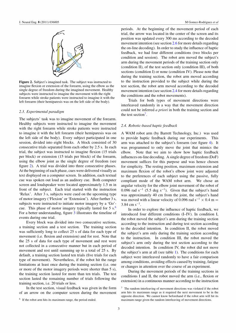
Figure 2. Subject’s imagined task. The subject was instructed toimagine flexion or extension of the forearm, using the elbow as thesingle degree of freedom during the imagined movement. Healthysubjects were instructed to imagine the movement with the rightforearm while stroke patients were instructed to imagine it with theleft forearm (their hemiparesis was on the left side of the body).
2.3. Experimental paradigm
The subjects’ task was to imagine movement of the forearm.Healthy subjects were instructed to imagine the movementwith the right forearm while stroke patients were instructedto imagine it with the left forearm (their hemiparesis was onthe left side of the body). Every subject participated in onesession, divided into eight blocks. A block consisted of 30consecutive trials separated from each other by 2.5 s. In eachtrial, the subject was instructed to imagine flexion (15 trialsper block) or extension (15 trials per block) of the forearm,using the elbow joint as the single degree of freedom (seefigure 2). A trial was divided into three consecutive phases.At the beginning of each phase, cues were delivered visually astext displayed on a computer screen. In addition, each textualcue was spoken out loud as an auditory cue. Both computerscreen and loudspeaker were located approximately 1.5 m infront of the subject. Each trial started with the instruction‘Relax’. After 3 s, subjects were cued on the upcoming typeof motor imagery (‘Flexion’ or ‘Extension’). After further 3 s,subjects were instructed to initiate motor imagery by a ‘Go’-cue. This phase of motor imagery typically lasted for 5 s.6
For a better understanding, figure 3 illustrates the timeline ofevents during one trial.
Every block was divided into two consecutive sections,a training section and a test section. The training sectionwas sufficiently long to collect 25 s of data for each type ofmovement (i.e. flexion and extension) and for rest. Note thatthe 25 s of data for each type of movement and rest werenot collected in a consecutive manner but in each period ofmovement and rest until summing up to a total of 25 s. Bydefault, a training section lasted ten trials (five trials for eachtype of movement). Nevertheless, if the robot hit the rangelimitations at least once during the training section (i.e. oneor more of the motor imagery periods were shorter than 5 s),the training section lasted for more than ten trials. The testsection lasted the remaining number of trials following thetraining section, i.e. 20 trials or less.
In the test section, visual feedback was given in the formof an arrow on the computer screen during the movement
6 If the robot arm hits its maximum range, the period ended.
periods. At the beginning of the movement period of eachtrial, the arrow was located in the center of the screen and itsposition was updated every 300 ms according to the decodedmovement intention (see section 2.6 for more details regardingthe on-line decoding). In order to study the influence of hapticfeedback, we had four different conditions (two blocks percondition and session). The robot arm moved the subject’sarm during the movement periods of the training section only(condition II), of the test section only (condition III), of bothsections (condition I) or none (condition IV). Please note thatduring the training section, the robot arm moved accordingto the instruction provided to the subject while during thetest section, the robot arm moved according to the decodedmovement intention (see section 2.4 for more details regardingthe conditions and the robot movements).
Trials for both types of movement directions wereinterleaved randomly in a way that the movement directioncould not be inferred a priori in both the training section andthe test section7.
2.4. Robotic-based haptic feedback
A WAM robot arm (by Barrett Technology, Inc.) was usedto provide haptic feedback during our experiments. Thisarm was attached to the subject’s forearm (see figure 4). Itwas programmed to only move the joint that mimics theelbow. Note that we aim to show how haptic feedbackinfluences on-line decoding. A single degree of freedom (DoF)movement suffices for this purpose and was hence chosenfor simplicity. The resting position, maximum extension andmaximum flexion of the robot’s elbow joint were adjustedto the preferences of each subject using the passive, fullycompliant mode of the WAM robot arm. We chose anangular velocity for the elbow joint movement of the robot of0.096 rad s−1 (5.5 deg s−1). Given that the subject’s handwas approximately 40 cm from the joint, the subject’s handwas moved with a linear velocity of 0.096 rad s−1 × 0.4 m =3.84 cm s−1.
In order to explore the influence of haptic feedback, weintroduced four different conditions (I–IV). In condition I,the robot moved the subject’s arm during the training sectionaccording to the instruction and during test section accordingto the decoded intention. In condition II, the robot movedthe subject’s arm only during the training section accordingto the instruction. In condition III, the robot moved thesubject’s arm only during the test section according to thedecoded intention. In condition IV, the robot did not movethe subject’s arm at all (see table 1). The conditions for eachsubject were interleaved randomly to have a fair comparisonamong conditions, avoiding effects caused by training, fatigueor changes in attention over the course of an experiment.
During the movement periods of the training sections inconditions I and II, the robot moved the arm (i.e., flexion orextension) in a continuous manner according to the instruction
7 The random interleaving of movement directions was violated if the robotarm hits its maximum range as it required the next movement to be in theopposite direction. We cannot know beforehand if the robot arm will hit itsmaximum range given the random interleaving of movement directions.
3

J. Neural Eng. 8 (2011) 036005 M Gomez-Rodriguez et al
Figure 3. Timeline of events during a trial. Please note that the spoken and textual cues are one-time events, the movement of the robotduring the movement period of the training sections is continuous, while the haptic and visual feedback are events that are updated every300 ms.
Figure 4. Experimental setup. A robot arm moves the subject’sforearm with the elbow as the single degree of freedom (DoF). Theresting position, maximum extension and maximum flexion of therobot arm could be adjusted depending on the subject. The singlejoint of the robot had an angular velocity of 0.096 rad s−1 (5.5 degs−1), i.e. every 300 ms of movement the joint rotated 0.0288 rad(1.65 deg). Given that the subject’s hand was approximately 40 cmfrom the joint, the hand was being moved with a linear velocity of0.096 rad s−1 × 0.4 m = 3.84 cm s−1.
Table 1. Conditions to explore haptic feedback.
Condition Training Test (always visual feedback)
I Robot movesa Robot movesb
II Robot movesa Robot does not moveIII Robot does not move Robot movesb
IV Robot does not move Robot does not move
Please note that the robot can move aaccording to the instruction intraining periods or baccording to the decoded movement intention intest periods.
given to the subject. During the movement periods of thetest sections, we decoded every 300 ms whether a movementintent was present, and updated the state of the robot arm(moving/resting) accordingly. The haptic feedback was givenduring each movement period in a synchronized manner
with the subject’s intention to move the arm, and not atthe end of every trial as done previously [22]. As statedpreviously, moving the robot throughout each trial, andnot at the end of it, is a key point in terms of a futureneurorehabilitation therapy, because it is a strategy that is likelyto result in increased cortical plasticity due to Hebbian-typelearning [15–17].
Finally, note that if the robot arm hit its maximum rangeduring a movement period, the movement period ended. Inthis case, the random interleaving of movement directions wasviolated and the next movement period was forced to be in theopposite direction.
2.5. Signal analysis
First, a center-surround spatial sharpening filter or surfaceLaplacian [26] was applied to the electrodes that were notlocated at the borders. Then, the signals of all electrodeswere bandpass filtered (2–115 Hz). As 20 online featuresper electrode, we used the normalized average power spectraldensities in 2 Hz frequency bins in the frequency range(2–42 Hz), as previously used in motor imagery with EEGs[4]. Welch’s method was used to compute an estimate ofthe power spectral density (PSD). Welch’s method divided thetime signal into time windows of length 500 ms with a 200 msoverlap.
The estimate was determined based on incrementallybigger time segments. In each movement or resting period, thefirst PSD estimate was computed using 500 ms and from thatmoment on every 300 ms, allowing an on-line classificationevery 300 ms (see section 2.6). Please note that the secondestimate based on 800 ms fits exactly two 500 ms windowswith 200 ms overlap (second window starts at 300 ms), thethird estimate based on 1100 ms fits three 500 ms windowswith 200 ms overlap (second window starts at 300 ms andthird window starts at 600 ms), and so on. As the movementor resting period advanced, the PSD was estimated from largersegments (i.e. all the signals of the period up to that point),leading to more reliable and less noisy estimates.
4

J. Neural Eng. 8 (2011) 036005 M Gomez-Rodriguez et al
2.6. On-line decoding
On-line classification was carried out to discriminate flexionversus resting, and to discriminate extension versus resting.As explained in sections 2.3 and 2.4, on-line visual feedbackand haptic feedback were provided. For each block, twolinear support vector machine (SVM) classifiers [27] weregenerated on-the-fly after the training section. As statedbefore, a training section consisted of a default of ten trials.However, if the robot hit the range limitations at least once,the training section lasted more trials. We had multipletime windows per movement period (i.e. (5 s–500 ms)/300 ms = 15) and resting period (i.e. (3 s–500 ms)/300 ms = 8). Each SVM was trained with 75 movementsamples and 75 resting samples (note that identical restingsamples were used for both classifiers and we had 5 spareresting samples that we did not use). Both classifiers wereused only in the test section following the training sectionwithin the same block. One classifier distinguished betweenflexion versus resting and the other one classified betweenextension versus resting. Due to the 20 frequency bins foreach of the 35 recorded channels, we had a 700-dimensionalfeature vector for each data point.
The labels yi ∈ {−1, 1} indicate resting or movement(both flexion and extension). Hence, during the trainingsection we obtain a set of tuples {(xi, yi), i = 1, . . . , n}. Asa classifier, a linear SVM classifier (w, b) was determined bysolving the following convex optimization problem for each ofthe two cases (i.e. flexion versus resting, and extension versusresting) given by
minw,ε
1
2‖w‖2 + C
n∑i=1
εi, (1)
subject to yi(w · xi − b) � 1 − εi, (2)
εi � 0. (3)
Linear SVM classifiers are characterized by providing decisionhyperplanes that maximize the margin between the two classesin the feature space. The slack variables εi allow for mislabeledexamples or outliers. The regularization parameter C wasestimated by tenfold cross-validation within the training set[28]. The outputs of each classifier over the training setwere used to fit the parameters of a sigmoid function thatmapped the classifier outputs zi onto probabilities p(zi). Anoutput probability p(zi) = 1 indicated 100% confidence whenpredicting movement (flexion or extension) and p(zi) = 0indicated 100% confidence when predicting resting.
2.7. Conditions comparison
We studied the differences among the different schemes ofhaptic feedback in three different ways. First, we computedthe ‘negative cross-entropy loss’ or NLP loss [29]. Second,we performed a Kruskal–Wallis one-way analysis of variance[30] over the NLP losses. Finally, we computed the area underthe receiver operating characteristic (ROC) curve (AUC) [31].Coherent empirical evidence coming in from all of these threemeasures will strengthen our conclusions on the differencesamong the different schemes of haptic feedback.
2.7.1. The negative cross-entropy loss [29]. To be able toanalyze the confidence of the probabilistic outputs in restingand movement periods together, the NLP loss of each trial wasused as a metric of discriminative power. The NLP loss isdefined as follows:
L = −1
n
⎡⎣ ∑
i:yi=1
log p(yi = 1|xi)
+∑
i:yi=0
log(1 − p(yi = 1|xi))
⎤⎦ , (4)
where yi is the real label (flexion or extension depending onthe trial and rest), yi is the predicted label, p(yi = 1|xi) is theprobability of movement given by the classifiers for a featurevector xi and p(yi = 0|xi) = 1−p(yi = 1|xi) is the probabilityof resting given by the classifiers for a feature vector xi.
This loss penalizes both over-confident and under-confident predictions. Note when the loss becomes larger,the model becomes increasingly certain that the point belongsto the wrong class. The theoretical minimum of this loss iszero and could be achieved if every label of every example(i.e. resting or movement) was predicted correctly with 100%confidence. When one predicts a label incorrectly, the lossis proportional to the confidence of the prediction. For thecomputation of the NLP, we consider the probabilistic outputsof both binary on-line classifiers together to have a biggernumber of samples for the subsequent statistical analysis.
2.7.2. The Kruskal–Wallis one-way analysis of variance[30]. We performed a Kruskal–Wallis one-way analysisof the variance over the NLP loss of each trial andsubject, with a Bonferroni adjustment to compensate formultiple comparisons [32]. The Kruskal–Wallis one-wayanalysis of variance is identical to a traditional one-wayanalysis of variance with the data replaced by their ranks.Moreover, it is a non-parametric method that does not assumenormal populations, unlike the analogous one-way analysis ofvariance. It is an extension of the Wilcoxon–Mann–Whitneytest [33] to three or more groups and it requires independencebetween sample points.
2.7.3. The ROC curve and the AUC [31]. The ROC curveis a curve of the classifier’s hit rate against its false alarmrate. As one varies the threshold of the classifier, one movesalong a curve in this two-dimensional space. A change inthe threshold for classifying flexion (extension) versus restingmay give more flexion (extension) samples classified as flexion(extension), or hits, and also more resting samples classifiedas flexion (extension), or false alarms, and vice versa. Thearea under the curve (AUC) is a very informative statistic forthe evaluation of performance of classification and rankingalgorithms. It relies only on the ordering of the points and it isfree of any parametric assumptions about the shape of the classdistributions. Again we aggregated flexion versus resting andextension versus resting to obtain more reliable results, giventhe bigger number of samples.
5
J. Neural Eng. 8 (2011) 036005 M Gomez-Rodriguez et al
3. Results
We give important insights on the effects resulting fromartificially closing the sensorimotor feedback loop by studyingthe differences in on-line decoding among conditions (seetable 1 for a definition of the conditions). First, we investigatethe informativeness of different features, i.e. the discriminativepower of every electrode under the presence and absenceof robot movement. Second, we study the differences inclassification performance among conditions using the toolsdescribed in section 2.7.
3.1. Spatial and spectral features
First, we compare the training periods where the robot armguided the subject’s arm and training periods in which it didnot. This comparison is based on the classifier weights perelectrode averaged over the frequency bands 8–16 Hz and18–28 Hz. We focus on these two frequency ranges becausethe first one contains the μ rhythm (9–14 Hz) and the secondrange contains the β rhythm (18–24 Hz). Both the μ andβ rhythms have been reported as highly discriminative inprevious motor imagery experiments [34]. Subsequently, wecompute the average classifier weights and power spectra perfrequency bin for the electrodes C3, CP3 and C4 in healthysubjects.
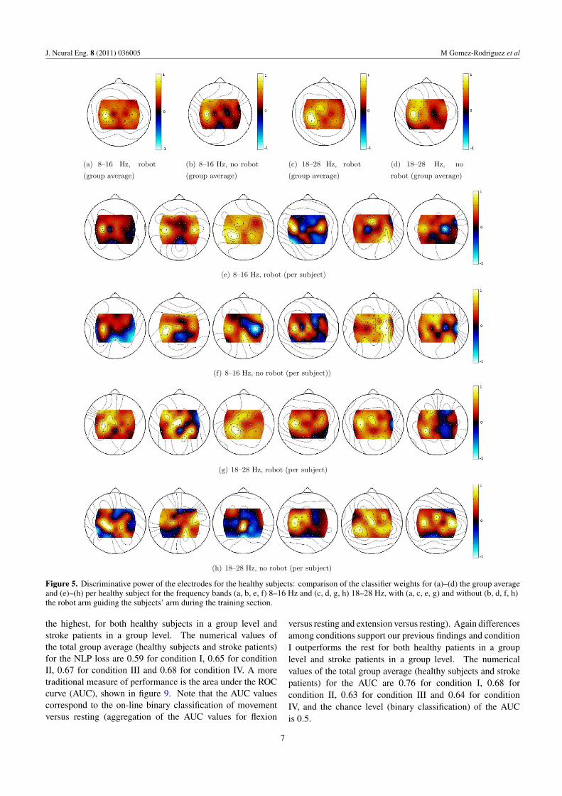
3.1.1. Healthy subjects. Figure 5 shows the classifier weightsper electrode averaged over the frequency bands 8–16 Hz and18–28 Hz for the healthy subjects. Note that healthy subjectswere instructed to perform motor imagery of the right forearm.In the group level in figures 5(a)–(d), we observe empiricalevidence of the following facts. First, electrodes over themotor area representing the right arm, i.e. C3, CP3, FC3,FC1, . . . , get larger weights (i.e. have a higher discriminativepower) when the robot arm guided the subject’s arm duringthe training period in both frequency bands. Second, thereis a higher discriminative power of the sensorimotor areaduring training periods in which the robot arm guided thesubject’s arm for the frequency band that contains the β
rhythm. Third, the spatial distribution of the weights inthe classifiers indicates that the classifiers employed neuralactivity, as the weights in the peripheral locations (i.e. F5, F3,F1, Fz, F2, F4, F6, FC6, C6, CP6, P6, P4, P2, Pz, P1, P3,P5, CP5, C5 and FC5) are low. Hence, it is not likely thatelectromyographic (EMG) activity coming from movementsof the head or the face plays a major role, as such movementsare likely to result in strongest weights in more peripherallocations [35].
Figures 7 and 8 show the average classifier weights andpower spectra per frequency bin for the electrodes C3, CP3and C4. Looking at the figures of the average classifierweights, we observe a shift in discriminative power towardhigher frequencies, i.e. from μ rhythm desynchronization toβ rhythm desynchronization, when the robot arm guided thesubject’s arm during the training period. In terms of the powerspectra, we observe a bigger ERD/ERS modulation when therobot was moving the subject’s arm. This finding also indicates
an increase in BCI-decoding performance achieved bycondition I.
3.1.2. Stroke patients. Figure 6 shows the classifier weightsper electrode averaged over the frequency bands 8–16 Hz and18–28 Hz for the stroke patients. Note that in this case strokepatients were instructed to perform motor imagery of the leftforearm. In the group level in figures 6(a)–(d), we observeimportant differences with respect to the group level resultsfor healthy subjects. First, in this case, we observe a higherdiscriminative power of sensorimotor areas in the μ-frequencyrange during training periods in which the robot arm guidedthe subject’s arm in comparison to those in which it did not.This indicates that in agreement with previous studies [36],the stroke patients could experience proprioception. Second,electrodes over the motor area representing the left arm, i.e. C4,CP4, . . . , are less informative than electrodes over other areas(i.e. the motor area representing the left leg or even electrodesover the ipsilateral side) for decoding movement intentionwhen the robot arm did not guide the subject’s arm duringthe training session. In terms of future rehabilitation, thislast point is undesirable and, therefore, we decide to re-buildthe classifiers for the stroke patients using only the spectralfeatures coming from electrodes in the surroundings of the leftarm representation in the cortex (specifically, FC2, FC4, FC6,C2, C4, C6, CP2, CP4 and CP6) for the performance analysis.Future studies with more stroke patients are necessary tostrengthen the empirical evidence of the discriminative powerof the spatial and spectral features.
3.2. Performance
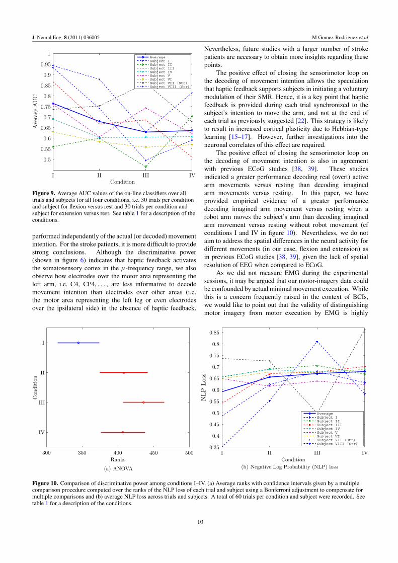
First, we perform a Kruskal–Wallis one-way analysis ofvariance at significance level α = 0.05 over the NLP loss. Itrejects the null-hypothesis that the classifier decision values’medians were equal for all conditions with a p-value of3.41 × 10−6. Subsequently, we perform a multiple t-testcomparison procedure also at a significance level α = 0.05,with a Bonferroni adjustment to compensate for multiplecomparisons, over the rank values of every pair of conditions.Figure 10(a) shows the approximate confidence intervals andthe average rank per condition of haptic feedback for themultiple comparison procedure. It rejects the null hypothesisthat the classifier decision values’ means are equal forcondition I in comparison with the remaining conditions. Theresults suggest an improvement in classification performancewhen comparing arm movement intention versus restingfor the case where the robot provides robot-based hapticreinforcement during the training and the test periods, i.e.condition I outperforms the rest. Note that during the test, therobot arm was programmed to move or stop according to thedecoded intention in an on-line manner during the movementperiods.
Average NLP loss values for every condition of hapticfeedback as well as for every subject are shown infigure 10(b). The differences among conditions are consistentwith the findings discussed during the analysis of the variance.In condition I, the classification performance appears to be
6
J. Neural Eng. 8 (2011) 036005 M Gomez-Rodriguez et al
(a) 8–16 Hz, robot
(group average)
(b) 8–16 Hz, no robot
(group average)
(c) 18–28 Hz, robot
(group average)
(d) 18–28 Hz, no
robot (group average)
(e) 8–16 Hz, robot (per subject)
(f) 8–16 Hz, no robot (per subject))
(g) 18–28 Hz, robot (per subject)
(h) 18–28 Hz, no robot (per subject)
Figure 5. Discriminative power of the electrodes for the healthy subjects: comparison of the classifier weights for (a)–(d) the group averageand (e)–(h) per healthy subject for the frequency bands (a, b, e, f) 8–16 Hz and (c, d, g, h) 18–28 Hz, with (a, c, e, g) and without (b, d, f, h)the robot arm guiding the subjects’ arm during the training section.
the highest, for both healthy subjects in a group level andstroke patients in a group level. The numerical values ofthe total group average (healthy subjects and stroke patients)for the NLP loss are 0.59 for condition I, 0.65 for conditionII, 0.67 for condition III and 0.68 for condition IV. A moretraditional measure of performance is the area under the ROCcurve (AUC), shown in figure 9. Note that the AUC valuescorrespond to the on-line binary classification of movementversus resting (aggregation of the AUC values for flexion
versus resting and extension versus resting). Again differencesamong conditions support our previous findings and conditionI outperforms the rest for both healthy patients in a grouplevel and stroke patients in a group level. The numericalvalues of the total group average (healthy subjects and strokepatients) for the AUC are 0.76 for condition I, 0.68 forcondition II, 0.63 for condition III and 0.64 for conditionIV, and the chance level (binary classification) of the AUCis 0.5.
7

J. Neural Eng. 8 (2011) 036005 M Gomez-Rodriguez et al
(a) 8–16 Hz, robot
(group average)
(b) 8–16 Hz, no
robot (group average)
(c) 18–28 Hz, robot
(group average)
(d) 18–28 Hz, no
robot (group average)
(e) 8–16 Hz, robot (per subject) (f) 8–16 Hz, no robot (per subject))
(g) 18–28 Hz, robot (per subject) (h) 18–28 Hz, no robot (per subject)
Figure 6. Discriminative power of the electrodes for the stroke patients: comparison of the classifier weights for (a–d) the group averageand (e–h) per stroke patient for the frequency bands (a, b, e, f) 8–16 Hz and (c, d, g, h) 18–28 Hz, with (a, c, e, g) and without (b, d, f, h) therobot arm guiding the subjects’ arm during the training section.
-0.2
0
0.2
0.4
0.6
0.8
1
1.2
10 15 20 25Frequency (Hz)
Robot arm moves during training
Robot arm does not move during training
(a) Classifier weights for electrode C3
-0.2
-0.1
0
0.2
0.1
0.3
0.4
0.5
0.6
0.7
0.8
10 15 20 25Frequency (Hz)
Robot arm moves during training
Robot arm does not move during training
(b) Classifier weights for electrode CP3
Figure 7. Discriminative power of the frequency components of the electrodes C3 and CP3 for healthy subjects: classifier weights for theelectrodes C3 and CP3 in the frequency band 8–30 Hz for the classifiers generated when the robot arm guides the subjects’ arm and whenthe robot arm is not moving during training. Refer to figure 1 for electrode grid spatial configuration.
8

J. Neural Eng. 8 (2011) 036005 M Gomez-Rodriguez et al
restmovement
0 5 10 15 20 25
Log
Pow
er
Frequency (Hz)
(a) C3, training, robot
restmovement
0 5 10 15 20 25
Log
Pow
er
Frequency (Hz)
(b) CP3, training, robot
restmovement
0 5 10 15 20 25
Log
Pow
er
Frequency (Hz)
(c) C4, training, robot
restmovement
0 5 10 15 20 25
Log
Pow
er
Frequency (Hz)
(d) C3, training, no robot
restmovement
0 5 10 15 20 25
Log
Pow
er
Frequency (Hz)
(e) CP3, training, no robot
restmovement
0 5 10 15 20 25
Log
Pow
er
Frequency (Hz)
(f) C4, training, no robot
restmovement
0 5 10 15 20 25
Log
Pow
er
Frequency (Hz)
(g) C3, test, robot
restmovement
0 5 10 15 20 25
Log
Pow
er
Frequency (Hz)
(h) CP3, test, robot
restmovement
0 5 10 15 20 25
Log
Pow
er
Frequency (Hz)
(i) C4, test, robot
restmovement
0 5 10 15 20 25
Log
Pow
er
Frequency (Hz)(j) C3, test, no robot
restmovement
0 5 10 15 20 25
Log
Pow
er
Frequency (Hz)(k) CP3, test, no robot
restmovement
0 5 10 15 20 25
Log
Pow
er
Frequency (Hz)(l) C4, test, no robot
Figure 8. Power spectra of the electrodes C3, C4 and CP3 for healthy subjects: power spectra in movement periods and the rest periods forthe electrodes C3, CP3 and C4 in the frequency band 2–30 Hz for the training and test sections with and without the robot arm guiding thesubjects’ arm. Refer to figure 1 for the electrode grid spatial configuration.
4. Discussion
In this paper, we have demonstrated that artificially closing thesensorimotor feedback loop facilitates decoding of movementintention in both healthy subjects and stroke patients. Thisresult indicates the feasibility of an integrated stroke therapythat combines robot-assisted physical therapy with decoding ofmovement intention by means of a BCI. Specifically, we haveprovided evidence that the strength of the sensorimotor rhythm(SMR), as measured by the class probability estimated fromthe outputs of the SVM, is modulated by the haptic robot-based feedback. Furthermore, our results suggest that thismodulation of the SMR is actually beneficial for decodingof arm movement intention. An increased classification
accuracy is exhibited by comparing performance with hapticfeedback to no haptic feedback (cf conditions I and IV infigure 10).
For healthy subjects, the discriminative power andpower spectra indicate that haptic feedback activates thesomatosensory cortex and increases ERD/ERS modulationin the β-frequency range. Both discriminative power andpower spectra are shown in figures 5 and 7. Interestingly,this observation is in agreement with previous reports onthe effect of haptic stimulation in non-human primates[37]. In addition, our results are also in agreement withprevious reports on the effect of passive haptic stimulation[18, 19, 21], despite that in these studies haptic stimulation was
9
J. Neural Eng. 8 (2011) 036005 M Gomez-Rodriguez et al
AverageSubject ISubject IISubject IIISubject IVSubject VSubject VISubject VII (Str)Subject VIII (Str)
0.5
0.55
0.6
0.65
0.7
0.75
0.8
0.85
0.9
0.95
1
Condition
Ave
rage
AU
C
I II III IV
Figure 9. Average AUC values of the on-line classifiers over alltrials and subjects for all four conditions, i.e. 30 trials per conditionand subject for flexion versus rest and 30 trials per condition andsubject for extension versus rest. See table 1 for a description of theconditions.
performed independently of the actual (or decoded) movementintention. For the stroke patients, it is more difficult to providestrong conclusions. Although the discriminative power(shown in figure 6) indicates that haptic feedback activatesthe somatosensory cortex in the μ-frequency range, we alsoobserve how electrodes over the motor area representing theleft arm, i.e. C4, CP4, . . . , are less informative to decodemovement intention than electrodes over other areas (i.e.the motor area representing the left leg or even electrodesover the ipsilateral side) in the absence of haptic feedback.
300 350 400 450 500
Con
diti
on
Ranks
I
II
III
IV
(a) ANOVA
AverageSubject ISubject IISubject IIISubject IVSubject VSubject VISubject VII (Str)Subject VIII (Str)
0.35
0.4
0.45
0.5
0.55
0.6
0.65
0.7
0.75
0.8
0.85
ConditionI II III IV
NLP
Los
s
(b) Negative Log Probability (NLP) loss
Figure 10. Comparison of discriminative power among conditions I–IV. (a) Average ranks with confidence intervals given by a multiplecomparison procedure computed over the ranks of the NLP loss of each trial and subject using a Bonferroni adjustment to compensate formultiple comparisons and (b) average NLP loss across trials and subjects. A total of 60 trials per condition and subject were recorded. Seetable 1 for a description of the conditions.
Nevertheless, future studies with a larger number of strokepatients are necessary to obtain more insights regarding thesepoints.
The positive effect of closing the sensorimotor loop onthe decoding of movement intention allows the speculationthat haptic feedback supports subjects in initiating a voluntarymodulation of their SMR. Hence, it is a key point that hapticfeedback is provided during each trial synchronized to thesubject’s intention to move the arm, and not at the end ofeach trial as previously suggested [22]. This strategy is likelyto result in increased cortical plasticity due to Hebbian-typelearning [15–17]. However, further investigations into theneuronal correlates of this effect are required.
The positive effect of closing the sensorimotor loop onthe decoding of movement intention is also in agreementwith previous ECoG studies [38, 39]. These studiesindicated a greater performance decoding real (overt) activearm movements versus resting than decoding imaginedarm movements versus resting. In this paper, we haveprovided empirical evidence of a greater performancedecoding imagined arm movement versus resting when arobot arm moves the subject’s arm than decoding imaginedarm movement versus resting without robot movement (cfconditions I and IV in figure 10). Nevertheless, we do notaim to address the spatial differences in the neural activity fordifferent movements (in our case, flexion and extension) asin previous ECoG studies [38, 39], given the lack of spatialresolution of EEG when compared to ECoG.
As we did not measure EMG during the experimentalsessions, it may be argued that our motor-imagery data couldbe confounded by actual minimal movement execution. Whilethis is a concern frequently raised in the context of BCIs,we would like to point out that the validity of distinguishingmotor imagery from motor execution by EMG is highly
10

J. Neural Eng. 8 (2011) 036005 M Gomez-Rodriguez et al
disputable. Motor imagery and motor execution share thesame neuronal substrate, with both imagery and executionresulting in increased muscle tone [40]. Minor movementsand motor imagery may thus result in indistinguishable EMGsignals. Accordingly, utilizing EMG measurements to controlfor motor execution suggests a certainty in excluding potentialconfounders that is not supported by empirical evidence.While this is a major concern when BCIs are used forcommunication, we wish to point out that this is not the casein our study. We employed motor imagery in healthy subjectsto simulate stroke patients, where movement intent is onlyaccompanied by minimal or even no movement execution. Assuch, we do not consider the potential presence of minimalmovements in our healthy subjects to be a major concern, assuch movements are also likely to be present in stroke patientswith only residual motor control.
The presented work is substantially different fromprevious studies that achieved comparable accuracy in full trialbinary or four-class classification of motor imagery withoutthe use of haptic feedback [41–43]. On-line classificationof motor imagery based on shorter time windows than a fulltrial is a considerably more difficult problem than full trialclassification. Our main take home message is that evenif everything else is identical, haptic feedback improves theaccuracy.
The presented results indicate the possibility of a futurestroke rehabilitation based on robot-assisted physical therapywith BCI-based decoding of movement intention. However,it needs to be pointed out that the support provided by thisstudy hinges on the assumption that the results presentedhere with healthy subjects and two stroke patients can betransferred to a bigger set of stroke patients with differentdegrees of hemiparesis and neurological conditions. Thebeneficial effect reported here is likely to depend on thepresence of proprioception. Nevertheless, there is no a priorireason why stroke patients should not have proprioception.Both stroke patients in this study exhibited proprioception andprevious studies have shown that most stroke patients recoverproprioception 8 weeks post-stroke despite remaining motordisabilities [36].
Besides the relevance of our results for potential stroketherapy, it is furthermore noteworthy that the positive influenceof haptic feedback on decoding accuracy may also bebeneficial for other subject groups. For example, subjectsin late stages of ALS appear not to be capable of modulatingtheir SMR sufficiently, as indicated by the fact that so farno communication with a completely locked-in subject hasbeen established by means of a BCI. While the extent ofsensory feedback in late stages of ALS remains unclear,haptic feedback might also support these subjects in initiatingvolitional modulation of their SMR.
Acknowledgments
Manuel Gomez Rodriguez has been supported in part by aFundacion Barrie de la Maza Graduate Fellowship. Theauthors thank Bernd Battes for technical support during theexperiments.
References
[1] Farwell L A and Donchin E 1988 Talking off the top of yourhead: toward a mental prosthesis utilizing event-relatedbrain potentials Electroencephalogr. Clin. Neurophysiol.70 510–23
[2] Birbaumer N, Ghanayim N, Hinterberger T, Iversen I,Kotchoubey B, Kubler A, Perelmouter J, Taub E and Flor H1999 A spelling device for the paralysed Nature 398 297–8
[3] Pfurtscheller G, Neuper C, Flotzinger D and Pregenzer M1997 EEG-based discrimination between imagination ofright and left hand movement Electroencephalogr. Clin.Neurophysiol. 103 642–51
[4] Grosse-Wentrup M, Liefhold C, Gramann K and Buss M 2009Beamforming in noninvasive brain–computer interfacesIEEE Trans. Biomed. Eng. 56 1209–19
[5] Blankertz B, Tomioka R, Lemm S, Kawanabe M andMuller K-R 2008 Optimizing spatial filters for robust EEGsingle-trial analysis IEEE Signal Process. Mag.25 41–56
[6] Kubler A, Nijboer F, Mellinger J, Vaughan T M, Pawelzik H,Schalk G, McFarland D J, Birbaumer N and Wolpaw J R2005 Patients with ALS can use sensorimotor rhythms tooperate a brain–computer interface Neurology 64 1775–7
[7] Dijkerman H C, Ietswaart M, Johnston M and MacWalter R S2004 Does motor imagery training improve hand function inchronic stroke patients? A pilot study Clin. Rehabil. 18 538
[8] Page S J, Levine P and Leonard A 2007 Mental practice inchronic stroke: results of a randomized, placebo-controlledtrial Stroke 38 1293
[9] Tanaka K, Matsunaga K and Wang H O 2005Electroencephalogram-based control of an electricwheelchair IEEE Trans. Robot. 21 762–6
[10] Millan J R, Renkens F, Mourino J and Gerstner W2004 Noninvasive brain-actuated control of amobile robot by human EEG IEEE Trans. Biomed. Eng.51 1026–33
[11] Daly J J and Wolpaw J R 2008 Brain–computer interfaces inneurological rehabilitation Lancet Neurol. 7 1032–43
[12] van Peppen R P S, Kwakkel G, Wood-Dauphine S,Hendriks H J M, van der Wees Ph J and Dekker J2004 The impact of physical therapy on functionaloutcomes after stroke: What’s the evidence? Clin. Rehabil.18 833–62
[13] Riener R, Nef T and Colombo G 2005 Robot-aidedneurorehabilitation of the upper extremities Med. Biol. Eng.Comput. 43 2–10
[14] Buch E, Weber C, Cohen L G, Braun C, Dimyan M A, Ard T,Mellinger J, Caria A, Soekadar S and Fourkas A 2008Think to move: a neuromagnetic brain–computer interface(BCI) system for chronic stroke Stroke 39 910
[15] Wang W, Collinger J L, Perez M A, Tyler-Kabara E C,Cohen L G, Birbaumer N, Brose S W, Schwartz A B,Boninger M L and Weber D J 2010 Neural interfacetechnology for rehabilitation: exploiting and promotingneuroplasticity Phys. Med. Rehabil. Clin. North Am.21 157–78
[16] Murphy T H and Corbett D 2009 Plasticity during strokerecovery: from synapse to behaviour Nature Rev. Neurosci.10 861–72
[17] Kalra L 2010 Stroke rehabilitation 2009: old chestnuts andnew insights Stroke 41 e88
[18] Muller G R, Neuper C, Rupp R, Keinrath C, Gerner H Jand Pfurtscheller G 2003 Event-related beta EEG changesduring wrist movements induced by functional electricalstimulation of forearm muscles in man Neurosci. Lett.340 143–7
[19] Muller-Putz G R, Zimmermann D, Graimann B, Nestinger K,Korisek G and Pfurtscheller G 2007 Event-related beta EEG
11

J. Neural Eng. 8 (2011) 036005 M Gomez-Rodriguez et al
changes during passive and attempted foot movements inparaplegic patients Brain Res. 1137 84–91
[20] Waldert S, Preissl H, Demandt E, Braun C, Birbaumer N,Aertsen A and Mehring C 2008 Hand movement directiondecoded from MEG and EEG J. Neurosci. 28 1000–8
[21] Ramos A, Halder S and Birbaumer N 2009 Proprioceptivefeedback in BCI Proc. 4th Int. IEEE/EMBS Conf. on NeuralEngineering pp 279–82
[22] Ang K K, Guan C, Chua S G, Ang B T, Kuah C, Wang C,Phua K S, Chin Z Y and Zhang H 2009 A clinical study ofmotor imagery-based brain–computer interface for upperlimb robotic rehabilitation Annu. Int. Conf. IEEEEngineering in Medicine and Biology Society(EMBC) pp 5981–4
[23] Gomez Rodriguez M, Peters J, Hill J, Scholkopf B,Gharabaghi A and Grosse-Wentrup M 2010 Closing thesensorimotor loop: haptic feedback facilitates decoding ofarm movement imagery Proc. IEEE Int. Conf. on Systems,Man, and Cybernetics. Workshop in Shared Control forBrain–Machine Interfaces (October 2010) pp 121–6
[24] Schalk G, McFarland D J, Hinterberger T, Birbaumer Nand Wolpaw J R 2004 BCI 2000: a general-purposebrain–computer interface (BCI) system IEEE Trans.Biomed. Eng. 51 1034–43
[25] http://bci2000.org/downloads/BCPy2000/[26] McFarland D J, McCane L M, David S V and Wolpaw J R
1997 Spatial filter selection for EEG-based communicationElectroencephalogr. Clin. Neurophysiol. 103 386–94
[27] Scholkopf B and Smola A J 2002 Learning with Kernels:Support Vector Machines, Regularization, Optimization,and Beyond (Cambridge, MA: MIT Press)
[28] Platt J C 1999 Probabilistic outputs for support vectormachines and comparisons to regularized likelihoodmethods Advances in Large Margin Classifiers (Cambridge,MA: MIT Press) pp 61–74
[29] Quinonero-Candela J, Rasmussen C, Sinz F, Bousquet Oand Scholkopf B 2006 Evaluating predictive uncertaintychallenge Lect. Notes Comput. Sci. 3944 1–27
[30] Kruskal W H and Wallis W A 1952 Use of ranks inone-criterion variance analysis J. Am. Stat. Assoc.47 583–621
[31] Hill N J, Lal T N, Tangermann M, Hinterberger T, Widman G,Elger C E, Scholkopf B and Birbaumer N 2007 Classifying
event-related desynchronization in EEG, ECoG and MEGsignals Lect. Notes Comput. Sci. 4174 404–13
[32] Hochberg Y and Tamhane A C 1987 Multiple ComparisonProcedures (New York: Wiley)
[33] Mann H B and Whitney D R 1947 On a test of whether one oftwo random variables is stochastically larger than the otherAnn. Math. Stat. 18 50–60
[34] Wolpaw J R, Birbaumer N, Heetderks W J, McFarland D J,Peckham P H, Schalk G, Donchin E, Quatrano L A,Robinson C J and Vaughan T M 2000 Brain–computerinterface technology: a review of the first internationalmeeting IEEE Trans. Rehabil. Eng. 8 164–73
[35] Goncharova I I, McFarland D J, Vaughan T M and Wolpaw J R2003 EMG contamination of EEG: spectral andtopographical characteristics Clin. Neurophysiol.114 1580–93
[36] Smith D L, Akhtar A J and Garraway W M 1983Proprioception and spatial neglect after stroke Age Ageing12 63
[37] Suminski A J, Tkach D C, Fagg A H and Hatsopoulos N G2010 Incorporating feedback from multiple sensorymodalities enhances brain–machine interface controlJ. Neurosci. 30 16777
[38] Shenoy P, Miller K J, Ojemann J G and Rao R P N 2008Generalized features for electrocorticographic BCIs IEEETrans. Biomed. Eng. 55 273
[39] Miller K J, Schalk G, Fetz E E, den Nijs M, Ojemann J Gand Rao R P N 2010 Cortical activity during motorexecution, motor imagery, and imagery-based onlinefeedback Proc. Natl. Acad. Sci. 107 4430
[40] Jeannerod M 1995 Mental imagery in the motor contextNeuropsychologia 33 1419–32
[41] Huang D, Lin P, Fei D Y, Chen X and Bai O 2009 Decodinghuman motor activity from EEG single trials for a discretetwo-dimensional cursor control J. Neural Eng. 6 046005
[42] Babiloni F, Cincotti F, Bianchi L, Pirri G, Millan J, Mourino J,Salineri S and Marciani M G 2001 Recognition of imaginedhand movements with low resolution surface Laplacian andlinear classifiers Med. Eng. Phys. 23 323–8
[43] McFarland D J, Sarnacki W A, Vaughan T M and Wolpaw J R2005 Brain–computer interface (BCI) operation: signal andnoise during early training sessions Clin. Neurophysiol.116 56–62
12