cloning of a marine cyanobacterial promoter for foreign gene expression using a promoter probe...
TRANSCRIPT

�9 1996 by Humana Press Inc. All rights of any nature whatsoever reserved. 0273-2289196/5903--0349508.00
Cloning of a Marine Cyanobacterial Promoter for Foreign Gene Expression
Using a Promoter Probe Vector
KOJI SODE,* NAOAKI HATANO,
AND MASAHIRO TATARA
Department of Biotechnology, Faculty of Technology, Tokyo University of Agriculture and Technology,
2.24.16 Naka-cho, Koganei-shi, Tokyo 184, Japan
Received February 21, 1995; Accepted August 9, 1995
ABSTRACT
A marine cyanobacterial promoter was cloned to allow efficient foreign gene expression. This was carried out using chloramphenicol acetyl transferase (CAT) as a marker protein. For rapid and simple mea- surement of CAT activity, a method based on a fluorescently labeled substrate was improved by utilizing HPLC equipped with a flow- through fluorescent spectrophotometer. This method was used in con- junction with a newly constructed promoter probe vector. Cyano- bacterial transformants, harboring plasmid containing a cloned 2-kbp marine cyanobacterial genomic fragment, showed a 10-fold higher CAT activity, compared with that achieved using the kanamycin-resistant gene promoter. From the sequence analysis of the cloned fragment, a putative promoter region was found.
Index Entries: Marine cyanobacteria; promoter; foreign gene ex- pression; fluorescent CAT assay.
INTRODUCTION
Much attention is being paid on the development of bioreactor systems and gene manipulation techniques for marine cyanobacteria, to allow the development of clean bioprocesses based on their photosynthetic activ- ity. Recombinant DNA techniques for marine cyanobacteria may expand
*Author to whom all correspondence and reprint requests should be addressed.
Applied Biochemistry and Biotechnology 349 Vol. 59, 1996

350 Sode, Hatano, and Tatara
their potential in the production of useful materials, such as metabolites (1,2), cell constituents (3,4), and recombinant gene products. In addition, they have potential for solar light energy conversion, for example, hydro- gen production (5) and, more recently, for biological carbon dioxide fixa- tion systems (6).
We have been working on the development of gene manipulation sys- tems for marine cyanobacteria based on transconjugation (7). We have used the broad host range vector, pKT230 (8) as the plasmid for foreign gene expression. Plasmid stability as well as pseudo-continuous culture were tested to illustrate their potential as suitable host-vector for foreign gene expression (9,10).
Chloramphenicol acetyl transferase (CAT) is widely used as an indica- tor for the measurement of expression efficiency, by using the CAT struc- tural gene fused downstream of an appropriate promoter. With recent advances in the development of nonradioisotopic labeling techniques, a fluorescently labeled chloramphenicol (Bodipy TM chloramphenicol) is extensively utilized. We previously reported CAT expression in marine cyanobacteria under various conditions (10,11). However, because con- ventional procedures for CAT activity measurement using thin-layer chro- matography combined with fluorescent spectrophotometer are compli- cated (12), development of a simple system is essential to advance the study of foreign gene expression using CAT as the reporter protein.
Here we report the cloning of a highly active marine cyanobacterial promoter region. To achieve this, we first improved the method for CAT activity measurement based on detection of fluorescently labeled chlor- amphenicol using HPLC. A promoter probe vector based on pKT230 with promoterless CAT gene was also constructed. A marine cyanobacterial genomic library was cloned into this vector and recombinants expressing high levels of CAT were isolated.
MATERIALS AND METHODS
Chemicals
Restriction enzymes and other DNA modification enzymes were pur- chased from Toyobo (Osaka, Japan). Fluorescently labeled chlorampheni- col, Bodipy TM chloramphenicol was purchased from Molecular Probes (Eugene, OR). Authentic CAT was purchased from Pharmacia (Uppsala, Sweden). All other chemicals used in this study were of analytical grade.
Bacterial Strains and Plasmids
As the host cyanobacterial strain, a marine cyanobacterium, Synecho- coccus sp. NKBG 15041c was used (7). The freshwater unicellular cyano- bacterial strain, Anacystis nidulans R2 (Synechococcus sp. PCC 7942), was obtained from the American Type Culture Collection (ATCC). As the donor
Applied Biochemistry and Biotechnology Vol. 59, 1996

Cloning Cyanobacterial Promoter �9 . .
Digested by Hindlll X
Sac , S
\ Digested by Pvul and S a c k \
Blunt end X
351
Hlndlll Scal ~ TG EcoRl Ncol
l I lndlll
CAT structure gene
2 Shlne-Dalgarno sequences
C A T gene Hindlll fragment
/ S -EooR, Pstl! / -~Hpal
Xhal ~
PSI' Accl
Digested by BamHI and EcoRI, Blunt end
__.
O~ mo Hindll,
~..,~-.. _ / Xho[ ~~-Accl
PSU I Fig. 1. Construction of promoter probe vector pKICAT based on broad
host range vector pKT230.
strain for transconjugation, Escherichia coli $17-1 was used (13). A promoter probe vector, pKICAT, was constructed as follows (Fig. 1). The CAT structural gene, which was obtained as CAT GenBlock (Pharmacia), was digested with HindIII and cloned into HindIII site of an expression vector, pTrc99A (Pharmacia). From this plasmid, a fragment was obtained by Sad
Applied Biochemistry and Biotechnology Vol. 59, 1996

352 Sode, Hatano, and Tatara
and PvuI digestion followed by blunt ending, using T4 DNA polymerase (Takara, Kyoto, Japan). The resulting fragment contained a multicloning site, CAT gene, and terminator (rrnB). Plasmid pKT230 was digested with BamHI and EcoRI, and ligated with the CAT fragment obtained as above. The constructed plasmid was named as pK1CAT (Fig. 1).
Cloning of Marine Cyanobacteriai Promoter Regions
Total genomic DNA from NKBG 15041c was extracted according to Porter et al. (14), and digested by either KpnI, Sa/I or XbaI. Restricted genomic DNA was cloned into pK1CAT, digested by each corresponding restriction enzyme, and cloned into E. coli $17-1. Transformants were selected on the LB agar plates in the presence of 25/~g/mL of kanamycin (Kin). Colonies which appeared on the selection plates were picked up and directly used for transconjugation as described previously (7). The first selection of marine cyanobacterial transconjugants was carried out in a liquid medium (marine BG11 medium) containing 10 ag/mL of Km. Cells growing in this selection medium were then transferred into media with various concentrations of Km and chloramphenicol (Cm), as shown in Table 1.
Southern Blot and DNA Sequence Analysis DNA hybridization experiments were carried out using the noniso-
topic labeling system [Digoxigenin-11-d uridine triphosphate (UTP), anti- digoxigenin enzyme-linked immunosorbent assay (ELISA)] (Boehringer, Mannheim, Germany). The hybridized digoxigenin-labeled DNA probe was detected using the chemiluminescent substrate, 3-(2'-Spiroadamant- ane)-4-methoxy-4-(3"-phosphoryloxy)-phenyl-l,2-dioxetane (AMPpDR), according to the manufacturer's instructions.
Cloned DNA fragments from marine cyanobacteria were sequenced using an automated DNA sequencer (Shimadzu DSQ-1, Kyoto, Japan).
Fluorescent CAT Analysis Using HPLC CAT expression levels were determined using fluorescently-labeled
chloramphenicol combined with the improved method. The CAT reaction with fluorescently-labeled chloramphenicol was carried out according to manufacturer's instructions, with a slight modification. The authentic CAT was diluted with 100 mM Tris-HC1 (pH 8.0). Sixty microliters of the diluted authentic CAT was mixed with 10/~L of Bodipy TM chloramphenicol substrate reagent and incubated at 37~ for 5 min. Ten microliters of 4 mM acetyl CoA was added and incubated for a fixed period of time between 1 min and 1 h, depending on the CAT concentration. After incubation, 400 #L of ice-cold ethyl acetate was added. Each sample was vortexed for about 20 s and the samples centrifuged for 3 min to separate the liquid phase. The top 350 #L of ethyl acetate was removed and transferred to a
Applied Biochemistry and Biotechnology Vol. 59, 1996

Cloning Cyanobacterial Promoter 353
LJ
0
~0
o g~
o
.o
N
N N
~ ~ +
~ ~ +
I -I- -k + +
I + + + + +
I + + + + + + + +
I + + + + + + + + +
+ -t- -t- -k -k -I- -k -I- -I- 4- 4- -I- + -t- -I- + + +
U ~
0 0 o 0
Z Z Z + Z
Applied Biochemistry and Biotechnology Vol. 59, 1996
354 Sode, Hatano, and Tatara
A 3 B 3
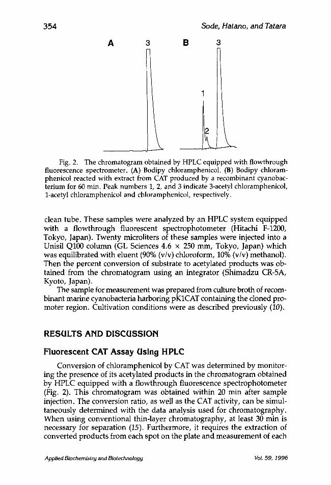
Fig. 2. The chromatogram obtained by HPLC equipped with flowthrough fluorescence spectrometer. (A) Bodipy chloramphenicol. (B) Bodipy chloram- phenicol reacted with extract from CAT produced by a recombinant cyanobac- terium for 60 min. Peak numbers 1, 2, and 3 indicate 3-acetyl chloramphenicol, 1-acetyl chloramphenicol and chloramphenicol, respectively.
clean tube. These samples were analyzed by an HPLC system equipped with a flowthrough fluorescent spectrophotometer (Hitachi F-1200, Tokyo, Japan). Twenty microliters of these samples were injected into a Unisil Q100 column (GL Sciences 4.6 x 250 mm, Tokyo, Japan) which was equilibrated with eluent (90% (v/v) chloroform, 10% (v/v) methanol). Then the percent conversion of substrate to acetylated products was ob- tained from the chromatogram using an integrator (Shimadzu CR-5A, Kyoto, Japan).
The sample for measurement was prepared from culture broth of recom- binant marine cyanobacteria harboring pKICAT containing the cloned pro- moter region. Cultivation conditions were as described previously (10).
RESULTS AND DISCUSSION
Fluorescent CAT Assay Using H PLC
Conversion of chloramphenicol by CAT was determined by monitor- ing the presence of its acetylated products in the chromatogram obtained by HPLC equipped with a flowthrough fluorescence spectrophotometer (Fig. 2). This chromatogram was obtained within 20 min after sample injection. The conversion ratio, as well as the CAT activity, can be simul- taneously determined with the data analysis used for chromatography. When using conventional thin-layer chromatography, at least 30 min is necessary for separation (15). Furthermore, it requires the extraction of converted products from each spot on the plate and measurement of each
Applied Biochemistry and Biotechnology Vol. 59, 1996

Cloning Cyanobacterial Promoter 355
20
i A// 10
0
A
.o
h- r., O .=
r.. 0 (3
B
2
1
0 0.0 0.1 0.2 0.000 0.001 0 .002 0 .003 0 .004
CAT (un=t) CAT (unit)
Fig. 3. Correlation between CAT activity and conversion ratio of Bodipy chloramphenicol. Ten nmol of Bodipy chloramphenicol and 40 #tool of acetyl coenzyme A were reacted at 37~ for I rain (A) or 60 rain (B). Reaction volume is 80 #L.
fluorescence, separately (12). Therefore, our method utilizing HPLC reduced time and effort required for CAT analysis.
When 0.7 U of CAT were used for the reaction, conversion was almost complete within 15 min. A good linear correlation between time and con- version ratio was observed within 5 min of incubation. To calculate the CAT activity of the sample, one should carry out the experiment where the conversion ratio is proportional with time. We varied the amount of CAT and time for incubation, and obtained the optimum incubation time to determine the CAT activity (Fig. 3). By making a dilution series of pur- ified CAT with defined activity, the dynamic range and sensitivity of this analyzing system were investigated. Each figure represents the reliability of this method in each range. The highest sensitivity achieved using this method was 4 x 10 -4 U, which was similar to that reported using 14C- labeled chloramphenicol as the indicator (16). Together with these figures, the wide range of sample concentrations can be measured with high sensitivity.
Cloning of Marine Cyanobacterial Promoter Using our constructed promoter probe vector, pKICAT (Fig. 1), the
cloning of marine cyanobacterial promoter was carried out. Transcon- jugants harboring marine cyanobacterial promoters were screened using liquid medium containing various concentrations of Cm and Km to enrich Cm-resistant transconjugants (Table 1). After 10 d of selection, the growth of transconjugants possibly harboring cyanobacterial genomic DNA frag- ments was observed in the presence of Cm, even at a concentration of 800 #g/mE In contrast, no growth was observed in host strain and also in the transconjugants not harboring cyanobacterial DNA fragment (pKICAT). In addition, no difference in the Km resistance was observed between
Applied Biochemistry and Biotechnology Vol. 59, 1996

356 Sode, Hatano, and Tatara
Ed-';?!>'~ /___. Iv.,,
Kpnl..~fragment nic oriV ~ ' . ~Acc, ~:AT /
_ p K I C A T c y a K mob t ~ r rnB 15.7kb .. . . / "~ t~.rminator ~ /
Hindlll'k'x~, .- r rep B~'accl ., X/x.NK-~m ' r e p / ~ , / /
X m a l / ~ =P~-/
Fig. 4. Structure of plasrnid pKICATcyaK harboring a 2-kbp marine cyanobacterial genomic fragment containing highly active promoter in NKBG 15041c.
pKICAT and those containing cyanobacterial DNA fragment. Such a high resistance to Cm might be due to the expression of CAT in the marine cyanobacteria. Of the three different libraries, transconjugants harboring promoter probe vectors containing KpnI-digested DNA fragment showed rapid growth. Using this library, we further characterized the marine cyanobacterial promoter. Plasmids were extracted from transconjugants (14) and transformed back into E. coli DH5oz to analyze their restriction pattern (Fig. 4). All plasmids extracted from E. coli DH5o~ harbored a 2-kbp KpnI fragment, indicating that transconjugant expressing high CAT activity contained a similar DNA fragment. Figure 5 shows the results of Southern blot analysis using the 2-kbp KpnI fragment as the probe (Fig. 5A) and agarose gel electrophoresis (Fig. 5B) of KpnI restriction fragments of the genomic DNA of NKBG 15041c, recombinant NKBG 15041c harboring this plasmid, and PCC 7942. Southern blot analysis revealed that this probe hybridized only with 2-kbp KpnI fragments that corresponded to marine cyanobacterial genomic DNA, but not PCC 7942. These results showed that the 2-kbp KpnI fragment was derived from NKBG 15041c, which did not show homology to the PCC 7942 genomic DNA. In further experi- ments, we designated this plasmid as pKICATcyaK. We have sequenced the cloned 2-kbp KpnI fragment. Figure 6 shows 240-based nucleotide sequence of cloned KpnI fragment with putative promoter region in its 3'- terminal region.
CAT Expression Using Marine Cyanobacterial Promoter Figure 7 shows the time-course of CAT expression (Fig. 7A) and
cell growth (Fig. 7B) of recombinant marine cyanobacteria harboring
Applied Biochemistry and Biotechnology Vol. 59, 1996

Cloning Cyanobacterial Promoter 357
Fig. 5. Southern hybridization analysis of 2-kbp KpnI fragment; (A) South- ern blot hybridized with 2-kbp KpnI fragment as a probe and (B) an agarose gel stained by EtBr. Lanes: 1, total DNA from recombinant NKBG 15041c harboring pKICATcyaK digested by KpnI; 2, total DNA from NKBG 15041c digested by KpnI; 3, total DNA from PCC 7942 digested by KpnI; 4, digoxigenin-labeled X- DNA digested by HindIII (marker).
1 I I 21 31 41 51 TGGTCCGATG CTANCGACAG GGGGACAATT ATCGCAGCAC TGGCAGCAAC TGACCCAAGC
61 71 81 91 101 111 GGGATCGCCC CAGAGAATGT GCGGATTATC TCGATCGTGG CCGCACCCCC AGCTTTGCAA
121 131 141 151 161 171 AAATTGAGTC AAGACTATCC CACTTTGCAA ATTTATACAG CGATGATTGA CCAAGATCTC
-35 181 191 201 211 221 231 AACGATCAAG GGTTTATTGT GCCGGGCCTG GGGGATGCGG GCGATCGCGC CTTTGGTACC
- 10 K p n l
241 251 261 271 281 291 CGGGGATCCT CTAGAGTCGA CCTGCAGGCA TGCAAGCAAG CTTCGACGAG ATTTTCAGGA
tIindlII 301 311 321 331 34t 351 GCTAAGGAAG CTAAAATGGA GAAAAAAATC ACTGGATATA CCACCGTTGA TATATCCCAA
-)cat
Fig. 6. Nucleotide sequence of putative promoter region from marine cyan- obacterial genomic DNA. Bases 1-239: cloned 3'-terminal genomic DNA from marine cyanobacteria NKBG 15041c. Bases 165-200: putative promoter region including-35 and -10 regions. Bases 316-: CAT structural gene.
Applied Biochemistry and Biotechnology Vol. 59, 1996

358 Sode, Hatano, and Tatara
5 0
4
"- ' : 3
11
0 10 e
0
0
10
Time (hour) 100
i
B _ o
200
0 100 200 Time (hour)
Fig. 7. Time course of CAT activity (A) and cell growth (B) in a batch cul- ture of recombinant NKBG 15041c harboring pKICATcyaK (11) and pKCAT2 (D), respectively.
pKICATcyaK. The CAT activity was determined by the improved fluores- cent analysis utilizing HPLC. The cell growth reached stationary phase after 150 h of cultivation. Maximum CAT activity was observed at 100 h, and the level was 10-fold higher than the pKCAT2 recombinant expressing CAT under Km-resistant gene (Kin r) promoter, which we had previously constructed (10). CAT activity gradually increased with cell growth until 100 h, then a decrease in CAT activity was observed. This pattern also had been observed in a culture using pKCAT2 (10). Since the cloning vector used in both studies was pKT230, an IncQ broad host range vector, the copy numbers in the same cyanobacterial strain should be similar in both cases. Therefore, the difference in the observed CAT expression level was due to the promoter activity, but not the the copy numbers, pKT230 includes the oriV region of plasmid RSF1010, which has copy numbers of 10-12 in E. coli (17). In cyanobacteria, however, the copy numbers of pKT230 should be lower, since we were able to detect this plasmid from cyanobacteria extract only by using hybridization analysis (7,9). In Table 2, the sequence of cloned putative promoter was compared with previously-
Applied Biochemistry and Biotechnology Vol. 59, 1996

Cloning Cyanobacterial Promoter
Table 2 Comparison of Cloned Putative Promoter Sequence with Other Promoters
359
Promoter - 35 - 10
Kanamycin resistance gene a Anabaena psbAI b
Synechococcus psbAII c
KpnI fragment [putative]
G ATGTI'ACATTG CACAA G ATAAAAATATATCATCAT A GTCTAGTAAATTTG CGTG AATTCATGTAAATTTTAT
GTTCTTTACAAAACTCAATCTG CTTGTTAGATrTTAC
GATTGACCAAGATCTCA ACGATCAAGGGTTTATTGT
aFrom Tn903 (18). bFrom Anabaena PCC 7120 (16). CFrom Synechococcus PCC 7942 (17).
reported cyanobacterial promoters (18,19) and Km r promoter (20), which was used in pKCAT2. The newly isolated putative promoter sequence from marine cyanobacteria did not show any homology with Kmr promoter, although more than 60 and 50% of homology were observed between Synechococcus psbAII and Anabaena psbAI promoters, respectively. Although several foreign genes have been expressed in cyanobacteria, little is known about the structure of promoter in cyanobacteria.
In conclusion, we demonstrated the cloning of highly active marine cyanobacterial promoter for foreign gene expression. The elucidation and characterization of detailed structure of the promoter region will allow a higher expression level of foreign gene products.
ACKNOWLEDGMENTS
This research was supported by an International Joint Research Grant from the New Energy and Industrial Technology Development Organiza- tion (NEDO). The authors also acknowledge Prof. T. Matsunaga and Dr. J. Grant Burgess, Tokyo University of Agriculture and Technology, for kind discussions.
REFERENCES
1. Matsunaga, T., Takeyama, H., Sudo, H., Oyama, N., Ariura, S., Takano, H., Hirano, M., Burgess, J. G., Sode, K., and Nakamura, N. (1991), Appl. Biochem. Biotechnol. 28/29, 157-167.
2. Matsunaga, T., Burgess, J. G., Yamada, N., Komatsu, K., Yoshida, S., and Wachi, Y. (1993), Appl. Microbiol. Biotechnol. 39, 250-253.
3. Manabe, E., Hirano, M., Takano, H., Ishikawa-Doi, N., Sode, K., and Matsunaga, T. (1992), Appl. Biochem. Biotechnol. 34135, 273-281.
4. Wake, H., Akasaka, A., Umetsu, H., Ozeki, Y., Shimomura, K., and Matsunaga, T. (1992), Appl. Microbiol. Biotechnol. 36, 684-688.
5. Mitsui, A., Kumazawa, S., Takahashi, A., Ikemoto, H., Cao, S., and Arai, T. (1986), Nature 323, 720-722.
Applied Biochernistry and Biotechnology Vol. 59, 1996

360 Sode, Hatano, and Tatara
6. Takano, H., Takeyama, H., Nakamura, N., Sode, K., Burgess, J. G., Manabe, E., Hirano, M., and Matsunaga, T. (1992), Appl. Biochem. Biotechnol. 34/35, 449-457.
7. Sode, K., Tatara, M., Takeyama, H., Burgess, J. G., and Matsunaga, T. (1992), Appl. Microbiol. Biotechnol. 37, 369-373.
8. Bagdasarian, M., Lurz, R., Ruckert, B., Franklin, F. C. H., Bagdasarian, M. M., Frey, J., and Timmis, K. N. (1981), Gene 16, 237-247.
9. Sode, K., Tatara, M., Ogawa, S., and Matsunaga, T. (1992), FEMS Microbiol. Lett. 99, 73-78.
10. Sode, K., Tatara, M., Hatano, N., and Matsunaga, T. (1994), J. Biotechnol. 33, 243-248. 11. Sode, K., Hatano, N., and Tatara, M. (1994), Biotechnol. Lett. 16, 973-976. 12. Young, S. L., Barbera, L., Kaynard, A. H., Haugland, R. P., Kang, H. C., Brinkley,
M., and Meiner, M. H. (1991), Anal. Biochem. 197, 401-407. 13. Simon, R., Priefer, U., and Puhler, A. (1983), Bio/technology 1, 784-791. 14. Porter, D. R. (1988), Methods Enzymol. 167, 703-712. 15. Gorman, C. M., Moffat, L. F., and Howard, B. H. (1982), MoL Cell. Biol. 2, 1044-1051. 16. Shaw, W. (1975), Meth. Enzymol. 43, 737-754. 1Z Hating, V., Scholz, P., Scherzinger, E., Frey, J., Derbyshire, K., Haffull, G., Willetts,
N. S., and Bagdasarian, M. (1985), Proc. Natl. Acad. Sci. USA 82, 6090-6094. 18. Elhai, J. (1993), FEMS Microbiol. Left 114, 179-184. 19. Bustos, S. A., Schaefer, M. R., and Golden, S. S. (1990), J. Bacteriol. 172, 1998-2004. 20. Grindley, N. D. F. and Joyce, C. M. (1980), Proc. Natl. Acad. Sci. USA 77, 7176-7180.
Applied Biochernistry and Biotechnology VoL 59, 1996