cloning and characterization of the scarlet gene of ...websites.uwlax.edu/biology/fly gene...
TRANSCRIPT

Copyright 0 1989 by the Genetics Society of America
Cloning and Characterization of the scarlet Gene of Drosophila melanogaster
Richard G. Tearle,*.’ John M. Belote,t’2 Michael McKeown,t” Bruce S. and Antony J. Howells*
*Department of Biochemistry, Faculty o f Science, Australian National University, Canberra, ACT, Australia and tDepartment o f Biology B-022, University of Calijornia at Sun Diego, La Jolla, Calijornia 92093
Manuscript received November 30, 1988 Accepted for publication March 13, 1989
ABSTRACT DNA from the scarlet (s t ) region of Drosophila melanogaster has been cloned by chromosome
walking, using the breakpoints of a new X-ray-induced third chromosome inversion (Zn(3LR)st-a27) which breaks in the scarlet (73A3.4) and rosy (87D13-14) regions. Two spontaneous mutants of st(st’ and st’p) contain insertions of non-st DNA located within 3.0 kb of the site of the inversion breakpoint used to isolate the gene, and a second scarlet inversion breaks within 6.5 kb of this site. However no changes detectable by Southern blotting were found in 5 X-ray-induced st mutants with cytologically normal third chromosomes. A 2.3-kb transcript arising from the st gene region (as defined by mutant analysis and DNA transformation) has been detected. This transcript is present throughout develop- ment at low levels, with a peak level during the early to mid-pupal stage. The size and amount of this transcript is altered in st’, and its amount is drastically reduced in stSP. Flies carrying the white’ mutation show normal levels of expression of the st transcript, suggesting that the w + gene does not regulate transcription of the st’ gene. Nucleotide homology between sequences from the st transcrip- tion unit and a fragment carrying coding information from the white gene has been detected. This suggests that the st and w proteins are related; they appear to belong to a family of membrane- spanning, ATP-binding proteins involved in the transport of pigment precursors into cells.
T HE normal red-brown eye color of Drosophila melanogaster is due to the presence of two types
of light screening pigments, a brown ommochrome (xanthommatin) and a series of red pteridines (drosop- terins) (PHILLIPS and FORREST 1980; SUMMERS, HOW- ELLS and PYLIOTIS 1982). The biosynthetic pathways leading to these two pigments are completely differ- ent, xanthommatin being derived from tryptophan and the drosopterins from guanosine triphosphate.
Four eye color genes-vermilion (v ) , cinnabar (cn), scarlet ( s t ) , and white (w)-encode products that are absolutely required for biosynthesis of xanthommatin. Two of these genes code for pathway enzymes, v for tryptophan oxygenase (BAGLIONI 1960; TARTOF 1969; BAILLIE and CHOVNICK 1971) and cn for kyn- urenine-3-hydroxylase (GHOSH and FORREST 1967; SULLIVAN, KITOS and SULLIVAN 1973). While the roles of st and w have not yet been satisfactorily explained, these genes do exhibit similarities in their expression. Mutations in w and st affect the expression of the pathway in the same organs: the Malpighian tubules of the larva and the compound eyes and ocelli of the
’ Present address: Department of Biochemistry, University of Adelaide,
Present address: Department of Biology, Biological Research Laborato-
’ Present address: Molecular Biology and Virology Laboratories, The Salk
Present address: Department of Biological Sciences, Stanford University,
S.A. 5000, Australia.
ries, Syracuse University, Syracuse, New York 13244.
Institute, P.O. Box 85800, San Diego, California 92138.
Stanford, California 94305.
Genetics 122: 595-606 (July, 1989)
adult (BEADLE and EPHRUSSI 1937), where they per- turb the ability of these tissues to take up, transport, or store xanthommatin precursors (SULLIVAN and SULLIVAN 1975; HOWELLS and RYALL 1975). In ad- dition, analysis of temperature-sensitive alleles of the two loci reveal a similar temporal requirement for the two gene products during development, 24-48 hr after pupariation (EPHRUSSI 1942; HOWELLS 1979). w and st differ in that w blocks the formation of both types of pigments whereas st affects just xanthomma- tin accumulation. We are undertaking an analysis of st and w to establish whether there is a common basis for their similar effects on ommochrome synthesis.
Here we report our initial characterization of the st gene. It was isolated by chromosome jumping, using an inversion whose breakpoints lie in salivary chro- mosome region 73A3.4 (the cytological location of s t ) and 87D13-14 (within the rosy-Ace chromosomal walk of BENDER, SPIERER and HOGNESS 1983). We have uncovered gross alterations in the DNA of several st mutants which we assume to be associated with the st lesions. A developmental profile of the st gene expres- sion has also been established and we have shown that lesions in the w gene do not affect the level of st transcripts. Nucleotide similarity between coding re- gions of the st and w genes has been detected, sug- gesting that the similarity in mutant phenotypes re- flects a common biochemical function for the w and st gene products.

596 R. G. Tearle et al.
MATERIALS AND METHODS
Sources of stocks: Many of the st mutants used in these experiments were uncovered in the mutant screens de- scribed below; these (and others) are listed in Table l . For the additional stocks used: st82C3 was obtained from M. M. GREEN; st' and st@ were obtained from the Bowling Green stock center. Of the wild-type strains, Amherst M56i was established by G. LEFEVRE, while Canberra has been main- tained in one of our laboratories (A. J. H.) for 16 yr.
Mutagenesis and cytogenetic analysis: Flies were raised at 25 O on a cornmeal-molasses-agar-yeast-propionic acid medium seeded with live yeast. For descriptions of the mutants used, see LINDSLEY and GRELL (1 968), LINDSLEY and ZIMM (1 986, 1987), or the indicated references.
To induce mutations in the st region with X-rays, adult males were collected from a Ki roe pP isogenic third chro- mosome line and aged for 2-7 days prior to irradiation. They were then subjected to an X-ray dose of 4000 r (Torrex 150 X-ray Inspection System, 120 kV, 5 mA, 0.5- mm plexiglass filter). Groups of 25 males were then placed in quarter pint bottles containing fresh media and about 40 d Z c 3 e virgin females. After 2-3 days, the flies were trans- ferred to fresh bottles for another 2-3 days. The F1 progeny from both sets of bottles were subsequently examined and those exhibiting the st phenotype were singly mated to Df(3L)st8'h'72/TM6B, Hu Tb e ca flies in order to confirm the existence of a new st allele or st region deficiency, and to make the irradiated chromosome heterozygous over TMBB, thus establishing a balanced stock.
For cytological analyses, flies carrying the chromosome of interest were crossed to Canton S flies, and the larvae reared at 18" in uncrowded, well-yeasted vials. Salivary glands from late 3rd instar larvae were dissected in 0.7% saline, fixed for 45 sec in 45% acetic acid, and transferred to a drop of lacto-acetic orcein stain on a coverslip. After 5-8 min a microscope slide was applied and the glands squashed. Chromosome spreads were observed under phase- contrast optics using a Zeiss Universal microscope. The chromosomal rearrangements listed in Table 1 are our determinations; some of these are not in agreement with previously published breakpoints.
Construction of the EMBL3-Zn(3LR)st-a27 library and chromosome walk A genomic library was prepared from flies homozygous for the Zn(3LR)st-a27 chromosome. Ge- nomic DNA was partially digested with MboI , size-fraction- ated and then ligated into the BamHI sites of the phage cloning vector EMBL3A (FRISCHAUF et al. 1983). Prepara- tion of fly and bacteriophage DNAs, DNA fragment isola- tion, Southern blot analyses, identification of recombinant clones, and enzymatic reactions (e .g . , restriction digests, ligations, and nick translations) were carried out using the protocols described in MANIATIS, FRITSCH and SAMBROOK (1 982), or the references therein.
In situ hybridization: Recombinant DNA was biotinyl- ated by nick translation using DNA polymerase I and Bio- 16-UTP (ENZO Biochemicals). Hybridization was carried out as described in LANCER-SAFER, LEVINE and WARD (1982). The hybridization signal was visualized using strep- tavidin-biotinylated horseradish peroxidase as described by the supplier (ENZO Biochemicals).
Southern blot analyses of fly DNA: Genomic DNA was isolated from adult flies as described in MIKLOS et al. (1984) or by a modification of the method described in COEN, THODAY and DOVER (1 982). Restriction digestion and elec- trophoresis of DNA were carried out as described in WALKER, HOWELLS and TEARLE (1986). Transfer of di- gested DNA to nylon membrane (Zetaprobe, Biorad) was
carried out as described in REED and MANN (1985). Hybrid- izations were performed in solutions of 1.5 X SSPE, 1% SDS, 0.5% BLOTTO (Diploma skim milk power), 500 rg/ ml salmon sperm DNA and (for genomic Southern analyses) 10% dextran sulfate at 68" for 16-24 hr. Filters were probed with recombinant phage or plasmid DNAs nick translated using ["PIdCTP (Amersham, 3000 Ci/mmol) to 1-2 X IO8 cpm/pg at a final concentration of 2-20 ng/mI, and washed (twice) in 0.1 X SSC, 1% SDS at 42-50" for 30 min.
Transcript analyses: Animals were collected at specific stages of development as described in RYALL and HOWELLS (1974) and stored at -45". RNA was prepared by the methods of CHIRCWIN et al. (1 982) or BINGHAM and ZACHAR (1985). Poly(A+) RNA was prepared from total RNA using oligo-dT cellulose as described in MANIATIS, FRITSCH and SAMBROOK (1 982) or by the use of Messenger Affinity Paper (Hybond-mAP) as described by the supplier (Amersham). Samples of RNA were glyoxylated and run on 0.8% or 1.5% agarose gels with circulating 10 mM Na-phosphate buffer, pH 7.0 (THOMAS 1983). Transfer to nylon membrane was carried out in 10 mM Na phosphate (pH 7.0) and the RNA fixed to the membrane by exposure to UV irradiation. Blots were hybridized in solutions of 50% formamide, 1.5 X SSPE, 1 % SDS, 0.5% BLOTTO, 10% dextran sulfate and 500 rg/ ml salmon sperm DNA at 60°, and probed with riboprobes synthesized from the T 7 promoter of pGEM-1 or pGEM-2 essentially as described in the protocol provided by Promega Biotec., using 20 PCi "P-UTP (Amersham, 800 Ci/mmol) per reaction. Filters were washed once in 0.1 X SSC, 0.1% SDS and once in 0.1 X SSC, 1.0% SDS; both washes at 60" for 15 min. Filters were sometimes subsequently treated with ribonuclease (1 pg/ml ribonuclease A in 2 X SSC for 15 min at 20") in order to reduce the level of nonspecifically bound probe.
DNA sequencing and sequence analysis: Restriction fragments for sequencing were ligated into M 13 sequencing vectors (mp8, mpl8 or mp19) and single-stranded DNA prepared as described by MESSING (1 983). All sequencing was carried out by the dideoxynucleotide chain-termination method developed by SANGER, NICKLEN and COULSEN (1977). Sequence data was analyzed using the DNA Inspec- tor I1 programs in a Macintosh Plus microcomputer.
RESULTS
Cytological localization of the scarlet gene: In order to help identify the location of the st gene, we carried out an X-ray mutagenesis experiment de- signed to induce and recover new st mutants (see MATERIALS AND METHODS). Approximately 40,000 FI individuals were scored for the bright red eye color phenotype characteristic of st. This screen resulted in the isolation of 2 1 new st- mutants (Table 1). A subset of these mutants had chromosome rearrangements involving the 73A region of chromosome 3 (2 inver- sions, 9 deficiencies, and 2 complex rearrangements that involved deletions of the 73A region). In addition to these, twelve chromosome deficiencies obtained from other sources (see Table 1) were examined cy- tologically and retested for complementation with st' and/or st82c3 alleles. T h e 22 deficiencies that remove the distal half of the chromosome doublet 73A3.4 are st-, whereas Df(3L)tra, which removes the proximal

Cloning and Characterization of scarlet 597
TABLE 1
Mutant alleles and chromosome rearrangements used in the characterization of the st locus
I. Mutant alleles not associated with chromosome rearrangements:
Allele Source Reference
st I Spontaneous 1 S t S P Spontaneous 1 stazk Spontaneous 2 st“7J< EMS-induced 3 stazd MR-induced 4 stZ4’ X-ray-induced 5 staZ4 X-ray-induced 5 st”” X-ray-induced 5 st b22 X-ray-induced 5 stgZOZ X-ray-induced 5
stkZ9 X-ray-induced 5 X-ray-induced 5
X-ray-induced 5 11. Deficiencies, inversions, duplication:
Rearrangement Cytology Reference
Df(3L)st4 Df(3L)st6 Df(3L)st7
Df(?L)st106 Df(?L)st100.62 Df(3L)st7P Of (3L)st8P Df(?L)st-klO
Df (3L)st
Df(?L)stlO3
Df(3L)st-k7
Df(3L)st-aZO Df(3L)st-bB Df(3L)st-b I I Df(3L)st-e4 Df(3L)st-eS
Df(3L)st-f13
Df(?L)st-g82
Df(3L)st-j7
Df(3L)st-g24
Df(?L)st-jll Df(?L)st-glB Df(3L)tra In(3LR)st-a27 In(?)st-k21 Dp(3;3)st+-gl8
72D7-11;73B7-C11 72E1.2;73A3.4 73A3.4;74A3
72E3;73A3.4 72F3-7373B3
72E4;73B4 73A3.4;72D1.2 73A3.4;74E1.2 72D7;74A3 73A3;73A4 72D5-11;74B2 72D10.11;73D1.2 72D5-10;73A5 72E5-F1173A2;72F1.2173C1.2
72Cl-D1;73A3.4
72D2-11;73B1.2 (associated with
73A1.2;73B1.2 72E2;73E1.2
73A4;74A1.2
73A3.4;centric heterochromatin
72E2175Al (duplicated for
73A2-3;74B3-5
73A1.2;73A7-9
(deficient for 73A2 to 73C1.2)
72D1.2;73A9-10
Tp(3;l)st-g82)
72E2;74F4-75A1.2
73A3.4i87D13-14
72E1.2174F4-72E2174F4-
72E1.2;74F4-75Al)
6 , 7 7 , 8 7, 8 7, 9 7, 9
7, 10 7, 10 4, 7 497
1 1 , 1 2 5, 7 5, 7 5, 7 5 , 7 5 , 7
5 , 7 5, 7 5, 7
5, 7 5, 7
13 14 5 5
13
1
T(K3)st Y170A1.2-74A4.5162B3-3L tip 15
The references are: (1) LINDSLEY and GRELL (1968), (2) R. G. TEARLE (unpublished data), (3) HOWELU (1979), (4) M. M. GREEN (unpublished data), (5) this report, (6) ASHBURNER et al. (1981), (7) LINDSLEY and ZIMM (1987), (8) VELISSARIOU and ASHBURNER ( 1 98 l ) , (9) ASHBURNER et al. ( 1 980), (10) J. J. BONNER (unpublished data), (1 1) T. C. KAUFMAN, (unpublished data), (12) BAKER and RIDGE (1 980), (1 3) BELOTE and MCKEOWN (1985), ( 1 4) B. S. BAKER (unpublished data), ( 1 5) J. KENNISON (unpublished data).
edge bu t not the distal two-thirds of this darkly stain- ing band, is st+. The two st inversions, Zn(?LR)st-a27
and Zn(3)st-k21, are broken in the middle of the 73A3.4 doublet. These results are in agreement with those of ASHBURNER et al. (1981), who also mapped the st locus to 73A3.4.
In addition to these chromosome rearrangements involving the st locus, we recovered eight new st “point mutants.” Here the term “point mutant” is not meant to imply that these are the result of single base changes, but rather it refers to mutants that are cy- tologically normal, and that complement mutations in the genes flanking st (1 (3 )7 jAb and I (3)7?Ac-g). Seven of these st alleles have phenotypes that are indistin- guishable from the original st’ mutant, while one mutant, stz4’ is a hypomorph. The eye color of newly emerged ~ ~ ~ ~ ; s t ’ / s t ~ ~ ’ adults is light yellow, in con- trast to the pure white eyes of wBwx;stl flies. All of the st “point mutants,” as well as the two st inversions are completely viable over a deficiency for st, confirming that the st+ gene performs no function essential for viability.
Among the chromosome aberrations isolated in this screen is a rearrangement, Dp(?;?)st+-g18, involving a tandem duplication plus an inversion of region 72E1.2 to 74F4-75A1. This st+ duplication chromo- some was recovered from an F1 germline mosaic that also carried the complementary deficiency chromo- some, Df(3L)st-g18, which is deleted for this same region (BELOTE and MCKEOWN 1985). Flies homozy- gous for Dp(?;?)st+-g18 and therefore carrying four doses of st+ exhibit an eye color that is indistinguish- able from flies with a single dose of st+ .
Cloning the scarlet region: The Zn(3LR)st-a27 in- version chromosome allowed us to “jump” into the 73A3.4 band at, or near, the st locus from the previ- ously cloned rosy (ry) gene region at 87D (BENDER, SPIERER and HOGNESS 1983). The cytological break- point of Zn(?LR)st-a27 is located in 87D13-14. DNA blotting (Figure 1A) shows that Zn(?LR)st-a27 is al- tered within the 4.1-kb EcoRI fragment from the ry region recombinant phage R2387 (provided by W. BENDER, Harvard University). No other alterations were detected in the region around this putative breakpoint. The results seen with respect to the EcoRI and BamHI digests are consistent with the expectation that Zn(?LR)st-a27 is the result of a 3imple inversion event with one of the breakpoints lying within the 3.4- kb BamHI fragment of XR2387. The HindIII diges- tion pattern is more complicated and suggests that the inverted chromosome also contains a small deletion that removes DNA containing the HindIII site at or near the inversion breakpoint (see below, Figure 1B).
We constructed a recombinant DNA phage library containing DNA isolated from Zn(3LR)st-a27 flies (see MATERIALS AND METHODS). We used the 4.1 -kb EcoRI fragment from XR2387 as a probe to screen this library for phage that contain the In(?LR)st-a27 re-

598 R. G . Tearle et al.
A. B. HindIII BumHI ECO RI
R H BSB H RB H BR RSRH h I I Ill I II I I I II v -" 87D13-14 Region
+ g + : : + " - """ 3R Distal
R H BSB
9- .nr c *
sta27-2
4- - Y
2.5- r. R R H B H R H
"I I t
- - - 73A3.4 Region t -
3L Distal 2 Kb
FIGURE 1 .-Inversion breakpoints of ln(3LR)st-a27. (A) Southern blot analysis of genomic DNA isolated from Ki roe p p (lanes labeled +) and In(3LR)st-a27 (lanes labeled staZ7), probed with the 4.1-kb EcoRl fragment from the rosy region clone XR2387 of BENDER, SPIERER and HCJGNFSS (1983). (B) Restriction maps of recombinant phage Xsta27-1 and Xsta27-2 with comparisons to the 87D13-14 and 73A3.4 chromosomal regions. B = BamHI, H = Hindlll, R = EcoRI, S = Sall. The 87D13-14 map is taken from Figure 3 of BENDER, SPIERER and HOGNFSS (1983). T h e region shown corresponds to -168 to -152 on their map. The 73A3.4 map is derived from our chromosome walk in a Canton S strain; the arrows below the map show the endpoints of a recombinant phage XstR4 which carries DNA from this region.
arrangement region. Two recombinant phage (Xsta27-1 and Xsta27-2) that hybridized to this probe were isolated. Their maps are shown in Figure 1B. Comparison of their restriction maps with the map of the 87D13-I4 region, as determined by BENDER, SPI- ERER and HOGNESS (1983), suggests that these two phage represent each of the inversion breakpoints of the In(3LR)st-a27 chromosome. Xsta27-1 contains the distal region of 73A3.4 fused to the proximal region of 87D13-14, and Xsta27-2 contains the proximal region of 73A3.4 fused to the distal portion of 87D13- 14. The restriction maps of these phage are consistent with our expectations based on the results shown in Figure 1A.
Given the above assumptions, the 4.5-kb EcoRI-Sal1 fragments of Xsta27-2 (the Sal1 site is at the junction of the insert and the vector arm) should represent DNA from the 73A3.4 region. This fragment was used to probe the wild-type Canton S library of MAN- IATIS et at. (1978) for phage containing DNA from 73A3.4 and the endpoints of the DNA of one such phage, XstR4, are shown on the map in Figure 1B. A comparison of the maps of XstR4 and XR2387 to the maps of the breakpoint-containing phage, Xsta27-1 and Xsta27-2, suggests that In(3LR)st-a27 also con- tains a deletion of 1 kb from region 87D13-14. This is consistent with the blotting data of Figure 1. In situ hybridization confirms that XstR4 DNA is derived from region 73A3.4 (data not shown).
DNA fragments from XstR4 were used as probes to isolate overlapping recombinant phage from the Can- ton S library, and the chromosomal walk was contin- ued in both directions until about 200 kb of contig- uous DNA was cloned. An additional 50 kb distal to our walk was provided to us by DEBORAH ANDREW, who had initiated a chromosome walk in salivary gland chromosome region 73192-3 (D. ANDREW and B. S. BAKER, unpublished data). A detailed description of the genetic and molecular characterization of this region are presented elsewhere (MCKEOWN, BELOTE and BAKER 1987; J. M. BELOTE, F. M. HOFFMAN, M. MCKEOWN and B. S. BAKER, in preparation). Another Canton S genomic DNA library (WALKER, HOWELLS and TEARLE 1986) was also screened with st region subclones and a restriction map of st region phage from both libraries is shown in Figure 2.
Southern blot analyses of wild-type strains: In order to characterize restriction site and fragment length polymorphisms compatible with wild type st function, we examined the DNA of several wild-type strains by DNA blotting. The blots were probed ini- tially with either XstR4, Xst4 or Xst25, thus covering a region of 29 kb (-1 1.2 to +17.9), and then with plasmid subclones to map the region more accurately. For convenience, restriction fragments are given codes indicating the restriction sites that define them and their length, e.g., H/Rl.4 for the 1.4 kb HindIIIl EcoRI fragment (co-ordinates 0.0 to 1.4). Figure 3A

Cloning and Characterization of scarlet 599
1JB8A stR4
st13 ) - st4
2JB8B
3JB10
R H R H H X R X R H XXB H R X H XHBH B X R H R BRS 1 I I l l I I I I I I I I I I I I I Ill I II I I II I
I -15 -10 -5 5 10 15 20 Kb
I qIn(3LRwa27 In(3bFtk21 -I
S PP st'
7 st' transcript (RNA blots) - S I + activity (transformants)
FIGURE 2.-DNA from the scarlet region. A restriction map of st region DNA from wild-type (Canton S) flies is shown. Restriction enzymes are abbreviated as in Figure 1 , plus X = XhoI. Position 0 is the HindIII site nearest the In(3LR)st-a27 breakpoint. The positions of recombinant phage from this region are shown above the map and those of various chromosomal rearrangements that disrupt st+ function are shown below the map. Horizontal lines below the map indicate the restriction fragments that hybridize to st RNA and also the fragment that has been tested successfully for st+ transforming ability.
gives an example of a blot, showing the results of using XstR4 to probe Canton S and Oregon R DNA.
Our Canton S strain is not isogenic for the 3rd chromosome, and is polymorphic for HindIII restric- tion sites at positions -1 1.7 and +8.8 (shown in pa- rentheses in Figure 2). Thus the presence of a HindIII site at -1 1.7 in some chromosomes results in the appearance of a minor 2.7-kb H/R band as well as the predominant 4.0-kb band (-13.0 to -9.O), while the absence of the HindIII site at +8.8 in a minority of chromosomes results in contiguous fragments of 0.5 kb and 3.7 kb being replaced by a 4.2-kb H/R band (+8.3 + 12.5). The st region of our Oregon R strain differs from that of Canton S in two respects: (1) there is an additional HindIII site at +4.0 and which results in the appearance of a 2.6-kb R/H fragment in place of the 4.0-kb Canton S fragment, and (2) there is a restriction fragment length polymorphism that makes the 4.0-kb H/R fragment seen in Canton S (-13.0 to -9.0) about 4.5 kb in length. We interpret this to be a length polymorphism rather than the loss of one restriction site and the gain of another. Our Canberra strain has a restriction pattern in the st region similar to that of Oregon R, while the Amherst M56i strain has a pattern similar to Canton S.
Southern blot analyses of st mutant strains: As the first approach toward defining the st+ functional unit, we examined several st mutants for DNA re- arrangements associated with the loss of st+ activity.
The most useful mutants for this purpose were the X- ray-induced alleles, since they arose in a defined iso- genic third chromosome line, Ki roe p p . Most inform- ative among the X-ray-induced mutants were the two st inversions. Zn(3LR)st-a27, which was used for the isolation of the st locus, has been discussed above. It has a chromosomal breakpoint in the region -0.8 to 0.0 on the molecular map. The st region breakpoint of Zn(3)st-k2I has not been mapped with the same accuracy, and is known only to lie somewhere between -7.3 and +1.4. Five other X-ray-induced st mutants (stz4', st@, stbz2, and staz4) that are not associated with cytologically visible rearrangements were exam- ined by Southern blotting and found to be indistin- guishable from the parental stock. Patterns of bands for three stg22 and stgZo2) are shown in Figure 3, B and C; although slightly different from Canton S, the patterns are identical to each other and to that obtained with DNA from Ki roe p p (the parental chromosome) (data not shown).
In addition to these newly induced mutants, we examined four other cytologically normal mutant al- leles: st', stsf', st8", and (Figure 3, B and C). Unfortunately, the parental chromosomes on which these mutations arose are not available for direct comparisons. Three of these mutations, st', stSP, and
are associated with changes within the same region disrupted by the st inversions.
The st' mutation is associated with the insertion of
St82c3
600 R. G. Tearle et al.
A. Hin dIIWco RI B. Hin dIIIEco RI
c. Bam HI/Xho I
6.4-
4.0- 3.2-
1.7-
0.9-
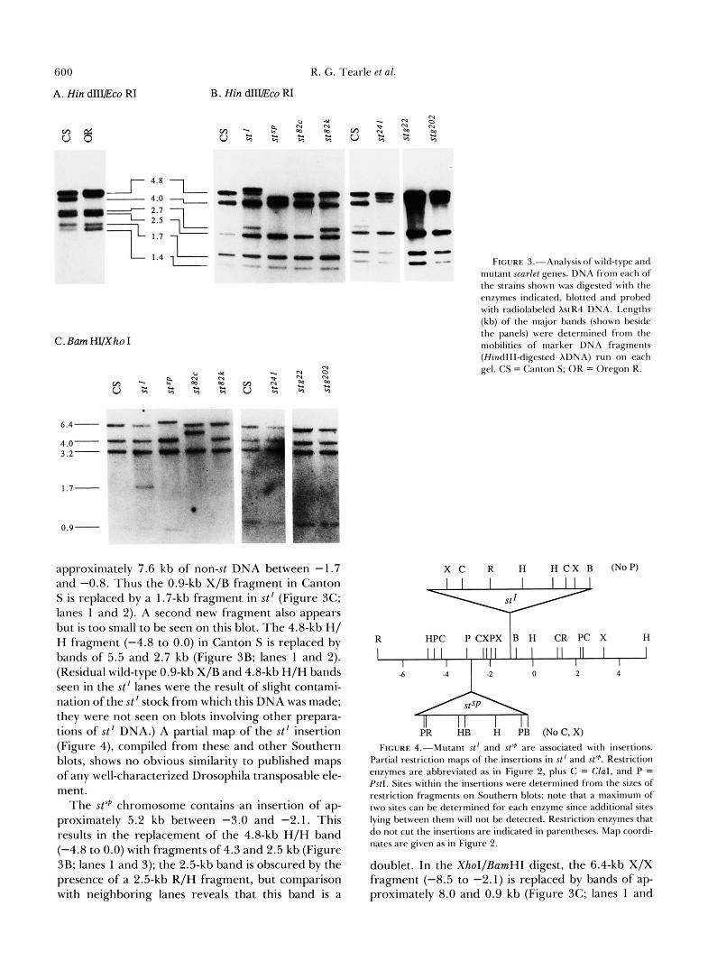
approximately 7.6 kb of non-st DNA between -1.7 and -0.8. Thus the 0.9-kb X/B fragment in Canton S is replaced by a 1.7-kb fragment in st' (Figure 3C; lanes 1 and 2). A second new fragment also appears but is too small to be seen on this blot. The 4.8-kb H/ H fragment (-4.8 to 0.0) in Canton S is replaced by bands of 5.5 and 2.7 kb (Figure 3B; lanes 1 and 2). (Residual wild-type 0.9-kb X/B and 4.8-kb H/H bands seen in the st' lanes were the result of slight contami- nation of the st' stock from which this DNA was made; they were not seen on blots involving other prepara- tions of st' DNA.) A partial map of the st' insertion (Figure 4), compiled from these and other Southern blots, shows no obvious similarity to published maps of any well-characterized Drosophila transposable ele- ment.
The stsP chromosome contains an insertion of ap- proximately 5.2 kb between -3.0 and -2.1. This results in the replacement of the 4.8-kb H/H band (-4.8 to 0.0) with fragments of 4.3 and 2.5 kb (Figure 3B; lanes 1 and 3); the 2.5-kb band is obscured by the presence of a 2.5-kb R/H fragment, but comparison with neighboring lanes reveals that this band is a
FIGURE J."Analysis of wild-type and mutant scarlef genes. DNA from each of the strains shown was digested with the enzymes indicated, blotted and probed with radiolabeled XstR4 DNA. Lengths (kb) of the major bands (shown beside the panels) were determined from the mobilities of marker DNA fragments (Hindlll-digested XDNA) run on each gel. CS = Canton S; OR = Oregon R.
R HPC P CXPX B H CR PC X H
I IIII I II I I I I I
-6 4 -2 0 2 4 A PR HB H PB (NoC, X)
FIGURE 4.-Mutant st' and sfJp are associated with insertions. Partial restriction maps of the insertions in st' and sf'p. Restriction enzymes are abbreviated as in Figure 2, plus C = Clal , and P = Pstl. Sites within the insertions were determined from the sizes of restriction fragments on Southern blots; note that a maximum of two sites can be determined for each enzyme since additional sites lying between them will not be detected. Restriction enzymes that do not cut the insertions are indicated in parentheses. Map coordi- nates are given as in Figure 2.
doublet. In the XhoI/BamHI digest, the 6.4-kb X/X fragment (-8.5 to -2.1) is replaced by bands of ap- proximately 8.0 and 0.9 kb (Figure 3C; lanes 1 and

Cloning and Characterization of scarlet 60 1
3). The partial restriction map of this insertion (Figure 4) shows no obvious similarity to published restriction maps of Drosophila transposable elements. The spot- ted phenotype of stSP is not particularly obvious in wild-type backgrounds; however, when combined with wBwx (to remove the drosopterins) stsp shows an eye pigment pattern reminiscent of wsP alleles-a pale yellow eye with spots of darker brown.
A putative MR-induced allele, (M. M. GREEN, personal communication), is missing the BamHI site at -0.8. Thus, the 0.9-kb X/B fragment (-1.7 to -0.8) and the adjacent 4.0-kb B/X fragment (-0.8 to +3.2) of Canton S are replaced by a band of 4.9 kb (Figure 3C; lanes 1 and 4). If the loss of the BamHI site is the result of a deletion, it must be rather small, since we can detect no change in the size of the 1.7- kb X/H fragment (-1.7 to 0.0) in the stazc3 mutant DNA (data not shown).
All of the st- rearrangements analyzed thus far are located between -7.3 and +1.4, suggesting that the st+ functional unit lies at least partly within this region. Recent DNA transformation studies (A. ELIZUR, R. G. TEARLE, R. B. SAINT and A. J. HOWELLS, unpub- lished data) have confirmed this by showing that a fragment extending from -7.3 to +1.4 is capable of supplying st+ function.
Transcription of scarlet: In order to characterize the size of the st transcription unit we probed blots of poly(A+) RNA from wild type third instar larvae with riboprobes made from various restriction fragments from across the region. The probes were strand spe- cific, with transcription being from left to right on the map shown in Figure 2. Figure 5A shows a number of such blots. These blots show that fragments located between the PstI site at -3.0 and the EcoRI site at + 1.4 hybridize to a 2.3 kb transcript. The fragment immediately to the left of -3.0 (1.5-kb P/P) fails to hybridize to any transcript but the next leftward frag- ment (2.4-kb R/H, -4.5 to -6.9) again hybridizes to a 2.3 kb transcript. Recent sequencing data (Y. HAUPT and A. J. HOWELLS, unpublished data) suggests that part of the st coding region is located in the 2.4-kb R/ H fragment and that the 1.5-kb P/P fragment is probably part of a relatively long intron. The frag- ment to the right of 0.0 (1.4-kb H/R, 0.0 to f1.4) weakly hybridizes to a 2.3-kb transcript, as well as to transcripts of about 2.6 and 3.6 kb. We interpret the latter two bands to be products of an adjacent proxi- mal transcription unit. The bands seen on these blots are resistant to ribonuclease digestion and are there- fore unlikely to be the result of spurious hybridization by the riboprobes. The results are consistent with the size of the gene inferred from our study of st- chro- mosomal rearrangements and with the transformation results mentioned above. The putative st transcript is present at substantially lower levels than white tran-
scripts in the same RNA samples from third instar larvae (data not shown).
The st' and stsP mutations involve the insertion of DNA into or near the st transcription unit. The mu- tations might alter the level or structure of the st RNA. Figure 5B shows blots of poly(A+) RNA from both st' and stSP pupae hybridized with region-specific probes. A faint st transcript (<5% of wildtype) is detected in the st"P sample using a probe covering the region -1.7 to -0.8; the higher level of background usually associated with the probe covering the region -0.8 to 0.0 prevents us from determining whether this fragment also detects the transcript in stSP. s t ' , on the other hand, gives rise to an RNA which is slightly smaller and less abundant than the wild-type st tran- script. This RNA is detected using a probe containing sequences to the left of the st' insertion (-1.7 to -0.8). Although the differences in length and abundance of the st' transcript relative to st+ are small, they are quite reproducible. A probe lying completely to the right of the st' insertion (-0.8 to 0.0) does not detect this RNA, indicating that the st' RNA is truncated near, or within, the st' insertion. The observation that the 2.3-kb RNA is affected by both of these sponta- neous st mutations provides strong evidence that this is the st gene transcript.
Developmental analyses using a temperature-sensi- tive st allele (HOWELLS 1979) indicate that the tem- perature-sensitive period for eye pigmentation is ap- proximately 24-48 hr after pupariation at 25 O . Other studies show that st+ must act in the larval Malpighian tubules. We have used RNA blotting to determine if the different developmental requirements for st+ ac- tivity are reflected in changes in expression of the st gene at the level of the RNA. Figure 5C shows RNA from different developmental stages hybridized to a probe from region -0.8 to -0.0. The major st+ tran- script is present at all stages. The level of st+ RNA is developmentally regulated such that it is present in about 1 0-fold higher levels in early to mid pupae than at any other stage. This corresponds well to the time at which the gene would need to be expressed for proper pigmentation of the eye.
Since the st+ and the w+ genes appear to play similar roles in the pathway leading to the deposition of xanthommatin, we wished to determine if st+ expres- sion is regulated by w+. In other words, is the w + gene function required for the expression of st+? We have addressed this question by carrying out an RNA blot analysis of the w' mutation. The blots shown in Figure 5D show that the level of st+ RNA does not change drastically in the presence of w l , suggesting that w + does not regulate the transcription of the st+ gene.
Homology between scarlet region DNA and white locus sequences: Because of the similar effects of st and w mutants on ommochrome deposition, we wished

602 R. G . Tearle et al.
A. B. ( 0
R HP P XPX B H R
-6 -4 -2 0 Kb " -
3 5 7 1 "- 2 4 6
1 2 3 4 5 6 7 -
mm
C.
"- D.
FIGURE 5.-RNA expression of the scarlet gene. (A) Location of the st transcription unit. An RNA blot (10 pg poly(A+) RNA/lane) was cut into strips and the strips hybridized to riboprobes made from restriction fragments from across the st region. After hybridization and washing, the filters were autoradiographed. The autoradiographs were aligned for photography. Positions of the fragments from which the riboprobes were made are shown below the restriction map. (B) Expression of st RNA in st mutants. Poly(A+) R N A (5 pg/lane) from wild type (Canton S) or st mutant early pupae was blotted and probed with riboprobes covering either region -1.7 to -0.8 (left three lanes) or -0.8 to 0.0 (right three lanes) (probes 5 and 6 respectively) (A). The same R N A preparations were used for the blots shown in (i) and (ii) and. for each blot, all three lanes were part of the same gel and were hybridized together. (C) Developmental expression of st. Poly(A+) RNA (10 pgllane) from various developmental stages was separated by electrophoresis, blotted and probed with a riboprobe covering the region -0.8 to 0.0 (probe 6) (A). (D) Expression of st RNA in the presence and absence of the w+ gene. Poly(A+) RNA from wild-type (w*) or mutant ( w ' ) third instar larvae was blotted and probed as in (C).
to determine if there is any similarity between the Xw45A2 was digested with various restriction enzymes coding or regulatory sequences of the two cloned loci. and probed with subclones from the st region (-4.8 A clone (Xw45A2) carrying thew+ gene and surround- to +3.4). A subclone carrying the 4.8-kb H/H st ing DNA (coordinates -5.2 to 9.2; LEVIS, BINCHAM fragment (-4.8 to 0.0) cross-hybridizes to a 1.1-kb S/ and RUBIN 1982) was isolated from the wild-type Xb fragment of thew locus (-0.7 to +0.4). A subclone Canton s library of MANIATIS et al. (1978). DNA from carrying the 2.0-kb S/B fragment (-0.7 to +I .3) of w

Cloning and Characterization of scarlet 603
6 4 2 0 -2.4 I 1 1 1 I I white
Gels hstR4 hw45A
w probe st probe hstR4 hw45A
7.0 5.6
3.5 2.7 2.2
1.4
FIGURE 6.-Cross-hybridization between scarlet and white. The top part of the figure shows restriction maps of phage clones carrying the w+ (Xw45A2) and st+ (XstR4) genes. They have been aligned so that transcription of both genes is from left to right. Exons of UI are shown as black bars under the w map. The approx- imate extent of the st transcription unit is shown as a horizontal line under the st map. Restriction enzymes are abbreviated as in Figure 4, plus Xb = Xbal. The major cross-hybridizing fragments are indicated by cross hatching. The bottom part of the figure shows DNA from the st or w clones digested with the restriction enzymes indicated. The stained gels (on the left) were blotted and probed, as indicated, with a w specific probe (-0.7 to +1.3) or a st specific probe (-4.8 to 0.0) to give the autoradiographs (on the right). The positions of marker fragments (Accl-digested XDNA) are indicated. Hybridization conditions were as given for Southern blots (MATERIALS AND METHODS); probes were labeled by nick translation.
cross-hybridizes to the 0.9-kb X/B fragment (-1.7 to -0.8) of st. This hybridization can be detected after hybridization (1.5 x SSPE at 68") and washing (0.1 X SSC at 60") under stringent conditions. The regions of st and w cross-hybridization are indicated on the restriction maps shown in Figure 6. The region lying downstream of the XhoI site (-1.7) of st was sequenced and a 320 bp region of this sequence showing the greatest homology to w is given in Figure 7; this sequence contains a single open reading-frame and the derived amino acid sequence is shown under the nucleotide sequence. Also given in Figure 7 is the
.sL CTTTCGGCGGCGCAA---ACTCGAATTGGTAGTGGCGATGAC~GAAGGTCCTT LeuSerAlaAlaGln---ThrArgIleGlySerGlyAspAspLysLysValLeu . . . . . . . .
frl CTCAGCAAATGTCAGCACACGATCATCGGTGTGCCCGGCAGGGTG~AGGTCTG LeuSerLysCysGlnHisThrIleIleGlyValProGlyArgValLysGlyLeu
st TCGGGGGGAGAACGCAAACGATTGGCATTCGCCGTGGAGCTGCTGAACAATCCG SerGlyGlyGluArgLysArgLeuAlaPheAlaValGluLeuLeuAs snPro
y TCCGGCGGAGAAAGGAAGCGTCTGGCATTCGCCTCCGAGGCACTAACCGATCCG SerGlyGlyGluArgLysArgLeuAlaPheAlaSerGluAlaLeuTh s Pro . . . . . . . . . . . . . . . . . . . . . . . .
.sL GTGATTCTATTTTGCGATGAGCCTACCACGGGACTGGACTCATACAGTGCCCAG
. . . . . - w CCGCTTCTGATCTGCGATGAGCCCACCTCCGGACTGGACTCATTTACCGCCCAC
st CAGCTGGTGGCCACGTTGTACGAGTTGGCCCAAAAGGGGCACCACCATACTGTGC GlnLeuValAlaThrLeuTyrGluLeuAlaGlnLysGlyThrThrIleLeuCys
SerValValGlnValLeuLysLysLeuSerGlnLysGlyLysThrValIleLeu frl AGCGTCGTCCAGGTGCTGAAGAAGCTGTCGCAG~GGGC~GACCGTCATCCTG
. . . . . . . . . . .
ST ACCATCCATCAGCCGAGTTCGCAGCTCTTCGATAACTTTAACAACGTAATGTTG ThrIleHisGlnProSerSerGlnLeuPheAspAsnPheAsnAsnValMetLeu
ThrIleHisGlnProSerSerGluLeuPheGluLeuPheAspLysIleLeuLeu y ACCATTCATCAGCCGTCTTCCGAGCTGTTTGAGCTCTTTGACAAGATCCTTCTG
. . . . . . . . . . . . . . . . . . . . . . . . .
st CTGGCCGATGGGCGAGTAGCCTTTACGGGATCACCTCAGCATGCGCTTAGTTTC LeuAlaASpGlyArgValAlaPheThrGlySerProGlnHisAlaLeuSerPhe
MetAlaGluGlyArgValAlaPheLeuGlyThrProSerGluAlaValAspPhe ATGGCCGAGGGCAGGGTAGCTTTCTTGGGCACTCCCAGCGAAGCCGTCGACTTC
. . . . . . . . . . . . . . . . . .
FIGURE 7.-Sequence homology between scarlet and white. The nucleotide sequence for sf starts 28 I nucleotides downstream from the Xhol site (coordinate -1.7): the w sequence is from O'HARE et al. (1984) (position -354 to -678). Colons between the deduced amino acids indicate identical residues; a single dot shows a con- servative substitution. The boxed region is strongly homologous to site B of the ATP-binding region of bacterial periplasmic transport proteins (AMFS 1986).
sequence of the homologous region from w (O'HARE et al. 1984), which shows that the overall homology between the two genes in this region is 59% (identical residues-at both the nucleotide and derived amino acid levels). This sequence includes one of the putative ATP-binding sites of the w protein (MOUNT 1987); the homology of the 100 nucleotides which encodes this site (and its immediate surroundings) is 72%, which probably accounts for the cross-hybridization between the two genes (as shown in Figure 6). Further DNA sequencing (A. ELIZUR, R. G . TEARLE, D. G . BOYLE and A. J. HOWELLS, in preparation) has shown that other smaller patches of homology not seen by hybridization studies are present in other parts of the putative coding regions.
DISCUSSION
The evidence that XstR4 contains the st gene is overwhelming. (1) Sequences from the clone corre- spond to the non-rosy region sequences of clones Xsta27-1 and Xsta27-2, which span the breakpoints of In(3LR)st-a27. This inversion chromosome, which is associated with a st mutant phenotype, has one break in 73A3.4, the cytological position of st ; the corre- spondence of the st mutant phenotype and the cyto- logical location implies strongly that the breakpoint is

604 R. G . Tearle et al.
either very close to, or within, the st gene. (2) In the DNA of four different mutant stocks showing a st phenotype, the restriction map of the region relative to wild type is altered. (3) Successful germline trans- formation of st’ individuals to st+ has been achieved following the injection of st’ embryos with a P-element transformation vector carrying an 8.7-kb fragment spanning coordinates -7.3 to +1.4 (A. ELIZUR, R. G. TEARLE, R. B. SAINT and A. J. HOWELLS, unpublished data). (4) A single 2.3-kb transcript is altered in st’ and greatly reduced in abundance in stsf’. (5 ) Some of the sequences from within the above transcription unit show significant sequence similarity to the white gene, another locus involved in ommochrome metab- olism.
The transformation experiments, together with the Northern blot analysis, indicate that the st transcrip- tion unit is confined to the region from -7.3 to +1.4. When DNA from Canton S is analyzed by Southern blotting using probes covering this region, few restric- tion site or fragment length polymorphisms are found. In contrast, DNA sequences which lie both distal (-14.1 to -9.0) and proximal (+1.4 to +12.5) to the st transcription unit show considerable polymorphism. The distal region is not only polymorphic in our Canton S strain but also shows different restriction patterns in all of the different wild type strains that we have examined. Obviously more detailed data is needed but it does seem that the regions that flank st are highly polymorphic. Such variation is not typical of the D. melanogaster genome in general, although some highly polymorphic regions, such as those con- taining the zeste locus, have been found (MARIANI, PIRROTTA and MANET 1985).
The developmental pattern of expression of the st gene at the RNA level agrees with biochemical and genetic data concerning this gene. Specifically, there are low levels of expression in larvae and adults and an obvious peak of expression in the pupal period. Tissue transplantation experiments (BEADLE, 1937a, b) suggest that the Malpighian tubules normally re- quire expression of both st and w for storage of ommochrome pigment precursors. The presence of st+ transcripts throughout larval life (albeit at a low level) probably reflects this expression in the Malpi- ghian tubules. This has been directly demonstrated for RNAs from the w gene (FJOSE et al. 1984).
Pigment deposition in the eyes commences about 48 hr after pupariation (SUMMERS, HOWELLS and PY- LIOTIS 1982). Genetic analysis using a temperature- sensitive st allele shows that st expression is required just prior to this time (about 24-48 hr post puparia- tion) for proper pigmentation of the eyes (HOWELLS 1979). This correlates with a tenfold increase in the level of st RNA early in the pupal period. Finer-scale analysis of st gene expression during pupal life (data
not shown) indicates that the peak level of st RNA corresponds to the temperature-sensitive period for the temperature-sensitive mutant ( i e . , 24-48 hr after pupariation). As for w (LEVIS, O’HARE and RUBIN 1984; PIRROTTA and BROCKL 1984), st RNA can be detected in adults. Whether this reflects stability of the mRNA made during pupal life or continued low level transcription has not been determined.
The transcriptional results obtained for st ’ are com- patible with the position of the non-st DNA insertion in this mutant. The use of two probes covering the -1.7 to 0.0 region was particularly informative. The probe covering the region -1.7 to -0.8 (which lies 5’ to the st’ insertion) detects a transcript which appears to be slightly smaller than that found in wild type. However a probe covering the region just 3’ to the st’ insertion fails to detect a major transcript. Thus, it seems likely that in st’, transcription initiates from the normal st promoter (probably located between -7.3 and -4.8) and terminates within the st’ insertion. Since the st’ transcript is polyadenylated, a polyade- nylation signal must be located within this insertion. Polyadenylated aberrant transcripts which terminate within inserted transposable elements have been found in several w mutants (PIRROTTA and BROCKL 1984; LEVIS, O’HARE and RUBIN 1984; ZACHAR, CHAPMAN and BINGHAM 1985; BINGHAM and CHAP- MAN 1986).
The stsP mutation is of interest because it gives a spotted pigment pattern reminiscent of the w5p alleles. This suggests that the lesion in st”P, like those in the wsP alleles, may affect regulatory rather than coding sequences. The severely reduced level of the st tran- script in 6-30 h stS$ pupae is consistent with this suggestion; however, unlike the situation with the wsp alleles, where the lesions lie 5’ to the coding region, the insertion in stSP appears to be located within an intron. This may mean that some of the regulatory sequences of the st gene are located within this intron; however it is also possible that the insertion has a cis- acting inhibitory effect on the st promoter or that an abberant stSP transcript is unstable and is rapidly de- graded.
As outlined in the Introduction, st and w mutants show marked similarities at the genetic and biochem- ical levels, raising the question of whether their prod- ucts perform similar functions, or whether there is a regulatory relationship between them. Our molecular analysis has revealed that there is considerable nucleo- tide similarity between the two genes. The sequencing data, which has confirmed the hybridization analysis, shows that the st/w homology is located in coding regions of these two genes and is just as strikingly apparent in the derived amino acid sequences. Al- though the functional significance of this similarity between the putative protein products of st and w

Cloning and Characterization of scarlet 605
must await further analysis, it would appear that the phenotypic similarity between lesions at the two loci reflects a common functional role for the proteins. MOUNT (1 987) has pointed out the sequence similarity between the putative w protein and a family of ATP- binding proteins involved in bacterial periplasmic transport systems. As shown in Figure 7, the strongest homology between the derived amino acid sequences of w and st includes a region thought to be involved in ATP binding. A role for the w and st proteins in the transport of xanthommatin precursors across the plasma membrane of pigment cells was proposed by SULLIVAN and SULLIVAN (1 975). The w gene is essen- tial for pteridine as well as ommochrome pigment production. The phenotypic analogue of st in the pteridine biosynthesis pathway appears to be the brown (bw) gene (SULLIVAN et al. 1980); recent molecular analysis of the bw gene has indicated that there is also strong amino acid homology between the protein products of the w and bw genes (DREESEN, JOHNSON and HENIKOFF 1988). As discussed by these authors, it seems likely that the w , bw and st gene products are members of a family of membrane-spanning, ATP- binding proteins which associate to provide the per- meases involved in the uptake of pigment precursors.
An alternative hypothesis concerning the pheno- typic similarities of lesions at st and w , that one gene regulates the expression of the other, is not supported by the data. The level of the s t transcript in third instar larvae is not affected by the w’ mutation (our data), and the level of w gene expression has previously been shown not to be affected in st’ adults (LEVIS, O’HARE and RUBIN 1984).
Given the similarities in the tissue-specific and tem- poral expression of the w and st genes (see Introduc- tion), we consider it possible that conserved regulatory as well as structural sequences might exist between the two genes. Such a possibility is exciting because of the implications it might have for the regulation and function of the ommochrome biosynthetic pathway in particular, and for the coordinate control of function- ally related genes in eukaryotes in general. Since the nucleotide sequence of the putative regulatory region upstream of w has been established (O’HARE et al. 1984) and analyzed in some detail (DAVISON et al. 1985; ZACHAR, CHAPMAN and BINGHAM 1985; PIR- ROTTA, STELLER and BOZZETTI 1985; LEVIS, HAZEL- RIGG and RUBIN 1986), we are now determining the nucleotide sequences of the upstream and intronic regions of st in order to identify any shared regulatory sequences.
We thank K. C. REED and A. ELIZUR for advice about methods and for valuable discussions. This work was supported by grants from the Australian Research Grants Scheme to A.J.H., and by grants from the U.S. National Institutes of Health (NIH) and the National Science Foundation to B.S.B. and J.M.B. R.G.T. was supported by an Australian Commonwealth Postgraduate Award
and M.M. was supported by an NIH postdoctoral training grant, and by a postdoctoral fellowship from the Helen Hay Whitney Foundation.
LITERATURE CITED
AMES, G. F.-L. 1986 Bacterial periplasmic transport systems: structure, mechanism, and evolution. Annu. Rev. Biochem. 55:
ASHBURNER, M., J. FAITHFULL, T. LITTLEWOOD, G. RICHARDS, S. SMITH, V. VELISSARIOU and R. WOODRUFF, 1980 New mu- tants. Drosophila Inform. Serv. 55: 193- 195.
ASHBURNER, M., P. ANGEL, C. DETWILER, J. FAITHFULL, D. GUBB, G. HARRINGTON, T . LITTLEWOOD, S. TSUBOTA, V. VELISSA- RIOU and V. WALKER, 1981 New mutants. Drosophila In- form. Serv. 5 6 186-191.
BAGLIONI, C., 1960 Genetic control of tryptophan pyrrolase in Drosophila melanogaster and Drosophila virilis. Heredity 15: 87- 96.
BAILLIE, D. L., and A. CHOVNICK, 1971 Studies on the genetic control of tryptophan pyrrolase in Drosophila melanogaster. Mol. Gen. Genet. 112: 341-353.
BAKER, B. S., and K. RIDGE, 1980 Sex and the single cell: on the action of major loci affecting sex determination in Drosophila melanogaster. Genetics 9 4 383-423.
BEADLE, G. W., 1937a Development of eye colors in Drosophila: Fat bodies and Malpighian tubules as sources of diffusible substances. Genetics 2 2 146-152.
BEADLE, G. W., 1937b Development of eye colors in Drosophila: Fat bodies and Malpighian tubules in relation to diffusible substances. Genetics 22: 587-61 1.
BEADLE, G. W., and B. EPHRUSSI, 1937 Development of eye colors in Drosophila: Diffusible substances and their interrelations. Genetics 22: 76-86.
BELOTE, J. M., and M. MCKEOWN, 1985 Post-replicative repair of an X-ray damaged chromosome following fertilization in Dro- sophila melanogaster. Drosophila Inform. Serv. 61: 33-34.
BENDER, W., P. SPIERER and D. S. HOGNFSS, 1983 Chromosomal walking and jumping to isolate DNA from the Ace and rosy loci and the bithorax complex in D. melanogaster. J. Mol. Biol. 168:
BINGHAM, P. M., and C. H. CHAPMAN, 1986 Evidence that white- blood is a novel type of temperature-sensitive mutation resulting from temperature dependent effects of a transposon insertion on formation of white transcripts. EMBO J. 5: 3343-3351.
BINGHAM, P. M., and Z. ZACHAR, 1985 Evidence that two muta- tions, wDzL and z’, affecting synapsis dependent genetic behavior of white are transcriptional regulatory mutants. Cell 4 0 819- 825.
CHIRGWIN, J. M., A. E. PRZYBYLA, R. J. MACDONALD and W. J. RUTTER, 1979 Isolation of biologically active ribonucleic acid from sources enriched in ribonuclease. Biochemistry 18: 5294- 5299.
COEN, E. S., J. M. THODAY and G. DOVER, 1982 Rate of turnover of structural variants in the rDNA family of Drosophila mela- nogaster. Nature 295: 564-568.
DAVISON, D., C. H. CHAPMAN, C. WEEDEN and P. M. BINGHAM, 1985 Genetic and physical studies of a portion of the white locus participating in transcriptional regulation and synapsis- dependent interactions in Drosophila adult tissues. Genetics 110: 479-494.
DREESEN, T. D., D. H. JOHNSON and S. HENIKOFF, 1988 The brown protein of Drosophila melanogaster is similar to the white protein and to components of active transport complexes. Mol. Cell. Biol. 8 5206-5215.
EPHRUSSI, B., 1942 Analysis of eye color differentiation in Dro- sophila. Cold Spring Harbor Symp. Quant. Biol. 10: 40-48.
FJOSE, A., L. C. POLITO, U. WEBER and W. J. GEHRING,
397-425.
17-33.

606 R. G. Tearle et al.
1984 Developmental expression of the white locus of Drosoph- ila melanogaster. EMBO J. 3: 2087-2094.
FRISCHAUF, A. M., H. LEHRACH, A. POUSTKA and N. MURRAY, 1983 Lambda replacement vectors carrying polylinker se- quences. J. Mol. Biol. 170: 827-842.
GHOSH, D., and H. S. FORREST, 1967 Enzymatic studies on the hydroxylation of kynurenine in Drosophila melanogaster. Ge- netics 55: 423-43 l .
HOWELLS, A. J., 1979 Isolation and biochemical analysis of a temperature-sensitive scarlet eye colour mutant of Drosophila melanogaster. Biochem. Genet. 17: 149-158.
HOWELLS, A. J., and R. L. RYALL, 1975 A biochemical study of the scarlet eye colour of Drosophila melanogaster. Biochem. Genet. 13: 273-282.
LANGER-SAFER, P. R., M. LEVINE and D. WARD, 1982 Im- munological method for mapping genes on Drosophila polytene chromosomes. Proc. Natl. Acad. Sci. USA 79: 4381-4385.
LEVIS, R., P. M. BINGHAM and G. M. RUBIN, 1982 Physical map of the white locus of D. melanogaster. Proc. Natl. Acad. Sci.
LEVIS, R., K. O’HARE and G. M. RUBIN, 1984 Effects of transpos- able element insertions on RNA encoded by the white gene of Drosophila. Cell 38: 471-481.
LEVIS, R., T . HAZELRIGG and G. M. RUBIN, 1985 Separable cis- acting control elements for expression of the white gene of Drosophila. EMBO J. 4: 3489-3499.
LINDSLEY, D. L., and E. H. GRELL, 1968 Genetic Variations of Drosophila melanogaster. Carnegie Inst. Wash. Publ. 627.
LINDSLEY, D. L., and G. ZIMM, 1986 The genome of Drosophila melanogaster. Part 2: Lethals; maps. Drosophila Inform. Serv. 64: 72-73.
LINDSLEY, D. L., and G. ZIMM, 1987 The genome of Drosophila melanogaster. Part 3: Rearrangements. Drosophila Inform. Serv. 65: 48.
MANIATIS, T., E. F. FRITSCH and J. SAMBROOK, 1982 Molecular Cloning: A Laboratory Manual. Cold Spring Harbor Laboratory, Cold Spring Harbor, N.Y.
MANIATIS, T., R. C. HARDISON, E. LACY, J. LAUER, C. O’CONNEL, D. QUON, G. K. SIM and A. EFSTRADIATIS, 1978 The isolation of structural genes from libraries of eukaryotic DNA. Cell 15: 687-701.
MARIANI, C., V. PIRROTTA and E. MANET, 1985 Isolation and characterization of the zeste locus of Drosophila. EMBO J. 4: 204552052,
MCKEOWN, M., J. M. BELOTE and B. S. BAKER, 1987 A molecular analysis of transformer, a gene in Drosophila melanogaster that controls female sexual differentiation. Cell 48: 489-499.
MESSING, J , , 1983 New MI3 vectors for cloning. Methods En- zymol. 101: 20-78.
MIKLOS, G. L. G., M. J. HEALY, P. PAIN, A. J. HOWELLS and R. J. RUSSELL, 1984 Molecular and genetic studies on the euchro- matin-heterochromatin transition region of the X chromosome of Drosophila melanogaster. I . A cloned entry point near to the uncoordinated locus. Chromosoma 8 9 218-227.
USA 79: 564-568.
MOUNT, S. M., 1987 Sequence similarity. Nature 325: 487. O’HARE, K., C. MURPHY, R. LEVIS and G. M. RUBIN, 1984 DNA
sequence of the white locus of Drosophila melanogaster. J. Mol. Biol. 180: 437-455.
PHILLIPS, J. P., and H. S. FORREST, 1980 Ommochromes and pteridines, pp. 542-623 in The Genetics and Biology of Drosoph- ila, Vol. 2d, edited by M. ASHBURNER and T . R. F. WRIGHT. Academic Press, New York.
PIRROTTA, V., and C. BROCKL, 1984 Transcription of the Dro- sophila white locus and some of its mutants. EMBO J. 3: 563- 568.
PIRROTTA, V., H. STELLER and M. P. BOZZETTI, 1985 Multiple upstream regulatory elements control the expression of the Drosophila white gene. EMBO J. 4: 3501-3508.
REED, K. C., and D. MANN, 1985 Rapid transfer of DNA from agarose gels to nylon membranes. Nucleic Acids Res. 13: 7207- 722 1.
RYALL, R. L., and A. J. HOWELLS, 1974 Ommochrome biosyn- thetic pathway of Drosophila melanogaster: Variations in the levels of enzyme activities and intermediates during adult de- velopment. Insect Biochem. 6 135-142.
SANGER, F., S. NICKLEN and A. R. COULSEN, 1977 DNA sequenc- ing with chain-terminating inhibitors. Proc. Natl. Acad. Sci. USA 74: 5463-5467.
SULLIVAN, D. T., R. J. KITOS and M. C. SULLIVAN, 1973 Developmental and genetic studies on kynurenine hy- droxylase from Drosophila melanogaster. Genetics 75: 65 1-66 1.
SULLIVAN, D. T. , and M. C. SULLIVAN, 1975 Transport defects as the physiological basis for eye color mutants of Drosophila melanogaster. Biochem. Genet. 13: 603-6 13.
SULLIVAN, D. T. , L. A. BELL, D. R. PATON and M. C. SULLIVAN, 1980 Genetic and functional analysis of tryptophan transport in Malpighian tubules of Drosophila. Biochem. Genet. 18: 1109-1 130.
SUMMERS, K. M., A. J. Howmuand N. A. PYLIOTIS, 1982 Biology of eye pigmentation in insects. Adv. Insect Physiol. 16: 119- 166.
TARTOF, K. D., 1969 Interacting gene systems: I . The regulation of tryptophan pyrrolase by the vermilion suppressor of vermilion system in Drosophila. Genetics 62: 781-795.
THOMAS, P. S., 1983 Hybridization of denatured RNA trans- ferred or dotted to nitrocellulose paper. Methods Enzymol. 100: 255-266.
VELISSARIOU, V., and M. ASHBURNER, 1981 Cytogenetic and ge- netic mapping of a salivary gland secretion protein in Drosophila melanogaster. Chromosoma 84: 173-1 85.
WALKER, A. R., A. J. HOWELLS and R. G. TEARLE, 1986 Cloning and characterization of the vermilion gene of D. melanogaster. Mol. Gen. Genet. 202: 102-107.
ZACHAR, Z. , C. H. CHAPMAN and P. M. BINGHAM, 1985 On the molecular basis of transvection effects and the regulation of transcription. Cold Spring Harbor Symp. Quant. Biol. 5 0 337- 346.
Communicating editor: V. G. FINNERTY