clinico-pathologic discussion of dilemmas in inflammatory...
TRANSCRIPT

Clinico-Pathologic Discussion of
Dilemmas in Inflammatory Bowel
DiseaseDisease
David Lewin MD
Department of Pathology
Lawrence W. Comerford MD, MS
Department of Medicine,
Division of Gastroenterology

Disclosures
• Dr. Lewin has no disclosures
• Dr. Comerford
UCB – Consultant
UCB – Research SupportUCB – Research Support
Abbott – Research Support
Centocor – Speakers Bureau

Additional Disclosures
• My wife is more attractive than I am
• None of my children listen to me
• I just submitted my 2010 taxes

Case-based presentation
#1 -Differentiating Ulcerative Colitis from
Crohn’s Colitis
#2 – Ileal Lesions – Crohn’s Disease?
#3 Dysplasia in Inflammatory Bowel Disease

Case #1 UC vs CD
21 year-old female college student presents with diarrhea (loose bowel movements daily) mild crampy abdominal pain, rectal bleeding for 2 months.
No recent travel or antibiotic use; Non-smokerNo recent travel or antibiotic use; Non-smoker
Mildly anemic Hg 11; Stool cultures are negative for enteric pathogens, C. difficile, and ova and parasites

Differential Diagnosis
• Infectious colitis
• Inflammatory Bowel Disease
• Ischemic colitis• Ischemic colitis
• Microscopic colitis
• SCAD
• Radiation colitis
• Drug-induced colitis
• Solitary rectal ulcer syndrome

DIFFERENTIAL DIAGNOSIS OF
INFECTIOUS AND ULCERATIVE
COLITIS

Endoscopy
• Colonoscopy: Diffuse ulcerated and friable
mucosa throughout entire colon; <
inflammation in rectum
• Unable to intubate terminal ileum• Unable to intubate terminal ileum
• Upper endoscopy normal


ANATOMIC EXTENT OF
ULCERATIVE COLITIS
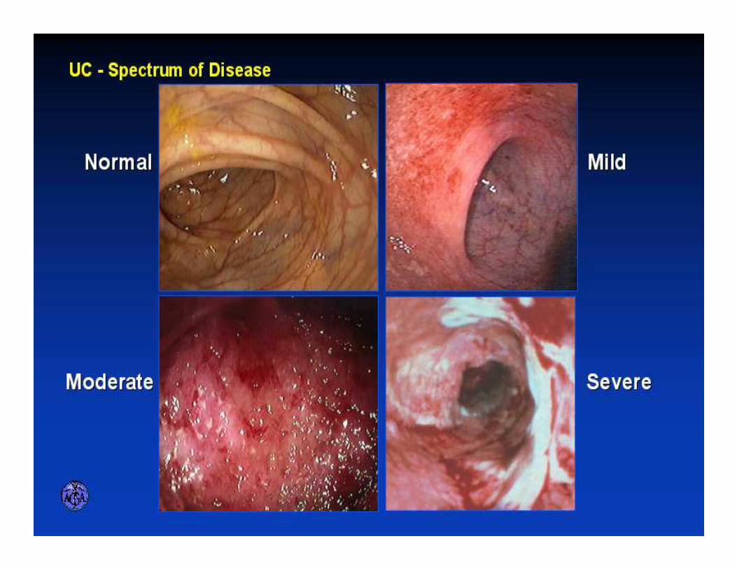
ENDOSCOPIC SPECTRUM OF
SEVERITY

Endoscopic Features
Distribution
Involves the rectum
Continuous pattern; no skip lesionsContinuous pattern; no skip lesions
More severe distally
May have a Cecal patch
Backwash ileitis

Crohn’s Disease
• Chronic inflammation potentially involving any
location of the alimentary tract
• 30% small bowel only, 40% ileocolic, 30% colon only
• Inflammatory Bowel Disease (IBD), ileitis, regional • Inflammatory Bowel Disease (IBD), ileitis, regional
enteritis, granulomatous enterocolitis
• Skip pattern, transmural (from mucosa to serosa)
• Stricture, fistula, abscess
• Unknown etiology
• No cure - Yet

ANATOMIC DISTRIBUTION
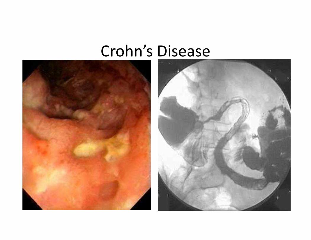
Crohn’s Disease

CLINICAL PATTERNS

Perirectal fistula

Crohn’s endoscopy
• Rectal sparing
• Skip patterns
• TI involvement
• Cobblestoning, ulceration• Cobblestoning, ulceration

Endoscopic Features
Rectal involvement UC > CD
Skip lesions CD > UC
Cobblestoning CD > UC
Pseudopolyps UC > CDPseudopolyps UC > CD
TI involvement CD > UC ( Backwash)
More severe distally UC> CD

Who are these guys?
Dr. Crohn with 2 Mt. Sinai gastroenterologists at 1928 ACP meeting

IBD
Genetics Immune
System
� Cytokine Imbalance
� Immunosuppressant and
anti-TNF-α therapy can
be effective.
� Prevalence differs among
ethnic groups
� Concordance rate for
monozygotic twins
(44%) > dizygotic twins
(3.8%)
� Link to genetically
Etiology of Inflammatory Bowel
Disease
IBD
Environmental
Influence
� Antigen Trigger (Bacteria, food Ag, or toxins)
� Hygiene or Urban Areas
� Smoking
� NSAIDS
� Link to genetically
susceptible hosts (e.g.
NOD 2/CARD 15)
Timmer. Digest Disease. 2003;21: 91–104.

GEOGRAPHICAL PREVALENCE
OF IBD
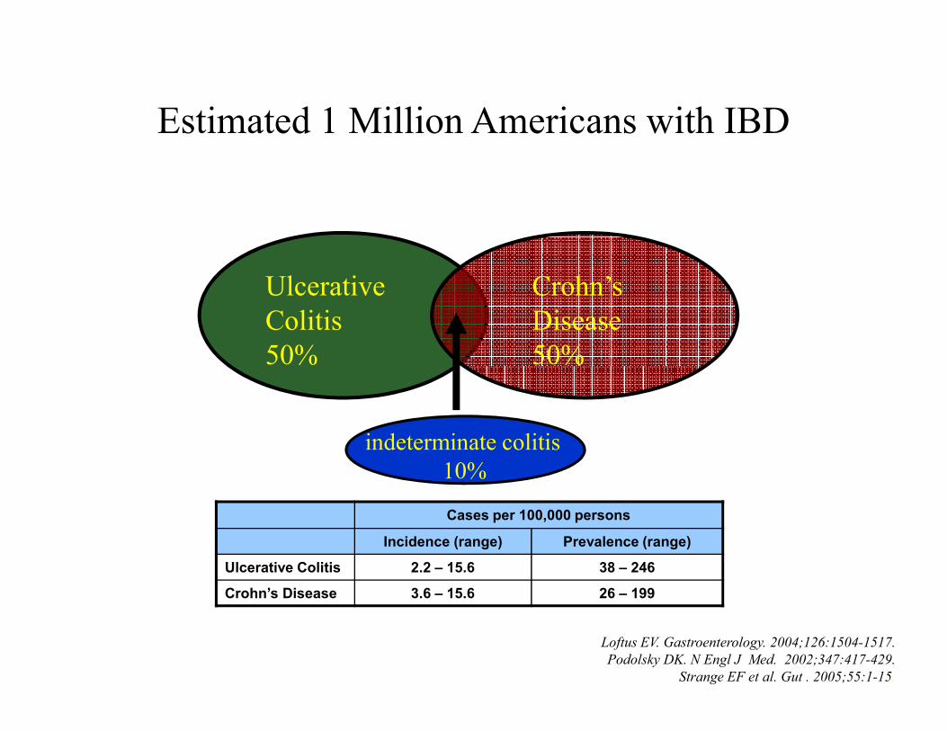
Estimated 1 Million Americans with IBD
Ulcerative
Colitis
50%
Crohn’s
Disease
50%
Loftus EV. Gastroenterology. 2004;126:1504-1517.
Podolsky DK. N Engl J Med. 2002;347:417-429.
Strange EF et al. Gut . 2005;55:1-15.
50% 50%
indeterminate colitis
10%
Cases per 100,000 persons
Incidence (range) Prevalence (range)
Ulcerative Colitis 2.2 – 15.6 38 – 246
Crohn’s Disease 3.6 – 15.6 26 – 199

Indeterminate colitis
• “IC” restricted to resected specimens
• “IBD Unclassified” - patients who appear to
have IBD colitis but who cannot be readily
classified when all clinical, radiological, classified when all clinical, radiological,
endoscopic, histologic, and serologic data
are taken into account

Indeterminate colitis
- Approximately 10% of cases
- 5 year Norway study of 40 IC cases1:
18/40 UC 7/40 CD 8/40 non-IBD
- Diagnostic problems common in early & - Diagnostic problems common in early &
severe disease
1 Moum B.., Scand J Gastroenterol 1996;31:362-366

Indeterminate colitis
CD vs UC
Radiology: SBFT, CTE, MRE
Small bowel endocapsuleSmall bowel endocapsule
Serology markers
Histology
Tincture of time

Potential Roles for
IBD Serologic Testing
• Assist in the initial diagnosis of IBD
– Presence of diagnostic uncertainty
– To assess need for endoscopy (esp. pediatrics)
• Differentiate Ulcerative Colitis (UC) from • Differentiate Ulcerative Colitis (UC) from Crohn’s Disease (CD)
– Indeterminate colitis
– Strengthen diagnosis prior to surgery
• Helps identify patients at risk for aggressive disease

PROMETHEUS® IBD Serology 7
• ELISA tests (Enzyme Linked Immunosorbent Assay)
– ASCA IgA
– ASCA IgG
– Anti-CBir1† IgA
– ANCA IgG– ANCA IgG
– Anti-OmpC* IgA
• IIF tests (Indirect Immunofluorescence)
– pANCA
– DNAse-sensitive pANCA*
†Patent pending.
*Proprietary and patented marker.
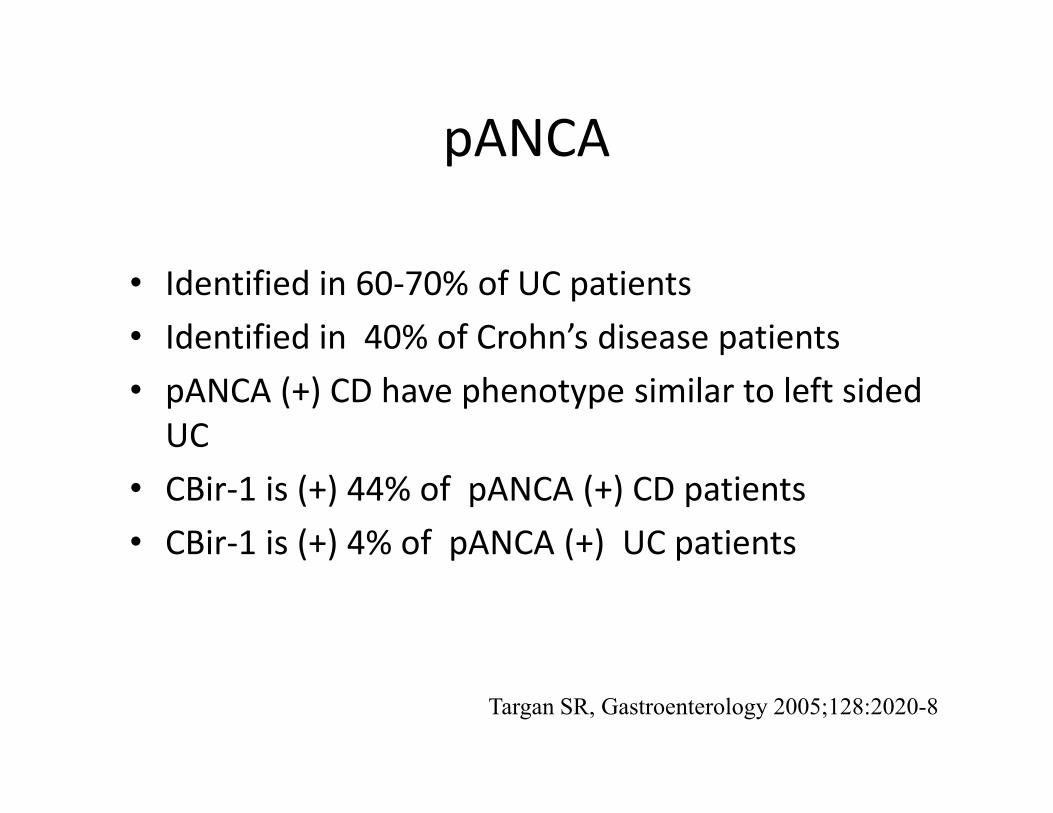
pANCA
• Identified in 60-70% of UC patients
• Identified in 40% of Crohn’s disease patients
• pANCA (+) CD have phenotype similar to left sided • pANCA (+) CD have phenotype similar to left sided
UC
• CBir-1 is (+) 44% of pANCA (+) CD patients
• CBir-1 is (+) 4% of pANCA (+) UC patients
Targan SR, Gastroenterology 2005;128:2020-8


Indeterminate colitis
Correct diagnosis important:
Surgical decisions ( IPAA)
Medical treatment
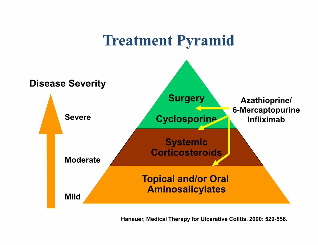
Treatment Pyramid
Severe
Surgery
Cyclosporine
Disease Severity
Azathioprine/
6-Mercaptopurine
Infliximab
Mild
Moderate
SystemicCorticosteroids
Topical and/or Oral Aminosalicylates
Cyclosporine Infliximab
Hanauer, Medical Therapy for Ulcerative Colitis. 2000: 529-556.

GROSS PATHOLOGY OF
ULCERATIVE COLITIS
“ Sometimes we have to save lives, not colons”
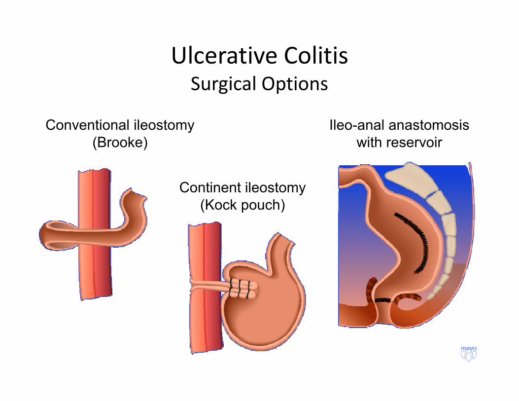
Ulcerative ColitisSurgical Options
Conventional ileostomy
(Brooke)
Continent ileostomy
(Kock pouch)
Ileo-anal anastomosis
with reservoir
(Kock pouch)

Indeterminate colitis
Severe Pouch complications1
CD 30-45% IC 20% UC 10%
Pouches requiring surgical correction2Pouches requiring surgical correction2
IC 50% UC 3% (n=235)
1 Geboes G. Inflamm Bowel Dis 2008;14:850-857
2 Koltun WA. Dis Colon Rectum 1991;34:857-860

ASLC V UC
• Plasmacytosis in the lamina propria extending to the mucosal base and mucosal distortion were present in all cases of UC and absent in all cases of ASLC.
– Histopathologic features always distinguished UC and ASLC
• Biopsy specimens are only diagnostic when obtained within the first 4 days
Nostrant et al. Gastroenterology 1987;92:318


ASLC V ACUTE ONSET IIBD
Findings ASLC IIBD
Normal Architecture 44 (85) 4 (8)
Distorted Crypt Architecture 0 34 (65)
Branched Glands 5 (10) 41 (79)
Villiform Surface 0 11 (21) Villiform Surface 0 11 (21)
Crypt Atrophy 1 (2) 5 (10)
Goblet Cell Mucin Depletion 36 (70) 48 (92)
Reactive Epithelial Hyperplasia 37 (73) 48 (92)
Surawicz et al. Gastroenterology 1994;107:755

ASLC V ACUTE ONSET IIBD
Findings ASLC IIBD
Acute and chronic
inflammation
16 (31) 49 (94)
inflammation
Crypt abscess 24 (37) 47 (90)
Basal plasmacytosis 3 (6) 40 (77)
Basal lymphoid aggregates 1 (2) 18 (35)
Isolated giant cells 1 (2) 13 (25)
Granulomatous crypt
abscess
8 (15) 19 (37)
Surawicz et al. Gastroenterology 1994;107:755
architect2.jpg

archdist1.jpg
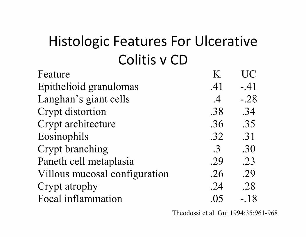
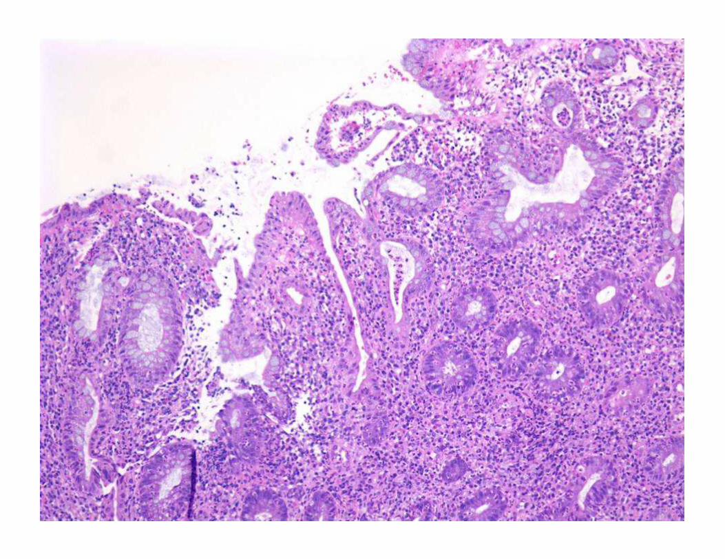
Histologic Features For Ulcerative
Colitis v CDFeature K UC
Epithelioid granulomas .41 -.41
Langhan’s giant cells .4 -.28
Crypt distortion .38 .34
Crypt architecture .36 .35 Crypt architecture .36 .35
Eosinophils .32 .31
Crypt branching .3 .30
Paneth cell metaplasia .29 .23
Villous mucosal configuration .26 .29
Crypt atrophy .24 .28
Focal inflammation .05 -.18
Theodossi et al. Gut 1994;35:961-968

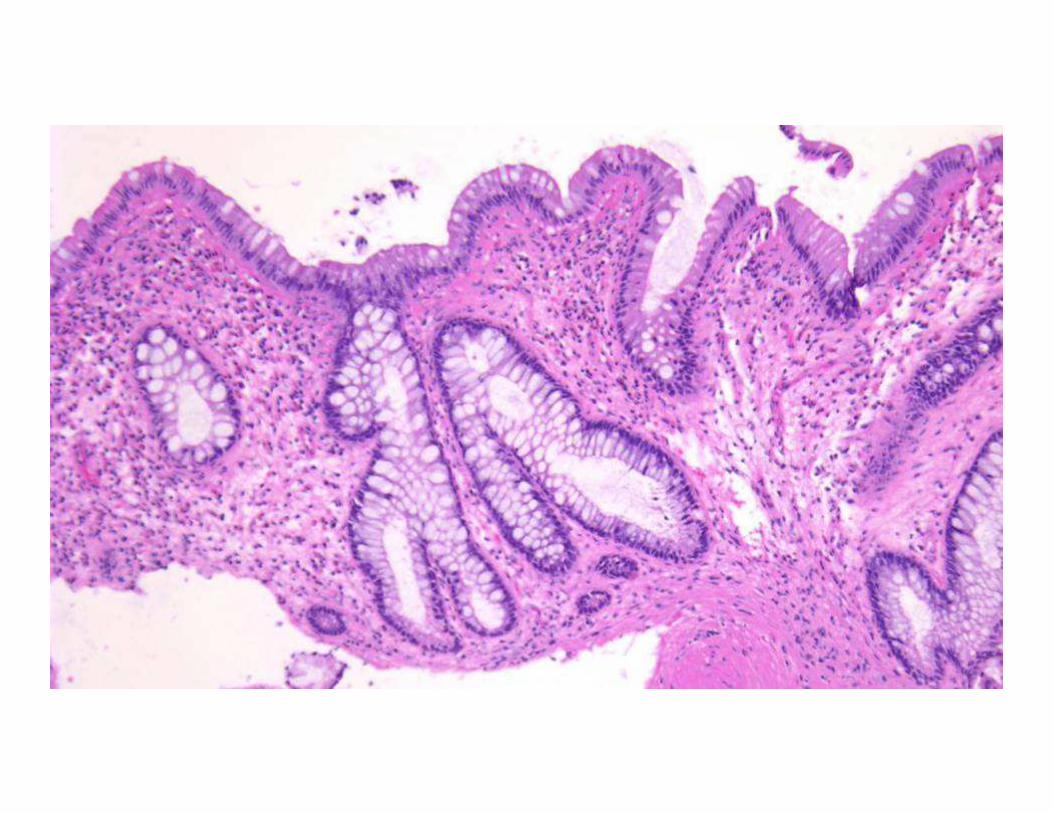
Diffuse (UC) v Patchy (CD)Diffuse (UC) v Patchy (CD)


pm40x.jpg

IBD SCORING SYSTEM
Feature IBD/normal UC/CD
Neutrophilic infiltration 1 -1
Epithelioid granulomas 1 -6
Crypt architecture abn 3 1 UC/CD score
Crypt architecture abn 3 1
Mucin depletion 1
Eosinophils 2 3
Paneth cell metaplasia 2
Diffuse inflammation 1 -2
Villous configuration 1
Theodossi et al. Gut 1994;35:961-968
<= -3 -2 to 2 => 3
<= 1 - “Normal -
2 to 4 CD Possible
IBD
UC
=> 5 CD IBD UC
IBD
/
nl score

Granulomas in Crohn’s Disease
• One of the best discriminating factors v UC
• Types of Granulomas
– Well-formed, non-necrotizing granuloma
• Diagnostic of CD
– Rule out TB and sarcoid– Rule out TB and sarcoid
– Pericryptal microgranuloma
• Occasionally seen in UC and other colitides
– Mucin Granuloma
• Ruptured crypt with associated giant cells, non-specific
• Can be seen with ulcerative colitis
• Granulomas only seen in 20 - 30% of Crohn’s biopsies

crohn4xcolgran.jpg


mucingranuloma1.jpg


Summary of Causes of Unusual Patterns of
Disease in UC
• Treatment effect
• Low-grade disease in remission
• Appendiceal involvement as a skip lesion
• Cecum/ascending colon inflammation in left-sided colitis
• Cecum/ascending colon inflammation in left-sided colitis
• Pediatric UC (initial presentation)
• “Backwash” ileitis
• Rare upper gi involvement (e.g., duodenitis)
• Fulminant colitis
Adapted from Table 10-1, Surgical Pathology of the GI Tract, Liver, Biliary Tract, and Pancreas. Ed. Odze RD, et al, 2004.

Before considering a pouch, make sure it is
UC and not Crohn’s
GI pathologist to review the biopsies
Evaluate small bowelEvaluate small bowel
IBD serology
Rectal exam

Case #2: Ileal Lesions
• 53 year old male
• Chronic diarrhea
• Multiple terminal ileal
ulcersulcers
• History of NSAIDs


Ileal Lesions
• Screening endosocopy (6,408 with terminal ileal intubation of 30,000 screening exams)
– Gross lesions in 1% (68)
• Ulcer (29), Lymphoid hyperplasia (23), Erythema (8), other (8)(8)
– Histopathology in 0.3% (22)
• Acute ileitis (17)
• Chronic active ileitis (5)
– Four had a change in management
• Conclusion: Ileal intubation not required in screening colonoscopy
Kennedy G et al. Surg Endoscopy 2008;22:2606

Ileal Lesions
• Normal (Abdominal pain, screening, constipation, rectal bleeding)
– 15/138 (11%) had changes
• No Crohn Disease
• Nonspecific Ileitis 2 (2.2%)• Nonspecific Ileitis 2 (2.2%)
• Nodular lymphoid hyperplasia 13 (14.3%)
• Chronic Nonbloody diarrhea
– 47/138 (34%) had changes
• Crohn Disease 9 (6.5%)
• Nonspecific Ileitis 18 (13%)
• Nodular lymphoid hyperplasia 33 (24%)
Morini S et al. Am J Gastroentrol 2003;98:1512.
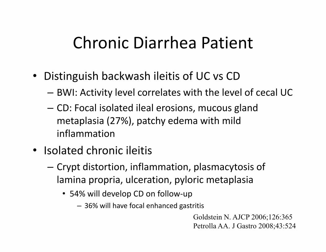
Chronic Diarrhea Patient
• Distinguish backwash ileitis of UC vs CD
– BWI: Activity level correlates with the level of cecal UC
– CD: Focal isolated ileal erosions, mucous gland
metaplasia (27%), patchy edema with mild metaplasia (27%), patchy edema with mild
inflammation
• Isolated chronic ileitis
– Crypt distortion, inflammation, plasmacytosis of
lamina propria, ulceration, pyloric metaplasia
• 54% will develop CD on follow-up
– 36% will have focal enhanced gastritis
Goldstein N. AJCP 2006;126:365
Petrolla AA. J Gastro 2008;43:524

Ileal Lesions
• Symptomatology and indication for endoscopy
predict the likelihood that ileitis will progress
to Crohn disease
– Asymptomatic ileitis (11 of 14 with features of – Asymptomatic ileitis (11 of 14 with features of
chronicity)
• None progressed to CD
– Symptomatic ileitis
• 8/10 with features of chronicity progressed
• 2/5 with focal active ileitis progressed
Courville EL et al. AJSP 2009;33:1341


Case #3 – Dysplasia in IBD
• 42 year female with history of Crohn Disease
• Originally diagnosed in 2009
• Treatment with prednisone, azathioprine, and
adalimumabadalimumab
– All discontinued due to side effects
• Currently on no medications
– 10 bowel movements per day
• Repeat colonoscopy performed

Endoscopic Findings
• The ICV was narrowed: could not intubate TI
• Proximal ascending colon, 5 cm distal to ICV large 4 cm sessile polyp surrounded in a field of inflamed mucosa concerning for a DALM lesion.
– Multiple biopsies taken of the polyp and placed in a separate jar.
– Biopsies were also taken of the adjacent mucosa and placed in – Biopsies were also taken of the adjacent mucosa and placed in a separate jar.
• Flat nodular area in the ascending colon a few centimeters distal to the polyp
– Biopsied and placed in the random right colon jar.
• Flat polyp 2 cm in size in the transverse colon.
• Patchy disease throughout colon; normal rectum
• Surveillance biopsies were taken in each of 4 quadrants every 10 cm throughout the colon.


5 cm Proximal Ascending Colon
Lesion

Biopsy Adjacent to 5 cm Lesion

Random Right Biopsies (Includes Flat
Nodular Area)
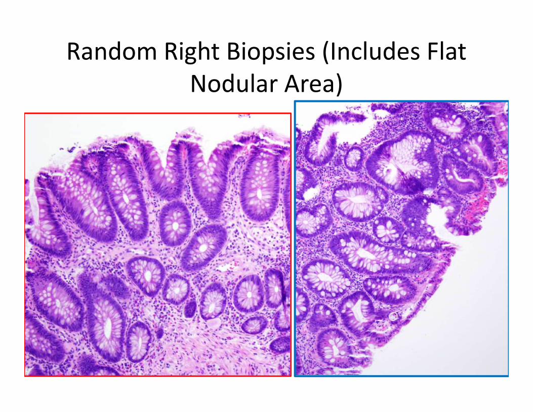
Random Right Biopsies (Includes Flat
Nodular Area)

Flat Polyp Transverse Colon

Random Left Colon

Random Left Colon
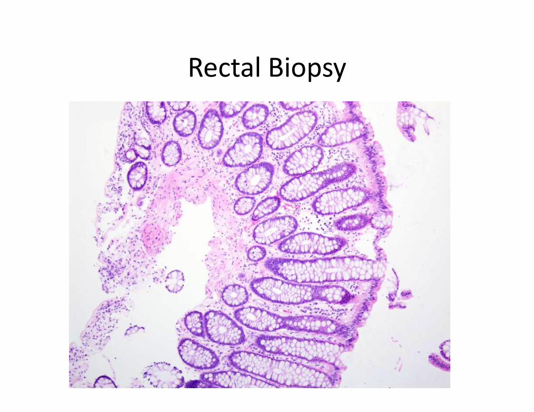
Rectal Biopsy

Colorectal Neoplasia in IBD
• UC and CD have an increased risk of
developing colorectal carcinoma
– Ulcerative colitis (UC)
• Risk 2% at 10 years, 8% at 20 years, 18% at 30 years• Risk 2% at 10 years, 8% at 20 years, 18% at 30 years
– Crohn disease (CD)
• Standardized incidence ratio for CRC 2.5 (95% CI, 1.7-
3.5)
• Relative risk 4.5 (95% CI, 1.3-14.9)

Risk Factors
• Disease duration
• Extent of disease
• Association of PSC
• Family history of sporadic CRC• Family history of sporadic CRC
• Colonic strictures (in UC), multiple
pseudopolyps
• Increased degree of macroscopic and
histologic inflammation

Screening/ Surveillance
• UC
– Screening at 8 to 10 years disease duration
– Surveillance
• 1-2 years in extensive colitis (proximal to splenic flexure) and left-sided (descending colon)left-sided (descending colon)
• Standard (non-uc) CRC surveillance in proctosigmoiditis (limited to rectum and sigmoid)
– No increase risk of colorectal cancer
• IBD + PSC
– Screening and surveillance (yearly) at time of PSC diagnosis

Biopsies
• Polyps (mass lesions)
– Resection of polyp if possible
– Biopsy of the mucosa next to the polyp in separate
jarjar
• Random biopsies
– No specific # recommended in guidelines,
“sufficient”
• Studies show minimum of 33 with extensive disease
• 4-quadrant biopsies every 10 cm

Gross Features of DALMs
• Adenoma-like
(endoscopically
resectable)
– Sessile/pedunculated
• Non-adenoma-like (not
endoscopically resectable)
– Usually sessile (broad based)
– Well circumscribed
– Smooth surface
– Visible borders
– Nonulcerated
– No stricture
– No mucosal tethering
– Poorly circumscribed
– Irregular surface
– Indistinct border
– Ulceration/necrosis
– Stricture
– Tethering
Farraye FA et al. Gastroenterology 2010;138:746–774

Adenoma-like
DALM
Non-adenoma-
like DALM
Farraye FA et al.
Gastroenterology
2010;138:746–774.

Farraye FA et al. Gastroenterology 2010;138:746–774.

ACG Practice Guidelines
• “From a practical perspective, therefore, it
matters little whether a mass lesion is called
an adenoma-like mass, or dysplasia-associated
lesion or mass; the important issue is to lesion or mass; the important issue is to
determine whether the lesion is completely
resectable and the rest of the colon is free of
dysplasia.”

DMSG CLASSIFICATION
• Negative for dysplasia (Neg)
• Indefinite for dysplasia (Indef)
• Low-grade dysplasia (LGD)
• High-grade dysplasia (HGD)
• Carcinoma (CA)

CLOSE
8
CLOSE
Farraye FA et al. Gastroenterology 2010;138:746–774.

Follow-up
• At resection:
– Partial colectomy (Crohn disease)
• Multiple polyps with low grade dysplasia and
multifocal flat low grade dysplasiamultifocal flat low grade dysplasia
– No invasive carcinoma

Resection Specimen: Villous Lesion

Flat Dysplasia: Resection Specimen

Flat Dysplasia: Resection Specimen

Summary
Clinico-pathologic Information
#1 -Differentiating Ulcerative Colitis from
Crohn’s Colitis
#2 – Ileal Lesions – Crohn’s Disease?
#3 Dysplasia in Inflammatory Bowel Disease