clinicalstudy - hindawi publishing...
TRANSCRIPT
Clinical StudyHistologic and Histomorphometric Comparison betweenSintered Nanohydroxyapatite and Anorganic Bovine Xenograftin Maxillary Sinus Grafting: A Split-Mouth RandomizedControlled Clinical Trial
Claudio Stacchi,1 Teresa Lombardi,2 Francesco Oreglia,3
Andrea Alberghini Maltoni,4 and Tonino Traini5
1Department of Medical, Surgical and Health Sciences, University of Trieste, Trieste, Italy2Private Practice, Cassano allo Ionio, Italy3Private Practice, Verona, Italy4Private Practice, Firenze, Italy5Department of Medical, Oral and Biotechnological Sciences, University of Chieti-Pescara, Chieti, Italy
Correspondence should be addressed to Claudio Stacchi; [email protected]
Received 13 January 2017; Accepted 25 May 2017; Published 6 August 2017
Academic Editor: Hassan Maghaireh
Copyright © 2017 Claudio Stacchi et al.This is an open access article distributed under the Creative Commons Attribution License,which permits unrestricted use, distribution, and reproduction in any medium, provided the original work is properly cited.
The presence of vital bone after maxillary sinus augmentation is crucial to enhance the quality of bone-implant interface, ensuringpredictable long-term results.The aims of this RCTwith split-mouth designwere the histologic andhistomorphometric comparisonof two different biomaterials in sinus elevation after 6 months of healing and the evaluation of the clinical outcomes of implantsinserted in the augmented areas after 12 months of prosthetic loading. Twenty-eight patients (10 females, 18 males) were treatedwith bilateral sinus floor elevation with lateral approach. Pure sintered nanohydroxyapatite (NHA) and anorganic bovine bone(ABB) were used as test and active control, respectively. After six months, 52 bone biopsies were harvested from 26 patients, and107 implants were inserted in the augmented areas. Histomorphometry showed that, in the two groups, vital bone percentages were34.9 ± 15% (NHA) and 38.5 ± 17% (ABB) (𝑝 = 0.428), marrow spaces percentages were 44.5 ± 18% (NHA) and 43.5 ± 23% (ABB)(𝑝 = 0.866), and residual graft percentages were 20.6 ± 13% (NHA) and 22.3 ± 12% (ABB) (𝑝 = 0.638). After 6 months of healing,no statistically significant difference was present in histomorphometric outcomes between NHA and ABB groups. Implant survivalrate in NHA group after 12 months of loading was 96.4%, showing no statistically significant differences with ABB group.
1. Introduction
Bone resorption and sinus pneumatization are commonoccurrences in the posterior maxilla after tooth extraction:they may cause both a quantitative reduction and qualitativedeterioration of bone, resulting in an inadequate bone vol-ume for dental implant placement [1]. Sinus floor elevationtechnique had been described more than 35 years ago[2] and extensively studied afterwards, demonstrating highpredictability in regenerating bone and allowing for reliableimplant supported rehabilitation [3, 4]. Accurate presurgi-cal planning is a mandatory step: sinus three-dimensional
conformation, eventual presence of Underwood septa, andprecise localization of the alveolar-antral artery should beassessed and carefully evaluated in order to minimize intra-operative complications and optimize surgical techniques [5–7].
In general, the quality of osseointegration of dentalimplants is directly related to the bone-implant contact: ina regenerated tissue, the quantity of newly formed bone isof paramount importance for successful integration of thefixture. Therefore, an adequate biomaterial choice is the firstcrucial step in bone-implant interface engineering to ensurepositive clinical long-term results.
HindawiBioMed Research InternationalVolume 2017, Article ID 9489825, 10 pageshttps://doi.org/10.1155/2017/9489825
2 BioMed Research International
Autogenous bone had been the first grafting material tobe used in sinus floor elevation, being considered as the goldstandard option for a long time [8], but its tendency of resorp-tion, its limited availability, and the necessity of a donorsite with associated increased morbidity for the patientshould be considered as major drawbacks. In the attempt toovercome these limitations, many biomaterials had been pro-posed and tested, such as allografts, 𝛽-tricalcium phosphate,calcium sulphate, and bone mineral matrix [9–15]. However,anorganic bovine bone (ABB) is probably the most widelyinvestigated bone substitute and, when used in sinus floorelevation, demonstrated satisfactory osteoconductive prop-erties and dimensional stability: from a clinical point ofview, implants inserted in ABB-grafted areas showed a highsurvival rate over time [16, 17].
Also, synthetic hydroxyapatites, when used as a bonegraft, showed a high degree of biocompatibility and supportfor cellular activity: they stimulate osteoconduction and aregenerally slowly replaced by the host bone after implantation[18]. Continuous improvements in synthesis, manufacturingtechnology, and purification have led to a variety of syntheticHA-based materials with different properties. Among them,synthetic hydroxyapatite with nanoscale porosities seemsto favor bone matrix proteins adhesion and to promotedifferentiation of osteoblast precursor cells [19, 20]. Evenif nanocrystalline HA embedded in a highly porous silicagel matrix was already tested as a material for sinus floorelevation [21, 22], a prospective study with a direct com-parison between the behavior in the maxillary sinus of puresintered nanohydroxyapatite (NHA) and ABB in terms ofosteoconductive potential was not performed yet.
The aim of this parallel-group, superiority randomizedclinical trial (RCT)with split-mouth designwas the histologicand histomorphometric comparison for the newly formedtissue after sinus floor elevation with lateral approach per-formed by using two different grafting materials: NHA as testand ABB as active control.
An additional aim was the evaluation of the clinicaloutcomes of dental implants inserted in the augmented areasafter 12 months of prosthetic loading. The planned follow-upfor this study is five years after prosthetic loading.
2. Materials and Methods
2.1. Study Design. The present study was a multicenter ran-domized controlled clinical trial with a split-mouth design,following CONSORT guidelines, and was conducted in fourclinical centers in accordance with theGoodClinical PracticeGuidelines (GCPs) and with the recommendations of theDeclaration of Helsinki as revised in Fortaleza (2013) forinvestigations with human subjects. The study protocol hadbeen approved by the relevant ethical committee (ComitatoEtico Calabria-Sezione Area Nord) and registered in a publicregister (NCT03077867).
Patients were thoroughly informed about the protocol,the treatment and its alternatives, the benefits, and thepossible risks and signed written informed consent for theparticipation in the study. This superiority trial tested thenull hypothesis of no differences in new bone formation
and dental implant survival between NHA bone grafts (testgroup) and ABB grafts (active control group) in atrophicmaxillae treated with sinus floor elevation with lateralapproach.
2.2. Study Population. Eligible participants were adult pa-tients (aged ≥ 18 years), with severe bilateral maxillaryatrophy (crestal height < 3mm, class V-VI of Cawood andHowell classification [23]) and needing sinus floor elevationto allow for fixed rehabilitation supported by osseointegratedimplants, inserted with a staged approach.
Exclusion criteria were
(1) acute myocardial infarction within the past 2 months;(2) uncontrolled coagulation disorders;(3) uncontrolled diabetes (HbA1c > 7.5%);(4) radiotherapy to the head/neck district within the past
24 months;(5) immunocompromised patients (HIV infection or
chemotherapy within the past 5 years);(6) present or past treatment with intravenous bisphos-
phonates;(7) allergy to bovine collagen;(8) presence of uncontrolled or untreated periodontal
disease;(9) presence of sinusal pathologies contraindicating sinus
floor elevation procedures;(10) psychological or psychiatric problems;(11) alcohol or drugs abuse;(12) patient not fully able to comply with the study
protocol;(13) Schneiderian membrane perforation during surgery.
2.3. Surgical Procedures. Surgical procedureswere performedin four centers by experienced operators (CS, TL, FO, andAAM). Patients were draped to guarantee maximum asepsisand perioral skin was disinfected by using iodopovidone 10%(Betadine, Medifarm, Italy). After performing local anesthe-sia by using articaine 4% with epinephrine 1 : 100.000 (Artin,Omnia, Italy) and raising a full-thickness flap, a window wasdesigned on the lateral wall of the sinus by using ultrasonicinstrumentation with the erosion technique (PiezosurgeryTouch, Mectron, Italy, and Piezotome, Acteon, France) [24]and the Schneiderian membrane was carefully elevatedusing ultrasonic inserts and manual curettes. After checkingthe integrity of the Schneiderian membrane with Valsalvamaneuver, the randomization sealed opaque envelope wasopened, revealing to the surgeon the grafting material to beused. The biomaterials selected for this study were sinteredNHA (Fisiograft Bone Granular, Ghimas, Italy) in the testsites and ABB (Bio-Oss, Geistlich, Switzerland) in the controlsites. After the completion of the grafting procedure, theantrostomy was covered by a resorbable bovine collagenmembrane (Bio-Gide, Geistlich, Switzerland), fixed with twopins (Micropin, Omnia, Italy), and flaps were sutured with

BioMed Research International 3
Sentineri technique [25] and single stitches using a syntheticmonofilament (PTFE, Omnia, Italy).
The contralateral sinus floor augmentation was per-formed in the same surgical session, with the same surgicalprotocol, inserting the grafting material not selected in thefirst intervention.
Patients were prescribed antibiotics for 6 days (amoxi-cillin 1 g twice a day or, in allergic patients, clarithromycin250mg three times a day) and NSAID (ibuprofen 600mg),when needed.
All patients were also advised to sneeze with the mouthopen and to avoid nose blowing for two weeks, to preventunnecessary pressure on the sinus membrane.
Sutures were removed 10 days after surgery. Postsurgicalvisits were scheduled at monthly intervals to check the courseof healing. After six months, bone-core biopsies were col-lected from the grafted areas using a trephine bur (3.5mmdiameter) during the implant bed preparation, and thendental implants (BnxEvo, Ghimas, Italy) were inserted in theharvesting sites. Bone-core specimenswere collectedwith theassistance of surgical templates based upon individual pros-thetic requirements. The surgical guides were also used toinsert the other programmed implants into the augmentedareas: they were left submerged for a four-month healingperiod, prior to being connected to healing abutments.Finally, implants were restored with screwed metal-ceramicprostheses and patients were followed up for twelve monthsafter loading.
2.4. Histological Analysis. Bone biopsies, left inside thetrephine burs, were carefully rinsed for 30–40 seconds witha cold 5% glucose solution to remove blood maintaining thecorrect osmolarity (278mOsm/L).
The specimens were then placed in Eppendorf tubes withan adequate volume (at least ten times the volume of thespecimen) of 10% formalin solution buffered with phosphateto pH 7.2.
Each specimen was stored in a separate container andlabeled. Both patient name and operator ID were notedon a separate sheet to identify the specimens. During theprocessing phase, both patient name and operator ID weredesignated by a numerical code.
The specimens were rinsed twice with phosphate-buffered saline and dehydratedwith a graded series of alcoholat 4∘C for seven days. Complete dehydration was thenobtained with absolute alcohol immersion for two additionaldays. Subsequently, the specimens were preinfiltrated in a50% resin/alcohol solution (Technovit 7200 VLC, Kulzer,Germany) for ten days and completely embedded in 100%resin (two changes) using a vacuum chamber for twenty addi-tional days or until the specimens have become transparent.
Finally, specimens were easily removed from the trephinebur using a custom-made plunger (thanks to the shrinkageconsequent to dehydration and resin infiltration) and thenoriented and polymerized.
After polymerization, the specimens were cut along thelongitudinal axis using a high-precision diamond disc atabout 50 microns (TT System, TMA2, Italy). The sectionswere ground under running water to about 30 ± 10 microns
using a series of polishing discs from 400 to 1200 grits,followed by a final polishwith 0.3-micron alumina in amicro-grinding system (TT System, TMA2, Italy).
The prepared sections were stained with Toluidine Blueand Azure II and counterstained with acid fuchsin or double-stained with Toluidine Blue with Pironine G at 1% andAzure II. The investigation was conducted in a transmittedbrightfield microscope (BX 51, Olympus America, USA)and under brightfield/circularly polarized light microscope(Axiolab, Zeiss, Germany) both connected to high-resolutiondigital cameras (FinePix S2 Pro, Fuji Photo Film, Japan).
Digital photomicrographs were used for histomorpho-metric analysis, which was performed by a trained andexperienced operator (TT). The following parameters weremeasured: (1) amount of tissue collected with the biopsiesover the obtained sections (size of samples); (2) amount ofvital bone as absolute value (mm2) and as relative value (vitalbone area/total sample size × 100); (3) marrow space (con-nective tissue) as absolute value (mm2) and as relative value(connective tissue area/total sample size × 100); (4) residualgrafting material as absolute value (mm2) and as relativevalue (biomaterial area/total sample size× 100). A histometricsoftware package with image capturing capabilities (Image-Pro Plus 6.0, Media Cybernetics Inc., USA) was used. Toensure accuracy, the software was calibrated for each exper-imental image using a feature named “Calibration Wizard,”which creates a linear remapping of the pixel numbers inmil-limeters. Intraexaminer variability was controlled by carryingout two measurements for each controlled index. When thedifference between the two performed readings exceeded 5%for the same index, the measurement was repeated.
2.5. Outcomes. This study evaluated the following outcomemeasures:
(i) Quality of the newly formed tissue: (1) new boneformation (percentage of newly formed bone areato total measured area), (2) residual graft particles(percentage of graft particles area to total measuredarea), and (3) marrow spaces (percentage of soft-tissue area to total measured area).
(ii) Implant failure: implantmobility and/or any situationsuggesting implant removal.
(iii) Biological and mechanical complications: any com-plication defined as an unexpected deviation from thenormal treatment outcome, both biological (mem-brane perforation, hemorrhagic events, sinusitis,peri-implantitis, etc.) and mechanical (implant frac-ture, prosthesis fracture, fixation screw loosening,etc.)
2.6. Sample Size and Statistical Power. Maxillary sinuses weredivided into two groups, depending on the grafting materialused: a sample size of 24 sinuses per group was neededto detect an effect size of 0.6 on the quantity of newlyformed bone (primary outcome), referred to as indicativeof a medium effect [26], between the groups (alpha levelset at 0.05 and power of 80%) (DSS Research, Fort Worth,USA). The effect size is defined as the difference in the given

4 BioMed Research International
Table 1: Patient characteristics at baseline.
Males 18 (64.3%)Females 10 (35.7%)Mean age (range) 60.1 (39–79)Nonsmoker 18 (64.3%)Light smoker (<10) 6 (21.4%)Heavy smoker (≥10) 4 (14.3%)Mean residual bone (SD) (range) (mm) 2.0 (0.7) (0.5–3)
outcome between the groups divided by the within-subjectsstandard deviations. Each clinical center treated 7 patientswith bilateral sinus floor elevation for a total of 56 augmentedsinuses to compensate eventual dropouts occurring duringthe study.
2.7. Randomization. A computer-generated table, distribut-ing right and left sinuses of each patient into two groups (testand control), was prepared using a balanced, randomly per-muted block approach (http://www.randomization.com/).The randomization codes were enclosed in numbered, sealed,opaque envelopes which were opened after Schneiderianmembrane elevation. Therefore, treatment allocation wasconcealed to the surgeons in charge of recruiting and treatingthe patients included in this clinical trial.
2.8. Statistical Analysis. Statistical analysis was performedby means of a computerized statistical package (SigmaStat3.5, SPSS Inc., Germany). Data were expressed as mean± SD and median (interquartile range), respectively, forparametric and nonparametric values. Items were analyzedwith descriptive statistics to assess whether they had anormal distribution; both equal variance and normality testswere used. Considering the two-arm superiority RCT studydesign, the hypothesis was tested using unpaired 𝑡-test in caseof normally distributed data, while Mann–Whitney 𝑈 testwas performed to compare nonparametric values. A 𝑝 value< 0.05 was considered statistically significant.
3. Results
3.1. Clinical Results. Twenty-eight patients (aged 60.1 ± 10.7years, range: 39–79 years, 10 females, 18 males) were enrolled,randomized, and treated with bilateral sinus floor elevationwith lateral approach. Each clinical center contributed with7 patients. Eighteen patients were nonsmokers, six lightsmokers, and four heavy smokers. Preoperative residualbone crest height ranged from 0.5 to 3mm (mean height:2.03±0.75mm).Themain baseline patient characteristics aresummarized in Table 1.
Three sinus membrane perforations occurred duringelevation in two patients, who dropped out from the study.However, membrane perforations were covered by multiplelayers of A-PRF membranes (PRF Duo, Mectron, Italy) andgrafting procedures were successfully completed in all ofthe three cases. No other intraoperative complications wererecorded. The healing period following sinus augmentationwas uneventful in all patients.
NHA NHA NHA
ABB ABB ABB
2.0mm
Figure 1: Sections of bone cores retrieved after 6 months of healing.NHA: sintered nanohydroxyapatite; ABB: anorganic bovine bone(Toluidine Blue and Azure II; original magnification: 12x).
Sixmonths after sinus floor elevation procedures, 52 bonebiopsies were harvested from 26 patients (26 biopsies intest sites, 26 in control sites), and a total of 107 implantswere inserted and submerged under the soft tissues (55implants in test sites, 52 in control sites). After four additionalmonths, at healing abutments connection, three implantsin three patients resulted to be not osseointegrated (2.8%cumulative failure rate): two implants were inserted in testsites (3.6% failure) and one implant was inserted in a controlsite (1.9% failure). Difference in implant failure rate betweentest and control groups was not statistically significant (𝑝 =0.32, unpaired 𝑡-test). After an accurate debridement of theimplant bed, removed fixtures were immediately replacedwith larger diameter implants, which were restored after fouradditional months of healing. Metal-ceramic screwed pros-theses were delivered and, at 12-month follow-up, all implantsand prostheses were successfully in function without theoccurrence of any biological or mechanical complication.
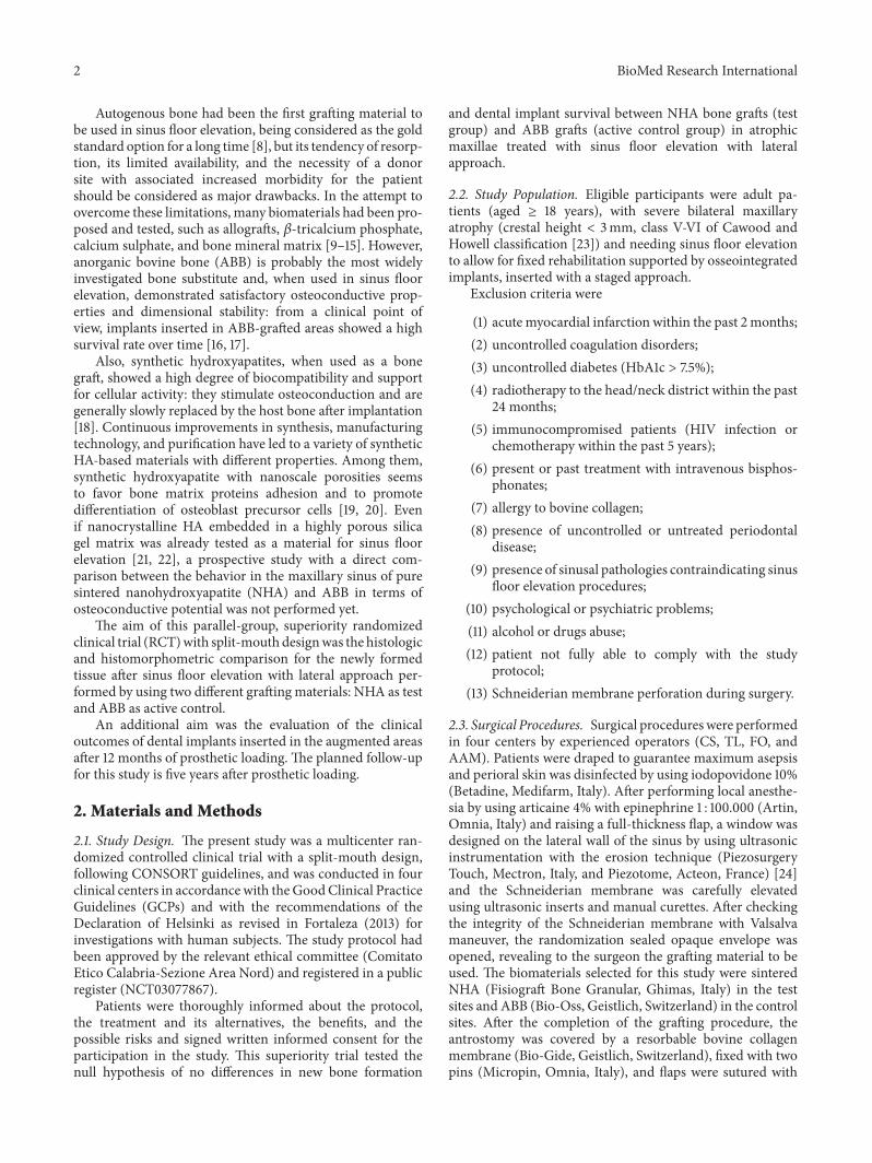
3.2. Histomorphometric Results. Thesections of the harvestedbiopsies had amean surface of 9.05± 2.7mm2 forNHAgroupand 10.31 ± 2.9mm2 for ABB group. The difference betweenthe two groups was not statistically significant (𝑝 = 0.116,unpaired 𝑡-test). Area of vital bone was 3.29 ± 2.1mm2 forNHAgroup and 4.12± 2.9mm2 forABBgroup.Thedifferencebetween the two groups was not statistically significant (𝑝 =0.213, Mann–Whitney 𝑈 test). Connective tissue area was3.82 ± 1.5mm2 for NHA group and 4.09 ± 2.3mm2 forABB group. The difference between the two groups was notstatistically significant (𝑝 = 0.869, Mann–Whitney 𝑈 test).The area occupied by residual grafting material was 1.92 ±1.4mm2 for NHA group and 2.09 ± 1.4mm2 for ABB group.The difference between the two groups was not statisticallysignificant (𝑝 = 0.516, Mann–Whitney 𝑈 test). The resultsare summarized in Table 2 and Figures 1 and 2.
The average percentage of vital bone was 34.9 ± 15% forNHA group and 38.5 ± 17% for ABB group. The differencebetween the two groups was not statistically significant (𝑝 =0.428, unpaired 𝑡-test). The average percentage of connectivetissue was 44.5 ± 18% for NHA group and 43.5 ± 23% forABB group. The difference between the two groups was notstatistically significant (𝑝 = 0.866, unpaired 𝑡-test). Theaverage percentage of residual grafting material was 20.6 ±13% forNHAgroup and 22.3±12% forABB group.Thediffer-ence between the two groups was not statistically significant
BioMed Research International 5
Table 2: Total area of the analyzed sections [mm2].
Samples Missing Mean Std. dev. Std. error CI of meanTotal area ABB 26 0 10,319 2,923 0,573 1,180Total area NHA 26 0 9,052 2,796 0,548 1,129Vital bone ABB 26 0 4,129 2,992 0,587 1,208Vital bone NHA 26 0 3,297 2,199 0,431 0,888Connective ABB 26 0 4,090 2,355 0,462 0,951Connective NHA 26 0 3,828 1,510 0,296 0,610Biomaterial ABB 26 0 2,099 1,421 0,279 0,574Biomaterial NHA 26 0 1,926 1,486 0,291 0,600
Range Max. Min. Median 25% 75%Total area ABB 12,190 18,340 6,150 9,920 8,720 11,120Total area NHA 10,600 14,480 3,880 8,550 7,210 11,000Vital bone ABB 15,820 16,950 1,130 3,705 2,530 5,040Vital bone NHA 9,700 9,960 0,260 2,750 1,840 4,630Connective ABB 7,920 8,470 0,550 3,815 1,920 6,230Connective NHA 6,320 6,920 0,600 3,900 2,960 4,940Biomaterial ABB 5,480 5,540 0,0600 1,795 1,200 3,110Biomaterial NHA 5,380 5,530 0,150 1,530 0,810 2,770
Skewness Kurtosis K-S dist. K-S prob. Sum Sum of squaresTotal area ABB 0,916 0,972 0,161 0,080 268,290 2981,976Total area NHA 0,260 −0,524 0,111 0,515 235,340 2325,652Vital bone ABB 3,279 13,938 0,221 0,002 107,360 667,054Vital bone NHA 1,232 2,081 0,123 0,380 85,730 403,549Connective ABB 0,139 −1,039 0,107 0,573 106,350 573,633Connective NHA −0,240 −0,184 0,137 0,228 99,530 437,977Biomaterial ABB 0,596 −0,208 0,138 0,222 54,570 165,038Biomaterial NHA 1,117 0,622 0,168 0,057 50,080 151,657
p = 0.116;
p = 0.213<
p = 0.869<
p = 0.516<
Vitalbone
Connectivetissue
Biomaterials
0
5
10
15
20
Are
a ext
ensio
n (m
m2)
ABB ABB ABB ABBNHA NHA NHANHASize of
samples
Figure 2: Samples area (mm2) and surface of vital bone, connectivetissue, and biomaterial remnants (mm2). NHA: sintered nanohy-droxyapatite; ABB: anorganic bovine bone. (a) Unpaired 𝑡-test; (b):Mann–Whitney 𝑈 test. Level of significance: 𝑝 < 0.05.
(𝑝 = 0.638, unpaired 𝑡-test). Results are summarized inTable 3 and Figure 3.
p = 0.428;
p = 0.866;
p = 0.638;
Vitalbone
Connectivetissue
BiomaterialsABB ABB ABBNHA NHANHA
0,0
0,2
0,4
0,6
0,8
1,0
Are
a rat
e×10
0(m
m2)
Figure 3: Histomorphometric measurements expressed in percent-age. NHA: sintered nanohydroxyapatite; ABB: anorganic bovinebone. (a) Unpaired 𝑡-test. Level of significance: 𝑝 < 0.05.
4. Discussion
In the clinical practice, the final purpose of bone regenerationis the formation of an adequate quantity of tissue of good

6 BioMed Research International
Table 3: Histomorphometric data expressed in percentage [%].
Samples Missing Mean Std. dev. Std. error CI of meanVital bone rate ABB 26 0 0,385 0,170 0,0333 0,0685Vital bone rate NHA 26 0 0,349 0,155 0,0303 0,0625Connective rate ABB 26 0 0,435 0,232 0,0456 0,0939Connective rate NHA 26 0 0,445 0,181 0,0354 0,0729Biomaterial rate ABB 26 0 0,223 0,128 0,0252 0,0518Biomaterial rate NHA 26 0 0,206 0,135 0,0265 0,0546
Range Max. Min. Median 25% 75%Vital bone rate ABB 0,810 0,924 0,114 0,379 0,263 0,469Vital bone rate NHA 0,661 0,688 0,0270 0,339 0,279 0,465Connective rate ABB 0,942 0,990 0,0480 0,462 0,293 0,560Connective rate NHA 0,709 0,786 0,0770 0,453 0,308 0,562Biomaterial rate ABB 0,442 0,448 0,00600 0,228 0,138 0,310Biomaterial rate NHA 0,524 0,550 0,0260 0,161 0,130 0,309
Skewness Kurtosis K-S dist. K-S prob. Sum Sum of squaresVital bone rate ABB 1,256 2,846 0,131 0,288 10,015 4,576Vital bone rate NHA −0,143 −0,0594 0,126 0,345 9,079 3,768Connective rate ABB 0,381 0,0977 0,0961 0,698 11,316 6,276Connective rate NHA −0,132 −0,392 0,102 0,625 11,570 5,964Biomaterial rate ABB 0,0123 −0,727 0,0758 0,858 5,800 1,706Biomaterial rate NHA 0,840 0,275 0,152 0,124 5,350 1,558
quality, in which to insert dental implants with a good long-term prognosis. Obviously, the biological behavior of the bio-materials is of primary interest. Kirkpatrick et al. underlinedthe differences between the regenerative processes that havethe teleological purpose of “restitutio ad integrum” of theaffected tissue and the repair process, which is a structuraladaptation to a function task [27]. It was reported that thebone regeneration process with alloplastic, xenograft, andallograft bone substitutes follows threemain phases: T1 whichis the “time of grafting,” with a predominant heterogeneousphase in suspension of blood clot and particles of biomateri-als; T2 which is the “time of repairing,” with a solid hetero-geneous composite phase of biomaterial remnants and newlyformed bone; T3 which is the “time of regeneration,” witha solid homogeneous phase of newly formed bone withoutbiomaterials remnants. The most common bone substitutebiomaterials do not reach the phase T3 in their clinicaluse [28]. However, many studies demonstrated that implantosseointegration process can be also obtained andmaintainedin augmented sinuses where residual graft particles were stillpresent, without a negative influence of biomaterial remnantson peri-implant bone regeneration [29–33].
Hence, even if autologous bone had been traditionallyconsidered as the gold standard to promote new boneregeneration, the choice of alternative biomaterials is nowthe preferred option in sinus floor elevation for three mainreasons: less morbidity, less resorption, and unlimited avail-ability. ABB is themost widespread biomaterial used for sinusgrafting and its behavior had been extensively investigatedover the years, showing satisfactory long-term results [16, 17].
However, disadvantages of xenografts should also be consid-ered: they include potential risk of prion disease transmission[34] and reaction of the host immune system [35]; in addition,some patients could refuse their use for religious motivationsor because they are in contrast with their lifestyle (e.g., vegansand vegetarians). In a recent study, allografts and xenograftselicited the highest refusal rates among the surveyed patients:15% of the patients said they would accept a xenograft underno circumstances, while 18% said they would accept this typeof bone graft only as a last resort [36].
The use of synthetic, alloplastic biomaterials could over-come these limitations: they have been studied for years andsuccessfully used in sinus augmentation, but a direct com-parison with xenografts, in a split-mouth design, has beenreported in the literature by very few and often underpoweredtrials [15, 37–39].
The results of the present RCT showed no statisticallysignificant differences between NHA and ABB groups interms of new bone formation and survival rate of implantsinserted in the augmented areas after 12 months of prostheticloading: therefore, the null hypothesis tested in this study wasaccepted.
The histometric comparison after 6 months of heal-ing showed that the osteoconductive potential of NHA isclinically and statistically comparable to ABB, even if itresulted in a slightly lower percentage of vital bone (34.9%against 38.5%), but showing also a lower percentage ofresidual grafting material (20.6% against 22.3%). Our dataare in accordance with other human studies performed byusing synthetic hydroxyapatites as sinus grafting material,

BioMed Research International 7
Figure 4: Biomaterial particles of both groups appeared to be surrounded and merged by newly formed bone. Several haversian systems (∗)were noted in the newly formed bone. NHA: sintered nanohydroxyapatite; ABB: anorganic bovine bone; VB: vital bone; MS: marrow spaces(Toluidine Blue and Azure II; original magnification: 100x).
Figure 5: The bone around both biomaterials was characterized by the presence of osteocytes (∗) embedded inside the mineralized bonematrix. In the test group, osteocytes were generally more numerous near the material surface. NHA: sintered nanohydroxyapatite; ABB:anorganic bovine bone; VB: vital bone; MS: marrow spaces (Toluidine Blue and Azure II; original magnification: 400x).
reporting new bone formation at six months ranging from32 to 38.5% [40–42]. These results are also comparable withthe histomorphometric outcomes of sinus augmentation per-formedbyusing solely autogenous bone (newbone formationat six months ranging from 36.8 to 41%) [41, 43–45].
After 6 months of healing, both biomaterials used inthe present study showed a good level of “osseointegration,”with an adequate extension of bonding surface betweenhost bone and the biomaterial particles. Graft remnantswere easily recognizable from the other components of theregenerated tissue and appeared to be merged by bridgesof new bone (Figure 4). Furthermore, the bone around thebiomaterial particles was characterized by numerous osteo-cytes embedded into the mineralized bone matrix. Thesecells, in the test group, were generally more numerous near
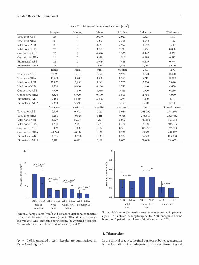
the biomaterial surface (Figure 5): this feature indicates botha considerable osteointegrative property due to stimulationof the osteoblastic activity and an osteoinductive propertyof the external surface of the biomaterial (Figure 6). Theseaspects, according to other studies [46–48], seem particularlyrelated to NHA structure. As described in detail by Kasai etal. [49], cells’ proliferation appeared to be stimulated, whenin contact with NHA paste, by the activation of epidermalgrowth factor receptor (EGFR) and its downstream targetsserine/threonine protein kinase (AKT) and signal regulatedkinases (ERK 1/2). Finally, as expected, both groups showedintense osteoconductive activities (Figure 7).
Implant survival rate in NHA group after 12 months ofloading (96.4%) showedno statistically significant differenceswith ABB group. This outcome is also comparable with
8 BioMed Research International
Figure 6: The new bone around some NHA particles presented osteons (∗), indicating a relative angiogenetic potential of the material.Vessels’ growth was present in pores of adequate dimension inside the biomaterial (black arrow). NHA: sintered nanohydroxyapatite; ABB:anorganic bovine bone; VB: vital bone; MS: marrow spaces (Toluidine Blue and Azure II; original magnification: 100x).
Figure 7: Under circularly polarized light, osteoconduction appeared clear with intimate contact between the new bone (VB) and bothbiomaterials (NHA and ABB). Moreover, the differences in the microstructure (mainly collagen fiber orientation) can be observed. Aroundsome NHA particles, the new bone presented osteons with vessels (HC).
results reported in recent systematic reviews for sinus graftingusing solely autogenous bone (97.2–97.4%) or different bonesubstitutes (98.2–98.6%) [50, 51].
Currently, the main limitation of the present study isthe relatively short time of follow-up: however, a long-termevaluation of the clinical outcomes in the patients enrolledin this trial had already been coordinated. Furthermore,additional investigations on the biomechanical performancesof different bone substitutes would help in determining theirappropriate clinical use.
5. Conclusions
The findings of the present RCT showed that both NHA andABB led to the formation of a regenerated tissue composedof more than 1/3 of vital bone after six months of healing,
without any statistically significant difference between testand control groups. NHA could be regarded as a suitablegrafting material for clinical cases needing bone augmenta-tion to allow dental implant placement.
The clinical implications of the present study include thepossibility of increasing the alternatives for the replacementof bone autografts, which not always represent a possibleor convenient option. Sintered NHA couples the benefitsof technological advancement with the safety of syntheticbiomaterials, preventing the potential risks of xenograftimplantation to the patient.
Conflicts of Interest
The authors declare no conflicts of interest related to thisstudy.

BioMed Research International 9
Acknowledgments
Theauthorswish to thankDr. Rosario Sentineri andDr. PaoloCusimano for their valuable help in conducting this study.
References
[1] A. Monje, F. Monje, R. Gonzalez-Garcıa et al., “Influenceof atrophic posterior maxilla ridge height on bone densityand microarchitecture,” Clinical Implant Dentistry and RelatedResearch, vol. 17, no. 1, pp. 111–119, 2015.
[2] P. J. Boyne andR.A. James, “Grafting of themaxillary sinus floorwith autogenousmarrow and bone,” Journal of Oral Surgery, vol.38, no. 8, pp. 613–616, 1980.
[3] M. Del Fabbro, T. Testori, L. Francetti, and R. Weinstein, “Sys-tematic review of survival rates for implants placed in thegrafted maxillary sinus,” International Journal of Periodonticsand Restorative Dentistry, vol. 24, no. 6, pp. 565–577, 2004.
[4] M. Esposito, M. G. Grusovin, J. Rees et al., “Effectivenessof sinus lift procedures for dental implant rehabilitation: aCochrane systematic review,” European Journal of Oral Implan-tology, vol. 3, no. 1, pp. 7–26, 2010.
[5] C. Stacchi, F. Andolsek, F. Berton, G. Perinetti, C. O. Navarra,and R. Di Lenarda, “Intraoperative complications during sinusfloor elevation with lateral approach: a systematic review,”International Journal of Oral andMaxillofacial Implants, vol. 32,no. 3, pp. e107–e118, 2017.
[6] P. Maridati, E. Stoffella, S. Speroni, M. Cicciu, and C. Maiorana,“Alveolar antral artery isolation during sinus lift procedure withthe double window technique,” Open Dentistry Journal, vol. 8,no. 8, pp. 95–103, 2014.
[7] D. Rancitelli, A. E. Borgonovo, M. Cicciu et al., “Maxillarysinus septa and anatomic correlation with the Schneiderianmembrane,” Journal of Craniofacial Surgery, vol. 26, no. 4, pp.1394–1398, 2015.
[8] S. A. Danesh-Sani, S. P. Engebretson, and M. N. Janal, “Histo-morphometric results of different grafting materials and effectof healing time on bone maturation after sinus floor augmenta-tion: a systematic review and meta-analysis,” Journal of Peri-odontal Research, vol. 52, no. 3, pp. 301–312, 2017.
[9] C. Stacchi, G. Orsini, D. Di Iorio, L. Breschi, and R. DiLenarda, “Clinical, histologic, and histomorphometric analysesof regenerated bone in maxillary sinus augmentation usingfresh frozen human bone allografts,” Journal of Periodontology,vol. 79, no. 9, pp. 1789–1796, 2008.
[10] J. Wu, B. Li, and X. Lin, “Histological outcomes of sinus aug-mentation for dental implants with calcium phosphate ordeproteinized bovine bone: a systematic review and meta-analysis,” International Journal of Oral and MaxillofacialSurgery, vol. 45, no. 11, pp. 1471–1477, 2016.
[11] M. Pettinicchio, R. Sammons, S. Caputi, A. Piattelli, and T.Traini, “Bone regeneration in sinus augmentation procedureswith calcium sulphate. Microstructure and microanayticalinvestigations,”AustralianDental Journal, vol. 57, no. 2, pp. 200–206, 2012.
[12] M. Nevins, F. Heinemann, U. W. Janke et al., “Equine-derivedbone mineral matrix for maxillary sinus floor augmentation: aclinical, radiographic, histologic, and histomorphometric caseseries,” International Journal of Periodontics and RestorativeDentistry, vol. 33, no. 4, pp. 483–489, 2013.
[13] G. Pinchasov and G. Juodzbalys, “Graft-free sinus augmenta-tion procedure: a literature review,” Journal of Oral and Maxil-lofacial Research, vol. 5, no. 1, 2014.
[14] L. Laino, G. Troiano, G. Giannatempo et al., “Sinus lift aug-mentation by using calcium sulphate. A retrospective 12monthsradiographic evaluation over 25 treated Italian patients,” OpenDentistry Journal, vol. 9, pp. 414–419, 2015.
[15] C. K. Dursun, E. Dursun, K. Eratalay et al., “Effect of poroustitanium granules on bone regeneration and primary stabilityin maxillary sinus: A human clinical, histomorphometric, andmicrocomputed tomography analyses,” Journal of CraniofacialSurgery, vol. 27, no. 2, pp. 391–397, 2016.
[16] C. E.A. Ferreira, A. B.Novaes Jr., V. I.Haraszthy,M. Bittencourt,C. B. Martinelli, and S. M. Luczyszyn, “A clinical study of 406sinus augmentationswith 100% anorganic bovine bone,” Journalof Periodontology, vol. 80, no. 12, pp. 1920–1927, 2009.
[17] D. Z. Lee, S. T. Chen, and I. B. Darby, “Maxillary sinus floorelevation and grafting with deproteinized bovine bone mineral:a clinical and histomorphometric study,” Clinical Oral ImplantsResearch, vol. 23, no. 8, pp. 918–924, 2012.
[18] R. E. Holmes, R. W. Bucholz, and V. Mooney, “Porous hydrox-yapatite as a bone-graft substitute in metaphyseal defects. Ahistometric study,” Journal of Bone and Joint Surgery, vol. 68,no. 6, pp. 904–911, 1986.
[19] T. Gerber, G. Holzhuter, W. Gotz, V. Bienengraber, K.-O.Henkel, and E. Rumpel, “Nanostructuring of biomaterials -A pathway to bone grafting substitute,” European Journal ofTrauma, vol. 32, no. 2, pp. 132–140, 2006.
[20] W. Gotz, T. Gerber, B. Michel, S. Lossdorfer, K.-O. Henkel,and F. Heinemann, “Immunohistochemical characterization ofnanocrystalline hydroxyapatite silica gel (NanoBone�) osteo-genesis: A study on biopsies from human jaws,” Clinical OralImplants Research, vol. 19, no. 10, pp. 1016–1026, 2008.
[21] L. Canullo, C. Dellavia, and F. Heinemann, “Maxillary sinusfloor augmentation using a nano-crystalline hydroxyapatitesilica gel: case series and 3-month preliminary histologicalresults,” Annals of Anatomy, vol. 194, no. 2, pp. 174–178, 2012.
[22] D. D. Bosshardt, M. M. Bornstein, J.-P. Carrel, D. Buser,and J.-P. Bernard, “Maxillary sinus grafting with a synthetic,nanocrystalline hydroxyapatite-silica gel in humans: histologicand histomorphometric results,” The International Journal ofPeriodontics & Restorative Dentistry, vol. 34, no. 2, pp. 259–267,2014.
[23] J. I. Cawood andR.A.Howell, “A classification of the edentulousjaws,” International Journal of Oral and Maxillofacial Surgery,vol. 17, no. 4, pp. 232–236, 1988.
[24] C. Stacchi, T. Vercellotti, A. Toschetti, S. Speroni, S. Salgar-ello, and R. Di Lenarda, “Intraoperative complications duringsinus floor elevation using two different ultrasonic approaches:A two-center, randomized, controlled clinical trial,” ClinicalImplant Dentistry and Related Research, vol. 17, supplement 1,pp. e117–e125, 2015.
[25] R. Sentineri, T. Lombardi, F. Berton, and C. Stacchi, “Laurell-Gottlow suture modified by sentineri for tight closure of awound with a single line of sutures,” British Journal of Oral andMaxillofacial Surgery, vol. 54, no. 1, pp. e18–e19, 2016.
[26] J. Cohen, “A power primer,” Psychological Bulletin, vol. 112, no.1, pp. 155–159, 1992.
[27] C. J. Kirkpatrick, V. Krump-Konvalinkova, R. E. Unger, F. Bit-tinger, M. Otto, and K. Peters, “Tissue response and biomaterialintegration: the efficacy of in vitro methods,” BiomolecularEngineering, vol. 19, no. 2-6, pp. 211–217, 2002.

10 BioMed Research International
[28] T. Traini, A. Piatelli, and S. Caputi, “Regeneration of humanbone using different bone substitute biomaterials,” ClinicalImplant Dentistry and Related Research, vol. 17, no. 1, pp. 150–162, 2015.
[29] P. Valentini, D. Abensur, D. Densari, J. N. Graziani, and C.Hammerle, “Histological evaluation of Bio-Oss in a 2-stagesinus floor elevation and implantation procedure. A human casereport,” Clinical Oral Implants Research, vol. 9, no. 1, pp. 59–64,1998.
[30] J. L. Rosenlicht and D. P. Tarnow, “Human histologic evidenceof integration of functionally loaded hydroxyapatite-coated im-plants placed simultaneously with sinus augmentation: a casereport 2 1/2 years postplacement,” Journal of Oral Implantology,vol. 25, no. 1, pp. 7–10, 1999.
[31] A. Scarano, G. Pecora, M. Piattelli, and A. Piattelli, “Osseoin-tegration in a sinus augmented with bovine porous bonemineral: histological results in an implant retrieved 4 years afterinsertion. A case report,” Journal of Periodontology, vol. 75, no.8, pp. 1161–1166, 2004.
[32] G. Iezzi, A. Scarano, C. Mangano, B. Cirotti, and A. Piattelli,“Histologic results from a human implant retrieved due tofracture 5 years after insertion in a sinus augmented with anor-ganic bovine bone,” Journal of Periodontology, vol. 79, no. 1, pp.192–198, 2008.
[33] G. Menicucci, F. Mussano, G. Schierano et al., “Healing proper-ties of implants inserted concomitantly with anorganic bovinebone. A histomorphometric human study,” Australian DentalJournal, vol. 58, no. 1, pp. 57–66, 2013.
[34] Y. Kim, H. Nowzari, and S. K. Rich, “Risk of prion disease trans-mission through bovine-derived bone substitutes: a systematicreview,” Clinical Implant Dentistry and Related Research, vol. 15,no. 5, pp. 645–653, 2013.
[35] S. R. Bannister andC.A. Powell, “Foreign body reaction to anor-ganic bovine bone and autogenous bone with platelet-richplasma in guided bone regeneration,” Journal of Periodontology,vol. 79, no. 6, pp. 1116–1120, 2008.
[36] R. F. Fernandez, C. Bucchi, P. Navarro, V. Beltran, and E.Borie, “Bone grafts utilized in dentistry: an analysis of patients’preferences,” BMCMedical Ethics, vol. 16, no. 1, 77 pages, 2015.
[37] G. L. de Lange, J. R. Overman, E. Farre-Guasch et al., “Ahistomorphometric and micro-computed tomography study ofbone regeneration in the maxillary sinus comparing biphasiccalcium phosphate and deproteinized cancellous bovine bonein a human split-mouth model,” Oral Surgery, Oral Medicine,Oral Pathology and Oral Radiology, vol. 117, no. 1, pp. 8–22, 2014.
[38] A. Shirmohammadi, L. Roshangar,M. T.Chitsazi, R. Pourabbas,M. Faramarzie, and N. Rahmanpour, “Comparative study onthe efficacy of anorganic bovine bone (Bio-Oss) and nanocrys-talline hydroxyapatite (Ostim) in maxillary sinus floor aug-mentation,” International Scholarly Research Notices, vol. 2014,Article ID 967091, 7 pages, 2014.
[39] J. Lorenz, A. Kubesch, T. Korzinskas et al., “TRAP-positivemultinucleated giant cells are foreign body giant cells ratherthan osteoclasts: results from a split-mouth study in humans,”Journal of Oral Implantology, vol. 41, no. 6, pp. e257–e266, 2015.
[40] C. Mangano, A. Scarano, G. Iezzi et al., “Maxillary sinus aug-mentation using an engineered porous hydroxyapatite: a clini-cal, histological, and transmission electronmicroscopy study inman,” Journal of Oral Implantology, vol. 32, no. 3, pp. 122–131,2006.
[41] A. Scarano, M. Degidi, G. Iezzi et al., “Maxillary sinus augmen-tation with different biomaterials: a comparative histologic and
histomorphometric study inman,” ImplantDentistry, vol. 15, no.2, pp. 197–207, 2006.
[42] S. M. Belouka and F. P. Strietzel, “Sinus floor elevation andaugmentation using synthetic nanocrystalline and nanoporoushydroxyapatite bone substitute materials: preliminary histo-logic results,” International Journal of Oral and MaxillofacialImplants, vol. 31, no. 6, pp. 1281–1291, 2016.
[43] E. S. Tadjoedin, G. L. De Lange, D. M. Lyaruu, L. Kuiper, andE. H. Burger, “High concentrations of bioactive glass material(BioGran) vs. autogenous bone for sinus floor elevation,”Clinical Oral Implants Research, vol. 13, no. 4, pp. 428–436, 2002.
[44] S. A. Danesh-Sani, S. S. Wallace, A. Movahed et al., “Maxillarysinus grafting with biphasic bone ceramic or autogenous bone:clinical, histologic, and histomorphometric results from arandomized controlled clinical trial,” Implant Dentistry, vol. 25,no. 5, pp. 588–593, 2016.
[45] H. D. Netto, M. D. Miranda Chaves, B. Aatrstrup, R. Guerra,and S. Olate, “Bone formation inmaxillary sinus lift using auto-genous bone graft at 2 and 6 months,” International Journal ofMorphology, vol. 34, no. 3, pp. 1069–1075, 2016.
[46] M. Yamada, T. Ueno, N. Tsukimura et al., “Bone integra-tion capability of nanopolymorphic crystalline hydroxyapatitecoated on titanium implants,” International Journal of Nano-medicine, vol. 7, pp. 859–873, 2012.
[47] V. P. Singh,D.G.Nayak, andD. Shah, “Clinical and radiographicevaluation of nano-crystalline hydroxyapatite bone graft (Sybo-graft) in combination with bioresorbable collagen membrane(Periocol) in periodontal intrabony defects,” Dental ResearchJournal (Isfahan), vol. 9, no. 1, pp. 60–67, 2012.
[48] Y. Qu, P. Wang, Y. Man, Y. Li, Y. Zuo, and J. Li, “Preliminarybiocompatible evaluation of nano-hydroxyapatite/polyamide66 composite porous membrane,” International Journal ofNanomedicine, vol. 5, no. 1, pp. 429–435, 2010.
[49] A. Kasai, B. Willershausen, C. Reichert, B. Rohrig, R. Smeets,and M. Schmidt, “Ability of nanocrystalline hydroxyapatitepaste to promote human ligament cell proliferation,” Journal ofOral Science, vol. 50, no. 3, pp. 279–285, 2008.
[50] D. Rickert, J. J. R. H. Slater, H. J. A. Meijer, A. Vissink, andG. M. Raghoebar, “Maxillary sinus lift with solely autogenousbone compared to a combination of autogenous bone andgrowth factors or (solely) bone substitutes. A systematic review,”International Journal of Oral and Maxillofacial Surgery, vol. 41,no. 2, pp. 160–167, 2012.
[51] B. Al-Nawas and E. Schiegnitz, “Augmentation proceduresusing bone substitute materials or autogenous bone—a sys-tematic review and meta-analysis,” European Journal of OralImplantology, vol. 7, no. 2, pp. S219–S234, 2014.

Submit your manuscripts athttps://www.hindawi.com
Stem CellsInternational
Hindawi Publishing Corporationhttp://www.hindawi.com Volume 2014
Hindawi Publishing Corporationhttp://www.hindawi.com Volume 2014
MEDIATORSINFLAMMATION
of
Hindawi Publishing Corporationhttp://www.hindawi.com Volume 2014
Behavioural Neurology
EndocrinologyInternational Journal of
Hindawi Publishing Corporationhttp://www.hindawi.com Volume 2014
Hindawi Publishing Corporationhttp://www.hindawi.com Volume 2014
Disease Markers
Hindawi Publishing Corporationhttp://www.hindawi.com Volume 2014
BioMed Research International
OncologyJournal of
Hindawi Publishing Corporationhttp://www.hindawi.com Volume 2014
Hindawi Publishing Corporationhttp://www.hindawi.com Volume 2014
Oxidative Medicine and Cellular Longevity
Hindawi Publishing Corporationhttp://www.hindawi.com Volume 2014
PPAR Research
The Scientific World JournalHindawi Publishing Corporation http://www.hindawi.com Volume 2014
Immunology ResearchHindawi Publishing Corporationhttp://www.hindawi.com Volume 2014
Journal of
ObesityJournal of
Hindawi Publishing Corporationhttp://www.hindawi.com Volume 2014
Hindawi Publishing Corporationhttp://www.hindawi.com Volume 2014
Computational and Mathematical Methods in Medicine
OphthalmologyJournal of
Hindawi Publishing Corporationhttp://www.hindawi.com Volume 2014
Diabetes ResearchJournal of
Hindawi Publishing Corporationhttp://www.hindawi.com Volume 2014
Hindawi Publishing Corporationhttp://www.hindawi.com Volume 2014
Research and TreatmentAIDS
Hindawi Publishing Corporationhttp://www.hindawi.com Volume 2014
Gastroenterology Research and Practice
Hindawi Publishing Corporationhttp://www.hindawi.com Volume 2014
Parkinson’s Disease
Evidence-Based Complementary and Alternative Medicine
Volume 2014Hindawi Publishing Corporationhttp://www.hindawi.com