clinical update and practical management of … update and practical management of diabetic foot and...
TRANSCRIPT

Clinical Update and Practical Management of Diabetic Foot and Venous Stasis Ulcers
September 2016
Dr. Tessa Laubscher, Family Physician and Co-chair LEW Pathway Clinical Implementation Committee
Ms. Carolyn Morin, Enterostomal Therapy Nurse, Saskatoon Health Region
Dr. Hugh Juma, Podiatrist, Saskatoon Health Region

Faculty/Presenter Disclosure
Presenters: Tessa Laubscher, Carolyn Morin, Hugh Juma
Relationships with Commercial Interests: None
Disclosure of Commercial Support: None
Potential for Conflict(s) of Interest: None

Pre-course Questionnaire
Current barriers to optimal management of venous stasis ulcers and diabetic foot ulcers
Objectives
At the end of this session participants will:
• Be able to describe key clinical features and differential diagnosis of lower extremity wounds.
• Be familiar with wound dressings, compression wrapping and orthotics/offloading devices.
• Be able to identify when urgent referral for specialty care is required.
• Be knowledgeable about the Saskatchewan Clinical Pathway for LEW – referral form, antibiotic protocols, levels of care.
Primary References
• Registered Nurses’ Association of Ontario (2013). Assessment and Management of Foot Ulcers for People with Diabetes (2nd ed.). Toronto, ON: Registered Nurses’ Association of Ontario.
• Health Service Executive, Ireland (2009). National best practice and evidence based guidelines for wound management.
• Scottish Intercollegiate Guidelines Network (2010). Management of chronic venous leg ulcers: a national clinical guideline.
• Hingorani, Anil et al. The management of diabetic foot: A clinical practice guideline by the Society for Vascular Surgery in collaboration with the American Podiatric Medical Association and the Society for Vascular Medicine. Journal of Vascular Surgery, 2016: Volume 63 , Issue 2 , 3S - 21S.
• O’Donnell, Thomas F. et al. Management of venous leg ulcers: Clinical practice guidelines of the Society for Vascular Surgery and the American Venous Forum. 2014: Journal of Vascular Surgery , Volume 60 , Issue 2 , 3S - 59S
Saskatchewan Lower Extremity Wound Pathway
Clinical pathway developed for the standardization and improved management of chronic wounds: ▫ Venous ulcers
▫ Arterial ulcers
▫ Diabetic ulcers – neuro-ischemic
▫ Mixed etiology ulcers
Does not include pressure ulcers.
Why is this important?
High number of acute care interventions:
• 189 amputations due to diabetic foot wounds in Saskatchewan in 2014/15.*
• Over 6,000 days of hospitalization for diabetic foot wounds in 2014/15.*
• Over 1,700 days of hospitalization for venous wounds.*
• ER visits/homecare hours – provincial data not currently available, but probably high!
* Saskatchewan Health Data
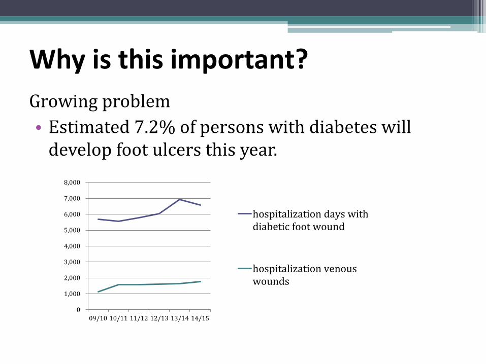
Growing problem
• Estimated 7.2% of persons with diabetes will develop foot ulcers this year.
Why is this important?
0
1,000
2,000
3,000
4,000
5,000
6,000
7,000
8,000
09/10 10/11 11/12 12/13 13/14 14/15
hospitalization days withdiabetic foot wound
hospitalization venouswounds

Why a Pathway?
Clinical Challenges identified:
• Inconsistent access to wound care services.
• Primary care providers lack information about local/regional resources for wound care or prevention.
• Wide variation in treatment plans and referral patterns for wound care patients.
• Number of acute care interventions (hospitalization, amputations) unacceptably high.

Potential benefits to health care providers
• Standardized protocols for assessment & treatment of wounds in primary care.
• Access to standardized wound care resources in the community.
• Clear criteria for specialist referral.
• Improved teamwork and communication among care providers.
• Prevention of wounds in people with diabetes and foot at high risk for ulceration.

• Improved teamwork and communication among care providers.
• Resources for patient education.
• Faster healing, avoid hospitalization or amputation.
Potential benefits to patients
Worldwide data suggests that 50% of diabetic foot amputations could be avoided with early identification and multidisciplinary clinical care. *
* Charing Cross International Vascular Symposium 2014

Most common chronic lower extremity wounds
Diabetic Foot Venous Ulcers – about 70-80% of LEW’s

Venous wounds

Venous Insufficiency – Stasis dermatitis and Venous Leg Ulcers (VLU)
• Lower leg between knee and ankle, usually proximal to medial malleolus – most commonly antero-medial calf.
• Gaiter or sock distribution.
• Lower leg edema (toes to knee) – worse by end of day; less with leg elevation.
• Sensation usually normal.
• Pedal pulses may be difficult to feel due to edema.

Venous Insufficiency – Stasis dermatitis and Venous Leg Ulcers (VLU)
Ulcer
• typically shallow ulcer base, dark red with yellow adherent slough
• irregular border
• large amount of wound exudate/drainage, especially when leg edematous
• relatively painless – achy, dull pain worsening as day progresses; increased pain if infected
• surrounding skin features of stasis dermatitis

Venous Insufficiency – Stasis dermatitis and Venous Leg Ulcers (VLU)
Stasis dermatitis
• Acute: erythematous maculopapular rash, vesicles, pruritic, skin edema
• Chronic: post-inflammatory skin changes/scarring, hemosiderin deposition in skin, dry scaling skin, erythema, dependant edema

Venous insufficiency/stasis

Cause of Venous Stasis Dermatitis/Ulcers
• In healthy leg veins, valves keep blood circulating up to heart.
• When valves become damaged/worn out, pressure increases in peripheral veins.
• Fluid leaks out of veins leading to swelling, irritation of the skin, and eventually skin breakdown.
• Once skin has broken down, ulcers are likely to recur if venous insufficiency not treated with compression stockings.

Venous Leg Ulcers
• Risk factors: ▫ Older age
▫ Varicose veins and incompetent venous valves
▫ Previous DVT
▫ Damage to lower leg veins – e.g. surgery, trauma
▫ Obesity
• Primary Prevention: ▫ Graduated compression (≥ 20mmHg) knee-high
stockings

Venous Leg Ulcer Treatment
1. Refer for compression bandaging ▫ First need to exclude significant PAD. ▫ Graduated compression wrapping if adequate arterial
flow to feet (ABPI ≥ 0.8)
2. Wound dressing ▫ Dressing to wound; protect skin surrounding ulcer ▫ Topical moderate potency corticosteroid if acute
dermatitis of surrounding skin ▫ Skin emollients – perfume and lanolin-free
3. Oral antibiotics if ulcer infected/cellulitis
4. Other drugs – statin*, pentoxifylline^
*Evangelista MT, Casintahan MF, Villafuerte LL. Simvastatin as a novel therapeutic agent for venous ulcers: a randomized, double-blind, placebo-controlled trials. Br J Dermatol 2014;170(5):1151-1157. ^Jull A, Arroll B, Parag V, Waters J. Pentoxifylline for treating venous leg ulcers. Cochrane Database Syst Rev 2007;3:CD001733.
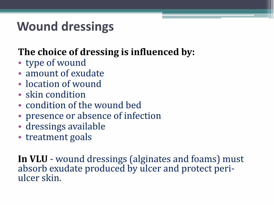
Wound dressings
The choice of dressing is influenced by: • type of wound • amount of exudate • location of wound • skin condition • condition of the wound bed • presence or absence of infection • dressings available • treatment goals In VLU - wound dressings (alginates and foams) must absorb exudate produced by ulcer and protect peri-ulcer skin.

Interpretation of ABI/TBI in Determining Compression
ABPI Value Interpretation/Clinical Significance Compression Therapy
>1.3 Abnormally high range, renders ABPI test - TBI indicated, contact wound clinician
Incompressible arteries
1.0 – 1.3 Normal High compression
0.8 - .99 Borderline to mild obstruction/peripheral arterial disease
High compression
0.71 – 0.79 Mild to moderate obstruction/ peripheral arterial disease
Modified compression
<0.7 Contact wound clinician or physician/NP Contra-indicated unless ordered by specialist
TBI Value Interpretation/Clinical Significance Compression Therapy
> 0.7 Normal High compression
0.41-0.69 Mild to moderate peripheral arterial disease Modified compression
< 0.4 Severe ischemia –contact wound clinician or physician/NP
Contra-indicated
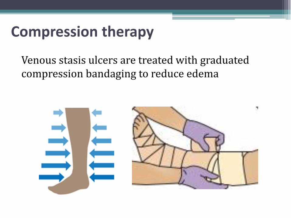
Compression therapy
Venous stasis ulcers are treated with graduated compression bandaging to reduce edema

Monitoring and surveillance
• Most venous stasis ulcers close in 3-4 months with optimal care.
• Non-healing wound requires specialist referral

Venous Ulcers - when to refer for specialist assessment
• Features of peripheral arterial disease (PAD) – preventing use of compression therapy
• Ulcer not healing adequately after 12 weeks of appropriate compression therapy
• Suspicion of malignancy
• Dermatitis not responding to topical steroids and compression therapy
• Frequent recurrence

Compression therapy – long term • Once the wound has healed, the wound care team will fit the
patient for graduated compression stockings (20-30 mmHg – knee or thigh high).
• A patient who has had a venous stasis ulcer can receive 2 pairs of
compression stockings every six months, for life. This is covered by SAIL.
• 50% of VLU recur in 10 years. • Graduated compression stockings
▫ 18-25 mm Hg: low compression for varicose veins and mild swelling ▫ 20-30 mm Hg: moderate compression for prevention/long-term management of
edema related to venous insufficiency ▫ 30-40 mm Hg: high compression for post-thrombotic venous insufficiency ▫ 50+ mm Hg: control of lymphedema
Primary care providers - important role in promoting adherence to compression therapy.

Venous wound care protocol - sample

Resources for patients and providers
http://www.sasksurgery.ca/provider/lowerextremitywound.html

Hands-on session with Q&A
• ABPI testing and interpretation
• Compression bandaging
• Wound Dressings used in treatment of venous leg ulcers
• Compression stockings
Diabetic wounds and the high-risk diabetic foot

Diabetic Foot Complications
• A consequence of: ▫ DPN – diabetic peripheral neuropathy -
sensory, motor and autonomic
▫ PAD - peripheral arterial disease – may be small and/or large vessel involvement
• DPN causes: ▫ loss of protective sensation in feet
▫ motor weakness in foot muscles resulting in deformities
▫ changes in skin blood circulation and skin growth
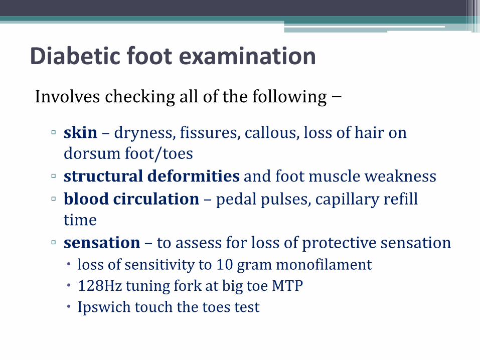
Diabetic foot examination
Involves checking all of the following –
▫ skin – dryness, fissures, callous, loss of hair on dorsum foot/toes
▫ structural deformities and foot muscle weakness
▫ blood circulation – pedal pulses, capillary refill time
▫ sensation – to assess for loss of protective sensation loss of sensitivity to 10 gram monofilament
128Hz tuning fork at big toe MTP
Ipswich touch the toes test

Screening for diabetic neuropathy
Peripheral sensory neuropathy – involves testing for loss of sensitivity to 10gram monofilament or loss of sensitivity to vibration (128Hz tuning fork) at the dorsum of the first toe • Loss of sensitivity to 10gram monofilament or 128Hz
tuning fork - associated with loss of protective sensation (LOPS) on the foot
• Practical tip – 25 lb fishing line cut into 4 cm lengths may be used for the 10gram monofilament test
Peripheral motor neuropathy – may be done by asking the patient to do the one foot stand test. This is a sensitive test for peripheral motor neuropathy in people with DM (increased risk of falls).
Peripheral autonomic neuropathy – look for skin and vasomotor changes in feet.

Sensory screening options Semmes-Weinstein Monofilament test
Reference: Craig A. B. et al. Foot Sensation Testing in the Patient With Diabetes: Introduction of the Quick & Easy Assessment Tool. WOUNDS. 2014; 26(8): 221-231
Vibratory sensation test - conventionally tested with a 128-Hz tuning fork at the interphalangeal joint of the hallux
Ipswich Touch the Toes test
Vibration perception threshold testing (VPT) - performed using a handheld device (instrument costs about $700)

Ipswich Touch-the-toes Test (IpTT)
IpTT and 10g MF – almost perfect agreement; both showed approx. 80% sensitivity and 90% specificity in identifying at-risk feet. IpTT useful screening test for sensory neuropathy; requires no equipment. Rayman et al. Diabetes Care 2011;
34:1517

Non-ulcerative diabetic foot complications
Skin changes
• Autonomic neuropathy affects the innervation of sweat glands resulting in dry skin and hyperkeratosis.
• Autonomic neuropathy affects blood circulation to the skin resulting in reduced nutritive blood flow and increased inflammatory changes in skin.
• Dry cracked skin and calluses may be lead to ulceration and limb-threatening infection, especially if patient also has PAD.
• Management – Patient education (self care and risk of ulceration/infection). Podiatry referral useful in high risk patients with LOPS, foot
deformities. Appropriate footwear.

Non-ulcerative diabetic foot complications Foot deformities
• Motor neuropathy / weakening of intrinsic foot muscles results in muscle imbalance and changes in foot structure and gait patterns.
• Toe deformities – claw toe, hammer toe, hallux rigidus, bunions, over-riding toes.
• These predispose to callus formation and ulceration of foot over pressure points.
• Management – podiatry - offloading, padding, custom orthoses or
shoes orthopedic surgery – joint fixation, arthroplasty,
amputation of toes.
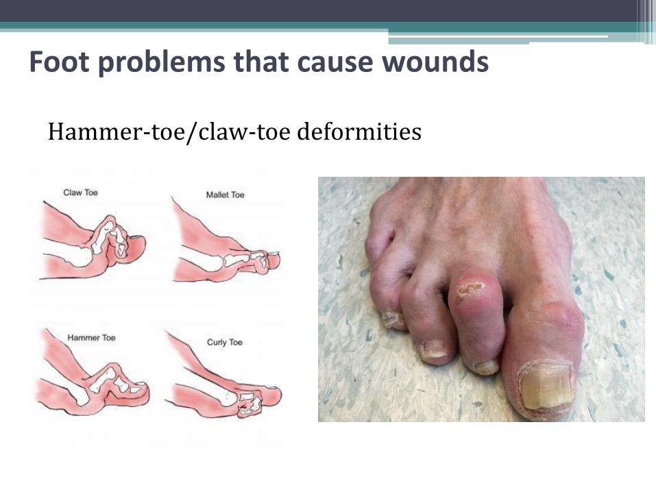
Foot problems that cause wounds
Hammer-toe/claw-toe deformities
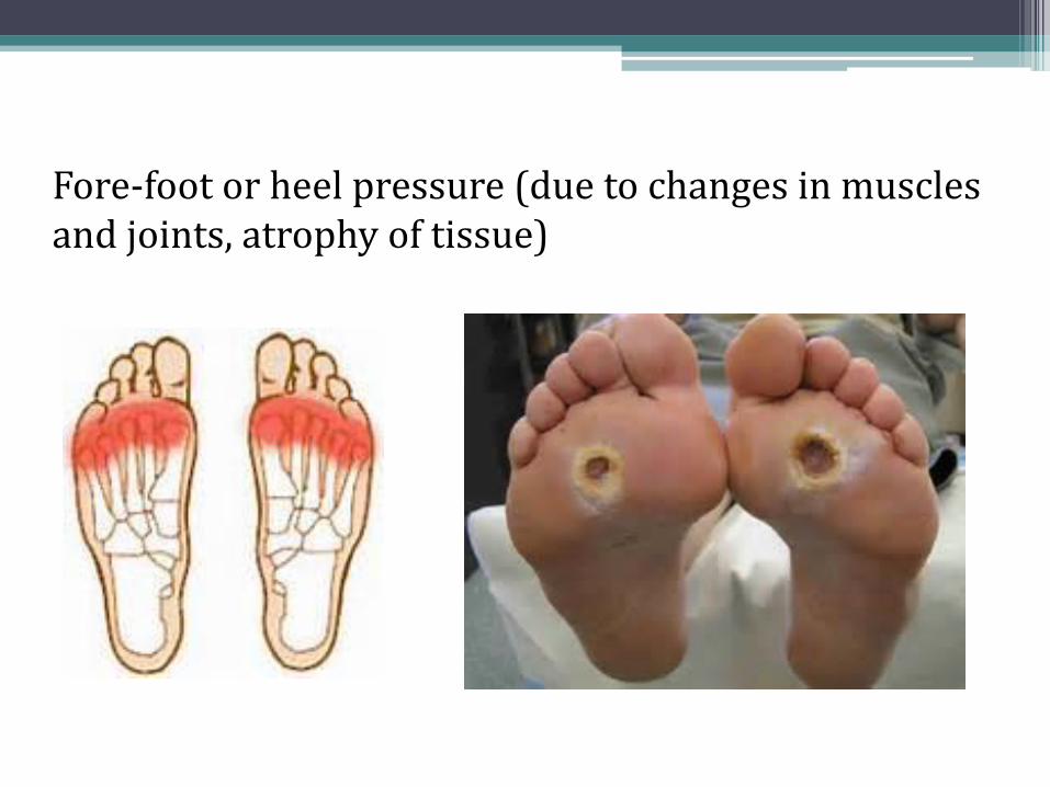
Fore-foot or heel pressure (due to changes in muscles and joints, atrophy of tissue)

Typical changes seen in diabetic foot
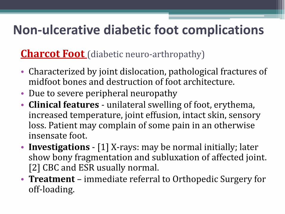
Non-ulcerative diabetic foot complications
Charcot Foot (diabetic neuro-arthropathy)
• Characterized by joint dislocation, pathological fractures of midfoot bones and destruction of foot architecture.
• Due to severe peripheral neuropathy • Clinical features - unilateral swelling of foot, erythema,
increased temperature, joint effusion, intact skin, sensory loss. Patient may complain of some pain in an otherwise insensate foot.
• Investigations - [1] X-rays: may be normal initially; later show bony fragmentation and subluxation of affected joint. [2] CBC and ESR usually normal.
• Treatment – immediate referral to Orthopedic Surgery for off-loading.

Charcot foot

Charcot foot with ulcer

Non-ulcerative diabetic foot complications
Peripheral Arterial Disease
• Diabetic foot PAD usually due to microvascular and/or macrovascular arterial disease.
• History - may have leg claudication, rest pain in foot, or no pain.
• Examination ▫ Decreased or absent pulses in foot.
▫ Signs of chronic vascular insufficiency – cool, dry skin, absence of hair, thickened nails, dependent rubor with pallor on elevation of foot.

Non-ulcerative diabetic foot complications
Peripheral Arterial Disease
• Diagnostic Testing
▫ Ankle-Brachial Pressure Index (ABPI) – ratio of SBP in ankles to SBP in upper arm; use BP cuff and hand-held Doppler. Normal ABPI is 0.9 – 1.3
ABPI <0.9 = PAD
ABPI <0.4 = severe arterial obstruction
ABPI >1.3 due to calcified blood vessels (common in diabetics)– cannot rely on this to assess PAD.
▫ Vascular studies and angiography – have to refer to Vascular Surgeon

Non-ulcerative diabetic foot complications Peripheral Arterial Disease
• Treatment Optimal glycemic and BP control Anti-platelet agent – ASA, other anti-platelet agents Lipid lowering drugs - Statins Exercise – walking program Smoking cessation Endovascular intervention or Surgical bypass - if
severe PAD, critical ischemia (rest foot or leg pain, non-healing ulcer, gangrene)
• Arterial Bypass success rates in people with diabetes:
▫ 45-60% remain patent 5 years post surgery ▫ 50% patient survival 5 years post surgery

Typical changes seen in diabetic foot PAD - dry gangrene

Etiology of Diabetic Foot Ulcers (DFU)
• Critical triad of neuropathy, deformity and minor trauma present in >60% of DFU.
• Neuropathy most important etiologic component.
• Minor trauma often preventable.
foot deformity
minor trauma
mechanical stress
neuropathy
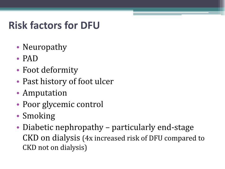
Risk factors for DFU
• Neuropathy
• PAD
• Foot deformity
• Past history of foot ulcer
• Amputation
• Poor glycemic control
• Smoking
• Diabetic nephropathy – particularly end-stage CKD on dialysis (4x increased risk of DFU compared to CKD not on dialysis)

Diabetic Foot Ulcers

Diabetic Foot Infection

Principles of DFU Management
1. Wound dressings - maintain moist wound healing environment
2. Off-load pressure from wound “It is not what one puts on a wound that heals it, but what one takes off.” Mainstay of wound healing in DFU is redistribution of pressure from the ulcerated skin.
3. Treat infection – LEW pathway antibiotic protocol
4. Debridement 5. Improve blood supply – endovascular
intervention/surgery if large vessel PAD 6. Optimal glycemic control – blood sugars less than
11mmol/L

Diabetic Foot Ulcer
Clinical Pearls
• >2cm of skin erythema around a diabetic lower limb ulcer may be indicative of limb-threatening infection (need to admit for IV antibiotics, limb elevation, wound care).
• Presence of pain in a previously insensate foot can be the first and most important indicator of severe infection or underlying osteomyelitis.
• Need to consider other diabetic complications such as CKD before selection and dosing of antibiotics for diabetic foot infections.
• Selection of antibiotic must be guided by previous/recent antibiotics used by patient.
• Poor glycemic control delays ulcer healing.

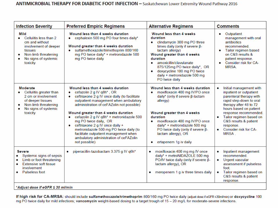
Antibiotic selection for infected DFU
KEY PRINCIPLES:
1. Duration of wound and bacteria ▫ short duration – mostly Staph and Strep ▫ duration > 4 weeks – likelihood of anaerobic bacteria
2. Severity of infection ▫ moderate or severe infection/cellulitis – need to consider
both Gram negative and positive bacteria 3. Renal function ▫ need to adjust antibiotic dose if eGFR <30 ml/min
4. Osteomyelitis ▫ probe-to-bone test ▫ MRI
5. CA-MRSA ▫ add septra or doxycycline if high risk

Family physician role in management of DFU
• Individuals with DFU tend to possess fewer cognitive resources than individuals with similar duration DM without foot ulcer.*
• Potentially problematic as management of DFU requires increased demands for self-treatment and adherence to treatment regimens that may be complex and of long duration.
• Family physician/nurse practitioner – important role in explaining and promoting adherence; advocating for patient.
Reference: Natovich R et al. Cognitive Dysfunction: Part and Parcel of the Diabetic Foot. Diabetes Care 2016 May; dc 152838.
Protecting and Healing the Diabetic Foot

• Probability of DFU healing without complications is 31% without use of offloading device; increasing to 64% when an offloading device effectively used.
• If individual has foot/lower limb amputated for DFU, other foot at high risk of developing DFU.
• Death following amputation for DFU estimated at 20-40% at one year, and 60-80% at 5years.
“Impact of Offloading Devices on the Cost of Diabetic Foot Ulcers in Ontario” – Canadian Diabetes Association, 2015

High risk foot - start with the shoe
• Most foot ulcers start with blisters, callous, bumps or scratches
• Proper footwear • can prevent wounds from starting • essential for wounds to heal
• Proper shoes: ▫ Good padding to absorb shock ▫ Good fit & smooth inside surfaces to reduce friction
inside the shoe ▫ Solid structure to stabilize and support ambulation ▫ Wide enough to accommodate common foot
deformities

Wide heel base and deep heel cup for stability
Slip-resistant, durable, flexible sole
Thick foam insole for extra shock absorption
Footbed provides cushioning and shock absorption – can be removed for custom inserts
Seamless lining reduces likelihood of friction
Soft interlining molds to foot, reduces friction
Laces or velcro for adjusting fit
Rocker sole reduces forefoot pressure
Wide, extra deep toe box
Breathable upper and lining
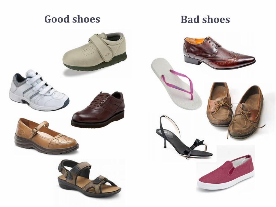
Good shoes Bad shoes

Coverage for ordinary proper shoes
• Most insurance plans do NOT provide coverage for ordinary shoes.
• Diabetic patients receiving social assistance benefits (including First Nations) can get extra allowance for footwear.
• Physician must provide patient with prescription.

Orthopedic shoes
• Orthopedic shoes and inserts can accommodate mild-moderate foot problems
• Measured and fitted by a pedorthist in
a shoe store (or an orthotist, podiatrist
if available).
Orthopedic shoes may have: More space in the toe box Room for extra inserts and/or padding in specific areas like toe or heel for offloading Sole modifications to stabilize gait
Orthopedic shoes and inserts can increase comfort and help to slow the progression of foot problems. But if cost is an issue, ordinary well-fitting shoes are a good alternative.

Coverage for orthopedic shoes
• Off-the-shelf orthopedic shoes and inserts may be covered by some private insurance plans (with prescription from physician).
• Orthopedic shoes start around $100
• Inserts around $60
• Orthotists and podiatrists charge patient for visit consultation in addition to purchase.
Custom shoes
Individuals with significant foot deformities who can no longer wear off-the-shelf shoes require custom shoes & inserts, or shoe modifications.
• Custom built from 3D cast of patient’s foot
• Can be built by orthotist, podiatrist, pedorthist.
Patients with significant deformity should also be referred to specialist in orthopedics or physiatry.
Coverage for custom shoes
• Custom shoes covered by many private insurance plans, with physician prescription.
• Coverage for First Nations patients is provided by Non-insured Health Benefits (NIHB).
• Coverage for low-income patients is provided by Sask Drug Plan Extended Benefits (Supplementary Benefits).
• Without coverage, patient is billed (cost about $500).
• Suppliers (pedorthists, orthotists and podiatrists) are usually knowledgeable about prior approval and billing processes.
• Advise patient to confirm insurance coverage before ordering product.

Suppliers of custom shoes
Public pedorthic programs - require prescription and appointment:
• Saskatoon (& Prince Albert) - Saskatchewan Abilities Council. Phone: 306-374-4400 Email: [email protected]
• Regina - Wascana Rehabilitation Centre.
Phone: 306-766-5730 or 306-766-5731
Private suppliers:
• Pedorthists - locate at www.cpedcs.ca
• Orthotists – find at www.opcanada.ca
• Podiatrists – find at www.saskpodiatry.org

When a wound occurs – Offloading
• Required to remove pressure from the wound.
• Proper offloading is as important as proper wound dressing.
• Should start as soon as possible.
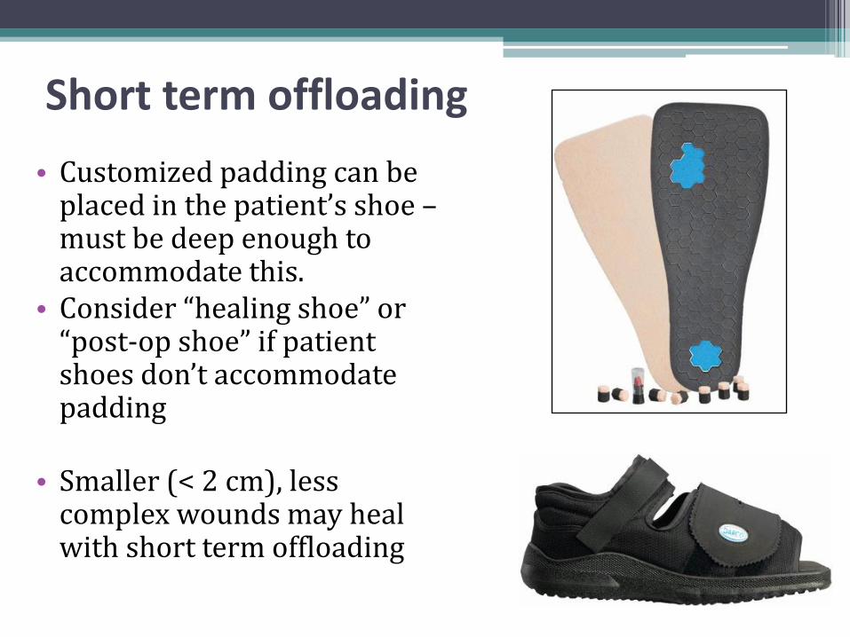
Short term offloading
• Customized padding can be placed in the patient’s shoe – must be deep enough to accommodate this.
• Consider “healing shoe” or “post-op shoe” if patient shoes don’t accommodate padding
• Smaller (< 2 cm), less
complex wounds may heal with short term offloading

Coverage for short term offloading
• No insurance coverage for these products.
• Padding may be supplied by homecare.
• Healing shoes may be provided by homecare but billed to patient.
• Healing shoes may be available for purchase from podiatrist, drugstore, shoe store or medical supply store, or order on-line.
• Cost starts at around $30.

Longer term offloading
Total Contact Cast
• Provides best wound healing
• Must be removed weekly for wound assessment and re-applied
• Should be applied by specially trained cast technician
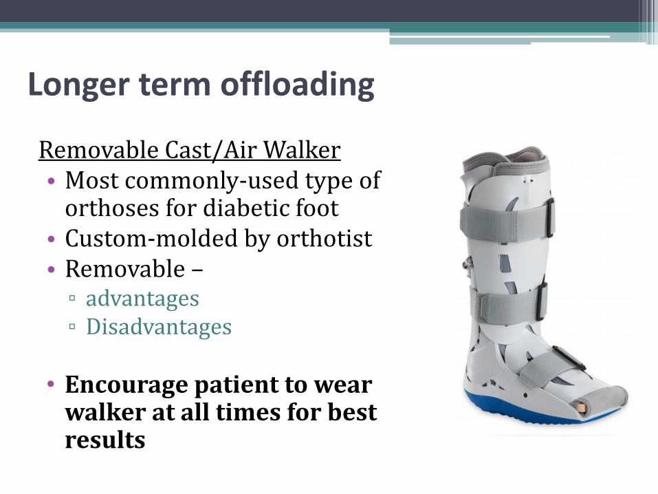
Longer term offloading
Removable Cast/Air Walker • Most commonly-used type of
orthoses for diabetic foot • Custom-molded by orthotist • Removable – ▫ advantages ▫ Disadvantages
• Encourage patient to wear walker at all times for best results
Coverage for longer term offloading
• All custom products are covered by private insurance/public insurance/NIHB.
• Patients who see a vascular surgeon for wound assessment and require long term offloading will be prescribed custom device or casting.
• Must be prescribed by specialist and fitted by an orthotist (Regina/Saskatoon).

Hands-on session with Q&A
• Diabetic foot ulcer wound dressings
• Offloading devices
• Orthopedic shoes

Saskatchewan LEW Pathway
How will the LEW Pathway improve the management of these wounds?
• Standardized referral form – direct referral to: local home care / wound care nurses tertiary care (vascular surgery)– in Saskatoon or Regina
• Referral forms within EMR
• Standardized lower leg assessment and wound care protocols to be followed by home care and wound care nurses.
• Capacity building - Wound care training program developed by Saskatchewan Polytechnic for home care nurses.
• Standardized antibiotic protocols and wound tracking tools.
• Enhanced communication between all providers, so family physician/NP remain informed.
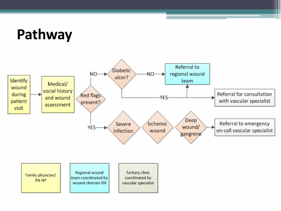
Pathway

Primary Care LEW Referral Form
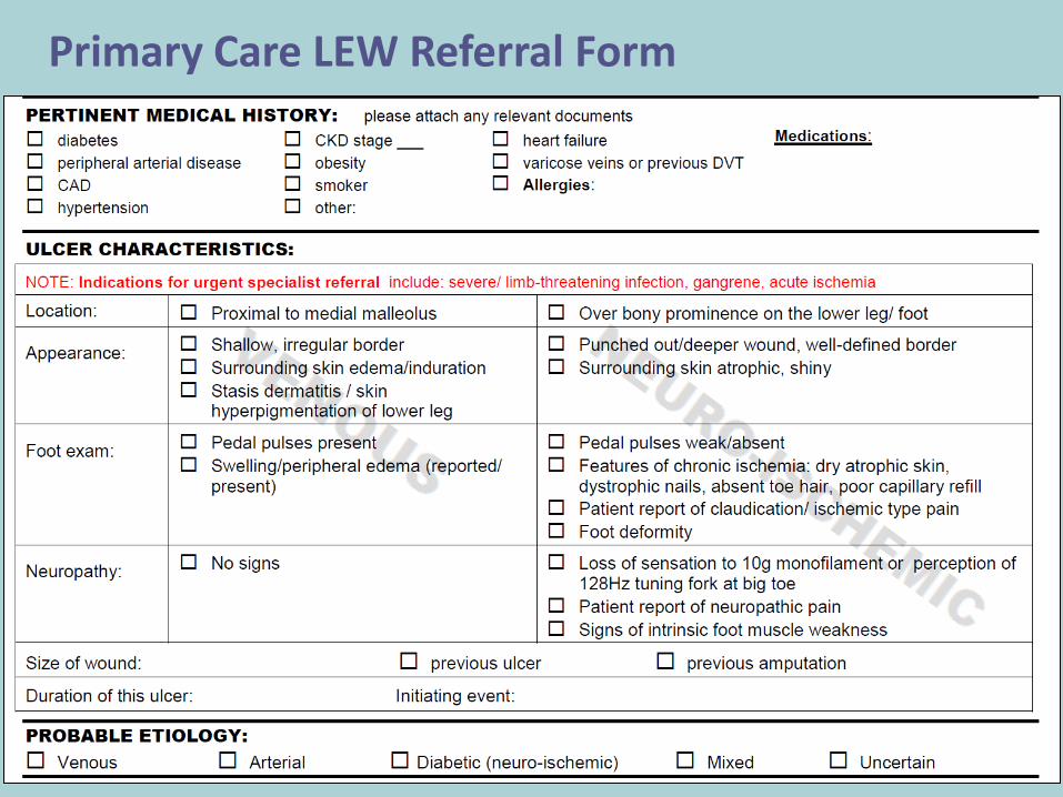
Primary Care LEW Referral Form

Primary Care LEW Referral Form

Communication from Wound Care Nurse to Primary Care Provider
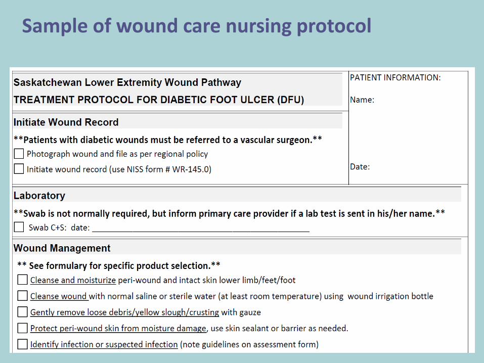
Sample of wound care nursing protocol

LEW Pathway website
http://www.sasksurgery.ca/provider/lowerextremitywound.html

THANK YOU
QUESTIONS Post-course Questionnaire