clinical study surgical guides (patient-specific
TRANSCRIPT
Hindawi Publishing CorporationSarcomaVolume 2013, Article ID 787653, 7 pageshttp://dx.doi.org/10.1155/2013/787653
Clinical StudySurgical Guides (Patient-Specific Instruments) for PediatricTibial Bone Sarcoma Resection and Allograft Reconstruction
Laura Bellanova,1,2 Laurent Paul,1,2 and Pierre-Louis Docquier1,2
1 Computer-Assisted Robotic Surgery (CARS), Institut de Recherche Experimentale et Clinique (IREC),Tour Pasteur +4, Avenue Mounier 53, 1200 Brussels, Belgium
2Department of Orthopaedic Surgery, Cliniques Universitaires Saint-Luc, (Universite Catholique de Louvain),Avenue Hippocrate 10, 1200 Brussels, Belgium
Correspondence should be addressed to Pierre-Louis Docquier; [email protected]
Received 21 October 2012; Revised 19 January 2013; Accepted 10 February 2013
Academic Editor: Hans Rechl
Copyright © 2013 Laura Bellanova et al.This is an open access article distributed under theCreative CommonsAttribution License,which permits unrestricted use, distribution, and reproduction in any medium, provided the original work is properly cited.
To achieve local control of malignant pediatric bone tumors and to provide satisfactory oncological results, adequate resectionmargins are mandatory. The local recurrence rate is directly related to inappropriate excision margins. The present study describesa method for decreasing the resection margin width and ensuring that the margins are adequate.This method was developed in thetibia, which is a common site for the most frequent primary bone sarcomas in children. Magnetic resonance imaging (MRI) andcomputerized tomography (CT) were used for preoperative planning to define the cutting planes for the tumors: each tumor wassegmented on MRI, and the volume of the tumor was coregistered with CT. After preoperative planning, a surgical guide (patient-specific instrument) that was fitted to a unique position on the tibia was manufactured by rapid prototyping. A second instrumentwas manufactured to adjust the bone allograft to fit the resection gap accurately. Pathologic evaluation of the resected specimensshowed tumor-free resection margins in all four cases.The technologies described in this paper may improve the surgical accuracyand patient safety in surgical oncology. In addition, these techniques may decrease operating time and allow for reconstructionwith a well-matched allograft to obtain stable osteosynthesis.
1. Introduction
The tibia is a common site for the most frequent malignantprimary bone tumors in children, osteosarcoma (tibia isaffected in 27% of cases), and Ewing’s sarcoma (8% of cases)[1, 2]. Improvements in diagnosis and therapeutic techniqueshave increased interest in limb-salvage surgery. However,several studies have suggested that limb-salvage surgerymay increase local recurrence in the case of inappropriateexcision margins [3]. To achieve local control of diseaseand to improve oncological results, wide resection marginsare mandatory. However, a wide surgical excision resultsin a large residual bone defect that requires restoration[4]. We propose a new technique to decrease the widthof the excision margins with patient-specific instruments(PSIs). In this method, the resection is carefully plannedprior to surgery, based on magnetic resonance imaging(MRI) and computed tomography (CT), which are used
to define the trajectories of the resection and createPSIs.
The function of the reconstructed limb is of major inter-est, especially in young and physically active patients whoplace high demands on their limbs. The limb reconstructionmust also be durable because life expectancy for many ofthese patients is several decades [4]. Limb reconstruction forsuch large defects can be performed by various techniques,including endoprosthetic reconstruction, osteoarticular allo-grafting, vascularized autografting, bone transport with dis-traction osteogenesis, or reimplantation of the tumor-bearingbone segment after the devitalization of the tumor cells (byheating, freezing, or extracorporeal irradiation) [5].
Massive bone allografting presents several drawbacks: rel-atively long rehabilitation due to immobilization and partialloss of weight bearing, difficulties in obtaining size-matchedallografts for small patients, an absence of expandability, anda relatively high incidence of complications [6]. Despite these

2 Sarcoma
drawbacks, allografting offers several advantages, includingthe ability to reattach the ligamentous and tendinous struc-tures of the host to the graft. An accurate reconstructionof the soft-tissue attachments at the host-allograft junctioncan lead to improved results [7]. Other advantages includethe biologic incorporation (at least partial) of the graft andthe preservation of the joint, the juxta-articular bone, andthe growth plates [8]. These advantages make massive boneallografting convenient for intercalary, osteoarticular, andarthrodesis operations, as well as for allograft-prostheticcomposite reconstruction in an extra-articular resection.In fact, bone allografting is the most common option forintercalary reconstruction, with a survival rate as high as 75%to 89% at 10 years [6, 9–12].
The concept of using a patient-specific template wasintroduced in the 1990s by Radermacher et al. [13] for pediclescrew placement, total knee arthroplasty, decompression ofcervical spine, and triple osteotomy of the pelvis. Theyperformed CT-based preoperative planning and conceived atemplate to fit the bone surface. The template was manufac-tured by milling because rapid prototyping was not yet welldeveloped and was far more expensive. Later, Salako et al.used guided intrapedicular screws to install instrumentationon the spine. This mechanism allowed the surgeon to drillin the optimal direction, and it decreased the rate of screwmisplacement [14]. In maxillofacial reconstruction surgery,the use of a PSI to guide the osteotomies allowed thesurgeon to avoid important structures, such as dental nervesand components of the vascular anatomy. This concept wasused by Leiggener et al. [15] for mandible reconstructionwith a free fibula osseous flap. Using CT angiography, theauthors manufactured a guide by rapid prototyping (SLS).They performed a complex mandible reconstruction withthis method, choosing the best sites on the donor (fibula)and recipient (mandible) with regard to function, aesthetics,and blood supply. Modabber et al. [16] concluded thatthis technique significantly decreased shaping time duringsurgery and will likely impact the survival of the flap. Severalauthors have used a patient-specific template technique totreat other conditions, such as a cubitus varus deformity, amalunited forearm fracture, and a distal-radial fracture com-bined with tibial deformities. After preoperative planning, arapid-prototyped model was manufactured to correct eachdeformity. This technique improves the accuracy and ease ofthe surgical act [17, 18].
In the present paper, we describe the use of PSIs forresecting aggressive tibial sarcomas and reconstructing theanatomy with an intercalary or osteoarticular allograft.First, we detail the preoperative planning process. Next, wedescribe how PSIs are used to perform the resections and toadjust the allografts. Finally, we present several clinical casesand their outcomes.
2. Materials and Methods
2.1. Preoperative Planning for Tumor Resection. Preoperativeimages of the patients were acquired for diagnoses. Anatom-
ical images were obtained by CT from a Brilliance 40 CTscanner (Philips, the Netherlands; 0.5mm spacing betweenslices, 1mm slice thickness, 120 kV peak voltage, and 99-mAtube current) andbyMRI froma 1.5 TNTScan Intera (Philips,the Netherlands, 4mm spacing between slices, 3mm slicethickness, 550ms TR, and 14ms TE).
Preoperative planning required delineation of eachtumor. An MRI series that clearly showed the boundaries ofthe tumor was selected. The tumor was manually delineatedon each slice on which it was visible, using the open-sourcesoftware ITK-Snap 2.0 (http://www.itksnap.org/) [19]. Theobtained delineation, referred to as the tumor volume, wassaved for later use.
A multimodal registration algorithm was used to merge,simultaneously, the MRI series with the CT images (imagefusion) and the tumor volume (Figure 1). 3D models of thebone and the tumor volume were extracted from the 2Dslices, and a combined 3D image of the tibia and tumor(red in Figure 1) was obtained. The 3D models were used toposition the resection planes (target planes) that representedthe trajectories of the saw blade. With the assistance of ahaptic device and a specific software program developedin the author’s laboratory, the planes were initially broughtinto contact with the tumor and then translated back witha surgeon-defined security margin of at least 5mm. Thismethod ensured a controlled safe margin during the surgery.The resection plane data were saved for use in the next twostages of preoperative planning.
2.2. Preoperative Planning for Allograft Cutting. To selectthe best-fitting allograft among the tibial allografts from thelocal bone bank, CT images of all available tibia allograftswere acquired (Somatom Definition AS, Siemens; 0.35-mmslice thickness, 0.7mm spacing between slices, 120 kV peakvoltage, and 99mA tube current). A monomodal registrationwas performed betweenCT scans from the tibia of the patientand the various allografts. The optimal allograft was chosenas the one that best bridged the bone defect created by theresection. Particular attention was paid to the articular sur-face in the case of an osteochondral allograft. The resectionplanes defined in the previous stage of preoperative planningwere transferred toward the allograft with the registrationalgorithm (Figure 3). This process ensured that the resectionplanes were identical for the patient and for the allograft, witha well-matched allograft to fit the bone defect.
2.3. Patient-Specific Instruments. Two PSIs were created foreach patient: one for the tumor resection and a second onefor the allograft cutting.ThePSIs were virtually designedwitha computer-aided design software package (Blender 2.63.11).Each PSI was endowed with a specific surface that could be fitonto a unique position on the bone surface. The instrumentscontained small holes that were specifically designed for2mm Kirschner wires (K-wires). These K-wires allowed theinstrument to be pinned onto the bone surface. A flat surfacematerialized the resection planes and permitted the clinicianto guide the saw blade during the osteotomies.

Sarcoma 3
(a) (b) (c)
(d) (e) (f)
Figure 1: 11-year-old patient with tibia metastasis of a femoral osteosarcoma. (a) MRI showing the osteosarcoma invading the proximal tibia.(b) Tumor is delineated in blue on each slice of MRI where it is visible. (d) CT scan. (e) Merging of MRI and CT. (f) MRI-CT merging withthe delineation of the tumor. (c) 3D reconstruction of the tibia with the delineated tumor visible (red).
(a) (b)
Figure 2: Images from the patient shown in Figure 1. (a) Target resection planes have been designed according to the osteosarcoma (red)with a safe margin.The green zone is included between the two planes. A 5mm distance (margin) between the plane and tumor was selectedto spare epiphysis, whereas a 10mm margin was chosen distally. K-wires are represented by green cylinders. A PSI was created to guide thesaw blade and to respect the planes. (b) PSI has been postitioned at the surface of the exposed tibia, and the surgical saw has been placed onthe PSI to follow the target planes.
The PSIs were produced by rapid prototyping with Selec-tive Laser Sintering (SLS) technology in a biocompatiblematerial (Polyamide, Figure 2). This additive manufacturingtechnology consists of building a 3D object by adding
material layer by layer. A laser draws a 2D shape on the surfaceof a powder bed, locally fusing the powder to create a solidsection. A new layer of powder is spread on top of the surface,and the process is repeated until the entire object is built.

4 Sarcoma
(a) (b) (c)
(d) (e)
Figure 3: Images from the patient shown in Figures 1 and 2. (a) Merging of the allograft CT and the tibial CT of the patient. (b) Target planeshave been transferred to the allograft. The pink zone is the planned cut allograft. (c) A PSI has been virtually created (blue) and is shownpinned to the bone with two K-wires (green cylinders). (d) PSI has been positioned on the surface of the tibial allograft. (e) Allograft aftercutting with the saw.
2.4. Patient Series (Table 1). Four patients were operated onfor tibial bone sarcoma resection and allograft reconstructionwith PSIs. Two patients presented with primary tibia sarco-mas (one with osteosarcoma and one with Ewing’s sarcoma),one patient had a local recurrence of tibial osteosarcoma,and one patient had a tibial metastasis from a contralateralfemoral osteosarcoma. The ages at operation ranged from 9years and 9months to 18 years and 2months. Imaging assess-ment consisted of plain radiograph, CT, MRI, and positronemission tomography (PET)-CT with fluorodeoxyglucose-18. The latter revealed no distant metastases. Neurologicaldeficits and systemic symptoms, such as fever or weight loss,were absent. The diagnosis was confirmed by an incisionalbiopsy and histological evaluation of the tissue. Patientsreceived multiagent, neoadjuvant, and adjuvant chemother-apy, according to the Euramos, Euro-Ewing, OS2005, andOSII-TTP protocols.
2.5. Assisted Surgery. The patients were placed in decubitus.A medial-tibial approach was used in three patients and alateral approach in the last patient (because the biopsy hadbeen performed laterally).The open-surgical biopsy tract wasexcised with the tumor, as an ellipse. A progressive soft-tissue dissection was performed to isolate the tumor with theplanned margin and with the preservation of the anteriortibial tuberosity. Both PSIs (one for the patient and onefor the allograft) were sterilized by standard autoclaving theday before the surgery. The resection PSI was positionedon the tibia and rigidly fixed with two K-wires at proximaland distal locations (Figure 2). A safe margin of 5mm waschosen (3.5mm after resection because of material loss dueto saw blade cutting, i.e., the kerf). Proximal and distalosteotomies were performed with a surgical saw that wasguided by the PSI. The tibial resection lengths ranged from8 to 16.4 cm.
Sarcoma 5
(a) (b) (c)
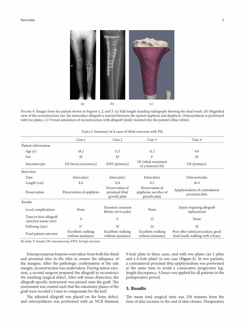
Figure 4: Images from the patient shown in Figures 1, 2, and 3. (a) Full-length standing radiograph showing the final result. (b) Magnifiedview of the reconstruction site: the intercalary allograft is inserted between the spared epiphysis and diaphysis. Osteosynthesis is performedwith two plates. (c) Virtual simulation of reconstruction with allograft (pink) inserted into the patient’s tibia (white).
Table 1: Summary of 4 cases of tibial resection with PSI.
Case 1 Case 2 Case 3 Case 4Patient information
Age (y) 18.2 12.5 11.2 9.8Sex M M F M
Sarcoma type OS (local occurrence) EWS (primary) OS (tibial metastasisof a femoral OS) OS (primary)
ResectionType Intercalary Intercalary Intercalary OsteoarticularLength (cm) 8.0 11.8 8.2 16.4
Preservation Preservation of epiphysisPreservation ofproximal tibialgrowth plate
Preservation ofepiphysis; sacrifice of
growth plate
Epiphysiodesis of contralateralproximal tibia
Results
Local complications None Transient commonfibular nerve palsy None Sepsis requiring allograft
replacementTime to host-allograftjunction union (mo) 4 9 12 None
Followup (mo) 17 19 14 10
Final patient outcome Excellent; walkingwithout assistance
Excellent; walkingwithout assistance
Excellent; walkingwithout assistance
Poor after initial procedure; goodfinal result; walking with a brace
M: male; F: female; OS: osteosarcoma; EWS: Ewing’s sarcoma.
Extemporaneous biopsies were taken from both the distaland proximal sites in the tibia to ensure the adequacy ofthe margins. After the pathologic confirmation of the safemargin, reconstruction was undertaken. During tumor exci-sion, a second surgeon prepared the allograft to reconstructthe resulting surgical defect. After soft-tissue dissection, theallograft-specific instrument was pinned onto the graft. Theinstrument was created such that the osteotomy planes of thegraft were receded 1.5mm to compensate for the kerf.
The adjusted allograft was placed on the bone defect,and osteosynthesis was performed with an NCB titanium
9-hole plate in three cases, and with two plates (an L-plateand a 6-hole plate) in one case (Figure 4). In two patients,a contralateral proximal tibia epiphysiodesis was performedat the same time to avoid a consecutive progressive leg-length discrepancy. A brace was applied for all patients in thepostoperative period.
3. Results
The mean total surgical time was 250 minutes from thetime of skin incision to the end of skin closure. Preoperative

6 Sarcoma
preparation, including general anesthesia, epidural catheterinsertion, patient positioning, and draping, lasted a mean of73.5 minutes.
One patient developed transient common fibular nervepalsy, which was confirmed by electromyography. Skinnecrosis and wound dehiscence occurred in the patientwith the osteoarticular allograft at one postoperative month,necessitating a lateral gastrocnemius flap. The allograft wasultimately found to be infected by Enterococcus faeciumand Pseudomonas and was explanted 3 months after thereconstruction. An antibiotic-impregnated cement spacerwas inserted, and a new reconstruction was performed 4months later with a new PSI to guide the allograft cutting.
Histological examination of the removed sarcoma con-firmed osteosarcoma in three patients and Ewing’s sarcomain one patient. All resection margins appeared to be tumor-free. Postoperative radiograph, CT scan, and MRI resultsrevealed satisfactory host-graft contact and no evidence ofrecurrent disease. The explanted, infected allograft was notconsolidated with the host tissue. For the other three patients,radiological union was obtained at the graft-host junctionat 4, 9, and 12 months. Partial weight bearing was allowedafter 6 weeks and full weight bearing after 3 months, exceptfor one patient for whom partial weight bearing was allowedimmediately.
4. Discussion
This paper reports a novel method of bone sarcoma surgerythat is supported by the use of PSIs. The PSI assistancewas used not only for tumor resection but also for massiveallograft cutting, allowing for optimal reconstruction. Thistechnique was applied to 4 patients.
In surgical oncology, obtaining a wide margin during atumor resection is crucial to avoid local recurrence. However,limb-salvage surgery requires the preservation of a func-tioning limb at the expense of obtaining safe margins [3].Accurate preoperative localization of the tumor provides fullcontrol over the safe margins, and PSIs improve the accuracyof the resection during the surgery.The combination of thesetechniques allows resection with adequate but minimal safemargins, thus preventing unnecessary resection and preserv-ing, when needed, articular cartilage in young patients. Inone of the patients presented here, the target margins weredefined at 3.5mm, which allowed for the preservation of thegrowth plate (Figure 5). This outcome would not have beenpossible without the assistance of PSIs.
The literature reports discrepant results for tibia allograft-ing reconstruction, ranging from excellent outcomes withfull incorporation and osteointegration of the allograft anddurable joint function to a high rate of failure and complica-tions, including accelerated and advanced arthritis, fractures,nonunion, and infection [20]. The PSI technique allows theallograft to be cut with a high degree of accuracy, producinga transplant of the optimal shape to bridge the bone defect.Moreover, the allograft can be adjusted simultaneously (byanother surgeon) or prior to the tumor resection, thereby
(a) (b)
Figure 5: 12-year-old boy with EWS sarcoma of the proximal tibia.Result of the reconstruction with preservation of the growth plate:radiographic anteroposterior and lateral view.
decreasing the operating time and improving patient safety.The precise tumor and allograft cuttings obtained by PSIsyield strong contact at host-graft junctions, resulting in astable osteosynthesis. The mechanical stability of the graftfacilitates improved and more rapid healing and bone fusiondue to the increased growth of blood vessels into the graft [21].
The production of PSIs requires medical images to besent to an engineer who performs the preoperative planning.This process presents a challenge because it is crucial tomaintain the security of the medical data of the patient.In addition, an open communication between the surgeonand the engineer is important. Furthermore, the engineermust have a strong clinical background to understand themedical context and the prerequisites of the PSI that will begenerated. The PSI must be localized to a unique site on thebone surface that will be exposed by the surgical approachwithout adding unnecessary surgical approaches, skin inci-sions, or dissection. Surgeons will be asked to anticipate theconstraints of the surgery (e.g., surgical approach, accessto bone surface, and presence of soft tissue). The engineermust respect these clinical data and determine a trade-offbetween the invasiveness of the PSI and its stability on thebone surface. In our series of patients, accurate positioningof the instruments was easily achieved for each case, and theinstruments were stable.
5. Conclusions
The techniques described in this paper may help to improvepatient safety and surgical accuracy for the resection ofbone sarcomas of the tibia and for the accompanying recon-struction at these sites. In addition, these techniques maypotentially benefit surgery for bone sarcomas at other sitessuch as the pelvis.

Sarcoma 7
Acknowledgment
Funds were received from the Fondation Belge Contre leCancer (Grant SCIE 2006/20).
References
[1] K. Winkler, S. Bielack, B. Kempf-Bielack et al., “Neoadjuvanttherapy for localized extremity osteosarcoma. Experience ofthe Cooperative Osteosarcoma Study Group COSS with 925patients,” Klinische Padiatrie, vol. 211, no. 4, pp. 260–270, 1999.
[2] M. Paulussen, S. Ahrens, J. Dunst et al., “Localized Ewing tumorof bone: final results of the Cooperative Ewing’s Sarcoma StudyCESS 86,” Journal of Clinical Oncology, vol. 19, no. 6, pp. 1818–1829, 2001.
[3] R. J. Grimer, “Surgical options for children with osteosarcoma,”The Lancet Oncology, vol. 6, no. 2, pp. 85–92, 2005.
[4] D. L. Muscolo, M. A. Ayerza, L. A. Aponte-Tinao, and M.Ranalletta, “Use of distal femoral osteoarticular allografts inlimb salvage surgery. Surgical technique,” The Journal of Boneand Joint Surgery, vol. 88, supplement 1, part 2, pp. 305–321,2006.
[5] P. F. Choong and F. H. Sim, “Limb-sparing surgery for bonetumors: new developments,” Seminars in Surgical Oncology, vol.13, no. 1, pp. 64–69, 1997.
[6] B. E. Brigman, F. J. Hornicek,M. C. Gebhardt, andH. J.Mankin,“Allografts about the knee in young patients with high-gradesarcoma,” Clinical Orthopaedics and Related Research, no. 421,pp. 232–239, 2004.
[7] D. L. Muscolo, M. A. Ayerza, and L. A. Aponte-Tinao, “Massiveallograft use in orthopedic oncology,” Orthopedic Clinics ofNorth America, vol. 37, no. 1, pp. 65–74, 2006.
[8] M. C. Gebhardt, D. I. Flugstad, D. S. Springfield, and H.J. Mankin, “The use of bone allografts for limb salvage inhigh-grade extremity osteosarcoma,” Clinical Orthopaedics andRelated Research, no. 270, pp. 181–196, 1991.
[9] E. W. Brien, R. M. Terek, J. H. Healey, and J. M. Lane, “Allograftreconstruction after proximal tibial resection for bone tumors:an analysis of function and outcome comparing allograft andprosthetic reconstructions,” Clinical Orthopaedics and RelatedResearch, no. 303, pp. 116–127, 1994.
[10] D. Donati, M. di Liddo, M. Zavatta et al., “Massive boneallograft reconstruction in high-grade osteosarcoma,” ClinicalOrthopaedics and Related Research, no. 377, pp. 186–194, 2000.
[11] B. A. Alman, A. de Bari, and J. I. Krajbich, “Massive allografts inthe treatment of osteosarcoma and Ewing sarcoma in childrenand adolescents,” Journal of Bone and Joint Surgery A, vol. 77, no.1, pp. 54–64, 1995.
[12] S. Gitelis, D. Heligman, G. Quill, and P. Piasecki, “The use oflarge allografts for tumor reconstruction and salvage of thefailed total hip arthroplasty,” Clinical Orthopaedics and RelatedResearch, no. 231, pp. 62–70, 1988.
[13] K. Radermacher, F. Portheine, M. Anton et al., “Computerassisted orthopaedic surgery with image based individual tem-plates,” Clinical Orthopaedics and Related Research, no. 354, pp.28–38, 1998.
[14] F. Salako, C.-E. Aubin, C. Fortin, and H. Labelle, “Feasibilitystudy of patient-specific surgical templates for the fixation ofpedicle screws,” Studies in Health Technology and Informatics,vol. 88, pp. 419–422, 2002.
[15] C. Leiggener, E. Messo, A. Thor, H.-F. Zeilhofer, and J.-M.Hirsch, “A selective laser sintering guide for transferring avirtual plan to real time surgery in composite mandibularreconstruction with free fibula osseous flaps,” InternationalJournal of Oral andMaxillofacial Surgery, vol. 38, no. 2, pp. 187–192, 2009.
[16] A. Modabber, C. Legros, M. Rana, M. Gerressen, D. Riedi-ger, and A. Ghassemi, “Evaluation of computer-assisted jawreconstruction with free vascularized fibular flap comparedto conventional surgery: a clinical pilot study,” InternationalJournal of Medical Robotics and Computer Assisted Surgery, vol.8, no. 2, pp. 215–220, 2012.
[17] Y. Z. Zhang, S. Lu, B. Chen, J. M. Zhao, R. Liu, and G. X. Pei,“Application of computer-aided design osteotomy template fortreatment of cubitus varus deformity in teenagers: a pilot study,”Journal of Shoulder and Elbow Surgery, vol. 20, no. 1, pp. 51–56,2011.
[18] T. Murase, K. Oka, H. Moritomo, A. Goto, H. Yoshikawa,and K. Sugamoto, “Three-dimensional corrective osteotomyof malunited fractures of the upper extremity with use of acomputer simulation system,” Journal of Bone and Joint SurgeryA, vol. 90, no. 11, pp. 2375–2389, 2008.
[19] P. A. Yushkevich, J. Piven, H. C. Hazlett et al., “User-guided 3Dactive contour segmentation of anatomical structures: signifi-cantly improved efficiency and reliability,” NeuroImage, vol. 31,no. 3, pp. 1116–1128, 2006.
[20] W.Mnaymneh, T. I. Malinin, R. D. Lackman, F. J. Hornicek, andL. Ghandur-Mnaymneh, “Massive distal femoral osteoarticularallografts after resection of bone tumors,” Clinical Orthopaedicsand Related Research, no. 303, pp. 103–115, 1994.
[21] S. Stevenson, S. E. Emery, andV.M.Goldberg, “Factors affectingbone graft incorporation,” Clinical Orthopaedics and RelatedResearch, vol. 324, pp. 66–74, 1996.

Submit your manuscripts athttp://www.hindawi.com
Stem CellsInternational
Hindawi Publishing Corporationhttp://www.hindawi.com Volume 2014
Hindawi Publishing Corporationhttp://www.hindawi.com Volume 2014
MEDIATORSINFLAMMATION
of
Hindawi Publishing Corporationhttp://www.hindawi.com Volume 2014
Behavioural Neurology
EndocrinologyInternational Journal of
Hindawi Publishing Corporationhttp://www.hindawi.com Volume 2014
Hindawi Publishing Corporationhttp://www.hindawi.com Volume 2014
Disease Markers
Hindawi Publishing Corporationhttp://www.hindawi.com Volume 2014
BioMed Research International
OncologyJournal of
Hindawi Publishing Corporationhttp://www.hindawi.com Volume 2014
Hindawi Publishing Corporationhttp://www.hindawi.com Volume 2014
Oxidative Medicine and Cellular Longevity
Hindawi Publishing Corporationhttp://www.hindawi.com Volume 2014
PPAR Research
The Scientific World JournalHindawi Publishing Corporation http://www.hindawi.com Volume 2014
Immunology ResearchHindawi Publishing Corporationhttp://www.hindawi.com Volume 2014
Journal of
ObesityJournal of
Hindawi Publishing Corporationhttp://www.hindawi.com Volume 2014
Hindawi Publishing Corporationhttp://www.hindawi.com Volume 2014
Computational and Mathematical Methods in Medicine
OphthalmologyJournal of
Hindawi Publishing Corporationhttp://www.hindawi.com Volume 2014
Diabetes ResearchJournal of
Hindawi Publishing Corporationhttp://www.hindawi.com Volume 2014
Hindawi Publishing Corporationhttp://www.hindawi.com Volume 2014
Research and TreatmentAIDS
Hindawi Publishing Corporationhttp://www.hindawi.com Volume 2014
Gastroenterology Research and Practice
Hindawi Publishing Corporationhttp://www.hindawi.com Volume 2014
Parkinson’s Disease
Evidence-Based Complementary and Alternative Medicine
Volume 2014Hindawi Publishing Corporationhttp://www.hindawi.com