clinical study on modified boron newly diagnosed glioblastoma
TRANSCRIPT

16
Clinical Study on Modified Boron Neutron Capture Therapy for
Newly Diagnosed Glioblastoma
Shinji Kawabata1,*, Yoko Matsushita1, Motomasa Furuse1, Shin-Ichi Miyatake1, Toshihiko Kuroiwa1 and Koji Ono2
1Department of Neurosurgery, Osaka Medical College, Takatsuki, Osaka
2Kyoto University Research Reactor Institute, Kumatori, Osaka
Japan
1. Introduction
Boron neutron capture therapy (BNCT) is based on the nuclear capture and fission reactions
that occur when non-radioactive boron-10 (10B) is irradiated with neutrons of the appropriate
energy to yield high energy alpha particles (4He) and recoiling lithium-7 (7Li) nuclei. Since
these particles have pathlengths of approximately one cell diameter, their lethality primarily is
limited to boron containing cells. BNCT, therefore, can be regarded as both a biologically and a
physically targeted type of radiation therapy (Fig. 1). Its success is dependent upon the
selective delivery of sufficient amounts of 10B to cancer cells with only small amounts localized
in the surrounding normal tissues. A wide variety of boron delivery agents have
Fig. 1. The principle of boron neutron capture therapy (BNCT).
www.intechopen.com

Advances in the Biology, Imaging and Therapies for Glioblastoma
326
been synthesized (Hiramatsu et al. 2011, Miyata et al. 2011, Wu et al. 2007, Yang et al. 2006), but only two of these currently are being used in clinically. The first, which has been used primarily in Japan, is sodium borocaptate or BSH, and the second is a dihydroxyboryl derivative of phenylalanine referred to as boronophenylalanine or BPA (Barth et al. 2005). The latter has been used in clinical trials in Japan, Europe and the United States, primarily for the treatment of high grade gliomas, and more recently for recurrent tumors of the head and neck region (Ariyoshi et al. 2007, Haginomori et al. 2009, Kimura et al. 2009). Following i.v. administration of either BPA or BSH by i.v. infusion, the tumor site is irradiated with neutrons, the source of which is a nuclear reactor. Recently, BNCT studies carried out by us at Osaka Medical College (OMC) and Kyoto University Research Reactor Institute (KURRI), in which BPA and BSH were administered in combination (Kawabata et al. 2003, Miyatake et al. 2005, Miyatake et al. 2009) for the patients with recurrent tumor after irradiation, or BNCT followed by an X-ray boost showed favorable responses in patients with newly-diagnosed glioblastoma (GB) and especially those in high risk groups (Kawabata et al. 2009a).
2. Clinical studies of our modified boron neutron capture therapy
Since the 1950s, BNCT has been used to treat malignant gliomas, although the results have
not been satisfactory. We modified the therapy in several ways to resolve problems
previously existing, and applied this modified BNCT to malignant gliomas beginning in
2002 by using KURRI (Kawabata et al. 2003). First, we utilized an epithermal rather than a
thermal beam to improve the distribution of thermal neutrons in deep sites. Second, we
used both of the boron compounds that are currently available worldwide for BNCT:
sodium borocaptate (BSH) and boronophenylalanine (BPA). These compounds reach
different subpopulations of tumor cells and accumulate in them in a different fashion.
2.1 Survival benefit from modified boron neutron capture therapy in combination with external beam fractionated X-ray treatment
We evaluate the clinical results of a form of tumor selective particle radiation, BNCT in
combination with external beam fractionated X-ray treatment (XRT) for newly-diagnosed
GB patients. Between 2002 and 2006, we treated 21 patients of newly-diagnosed GB with
BNCT utilizing sodium borocaptate and boronophenylalanine simultaneously. The first 10
were treated with only BNCT (protocol 1), and the last 11 were treated with BNCT followed
by fractionated XRT of 20 to 30 Gy (protocol 2) to reduce the possibility of local tumor
recurrence. No chemotherapy was applied until tumor progression was observed.
2.1.1 Methods and the study design of our modified BNCT with XRT
As mentioned previously, we modified the protocol of BNCT as follows: First, we started using epithermal neutrons instead of thermal neutrons to obtain good penetration for deep-seated lesions. Second, we simultaneously used 2 different boron compounds (BSH and BPA) with different accumulation mechanisms to the tumor cells (Ono et al. 1999, Yokoyama et al. 2006). Third, we utilized a dose simulation work station, the Simulation Environments for Radiotherapy Applications (SERA; Idaho National Laboratory, Idaho Falls, ID) or the JAERI computational dosimetry system (JCDS). Fourth, 18F-labeled BPA positron emission tomography (18F-BPA-PET) (Fig.2) was applied for the estimation of the boron compound
www.intechopen.com

Clinical Study on Modified Boron Neutron Capture Therapy for Newly Diagnosed Glioblastoma
327
accumulation prior to neutron irradiation (Imahori et al. 1998). Fifth, we filled the tumor removed cavity with air to obtain enough neutron flux, especially for the bottom of deep-seated tumors (Sakurai et al. 2006). Sixth, we developed a central shielding method with a lithium plate at the center of the irradiation field to obtain uniform neutron distribution and increase the neutron flux relativey at the periphery in the radiation field (Ono 2006, Ono et al. 2000). With these modifications, even patients with deep-seated tumors can be treated by BNCT without craniotomy with a short hospital stay. In the present study, the revised protocol was used as a new protocol as follows.
Fig. 2. 18F labeled BPA positron emmision tomography (18F - BPA PET) has been applied for the estimation of the boron compound accumulation prior to BNCT. The tracer is fluororide labelled boron compound. This PET ensures the effectiveness of BNCT. 18F-BPA accumulates well and distributes precisely in the tumor lesion and the infiltrating tumor zone.
Twelve hours before the neutron irradiation, the patients were administered 100mg/kg of BSH intravenously for one hour. 700mg/kg of BPA was infused continuously to the patients for 6 hours before the irradiation, and they were positioned for neutron irradiation in the reactor (KURRI or JRR-4 (Japan Atomic Energy Agency Research Reactor 4)). Just after termination of continuous BPA infusion for 6hrs, neutrons were irradiated. We used the dose-planning workstation to calculate the radiation dose for tumors from the 18F-BPA-PET data and blood 10B concentrations obtained every 2 hours after BSH administration. We used an epithermal neutron beam. Following this, a 2 Gy daily fraction of XRT was applied, for a total of 20 to 30Gy. The total dose of XRT was decided based on the BNCT dose for the normal brain. In Protocol 1, we aimed to apply more than 30 gray-equivalent (Gy-Eq) for gross tumor volume (GTV) and less than 12 Gy-Eq for normal brain, as BNCT. In Protocol 2, we aimed to apply more than 40 Gy-Eq for GTV and less than 15 Gy-Eq for normal brain. No chemotherapy was applied for any of the patients until the tumor progression was confirmed histologically or by 18F-BPA-PET (Miyashita et al. 2008). Survival time from histologically diagnosed as GB was compared with the survival time for the institutional historical controls who were treated by surgical removal followed by XRT and chemotherapy (mainly nitrosourea) from 1990 to 2006 in OMC. In this historical control
www.intechopen.com

Advances in the Biology, Imaging and Therapies for Glioblastoma
328
group, all cases were operated on to aim for the maximum tumor removal, the same as for the cases of the BNCT group. These Kaplan–Meier curves were calculated and the Log-rank test was used for statistical analysis. For the 21 patients who received BNCT, survival time was also compared with the corresponding recursive partitioning analysis (RPA) subclasses by the Radiation Therapy Oncology Group (RTOG) (Curran et al. 1993), as the international historical control, and European Organisation for Research and Treatment of Cancer -National Cancer Institute of Canada (EORTC-NCIC) trial (Stupp et al. 2009).
2.1.2 Results from our past study
Patients treated with BNCT (n=21) had a median ST of 15.6 months (95% confidence interval
(CI): 12.2-23.9) after diagnosis (Fig. 3). Here the date of diagnosis is the initial debulking
surgery date. This was significantly longer than the median survival time (MST) for the
historical controls at our institute who were treated with surgical removal followed by XRT
and chemotherapy (n=27, MST was 10.3 months (95% CI: 7.4-13.2), log-rank test p = 0.0035).
The survival time from the date of diagnosis was calculated using the Kaplan-Meier
method. The MST of the protocol 2 was 23.5 months (95% CI: 10.2 - undetermined) after
diagnosis (n=11), and that of the protocol 1 patients (n=10) was 14.1 months (95% CI: 9.9-
18.5), although the difference was not statistically significant (Fig. 4).
Fig. 3. Cumulative survival data for all newly diagnosed glioblastoma (WHO grade 4, n=21). Blue line is our recent historical control treated by external beam X-ray irradiation. The median survival time (ST) of boron neutron capture therapy (BNCT) group (red line) is 15.6 months. There is statistical significance between both group in Log-rank test (p=0.0035).
www.intechopen.com

Clinical Study on Modified Boron Neutron Capture Therapy for Newly Diagnosed Glioblastoma
329
The RPA class distribution of 21 patients treated with BNCT at the initial diagnosis was as
follows: Class III = 6 (29%); Class IV = 6 (29%); Class V = 8 (38%); Class VI = 1 (5%). The
MSTs of the patients in classes III, IV, V, and VI were 23.5, 16.9, 13.2, and 9.8 months,
respectively (Table 1). In historical control, the RPA class distribution was as follows: Class
III = 3 (11%); Class IV = 14 (52%); Class V = 8 (30%); Class VI = 2 (7%). The distributions of
each RPA class in BNCT group and institutional historical control group are a little bit
different. We compare the survival of both groups in low risk RPA (class III and IV) and in
high risk RPA (class V and IV) separately. The MST of BNCT group in low risk group was
18.5 months (n=12, 95% CI: 13.7-36.1) and that of historical control was 13.0 months (n=17,
95% CI: 8.6-18.0). There is statistical significance in log-rank test (p=0.028). The MST of
BNCT group in high risk group was 12.2 months (n=9, 95% CI: 9.8-undetermined) and that
of historical control was 7.4 months (n=10, 95% CI: 2.7-10.3). There is also statistical
significance in log-rank test (p=0.0083). Therefore, it can be concluded that BNCT group
shows the long survival in comparison with historical control not mainly by the difference
of distribution of each RPA class in both groups. Our BNCT results for survival among the
newly-diagnosed GB cases were favorable in comparison with those obtained from the
corresponding RTOG - RPA subclasses.
Fig. 4. Cumulative survival data for all newly diagnosed glioblastoma (protocol 1 and 2). External beam X-ray irradiation boost after boron neutron capture therapy (BNCT) (protocol 2, red line) was indicated for later 11 cases. This improve the median survival time as 23.5 months (vs 14.1 months for BNCT only (protocol 1, dotted red line)).
www.intechopen.com

Advances in the Biology, Imaging and Therapies for Glioblastoma
330
RTOG RPA class
RTOG original
EORTC-NCIC (XRT/TMZ)
BNCT
XRT XRT TMZ/XRT XRT BNCT
III 17.9 14.8 18.7 13.4 23.5
IV 11.1 13.3 16.3 12.8 16.9
V 8.9 9.1 10.7 7.4 13.2
VI 4.6 Not reported 3.6 9.8
*Median survival time (months). RTOG: radiation therapy oncology group, EORTC: european organisation for research and treatment of cancer, NCIC: National Cancer Institute of Canada Clinical Trials, RPA: recursive partitioning analysis, XRT: X-ray radiation therapy, TMZ: Temozolomide, BNCT: boron neutron capture therapy.
Table 1. Comparison of survival data among RPA class in the RTOG database, EORTC-NCIC (XRT/TMZ) trial, and in our cases treated with BNCT.
2.2 Representative case treated by modified boron neutron capture therapy combination with fractionated X-ray irradiation
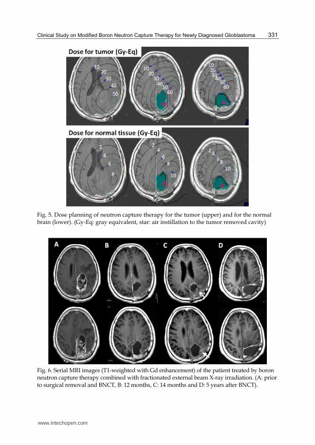
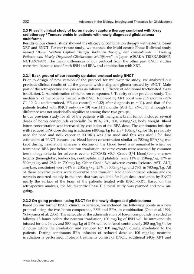
A 63-year old female had a left parietal tumor removed partially in a hospital in November 2004. The histopathological diagnosis was GB. She was introduced to our hospital for BNCT for the remaining lesion. While she waited for the BNCT, rapid growth of the tumor caused aggravations of right hemiparesis, total aphasia and consciousness disturbance, all of which caused us to remove the tumor again in our hospital prior to BNCT. The tumor was gross totally removed, and the patient received BNCT with air instillation in tumor removed cavity, followed by XRT. At the time of BNCT, the workstation calculates two different estimated doses, one is for the tumor and the other is for the normal brain (Fig. 5). Dose calculation shows estimated gray-equivalent (Gy-Eq) isodose lines from BNCT. This is mainly based on the decay of the irradiated neutron, boron concentration and biological effectiveness of each tissue. The deepest part of the tumor was 7.5 cm from the scalp in this case. The irradiated minimum tumor dose by BNCT was improved by the air-instilation (star in the Fig. 5) methods from 18.9 Gy-Eq (without air) to 26.9 Gy-Eq (with air). The maximum irradiated point of the normal brain was 2 cm from the scalp and was 12.7 Gy-Eq and scalp surface was 9.0 Gy-Eq. An additional 20 Gy was applied by XRT (daily fraction of 2 Gy x 10 days) aimed for the deep part of the mass but included surface of the tumor and the normal brain. No tumor recurrence on MRI and no neurological deficit were seen for 13 months after BNCT (Fig. 6). BNCT followed by fractionated XRT could control the tumor for 12 months (Fig. 6B). However, 14 months after BNCT, the patient suffered from right hemiparesis and MRI revealed a Gd-enhanced lesion from the surface area of the tumor removed cavity which was irradiated by BNCT with enough dose and also included into the boost XRT (Fig. 6C). Radiation necrosis was suspected as the pathological condition based on L/N ratio in BPA-PET, but we could not neglect the possibility of a recurrence of the GB. We performed a re-craniotomy to remove this lesion. The pathological diagnosis was radiation necrosis; no apparent tumor cells were found by pathologists. After the surgery, the right hemiparesis was improved, while no tumor progression was observed on MRI in the follow-up for more than 5 years (Fig. 6D). The patient received best medical treatment (anti-coagulants and vitamin E) for a year to prevent a further radiation necrosis.
www.intechopen.com
Clinical Study on Modified Boron Neutron Capture Therapy for Newly Diagnosed Glioblastoma
331
Fig. 5. Dose planning of neutron capture therapy for the tumor (upper) and for the normal brain (lower). (Gy-Eq: gray equivalent, star: air instillation to the tumor removed cavity)
Fig. 6. Serial MRI images (T1-weighted with Gd enhancement) of the patient treated by boron neutron capture therapy combined with fractionated external beam X-ray irradiation. (A: prior to surgical removal and BNCT, B: 12 months, C: 14 months and D: 5 years after BNCT).
www.intechopen.com
Advances in the Biology, Imaging and Therapies for Glioblastoma
332
2.3 Phase II clinical study of boron neutron capture therapy combined with X-ray radiotherapy / Temozolomide in patients with newly diagnosed glioblastoma multiforme
Results of our clinical study showed the efficacy of combination therapy with external beam XRT and BNCT. For our future study, we planned the Multi-centric Phase II clinical study named “Boron Neutron Capture Therapy, Radiation Therapy, and Temozolomide in Treating Patients with Newly Diagnosed Glioblastoma Multiforme” in Japan (OSAKA-TRIBRAIN0902, NCT00974987). The major differences of our protocol from the other past BNCT studies were simultaneous use of both BSH and BPA, and combination with XRT.
2.3.1 Back ground of our recently up-dated protocol using BNCT
Prior to design of new version of the protocol for multi-centric study, we analyzed our previous clinical results of all the patients with malignant glioma treated by BNCT. Main part of the retrospective analysis was as follows. 1. Efficacy of additional fractionated X-ray irradiation, 2. Administration of the boron compouns, 3. Toxicity of our previous study. The median ST of the patients treated with BNCT followed by XRT boost was 23.5 months (95% CI: 10. 2 – undetermined, HR (vs control) = 0.32) after diagnosis (n = 11), and that of the patients treated with BNCT only (n = 10) was 14.1 months (95% CI: 9.9–18.5), although the difference was not statistically significant among these two groups. In our previous study for all of the patients with malignant brain tumor included several
doses of boron compounds especially for BPA, 250, 500, 700mg/kg body weight. Blood
boron concentration was increased by escalation of the BPA dose. The continuous infusion
with reduced BPA dose during irradiation (400mg/kg for 2h + 100mg/kg for 1h, previously
used for head and neck cancer in KURRI) was also used and this was useful for dose
estimation of BNCT because the blood boron concentration similar as 700mg BPA/kg was
kept during irradiation whereas a decline of the blood level was remarkable when we
terminated BPA just before neutron irradiation. Adverse events were assessed by common
terminology criteria for adverse events (CTCAE) v3.0. Grade 3/4 blood/bone marrow
toxicity (hemoglobin, leukocytes, neutrophils, and platelets) were 11% in 250mg/kg, 17% in
500mg/kg, and 28% in 700mg/kg. Other Grade 3/4 adverse events (seizure, AST, ALT,
amylase, creatinine) were 64% in 250mg/kg, 25% in 500mg/kg, and 73% in 700mg/kg. All
of these adverse events were reversible and transient. Radiation induced edema and/or
necrosis occurred mainly in the area that was available for high-dose irradiation by BNCT
nearly the surface of the brain of the patients treated with BNCT+XRT. Based on this
retrospective analysis, the Multi-centric Phase II clinical study was planned and now on-
going.
2.3.2 On-going protocol using BNCT for the newly diagnosed glioblastoma
Based on our former BNCT clinical experience, we included the following points in a new
protocol using the two boron compounds, BSH and BPA, in combination (Ono et al. 1999,
Yokoyama et al. 2006). The schedule of the administration of boron compounds is settled as
follows; 13 hours before the neutron irradiation, 100 mg/kg of BSH will be intravenously
infused for one hour, and 500 mg/kg of BPA will be infused continuously 200 mg/kg/h for
2 hours before the irradiation and reduced for 100 mg/kg/h during irradiation to the
patients. During continuous BPA infusion of reduced dose as 100 mg/kg, neutrons
irradiation is performed. Protocol treatments consist of BNCT, additional 24Gy XRT and
www.intechopen.com

Clinical Study on Modified Boron Neutron Capture Therapy for Newly Diagnosed Glioblastoma
333
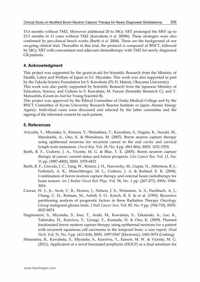
chemotherapy with TMZ. Prescription dose by BNCT is regulated as not to be more than
13Gy-Eq for normal brain. Additional XRT is given with 3 gradient such as 8, 16, 24Gy from
the surface of scalp to the bottom of tumor infiltrated zone (Fig. 7). Chemotherapy with
TMZ is applied concomitantly during XRT treatments and adjuvant chemotherapy with the
same agent is repeated in outpatient clinic (Fig. 8) (Stupp et al. 2005).
Fig. 7. Illustrated image of the protocol combined with boron neutron capture therapy (BNCT) and 3 gradient fractionated X-ray irradiation (XRT). Gy: gray, fr: fraction.
Based on our previous clinical study, the Hazard ratio of BNCT vs. XRT was simulated as
0.4, so the total estimated number of the patients who should be included in our new study
become 45 totally. Primary end point is overall survival and these patients will be followed
up for 2 years after the last patient treatment. The most important point in our protocol is
diagnosis and treatment of radiation effects such as swelling, radiation induced edema,
transient expansion of the tumor, pseudo- progression / response, and radiation necrosis. 18F-BPA-PET study is included for the diagnosis of these pathologies.
www.intechopen.com
Advances in the Biology, Imaging and Therapies for Glioblastoma
334
Fig. 8. Treatment protocol of BNCT combination with EBRT/TMZ for Newly diagnosed Malignant Glioma
3. Conclusion
Glioblastoma is currently not curable and the prognosis of it is very poor. A world-wide
standard care of newly-diagnosed GB is postoperative XRT with concomitant and adjuvant
chemotherapy with new alkylating agent TMZ(Stupp et al. 2005). This standard treatment
for newly diagnosed GB prolonged the median ST of patients from 12.1 months to 14.6
months in comparison with XRT alone, which is still pessimistic clinical result of this
disease. Therefore an alternative promising treatment should be developed for the
improvement of the prognosis of newly diagnosed GB. Several recent clinical studies on the
treatment of patients with GB by means of BNCT have reported encouraging results.
Careful analysis of survival data from a study, carried out in Sweden (Skold et al.) in which
BPA was administered at a higher dose over a longer period of time (Skold et al.), suggested
that a subset of patients had survival times that were at least as good as those obtained with
conventional therapy consisting of X-irradiation in combination with TMZ (Stupp et al.
2005).
On the other hand, BNCT is tumor-selective particle radiation. Tumor-seeking boron compounds boronophenylalanine (BPA) and sodium borocapate (BSH) can be delivered selectively in GB tissue with high contrast of accumulation in comparison with normal brain tissue. This tumor selective accumulation of boron compounds is followed by neutron irradiation, which produced high linear energy transfer particles (alpha particle and re-coiled Li nucleus). Thereafter these particles can destroy tumor cells selectively with high efficiency (Barth et al. 2005). The principal investigator of this clinical trial published the excellent survival data of 21 cases of newly diagnosed GB treated by BNCT with the MST of
www.intechopen.com
Clinical Study on Modified Boron Neutron Capture Therapy for Newly Diagnosed Glioblastoma
335
15.6 months without TMZ. Moreover additional 20 to 30Gy XRT prolonged the MST up to 23.5 months in 11 cases without TMZ (Kawabata et al. 2009b). These strategies were also confirmed by pre-clinical bench works (Barth et al. 2004). These are the background of our on-going clinical trial. Thereafter in this trial, the protocol is composed of BNCT, followed by 24Gy XRT with concomitant and adjuvant chemotherapy with TMZ for newly diagnosed GB patients.
4. Acknowledgment
This project was supported by the grant-in-aid for Scientific Research from the Ministry of Health, Labor and Welfare of Japan to S-I. Miyatake. This work was also supported in part by the Takeda Science Foundation for S. Kawabata (PI; H. Matsui, Okayama University). This work was also partly supported by Scientific Research from the Japanese Ministry of Education, Science, and Culture to S. Kawabata, M. Furuse (Scientific Research C), and Y. Matsushita (Grant-in-Aid for Young Scientist B). This project was approved by the Ethical Committee of Osaka Medical College and by the BNCT Committee of Kyoto University Research Reactor Institute or Japan Atomic Energy Agency. Individual cases were discussed and selected by the latter committee and the signing of the informed consent by each patient.
5. References
Ariyoshi, Y., Miyatake, S., Kimura, Y., Shimahara, T., Kawabata, S., Nagata, K., Suzuki, M.,
Maruhashi, A., Ono, K. & Shimahara, M. (2007). Boron neuron capture therapy
using epithermal neutrons for recurrent cancer in the oral cavity and cervical
lymph node metastasis. Oncol Rep, Vol. 18, No. 4 pp. (861-866), ISSN. 1021-335X
Barth, R. F., Coderre, J. A., Vicente, M. G. & Blue, T. E. (2005). Boron neutron capture
therapy of cancer: current status and future prospects. Clin Cancer Res, Vol. 11, No.
11 pp. (3987-4002), ISSN. 1078-0432
Barth, R. F., Grecula, J. C., Yang, W., Rotaru, J. H., Nawrocky, M., Gupta, N., Albertson, B. J.,
Ferketich, A. K., Moeschberger, M. L., Coderre, J. A. & Rofstad, E. K. (2004).
Combination of boron neutron capture therapy and external beam radiotherapy for
brain tumors. Int J Radiat Oncol Biol Phys, Vol. 58, No. 1 pp. (267-277), ISSN. 0360-
3016
Curran, W. J., Jr., Scott, C. B., Horton, J., Nelson, J. S., Weinstein, A. S., Fischbach, A. J.,
Chang, C. H., Rotman, M., Asbell, S. O., Krisch, R. E. & et al. (1993). Recursive
partitioning analysis of prognostic factors in three Radiation Therapy Oncology
Group malignant glioma trials. J Natl Cancer Inst, Vol. 85, No. 9 pp. (704-710), ISSN.
0027-8874
Haginomori, S., Miyatake, S., Inui, T., Araki, M., Kawabata, S., Takamaki, A., Lee, K.,
Takenaka, H., Kuroiwa, T., Uesugi, Y., Kumada, H. & Ono, K. (2009). Planned
fractionated boron neutron capture therapy using epithermal neutrons for a patient
with recurrent squamous cell carcinoma in the temporal bone: a case report. Head
Neck, Vol. 31, No. 3 pp. (412-418), ISSN. 1097-0347 (Electronic), 1043-3074 (Linking)
Hiramatsu, R., Kawabata, S., Miyatake, S., Kuroiwa, T., Easson, M. W. & Vicente, M. G.
(2011). Application of a novel boronated porphyrin (HOCP) as a dual sensitizer for
www.intechopen.com

Advances in the Biology, Imaging and Therapies for Glioblastoma
336
both PDT and BNCT. Lasers Surg Med, Vol. 43, No. 1 pp. (52-58), ISSN. 1096-9101
(Electronic), 0196-8092 (Linking)
Imahori, Y., Ueda, S., Ohmori, Y., Sakae, K., Kusuki, T., Kobayashi, T., Takagaki, M., Ono,
K., Ido, T. & Fujii, R. (1998). Positron emission tomography-based boron neutron
capture therapy using boronophenylalanine for high-grade gliomas: part II. Clin
Cancer Res, Vol. 4, No. 8 pp. (1833-1841), ISSN. 1078-0432
Kawabata, S., Miyatake, S., Kajimoto, Y., Kuroda, Y., Kuroiwa, T., Imahori, Y., Kirihata, M.,
Sakurai, Y., Kobayashi, T. & Ono, K. (2003). The early successful treatment of
glioblastoma patients with modified boron neutron capture therapy. Report of two
cases. J Neurooncol, Vol. 65, No. 2 pp. (159-165), ISSN. 0167-594X
Kawabata, S., Miyatake, S., Kuroiwa, T., Yokoyama, K., Doi, A., Iida, K., Miyata, S.,
Nonoguchi, N., Michiue, H., Takahashi, M., Inomata, T., Imahori, Y., Kirihata, M.,
Sakurai, Y., Maruhashi, A., Kumada, H. & Ono, K. (2009a). Boron neutron capture
therapy for newly diagnosed glioblastoma. J Radiat Res (Tokyo), Vol. 50, No. 1 pp.
(51-60), ISSN. 0449-3060
Kawabata, S., Miyatake, S., Nonoguchi, N., Hiramatsu, R., Iida, K., Miyata, S., Yokoyama,
K., Doi, A., Kuroda, Y., Kuroiwa, T., Michiue, H., Kumada, H., Kirihata, M.,
Imahori, Y., Maruhashi, A., Sakurai, Y., Suzuki, M., Masunaga, S. & Ono, K.
(2009b). Survival benefit from boron neutron capture therapy for the newly
diagnosed glioblastoma patients. Appl Radiat Isot, Vol. 67, No. 7-8 Suppl pp. (S15-
18), ISSN. 1872-9800 (Electronic), 0969-8043 (Linking)
Kimura, Y., Ariyoshi, Y., Miyatake, S., Shimahara, M., Kawabata, S. & Ono, K. (2009). Boron
neutron capture therapy for papillary cystadenocarcinoma in the upper lip: a case
report. Int J Oral Maxillofac Surg, Vol. 38, No. 3 pp. (293-295), ISSN. 1399-0020
(Electronic), 901-5027 (Linking)
Miyashita, M., Miyatake, S., Imahori, Y., Yokoyama, K., Kawabata, S., Kajimoto, Y., Shibata,
M. A., Otsuki, Y., Kirihata, M., Ono, K. & Kuroiwa, T. (2008). Evaluation of
fluoride-labeled boronophenylalanine-PET imaging for the study of radiation
effects in patients with glioblastomas. J Neurooncol, Vol. 89, No. 2 pp. (239-246),
ISSN. 0167-594X
Miyata, S., Kawabata, S., Hiramatsu, R., Doi, A., Ikeda, N., Yamashita, T., Kuroiwa, T.,
Kasaoka, S., Maruyama, K. & Miyatake, S. I. (2011). CT imaging of transferrin
targeting liposomes encapsulating both boron and iodine contrast agent by CED to
F98 rat glioma for boron neutron capture therapy. Neurosurgery, Vol., No., ISSN.
1524-4040 (Electronic), 0148-396X (Linking)
Miyatake, S., Kawabata, S., Kajimoto, Y., Aoki, A., Yokoyama, K., Yamada, M., Kuroiwa, T.,
Tsuji, M., Imahori, Y., Kirihata, M., Sakurai, Y., Masunaga, S., Nagata, K.,
Maruhashi, A. & Ono, K. (2005). Modified boron neutron capture therapy for
malignant gliomas performed using epithermal neutron and two boron compounds
with different accumulation mechanisms: an efficacy study based on findings on
neuroimages. J Neurosurg, Vol. 103, No. 6 pp. (1000-1009), ISSN. 0022-3085
Miyatake, S., Kawabata, S., Yokoyama, K., Kuroiwa, T., Michiue, H., Sakurai, Y., Kumada,
H., Suzuki, M., Maruhashi, A., Kirihata, M. & Ono, K. (2009). Survival benefit of
www.intechopen.com

Clinical Study on Modified Boron Neutron Capture Therapy for Newly Diagnosed Glioblastoma
337
Boron neutron capture therapy for recurrent malignant gliomas. J Neurooncol, Vol.
91, No. 2 pp. (199-206), ISSN. 0167-594X
Ono, K. (2006). central shielding. In Y. Nakagawa, T. Kobayashi & H. Fukuda (Eds.),
Advances in Neutron Capture Therapy 2006: "From the past to the future", Proceedings of
12th International Congress on Neutron Capture Therapy. Kagawa, Japan.
Ono, K., Masunaga, S., Suzuki, M., Kinashi, Y., Takagaki, M. & Akaboshi, M. (1999). The
combined effect of boronophenylalanine and borocaptate in boron neutron capture
therapy for SCCVII tumors in mice. Int J Radiat Oncol Biol Phys, Vol. 43, No. 2 pp.
(431-436), ISSN. 0360-3016
Ono, K., Sakurai, Y., Masunaga, S., Kinashi, Y., Takagaki, M. & Kobayashi, T. (2000).
Improvement of B-10 dose distribution in water phantom irradiated with
epithermal neutron beam and its assessment by colonyformation assay. Program &
Abstracts of the Ninth International Symposium on Neutron Capture Therapy for Cancer
(Kyoto University Research Reactor Institute). Kyoto University Research Reactor
Institute: Osaka, Japan.
Sakurai, Y., Ono, K., Miyatake, S. & Maruhashi, A. (2006). Improvement effect on the depth-
dose distribution by CSF drainage and air infusion of a tumour-removed cavity in
boron neutron capture therapy for malignant brain tumours. Phys Med Biol, Vol. 51,
No. 5 pp. (1173-1183), ISSN. 0031-9155
Skold, K., B, H. S., Diaz, A. Z., Giusti, V., Pellettieri, L. & Hopewell, J. W. (2010a). Boron
Neutron Capture Therapy for glioblastoma multiforme: advantage of prolonged
infusion of BPA-f. Acta Neurol Scand, Vol. 122, No. 1 pp. (58-62), ISSN. 1600-0404
(Electronic), 0001-6314 (Linking)
Skold, K., Gorlia, T., Pellettieri, L., Giusti, V., B, H. S. & Hopewell, J. W. (2010b). Boron
neutron capture therapy for newly diagnosed glioblastoma multiforme: an
assessment of clinical potential. Br J Radiol, Vol. 83, No. 991 pp. (596-603), ISSN.
1748-880X (Electronic), 0007-1285 (Linking)
Stupp, R., Hegi, M. E., Mason, W. P., van den Bent, M. J., Taphoorn, M. J., Janzer, R. C.,
Ludwin, S. K., Allgeier, A., Fisher, B., Belanger, K., Hau, P., Brandes, A. A.,
Gijtenbeek, J., Marosi, C., Vecht, C. J., Mokhtari, K., Wesseling, P., Villa, S.,
Eisenhauer, E., Gorlia, T., Weller, M., Lacombe, D., Cairncross, J. G. & Mirimanoff,
R. O. (2009). Effects of radiotherapy with concomitant and adjuvant temozolomide
versus radiotherapy alone on survival in glioblastoma in a randomised phase III
study: 5-year analysis of the EORTC-NCIC trial. Lancet Oncol, Vol. 10, No. 5 pp.
(459-466), ISSN. 1474-5488 (Electronic), 1470-2045 (Linking)
Stupp, R., Mason, W. P., van den Bent, M. J., Weller, M., Fisher, B., Taphoorn, M. J.,
Belanger, K., Brandes, A. A., Marosi, C., Bogdahn, U., Curschmann, J., Janzer, R. C.,
Ludwin, S. K., Gorlia, T., Allgeier, A., Lacombe, D., Cairncross, J. G., Eisenhauer, E.
& Mirimanoff, R. O. (2005). Radiotherapy plus concomitant and adjuvant
temozolomide for glioblastoma. N Engl J Med, Vol. 352, No. 10 pp. (987-996), ISSN.
1533-4406 (Electronic), 0028-4793 (Linking)
Wu, G., Yang, W., Barth, R. F., Kawabata, S., Swindall, M., Bandyopadhyaya, A. K., Tjarks,
W., Khorsandi, B., Blue, T. E., Ferketich, A. K., Yang, M., Christoforidis, G. A.,
Sferra, T. J., Binns, P. J., Riley, K. J., Ciesielski, M. J. & Fenstermaker, R. A. (2007).
www.intechopen.com

Advances in the Biology, Imaging and Therapies for Glioblastoma
338
Molecular targeting and treatment of an epidermal growth factor receptor-positive
glioma using boronated cetuximab. Clin Cancer Res, Vol. 13, No. 4 pp. (1260-1268),
ISSN. 1078-0432
Yang, W., Barth, R. F., Wu, G., Kawabata, S., Sferra, T. J., Bandyopadhyaya, A. K., Tjarks, W.,
Ferketich, A. K., Moeschberger, M. L., Binns, P. J., Riley, K. J., Coderre, J. A.,
Ciesielski, M. J., Fenstermaker, R. A. & Wikstrand, C. J. (2006). Molecular targeting
and treatment of EGFRvIII-positive gliomas using boronated monoclonal antibody
L8A4. Clin Cancer Res, Vol. 12, No. 12 pp. (3792-3802), ISSN. 1078-0432
Yokoyama, K., Miyatake, S., Kajimoto, Y., Kawabata, S., Doi, A., Yoshida, T., Asano, T.,
Kirihata, M., Ono, K. & Kuroiwa, T. (2006). Pharmacokinetic study of BSH and BPA
in simultaneous use for BNCT. J Neurooncol, Vol. 78, No. 3 pp. (227-232), ISSN.
0167-594X
www.intechopen.com

Advances in the Biology, Imaging and Therapies for GlioblastomaEdited by Prof. Clark Chen
ISBN 978-953-307-284-5Hard cover, 424 pagesPublisher InTechPublished online 09, November, 2011Published in print edition November, 2011
InTech EuropeUniversity Campus STeP Ri Slavka Krautzeka 83/A 51000 Rijeka, Croatia Phone: +385 (51) 770 447 Fax: +385 (51) 686 166www.intechopen.com
InTech ChinaUnit 405, Office Block, Hotel Equatorial Shanghai No.65, Yan An Road (West), Shanghai, 200040, China
Phone: +86-21-62489820 Fax: +86-21-62489821
This book is intended for physicians and scientists with interest in glioblastoma biology, imaging and therapy.Select topics in DNA repair are presented here to demonstrate novel paradigms as they relate to therapeuticstrategies. The book should serve as a supplementary text in courses and seminars as well as a generalreference.
How to referenceIn order to correctly reference this scholarly work, feel free to copy and paste the following:
Shinji Kawabata, Yoko Matsushita, Motomasa Furuse, Shin-Ichi Miyatake, Toshihiko Kuroiwa and Koji Ono(2011). Clinical Study on Modified Boron Neutron Capture Therapy for Newly Diagnosed Glioblastoma,Advances in the Biology, Imaging and Therapies for Glioblastoma, Prof. Clark Chen (Ed.), ISBN: 978-953-307-284-5, InTech, Available from: http://www.intechopen.com/books/advances-in-the-biology-imaging-and-therapies-for-glioblastoma/clinical-study-on-modified-boron-neutron-capture-therapy-for-newly-diagnosed-glioblastoma

© 2011 The Author(s). Licensee IntechOpen. This is an open access articledistributed under the terms of the Creative Commons Attribution 3.0License, which permits unrestricted use, distribution, and reproduction inany medium, provided the original work is properly cited.