clinical study influence of second-trimester ultrasound...
TRANSCRIPT
Clinical StudyInfluence of Second-Trimester Ultrasound Markers for DownSyndrome in Pregnant Women of Advanced Maternal Age
Mariza Rumi Kataguiri, Edward Araujo Júnior, Luiz Claudio Silva Bussamra,Luciano Marcondes Machado Nardozza, and Antonio Fernandes Moron
Department of Obstetrics, Paulista School of Medicine—Federal University of Sao Paulo (EPM-UNIFESP),Rua Carlos Weber, 956, apto. 113 Visage, Vila Leopoldina, 05303-000 Sao Paulo, Brazil
Correspondence should be addressed to Edward Araujo Junior; [email protected]
Received 24 January 2014; Revised 22 February 2014; Accepted 24 February 2014; Published 25 March 2014
Academic Editor: R. L. Deter
Copyright © 2014 Mariza Rumi Kataguiri et al. This is an open access article distributed under the Creative Commons AttributionLicense, which permits unrestricted use, distribution, and reproduction in any medium, provided the original work is properlycited.
The objective of the present study was to evaluate the influence of second-trimester ultrasound markers on the incidence ofDown syndrome among pregnant women of advanced maternal age. This was a retrospective cohort study on 889 singletonpregnancies between the 14th and 30th weeks, with maternal age ≥ 35 years, which would undergo genetic amniocentesis. Thesecond-trimester ultrasound assessed the following markers: increased nuchal fold thickness, cardiac hyperechogenic focus, mildventriculomegaly, choroid plexus cysts, uni- or bilateral renal pyelectasis, intestinal hyperechogenicity, single umbilical artery,short femur and humerus length, hand/foot alterations, structural fetal malformation, and congenital heart disease. To investigatedifferences between the groups with and without markers, nonparametric tests consisting of the chi-square test or Fisher’s exact testwere used. Moreover, odds ratios with their respective 95% confidence intervals were calculated. Out of the 889 pregnant women,131 (17.3%) presented markers and 758 (82.7%) did not present markers on the second-trimester ultrasound. Increased nuchal fold(𝑃 < 0.001) and structural malformation (𝑃 < 0.001) were the markers most associated with Down syndrome.The presence of onemarker increased the relative risk 10.5-fold, while the presence of two or more markers increased the risk 13.5-fold.The presence ofmarkers on the second-trimester ultrasound, especially thickened nuchal fold and structural malformation, increased the risk ofDown syndrome among pregnant women with advanced maternal age.
1. Introduction
Although chromosomal abnormalities occur at low fre-quency in the population, around 0.5% to 2% [1], theycontribute significantly to increased perinatal morbidity andmortality [2]. Trisomy is the most frequent chromosomalabnormality, especially of chromosome 21, that is, Downsyndrome. Since Down syndrome is difficult to diagnoseduring the prenatal period and because there is the possibilityof survival after birth, it contributes to increasing the statisticsof cases of mental retardation. Thus, prenatal screening isimportant, especially among women of advanced maternalage, that is, greater than or equal to 35 years [3].
The presence of certain alterations on the second-trimester ultrasound, called markers, enables increased sen-sitivity in screening for trisomy 21. This can reach up to 84%
and possibly surpass 90% when heart markers are included[4–7].
The challenge is to distinguish the presence of these smallalterations on the second-trimester ultrasound, betweenchromosomally abnormal and normal fetuses, consideringthat the latter may also present these markers at a rate ofaround 13% to 17%, which can be considered to be a highpercentage of false positives [8]. The importance of thischallenge is greater among pregnant women of advancedmaternal age, when the relationship with Down syndromebecomes closer [9]. If, on the one hand, these markers enablegood detection of aneuploidy, on the other, they lead toan unacceptably high rate of invasive procedures, therebyexposing chromosomally normal fetuses to a risk of deathor unnecessary complications [10]. Thus, second-trimesterultrasound can be used as a method to assist in evaluating
Hindawi Publishing CorporationJournal of PregnancyVolume 2014, Article ID 785730, 6 pageshttp://dx.doi.org/10.1155/2014/785730

2 Journal of Pregnancy
the risk of fetal aneuploidy, thereby making it possible for thecouple to better evaluate the need for an invasive examinationto assess the fetal karyotype [11].
The objective of the present study was to determineassociations that might exist between some second-trimesterultrasound markers and Down syndrome, among pregnantwomen of advance maternal age, who underwent fetal chro-mosomal analysis through amniocentesis.
2. Materials and Methods
This was a retrospective cohort study on 889 singletonpregnant women aged ≥ 35 years, who underwent geneticamniocentesis. The present study was approved by theResearch Ethics Committee of the Federal University of SaoPaulo (UNIFESP). All the patients who voluntarily agreed toparticipate signed a consent form.
The present study was carried out at the Departmentof Obstetrics of UNIFESP and at Santa Joana Hospital, SaoPaulo, SP, Brazil. Singleton pregnancies between their 14thand 30th weeks that would undergo genetic amniocentesisbecause of maternal age ≥ 35 years, with or without markersfor Down syndrome on the second-trimester ultrasound,were selected. Pregnant women with a history of genitalbleeding over the past seven days, suspected premature rup-ture of membranes, fetal heart rate abnormalities, suspectedcongenital infection (from ultrasound alterations), or use ofpotentially teratogenic drugs were excluded.
The second-trimester ultrasound was performed beforethe genetic amniocentesis, using the Logic 500 device(General Electric Medical System, Milwaukee, WI, USA)equipped with a convex transducer (3–5MHz). In thisexamination, the presence of the following markers wasevaluated: increased nuchal fold thickness (≥6mm), mildventriculomegaly (atrium of the lateral ventricle measuringbetween 12 and 15mm), choroid plexus cysts, cardiac hypere-chogenic focus, unilateral or bilateral renal pyelectasis (renalpelvis measuring ≥4mm between the 15th and 20th weeksand ≥5mm between the 21st and 30th weeks), intestinalhyperechogenicity, single umbilical artery, short femur length(observed/expected measurement < 0.90), short humeruslength (observed/expected measurement < 0.89), alterationsat the extremities such as the shape or position of hands orfeet, structural malformations, or congenital heart diseases.
To carry out the amniocentesis, a 20mL syringe anda 20G needle were used after applying local anesthesiaconsisting of 2% lidocaine. 20mL of clear liquid were col-lected, without blood contamination, and were immediatelyforwarded to the cytogenetic laboratory. Pregnant womenwith a negative Rh blood type, negative indirect Coombs test,or a Rh-positive partner received anti-D immunoglobulinwithin 72 hours after the procedure, so as to avoid maternalsensitization.
Thedatawere transferred to aworksheet in the Excel 2003software (Microsoft Corp., Redmond, WA, USA) and wereanalyzed using the SPSS version 13.0 software for Windows(SPSS Inc., Chicago, IL, USA). Probability calculations were
Table 1: Description of the cases of Down syndrome according tothe groups with and without ultrasound markers (𝑃 < 0.001).
Group of pregnant women Down syndromePresent Absent Total
With markers 19 14.6% 112 85.5% 131 100.0%Without markers 12 1.6% 746 98.4% 758 100.0%Total 31 3.5% 858 96.5% 889 100.0%
used to search for any relationships between fetal malfor-mations and/or second-trimester ultrasound markers fordetecting Down syndrome in women of advanced maternalage, so as to observe whether there were any associationsbetween the presence of markers (singly or in combination)and the presence of Down syndrome and, for each of themarkers found, the association of each type of marker withthe syndrome. Initially, a descriptive analysis was performed,presenting the data as absolute frequencies (𝑁) and relativefrequencies (%) for each category of the qualitative variablesof the patients’ profile. For the quantitative variables, means,standard deviations, and minimum and maximum valueswere calculated. To investigate possible associations betweenpairs of characteristics or qualitative variables, nonparamet-ric tests consisting of the chi-square test or Fisher’s exact testwhen necessary were used, with a significance level of 5%.Combined associations of the variables were assessed in amultivariate manner, by means of correspondence analysis.The relative risk calculations and odds ratios were consideredsignificant when the data were within the 95% confidenceinterval.
3. Results
Thematernal age ranged from 35 to 47 years, with a mean of38.8 ± 2.9 years for the 31 pregnant women with fetuses withDown syndrome and 38.6 ± 2.6 years for the 858 pregnantwomen with chromosomally normal fetuses, without anystatistical difference. Similarly, gestational age at the timeof amniocentesis did not present any significant differencebetween the two groups, with a mean of 17.7 ± 3.1 weeksfor the Down syndrome cases and 17.3 ± 2.4 weeks for thechromosomally normal fetuses.
Among the 889 pregnant women, 131 (17.3%) presentedmarkers and 758 (82.7%) did not present markers on thesecond-trimester ultrasound.
Table 1 presents the distribution of the 31 fetuses withDown syndrome and their correlation with the ultrasoundmarkers, which presented a statistically significant difference(𝑃 < 0.001).
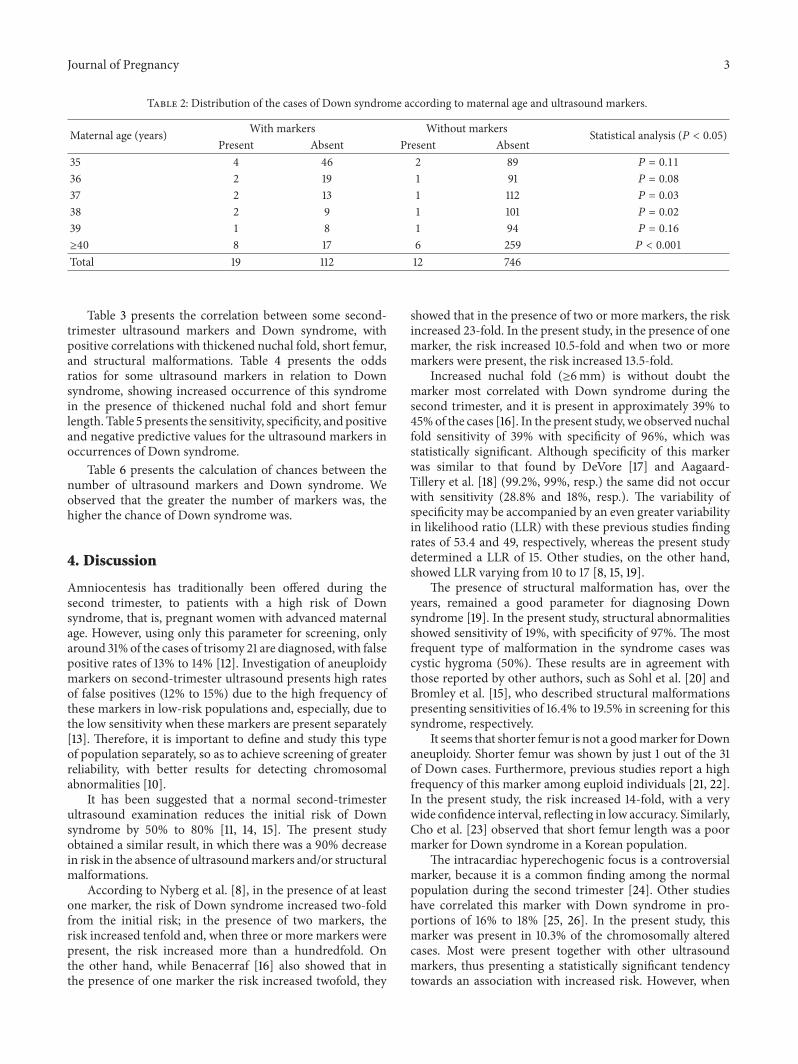
Table 2 presents the distribution of the Down syndromecases, between the two groups studied, with and withoutmarkers, according to maternal age, in which there was astatistically significant difference for the maternal ages of 37,38, and ≥40 years. The odds ratio calculation did not showany increased occurrence of Down syndrome among women≥40 years of age in the presence of ultrasound markers(OR = 1.74; 95% CI: 0.84–3.57).
Journal of Pregnancy 3
Table 2: Distribution of the cases of Down syndrome according to maternal age and ultrasound markers.
Maternal age (years) With markers Without markers Statistical analysis (𝑃 < 0.05)Present Absent Present Absent
35 4 46 2 89 𝑃 = 0.11
36 2 19 1 91 𝑃 = 0.08
37 2 13 1 112 𝑃 = 0.03
38 2 9 1 101 𝑃 = 0.02
39 1 8 1 94 𝑃 = 0.16
≥40 8 17 6 259 𝑃 < 0.001
Total 19 112 12 746
Table 3 presents the correlation between some second-trimester ultrasound markers and Down syndrome, withpositive correlations with thickened nuchal fold, short femur,and structural malformations. Table 4 presents the oddsratios for some ultrasound markers in relation to Downsyndrome, showing increased occurrence of this syndromein the presence of thickened nuchal fold and short femurlength. Table 5 presents the sensitivity, specificity, and positiveand negative predictive values for the ultrasound markers inoccurrences of Down syndrome.
Table 6 presents the calculation of chances between thenumber of ultrasound markers and Down syndrome. Weobserved that the greater the number of markers was, thehigher the chance of Down syndrome was.
4. Discussion
Amniocentesis has traditionally been offered during thesecond trimester, to patients with a high risk of Downsyndrome, that is, pregnant women with advanced maternalage. However, using only this parameter for screening, onlyaround 31% of the cases of trisomy 21 are diagnosed, with falsepositive rates of 13% to 14% [12]. Investigation of aneuploidymarkers on second-trimester ultrasound presents high ratesof false positives (12% to 15%) due to the high frequency ofthese markers in low-risk populations and, especially, due tothe low sensitivity when these markers are present separately[13]. Therefore, it is important to define and study this typeof population separately, so as to achieve screening of greaterreliability, with better results for detecting chromosomalabnormalities [10].
It has been suggested that a normal second-trimesterultrasound examination reduces the initial risk of Downsyndrome by 50% to 80% [11, 14, 15]. The present studyobtained a similar result, in which there was a 90% decreasein risk in the absence of ultrasoundmarkers and/or structuralmalformations.
According to Nyberg et al. [8], in the presence of at leastone marker, the risk of Down syndrome increased two-foldfrom the initial risk; in the presence of two markers, therisk increased tenfold and, when three or more markers werepresent, the risk increased more than a hundredfold. Onthe other hand, while Benacerraf [16] also showed that inthe presence of one marker the risk increased twofold, they
showed that in the presence of two or more markers, the riskincreased 23-fold. In the present study, in the presence of onemarker, the risk increased 10.5-fold and when two or moremarkers were present, the risk increased 13.5-fold.
Increased nuchal fold (≥6mm) is without doubt themarker most correlated with Down syndrome during thesecond trimester, and it is present in approximately 39% to45%of the cases [16]. In the present study, we observed nuchalfold sensitivity of 39% with specificity of 96%, which wasstatistically significant. Although specificity of this markerwas similar to that found by DeVore [17] and Aagaard-Tillery et al. [18] (99.2%, 99%, resp.) the same did not occurwith sensitivity (28.8% and 18%, resp.). The variability ofspecificity may be accompanied by an even greater variabilityin likelihood ratio (LLR) with these previous studies findingrates of 53.4 and 49, respectively, whereas the present studydetermined a LLR of 15. Other studies, on the other hand,showed LLR varying from 10 to 17 [8, 15, 19].
The presence of structural malformation has, over theyears, remained a good parameter for diagnosing Downsyndrome [19]. In the present study, structural abnormalitiesshowed sensitivity of 19%, with specificity of 97%. The mostfrequent type of malformation in the syndrome cases wascystic hygroma (50%). These results are in agreement withthose reported by other authors, such as Sohl et al. [20] andBromley et al. [15], who described structural malformationspresenting sensitivities of 16.4% to 19.5% in screening for thissyndrome, respectively.
It seems that shorter femur is not a goodmarker forDownaneuploidy. Shorter femur was shown by just 1 out of the 31of Down cases. Furthermore, previous studies report a highfrequency of this marker among euploid individuals [21, 22].In the present study, the risk increased 14-fold, with a verywide confidence interval, reflecting in low accuracy. Similarly,Cho et al. [23] observed that short femur length was a poormarker for Down syndrome in a Korean population.
The intracardiac hyperechogenic focus is a controversialmarker, because it is a common finding among the normalpopulation during the second trimester [24]. Other studieshave correlated this marker with Down syndrome in pro-portions of 16% to 18% [25, 26]. In the present study, thismarker was present in 10.3% of the chromosomally alteredcases. Most were present together with other ultrasoundmarkers, thus presenting a statistically significant tendencytowards an association with increased risk. However, when

4 Journal of Pregnancy
Table 3: Correlation between ultrasound markers and Down syndrome.
Marker Down syndrome Statistical analysisPresent Absent
Nuchal fold thickness ≥ 6mm 12/31 35/858 𝑃 < 0.001
Intracardiac hyperechogenic focus 4/31 35/858 𝑃 = 0.05
Short femur length 1/31 2/858 𝑃 = 0.10
Structural malformation 6/31 26/858Duodenal Atresia 1 5
𝑃 < 0.001
Esophageal Atresia 1 4Cystic Hygroma 3 0Meningomyelocele 0 6Spina Bifida 0 4Congenital heart disease 1 2Hydrocephalus 0 5
Table 4: Odds ratios for the ultrasound markers for Down syndrome.
Marker Down syndrome Odds ratio Confidence interval (95%)Absent Present
Nuchal fold thickness ≥ 6mm Present 35 12 14.9 6.7–33.0Absent 823 19
Intracardiac hyperechogenic focus Present 35 4 3.5 1.1–10.5Absent 823 27
Short femur length Present 2 1 14.2 1.3–161.7Absent 856 30
Structural fetal malformation Present 26 6 7.7 2.9–20.3Absent 832 25
present separately, this marker did not present any significantassociation with Down syndrome.
The congenital heart disease (CHD) was present in onefetus with Down syndrome (1/31 = 3.2%) and in two euploidfetuses (2/858 = 0.23%). We included the cases of CHD in thegroup of structural malformations, because the number caseswere insufficient to correlate them with the Down syndrome.It has occurred because of the low gestational age at themoment of ultrasound exam (17 weeks), when the CHD ratedetection is smaller than after 20 weeks [27]. Furthermore,the ultrasound exams were realized by sonographers withoutexpertise in fetal echocardiography, justifying the low ratedetection. The main structural CHD in fetuses with Downsyndrome is atrioventricular septal defect [28]; however, thedetection rate of this CHD during prenatal ultrasound is low[29].
In the present study, we performed a statistical calculationcomparing the diagnosis of Down syndrome in the presenceand absence of ultrasound markers. In considering the cal-culation with two markers, the sensitivity reduced markedlyto 29.4%, but with an increase in specificity to 97%. Thus,we considered that second-trimester ultrasound had a highchance of screening for Down syndrome in the presence of atleast two markers in the population of pregnant women withadvanced maternal age.
The use of ultrasound markers during the second trimes-ter to screen for Down syndrome is subject to bias, since
there are still no definitive studies regarding the sensitivityand specificity of each marker or set of markers [30].However, through appropriate standardization of the typesof populations and the studies carried out, the analyses onthese markers will become better, because the interactions ofeach marker, separately or in association with other markers,will be evaluated, thereby resulting in a second-trimesterscreening for Down syndrome that is more reliable overall[31].
The accuracy of ultrasound as Down syndrome screeningexamination has been improved by combining its findings inthe first trimester of pregnancy with biochemical screeningin the first and second trimester (FASTER trial). Currently, amuch more accurate screening tool is available. Noninvasiveprenatal testing (NIPT) is able to detect trisomy 21 directlyon free fetus DNA fragments in mother’s blood stream. Ithas high accuracy; however, it is associated with highercosts and equipment availability, which is not universallyprovided, mainly in developing countries as Brazil. Thismakes genetic ultrasound a relatively reliable screening tool,thus supporting investigative work undertaken to better usethis tool.
5. Conclusion
In summary, increased nuchal fold and presence of structuralmalformation presented significant associations with Down

Journal of Pregnancy 5
Table 5: Sensitivity, specificity, positive predictive value (PPV), and negative predictive value (NPV) of the ultrasound markers for detectingDown syndrome.
Marker Sensitivity (%) Specificity (%) PPV (%) NPV (%)Nuchal fold thickness ≥ 6mm 38.7 95.9 25.5 97.7Short femur length 32.3 99.8 33.3 96.6Intracardiac hyperechogenic focus 12.9 95.9 10.3 96.8Structural fetal malformation 19.3 97.0 18.8 97.1
Table 6: Odds ratios between the number of ultrasound markers and Down syndrome.
Number of markers Down syndrome Odds ratio Confidence interval (95%)Present Absent
0 12 38.7 746 86.9 0.1 0.0–0.21 14 45.2 89 10.4 10.5 4.1–23.4≥2 5 16.1 23 2.7 13.5 3.8–46.2
syndrome, especially when associated with other ultrasoundmarkers, among pregnant women of advanced maternal age.
Conflict of Interests
The authors declare that there is no conflict of interestsregarding the publication of this paper.
References
[1] P. A. Baird, T. W. Anderson, H. B. Newcombe, and R. B. Lowry,“Genetic disorders in children and young adults: a populationstudy,”The American Journal of Human Genetics, vol. 42, no. 5,pp. 677–693, 1988.
[2] A. Sprigg, “Fetalmalformations diagnosed antenatally 1: generalprinciples,” British Journal of Hospital Medicine, vol. 54, no. 8,pp. 387–390, 1995.
[3] R. J. M. Snijders, K. Sundberg, W. Holzgreve, G. Henry, andK. H. Nicolaides, “Maternal age and gestation-specific risk fortrisomy 21,”Ultrasound in Obstetrics and Gynecology, vol. 13, no.3, pp. 167–170, 1999.
[4] B. R. Benacerraf, D. Neuberg, B. Bromley, and F. D. FrigolettoJr., “Sonographic scoring index for prenatal detection of chro-mosomal abnormalities,” Journal of Ultrasound inMedicine, vol.11, no. 9, pp. 449–458, 1992.
[5] D. A. Nyberg, D. A. Luthy, E. Y. Cheng, R. C. Sheley, R. G.Resta, and M. A. Williams, “Role of prenatal ultrasonographyin women with positive screen for Down syndrome on the basisof maternal serummarkers,”TheAmerican Journal of Obstetricsand Gynecology, vol. 173, no. 4, pp. 1030–1035, 1995.
[6] G. R. DeVore and R. Romero, “Genetic sonography: an optionfor women of advanced maternal age with negative triple-marker maternal serum screening results,” Journal of Ultra-sound in Medicine, vol. 22, no. 11, pp. 1191–1199, 2003.
[7] D. Wellesley, C. de Vigan, N. Baena et al., “Contributionof ultrasonographic examination to the prenatal detection oftrisomy 21: experience from 19 European registers,” Annales deGenetique, vol. 47, no. 4, pp. 373–380, 2004.
[8] D. A. Nyberg, V. L. Souter, A. El-Bastawissi, S. Young, F.Luthhardt, and D. A. Luthy, “Isolated sonographic markers fordetection of fetal Down syndrome in the second trimester of
pregnancy,” Journal of Ultrasound in Medicine, vol. 20, no. 10,pp. 1053–1063, 2001.
[9] A. M. Vintzileos, W. A. Campbell, E. R. Guzman, J. C. Smulian,D. A. Mclean, and C. V. Ananth, “Second-trimester ultrasoundmarkers for detection of trisomy 21: which markers are best?”Obstetrics and Gynecology, vol. 89, no. 6, pp. 941–944, 1997.
[10] D. A. Nyberg, D. A. Luthy, R. G. Resta, B. C. Nyberg, and M.A. Williams, “Age-adjusted ultrasound risk assessment for fetalDown’s syndrome during the second trimester: description ofthe method and analysis of 142 cases,” Ultrasound in Obstetricsand Gynecology, vol. 12, no. 1, pp. 8–14, 1998.
[11] D. A. Nyberg and V. L. Souter, “Use of genetic sonographyfor adjusting the risk for fetal Down syndrome,” Seminars inPerinatology, vol. 27, no. 2, pp. 130–144, 2003.
[12] J. F. X. Egan, P. Benn, A. F. Borgida, J. F. Rodis, W. A. Campbell,and A. M. Vintzileos, “Efficacy of screening for fetal Downsyndrome in the United States from 1974 to 1997,”Obstetrics andGynecology, vol. 96, no. 6, pp. 979–985, 2000.
[13] J. C. Hobbins, D. C. Lezotte, W. H. Persutte et al., “An 8-center study to evaluate the utility of midterm genetic sono-grams among high-risk pregnancies,” Journal of Ultrasound inMedicine, vol. 22, no. 1, pp. 33–38, 2003.
[14] A. M. Vintzileos, C. V. Ananth, J. C. Smulian, D. L. Day-Salvatore, T. Beazoglou, and R. A. Knuppel, “Cost-benefitanalysis of prenatal diagnosis for Down syndrome using theBritish or the American approach,” Obstetrics and Gynecology,vol. 95, no. 4, pp. 577–583, 2000.
[15] B. Bromley, E. Lieberman, T. D. Shipp, and B. R. Benacerraf,“The genetic sonogram: a method of risk assessment for Downsyndrome in the second trimester,” Journal of Ultrasound inMedicine, vol. 21, no. 10, pp. 1087–1096, 2002.
[16] B. R. Benacerraf, “Use of sonographic markers to determine therisk of Down syndrome in second-trimester fetuses,”Radiology,vol. 201, no. 3, pp. 619–620, 1996.
[17] G. R. DeVore, “Genetic sonography: the historical and clinicalrole of fetal echocardiography,” Ultrasound in Obstetrics andGynecology, vol. 35, no. 5, pp. 509–521, 2010.
[18] K. M. Aagaard-Tillery, F. D. Malone, D. A. Nyberg et al., “Roleof second-trimester genetic sonography after Down syndromescreening,” Obstetrics and Gynecology, vol. 114, no. 6, pp. 1189–1196, 2009.
[19] R. Smith-Bindman, P. Chu, and J. D. Goldberg, “Secondtrimester prenatal ultrasound for the detection of pregnancies

6 Journal of Pregnancy
at increased risk of Down syndrome,” Prenatal Diagnosis, vol.27, no. 6, pp. 535–544, 2007.
[20] B. D. Sohl, A. L. Scioscia, N. E. Budorick, and T. R. Moore,“Utility of minor ultrasonographic markers in the prediction ofabnormal fetal karyotype at a prenatal diagnostic center,” TheAmerican Journal of Obstetrics and Gynecology, vol. 181, no. 4,pp. 898–903, 1999.
[21] T. D. Shipp, B. Bromley, M. Mascola, and B. Benacerraf,“Variation in fetal femur length with respect to maternal race,”Journal of Ultrasound in Medicine, vol. 20, no. 2, pp. 141–144,2001.
[22] C. M. Kovac, J. A. Brown, C. C. Apodaca et al., “Mater-nal ethnicity and variation of fetal femur length calculationswhen screening for Down syndrome,” Journal of Ultrasound inMedicine, vol. 21, no. 7, pp. 719–722, 2002.
[23] H. J. Cho, H. S. Won, D. H. Ju, H. J. Roh, P. R. Lee, and A.Kim, “Evaluation of the usefulness of the fetal femur lengthwithrespect to gestational age to detect Down syndrome in Koreansubjects,” Prenatal Diagnosis, vol. 30, no. 8, pp. 734–738, 2010.
[24] T. D. Shipp, B. Bromley, E. Lieberman, and B. R. Benacerraf,“The frequency of the detection of fetal echogenic intracardiacfoci with respect to maternal race,”Ultrasound in Obstetrics andGynecology, vol. 15, no. 6, pp. 460–462, 2000.
[25] J. E. Manning, N. Ragavendra, J. Sayre et al., “Significance offetal intracardiac echogenic foci in relation to trisomy 21: aprospective sonographic study of high-risk pregnant women,”TheAmerican Journal of Roentgenology, vol. 170, no. 4, pp. 1083–1084, 1998.
[26] J. R. Wax, A. Cartin, M. G. Pinette, and J. Blackstone, “Areintracardiac echogenic foci markers of congenital heart diseasein the fetus with chromosomal abnormalities?” Journal ofUltrasound in Medicine, vol. 23, no. 7, pp. 895–898, 2004.
[27] G. R. DeVore, “The role of fetal echocardiography in geneticsonography,” Seminars in Perinatology, vol. 27, no. 2, pp. 160–172, 2003.
[28] R. Mogra, V. Zidere, and L. D. Allan, “Prenatally detectablecongenital heart defects in fetuses with Down syndrome,”Ultrasound in Obstetrics and Gynecology, vol. 38, no. 3, pp. 320–324, 2011.
[29] H. ter Heide, J. D. R. Thomson, G. A. Wharton, and J. L. Gibbs,“Poor sensitivity of routine fetal anomaly ultrasound screeningfor antenatal detection of atrioventricular septal defect,” Heart,vol. 90, no. 8, pp. 916–917, 2004.
[30] M. H. Graupe, C. S. Naylor, N. H. Greene, D. E. Carlson, andL. Platt, “Trisomy 21: second-trimester ultrasound,” Clinics inPerinatology, vol. 28, no. 2, pp. 303–319, 2001.
[31] P. J. Schluter andG. Pritchard, “Mid trimester sonographic find-ings for the prediction of Down syndrome in a sonographicallyscreened population,” The American Journal of Obstetrics andGynecology, vol. 192, no. 1, pp. 10–16, 2005.

Submit your manuscripts athttp://www.hindawi.com
Stem CellsInternational
Hindawi Publishing Corporationhttp://www.hindawi.com Volume 2014
Hindawi Publishing Corporationhttp://www.hindawi.com Volume 2014
MEDIATORSINFLAMMATION
of
Hindawi Publishing Corporationhttp://www.hindawi.com Volume 2014
Behavioural Neurology
EndocrinologyInternational Journal of
Hindawi Publishing Corporationhttp://www.hindawi.com Volume 2014
Hindawi Publishing Corporationhttp://www.hindawi.com Volume 2014
Disease Markers
Hindawi Publishing Corporationhttp://www.hindawi.com Volume 2014
BioMed Research International
OncologyJournal of
Hindawi Publishing Corporationhttp://www.hindawi.com Volume 2014
Hindawi Publishing Corporationhttp://www.hindawi.com Volume 2014
Oxidative Medicine and Cellular Longevity
Hindawi Publishing Corporationhttp://www.hindawi.com Volume 2014
PPAR Research
The Scientific World JournalHindawi Publishing Corporation http://www.hindawi.com Volume 2014
Immunology ResearchHindawi Publishing Corporationhttp://www.hindawi.com Volume 2014
Journal of
ObesityJournal of
Hindawi Publishing Corporationhttp://www.hindawi.com Volume 2014
Hindawi Publishing Corporationhttp://www.hindawi.com Volume 2014
Computational and Mathematical Methods in Medicine
OphthalmologyJournal of
Hindawi Publishing Corporationhttp://www.hindawi.com Volume 2014
Diabetes ResearchJournal of
Hindawi Publishing Corporationhttp://www.hindawi.com Volume 2014
Hindawi Publishing Corporationhttp://www.hindawi.com Volume 2014
Research and TreatmentAIDS
Hindawi Publishing Corporationhttp://www.hindawi.com Volume 2014
Gastroenterology Research and Practice
Hindawi Publishing Corporationhttp://www.hindawi.com Volume 2014
Parkinson’s Disease
Evidence-Based Complementary and Alternative Medicine
Volume 2014Hindawi Publishing Corporationhttp://www.hindawi.com