clinical skills lecture notes textbook
TRANSCRIPT
Clinical SkillsROBERT TURNERMD, FRCPThe late Professor of Medicine and Director of the Diabetes Research Laboratories,University of Oxford,Oxford
CHRIS HATTONFRCP, FRCPathConsultant Haematologist,Department of Haematology,The John Radcliffe Hospital,Oxford
ROGER BLACKWOODMA, FRCPConsultant Physician,Wexham Park Hospital, Slough,and Honorary Consultant Physician atHammersmith Hospital, London
Fourth edition
LECTURE NOTES ON
BlackwellScience
Contents
Preface, v
Acknowledgements, vii
Introduction:The First Approach, 1
1 History Taking, 6
2 General Examination, 26
3 Examination of the Cardiovascular System, 50
4 Examination of the Chest, 76
5 Examination of the Abdomen, 87
6 Examination of the Mental State, 101
7 Examination of the Nervous System, 111
8 Assessment of Disability Including Care of the Elderly, 154
9 Basic Examination, Notes and Diagnostic Principles, 161
10 Presenting Cases and Communication, 172
11 Imaging Techniques and Clinical Investigations, 181
12 The 12-Lead Electrocardiogram, 235
13 Interpretation of Investigations, 268
14 Laboratory Results —Normal Values, 271
15 Common Emergency Treatments, 278
Appendices, 289
1: Jaeger reading chart, 289
2:Visual acuity 3m chart, 292
iii
3: Hodkinson ten point mental test score, 294
4: Barthel index of activities of daily living, 296
5: Cardiac arrest instructions, 298
Index, 300
Colour plates 1–6 between pp. 152 and 153
iv Contents
Preface
v
Clinical Skills was the inspiration of Profesor Robert Turner. Roger Blackwood was his senior registrar in Oxford when, together, theyplanned and wrote the first edition. Roger Blackwood took his clinicalskills into cardiology and has remained an inspirational teacher to manymedical students and MRCP candidates. Sadly, Robert Turner died suddenly in 1999 leaving the book bereft of its senior author. Robert Turner was an outstanding clinical scientist and clinician and most of thecontent and flavour of the book remain his.The main focus of the book iscareful history taking and clinical examination.Whilst these skills remainthe mainstay of all medical practice, clinical medicine is changing. Inceasedsophistication of imaging and diagnostic techniques is resulting in greaterdiagnostic accuracy; however, the first meeting with the patient remainsmuch the same.The ‘bedside manner’ is still important and your approachto appropriate imaging and diagnostic procedures largely depends on thesimple history and examination taken at the outset.
The preface to previous editions started with the statement that whena medical student first approaches a patient, he has to:
° Develop a suitable doctor–patient relationship
° Master many relevant skills and techniques
° Develop an enquiring and intelligent approachNothing has changed. I should add that we have stuck with the same
convention of using the he pronoun when rferring to doctors, medicalstudents or patients.This is not meant to offend anyone, simply economi-cal linguistic convention.
In this new edition we have added some new sections on imaging andsimple ‘bedside relevant’ pathology tests.We have updated a number ofthe other chapters and we are gretly indebted to friends and colleagueswho have helped us.We are particularly indebted to Dr Dennis Briley for

his help with the neurology section. Remember, the most important skillfor any doctor is to be able to take a good history and perform a carefulexamination. Good Luck.
Chris HattonOxford 2003
vi Preface
Acknowledgements
We are grateful to many colleagues and students who have made sugges-tions. The book has benefited from their suggestions, but any faults oromissions are those of the authors. Specific advice was received from:
History Bill Rosenberg,Tony HopeSkin Terence Ryan, Sue BurgeEyes Peggy FrithOrthopaedics Roger Smith, Chris BulstrodeSurgery Jack Collins, Michael GreenallHeart Oliver OrmerodLungs Donald LaneGastroenterology Derek JewellMental state Michael SharpeNeurology Michael Donaghy, Robin Kennett, Dennis
BrileyDisability Simon Winner, Sebastian FairweatherImaging Niall Moore, David Lindsell,Wattie Fletcher,
Andy Molyneux, Basil ShepstoneEvidence-based medicine David SackettTherapies Chris Garrard, Lucy Belson, Ben AszaliBiochemistry and Jonathan Kay, Nicki Metson, Louise Bowman
endocrinologyHistory and examination Becky Rippon
The visual acuity reading charts (Appendices 1 and 2) are reproducedcourtesy of Keeler Ltd; and the cardiac arrest instructions (Appendix 5)are redrawn courtesy of the European Resuscitation Council (© 1994).
vii

The colour plates are reproduced courtesy of Department of MedicalIllustration, Heatherwood and Wexham Park Hospitals Trust (Plates 1a–d,2d, 2f, 3a, 3c–e, 4b, 5b–f, 6a, 6c–f), King Edward VII Hospital, Windsor(Plates 1e, 1f, 2a, 2b, 4a, 4c, 4e), Department of Medical Illustration, JohnRadcliffe Hospital, Oxford (Plates 2e, 4d, 5a, 6b), Department of MedicalIllustration, Radcliffe Infirmary, Oxford (Plates 3f, 4f).
viii Acknowledgements
INTRODUCTION
The First Approach
General principles
General objectivesWhen the student (or doctor) approaches a patient there are four initial objectives:
° Obtain a professional rapport with the patient and gain hisconfidence.
° Obtain all relevant information which allows assessment ofthe illness, and provisional diagnoses.
° Obtain general information regarding the patient, his back-ground, social situation and problems. In particular it is nec-essary to find out how the illness has affected him, his family,friends, colleagues and his life.
The assessment of the patient as a whole is of utmost importance.
° Understand the patient’s own ideas about his problems, hismajor concerns and what he expects from the hospital admission, outpatient or general practice consultation.
Remember medicine is just as much about worry as disease.Whatever the illness, whether chest infection or cancer,anxiety about what may happen is often uppermost in the patient’s mind. Listen attentively.
The following notes provide a guide as to how one obtains the necessary information.
Specific objectivesIn taking a history or making an examination there are two comple-mentary aims:
1
° Obtain all possible information about a patient and his illness(a database).
° Solve the problem as to the diagnoses.
Analytical approachFor each symptom or sign one needs to think of a differential diagnosis, and of other relevant information (by history, examination orinvestigation) which one will need to support or refute these possible diagnoses. A good history combines these two facets, and one shouldnever approach the patient with just a set series of rote questions. How-ever, until one knows more medicine, one cannot know the possible significance of the information one gains, and the obvious change of questioning which this might entail.These notes provide background in-formation enabling a full history and examination to be taken. This pro-vides a necessary basis for a later, more inquisitive approach which shoulddevelop as knowledge about illnesses is acquired.
Self-relianceThe student must take his own history, make his own examina-tion and write his own clinical records.After 1 month he should besufficiently proficient that his notes could become the final hospitalrecord. The student should add a summary including his assessment of the problem list, provisional diagnoses and preliminary investigations.Initially these will be incomplete and occasionally incorrect. Neverthe-less, this exercise will help to inculcate an enquiring approach and to highlight areas in which further questioning, investigation or reading isneeded.
What is important when you start?At the basis of all medicine is clinical competence. No amount ofknowledge will make up for poor technique.
Over the first few weeks it is essential to learn the basic ABC of clinical medicine, covered in these notes:
° how to relate to patients
° how to take a good history efficiently, knowing which ques-tion to ask next and avoiding leading questions
2 Introduction:The First Approach
° how to examine patients in a logical manner, in a set routinewhich will mean you will not miss an unexpected sign
You will be surprised how often students can fail an exam, notbecause of lack of knowledge but because they have not mas-tered elementary clinical skills.These notes are written to tryand help you to identify what is important and to help relatefindings to common clinical situations.
There is nothing inherently difficult about clinical medicine. You willquickly become clinically competent if you:
° apply yourself
° initially learn by rote which skills are appropriate for each situation
Common senseCommon sense is the cornerstone of medicine
° Always be aware of the patient’s needs.
° Always evaluate what important information is needed:– to obtain the diagnosis– to give appropriate therapy– to ensure continuity of care at homeMany mistakes are made by being side-tracked by aspects that are not
important.
LearningYour clinical skills and knowledge can soon develop with good organization.
° Take advantage of seeing many patients in hospital, in clinics andin the community. It is particularly helpful to be present when patientsare being admitted as emergencies or are being seen in a clinic for thefirst time.
° Obtain a wide experience of clinical diseases, how they affectpatients and how they are managed.
Medicine is a practical subject and first-hand experience is invaluable. The more patients you can clerk yourself, thesooner you will become proficient and the more you will learn about patients and their diseases.
General Principles 3
Building up knowledgeAt first medicine seems a huge subject and each fact you learnseems to be an isolated piece of information. How will you everbe able to learn what is required?You will find after a few months thatthe bits of information do interrelate and that you are able to put new bitsof information into context. The pieces of the jigsaw puzzle begin to fit together and then your confidence will increase.Although you will needto learn many facts, it is equally important to acquire the attitude of ques-tioning, reasoning and knowing when and where to go to seek additionalinformation.
° Choose a medium-sized student’s textbook in which youread up about each disease you see or each problem you encounter.
Attaching knowledge to individual patients is a greathelp in acquiring and remembering facts. To practisemedicine without a textbook is like a sailor without a chart,whereas to study books rather than patients is like a sailorwho does not go to sea.
Understand the scientific background of disease, including the ad-vances that are being made and how these could be applied to improvecare.
° Regularly pick up and read the editorials or any articles whichinterest you in a general medical journal such as New England Journalof Medicine, Lancet or British Medical Journal.
Even if at first you are not able to put information into con-text, they will keep you in touch with new developments thatadd interest. Nevertheless, it is not sensible to delve toodeeply into any one subject when you are just beginning.
RelationshipsTraining to become a doctor includes the distinct challenges of learning:
° to have a natural, sincere, receptive and, when necessary,supportive relationship with patients and staff
° the optimum means of working with patients and colleaguesto facilitate good care
4 Introduction:The First Approach
Presentation of your findings andcommunication in generalChapter 10 indicates how you should present patients on ward rounds orat meetings.
Ancillary investigationsIntroductory information about several common clinical investigations isgiven in Chapter 11, together with a simple guide to reading an electro-cardiogram (ECG) in Chapter 12.
Treatment of illnessYou will soon witness various treatments being given. Chapter 15 detailsthe essentials of common emergency therapies that you will encounter.
Evidence-based medicine, statisticalanalyses and interpretation of testsMany advances in medicine are occurring. It is helpful to have the back-ground knowledge to allow evaluation of new information, clinical trialsand techniques. Chapter 13 provides overviews of interpretation of data.
Bon voyageIn training to become a doctor, you have:
° the privilege of developing supportive relationships with patients and staff
° the chance to develop special practical skills
° the opportunity to appreciate the academic developmentsthat are being madeWe wish you good luck with your career and the all-important master-
ing of basic clinical skills.
General Principles 5
CHAPTER 1
History Taking
General procedures
Approaching the patient
° Look the part of a doctor and put the patient at ease. Be confident and quietly friendly.
° Greet the patient:‘Good morning, Mr Smith’.
° Shake the patient’s hand or place your hand on his if he is ill.
° State your name and that you are a student doctor helpingstaff care for patients.
° Make sure the patient is comfortable.
° Explain that you wish to ask the patient questions to find outwhat happened to him.
Inform the patient how long you are likely to take and what toexpect. For example, after discussing what has happened tothe patient, you would like to examine him.
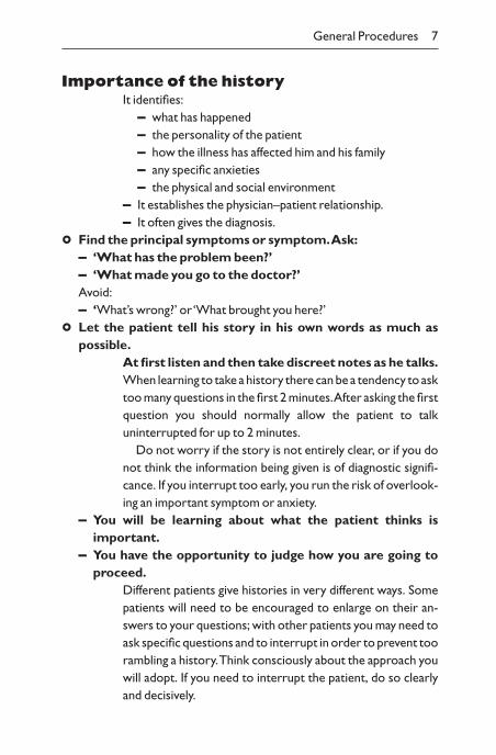
Usual sequence of events
6
History Investigations
Examination Diagnosis confirmed
Problem list Treatment
Differential diagnosis
Importance of the historyIt identifies:
– what has happened– the personality of the patient– how the illness has affected him and his family– any specific anxieties– the physical and social environment
– It establishes the physician–patient relationship.– It often gives the diagnosis.
° Find the principal symptoms or symptom.Ask:– ‘What has the problem been?’– ‘What made you go to the doctor?’Avoid:– ‘What’s wrong?’ or ‘What brought you here?’
° Let the patient tell his story in his own words as much as possible.
At first listen and then take discreet notes as he talks.When learning to take a history there can be a tendency to asktoo many questions in the first 2 minutes.After asking the firstquestion you should normally allow the patient to talk uninterrupted for up to 2 minutes.
Do not worry if the story is not entirely clear, or if you donot think the information being given is of diagnostic signifi-cance. If you interrupt too early, you run the risk of overlook-ing an important symptom or anxiety.
– You will be learning about what the patient thinks is important.
– You have the opportunity to judge how you are going toproceed.
Different patients give histories in very different ways. Somepatients will need to be encouraged to enlarge on their an-swers to your questions; with other patients you may need toask specific questions and to interrupt in order to prevent toorambling a history.Think consciously about the approach youwill adopt. If you need to interrupt the patient, do so clearlyand decisively.
General Procedures 7
° Try, if feasible, to conduct a conversation rather than an interrogation, following the patient’s train of thoughts.
You will usually need to ask follow-up questions on the mainsymptoms to obtain a full understanding of what they wereand of the chain of events.
° Obtain a full description of the patient’s principal complaints.
° Enquire about the sequence of symptoms and events.Beware pseudomedical terms, e.g.‘gastric flu’ —enquire whathappened
° Do not ask leading questions.A central aim in taking the history is to understand patients’symptoms from their own point of view. It is important not totarnish the patient’s history by your own expectations. Forexample, do not ask a patient whom you suspect might be thy-rotoxic: ‘Do you find hot weather uncomfortable?’ This invites the answer ‘Yes’ and then a positive answer becomes of little diagnostic value.Ask the open question: ‘Do you par-ticularly dislike either hot or cold weather?’
° Be sensitive to a patient’s mood and non-verbal responses.e.g. hesitancy in revealing emotional content.
° Be understanding, receptive and matter-of-fact without excessive sympathy.
° Rarely show surprise or reproach.
° Clarify symptoms and obtain a problem list.When the patient has finished describing the symptom or symptoms:– briefly summarize the symptoms– ask whether there are any other main problems
For example say ‘You have mentioned two problems: pain onthe left side of your tummy, and loose motions over the last 6weeks. Before we talk about those in more detail, are thereany other problems I should know about?’
Usual sequence of history– nature of principal complaints, e.g. chest pain, poor home
circumstances
8 Chapter 1:History Taking
– history of present complaint —details of current illness– enquiry of other symptoms (see Functional enquiry, p. 10)– past history– family history– personal and social history
° If one’s initial enquiries make it apparent that one section isof more importance than usual (e.g. previous relevant ill-nesses or operation), then relevant enquiries can be broughtforward to an earlier stage in the history (e.g. past historyafter finding principal complaints).
History of present illness
° Start your written history with a single sentence summing upwhat your patient is complaining of. It should be like the banner headline of a newspaper. For example:
c/o chest pain for 6 months
° Determine the chronology of the illness by asking:– ‘How and when did your illness begin?’ or– ‘When did you first notice anything wrong?’ or– ‘When did you last feel completely well?’
° Begin by stating when the patient was last perfectly well.Describe symptoms in chronological order of onset.
Both the date of onset and the length of time prior to admission should be recorded. Symptoms should never be dated by the day of the week as this later becomes meaningless.
° Obtain a detailed description of each symptom by asking:– ‘Tell me what the pain was like’, for example. Make sure you ask
about all symptoms, whether they seem relevant or not.
° With all symptoms obtain the following details:– duration– onset —sudden or gradual– what has happened since:
– constant or periodic– frequency– getting worse or better
General Procedures 9
– precipitating or relieving factors– associated symptoms
° If pain is a symptom also determine the following:– site– radiation
– character, e.g. ache, pressure, shooting, stabbing, dull– severity, e.g. ‘Did it interfere with what you were doing?
Does it keep you awake?’– have you ever had this pain before?– is the pain associated with nausea, sweating, e.g. angina?
Avoid technical language when describing a patient’s his-tory. Do not say ‘the patient complained of melaena’, rather:‘the patient complained of passing loose, black, tarry motions’.
Supplementary historyWhen patients are unable to give an adequate or reliable his-tory, the necessary information must be obtained from friendsor relations. A history from a person who has witnessed a sud-den event is often helpful.
Accordingly, the student should arrange with the housemanto be present when the relatives or witnesses are inter-viewed. This is particularly important with patients sufferingfrom disease of the central nervous system. The date andsource of such information should be written in the notes.
When necessary, arrange for an interpreter.Make use of GP’s letter and contact GP if necessary.
Functional enquiry
This is a checklist of symptoms not already discovered.Do not ask questions already covered in establishing the principal symptoms.This list may detect other symptoms.
° Modify your questioning according to the nature of the suspected disease, available time and circumstances.
If during the functional enquiry a positive answer is obtained,
10 Chapter 1:History Taking
full details must be elicited. Asterisks (*) denote questionswhich must nearly always be asked.
General questions
° Ask about the following points:– *Appetite: ‘What is your appetite like? Do you feel like eating?’– *Weight: ‘Have you lost or gained weight recently?’– *General well-being: ‘Do you feel well in yourself?’– Fatigue: ‘Are you more or less tired than you used to be?’– Fever or chills: ‘Have you felt hot or cold? Have you shivered?’– Night sweats:‘Have you noticed any sweating at night or any other
time?’– Aches or pains.– Rash: ‘Have you had any rash recently? Does it itch?’– Lumps and bumps.
Cardiovascular and respiratory system
° Ask about the following points:– *Chest pain: ‘Have you recently had any pain or discomfort in the
chest?’The most common causes of chest pain are:
Ischaemic heart disease: severe constricting, central chestpain radiating to the neck, jaw and left arm. Angina is this painprecipitated by exercise or emotion; relieved by rest. In a my-ocardial infarction the pain may come on at rest, be more severe and last hours.
Pleuritic pain: sharp, localized pain, usually lateral; worse oninspiration or cough.
Anxiety or panic attacks are a very common cause of chestpain. Enquire about circumstances that bring on an attack.
– *Shortness of breath: ‘Are you breathless at any time?’Breathlessness (dyspnoea) and chest pain must be accuratelydescribed.The degree of exercise which brings on the symp-toms must be noted (e.g. climbing one flight of stairs, after 0.5km (1/4 mile) walk).
– Shortness of breath on lying flat (orthopnoea): ‘Do you get
Functional Enquiry 11
breathless in bed? What do you do then? Does it get worse or better on sitting up? How many pillows do you use? Can you sleepwithout them?’
– Waking up breathless: ‘Do you wake at night with any symp-toms? Do you gasp for breath? What do you do then?’
Orthopnoea (breathless when lying flat) and paroxysmal noctur-nal dyspnoea (waking up breathless, relieved on sitting up) arefeatures of left heart failure.
– *Ankle swelling.Common in congestive cardiac failure (right heart failure).
– Palpitations: ‘Are you aware of your heart beating?’Palpitations may be:
– single thumps (ectopics)– slow or fast– regular or irregular
Ask the patient to tap them out.Paroxysmal tachycardia (sudden attacks of palpitations)
usually starts and finishes abruptly.– *Cough:‘Do you have a cough? Is it a dry cough or do you cough up
sputum? When do you cough?’– Sputum: ‘What colour is your sputum? How much do you cough
up?’Green sputum usually indicates an acute chest infection. Clearsputum daily during winter months suggests chronic bronchitis.Frothy sputum suggests left heart failure.
– *Blood in sputum (haemoptysis): ‘Have you coughed up blood?’Haemoptysis must be taken very seriously. Causes include:
carcinoma of bronchuspulmonary embolismmitral stenosistuberculosisbronchiectasis
– Black-outs (syncope): ‘Have you had any black-outs or faints? Didyou feel light-headed or did the room go round? Did you lose con-sciousness? Did you have any warning? Can you remember whathappened?’
12 Chapter 1:History Taking
– *Smoking: ‘Do you smoke? How many cigarettes do you smoke?’
Gastrointestinal system
° Ask about the following points:– Mouth ulcers– Nausea: ‘Are there times when you feel sick?’– Vomiting: ‘Do you vomit? What is it like?’
‘Coffee grounds’ vomit suggests altered blood.Old food suggests pyloric stenosis.If blood what colour is it —dark or bright red?
– Difficulty in swallowing (dysphagia): ‘Do you have difficulty swallowing? Where does it stick?’
For solids: often organic obstruction.For fluids: often neurological or psychological.
– Indigestion: ‘Do you have any discomfort in your stomach aftereating?’
– Abdominal pain: ‘Where is the pain? How is it connected tomeals or opening your bowels? What relieves the pain?’
– *Bowel habit: ‘Is your bowel habit regular? How many times doyou open your bowels per day? Do you have to open your bowels atnight?’ (often a sign of true pathology).
If diarrhoea is suggested, the number of motions per day andtheir nature (blood? pus? mucus?) must be established.
‘What are your motions like?’ The stools may be pale,bulky and float (fat in stool — steatorrhoea) or tarry from di-gested blood (melaena —usually from upper gastrointestinaltract).
Bright blood on the surface of a motion may be from haemorrhoids, whereas blood in a stool may signify cancer or inflammatory bowel disease.
– Jaundice: ‘Is your urine dark? Are your stools pale? What tabletshave you been taking recently? Have you had any recent injectionsor transfusions? Have you been abroad recently? How much alco-hol do you drink?’
Jaundice may be:
Functional Enquiry 13
– obstructive (dark urine pale stools) from:carcinoma of the head of the pancreas gallstones
– hepatocellular (dark urine, pale stools may develop)from:
ethanol (cirrhosis)drugs or transfusions (viral hepatitis)drug reactions or infections (travel abroad, viral hepatitisor amoebae)
– haemolytic (unconjugated bilirubin is bound to albuminand is not secreted in the urine)
Genitourinary system
° Ask about the following points:– Dysuria: pain on passing urine usually burning (often a sign of
infection).– Loin pain: ‘Any pain in your back?’
Pain in the loins suggests pyelonephritis.– *Urine: ‘Are your waterworks all right? Do you pass a lot of water
at night? Do you have any difficulty passing water? Is there blood inyour water?’ —haematuria.
Polyuria and nocturia occur in diabetes.Prostatism results in slow onset of urination, a poor streamand terminal dribbling.
– Sex: ‘Any problems with intercourse or making love?’– *Menstruation: ‘Any problems with your periods? Do you bleed
heavily? Do you bleed between periods?’Vaginal bleeding between periods or after the menopause raises the possibility of cervical or uterine cancer.
– Vaginal discharge.– Menstrual cycle: last menstrual period (LMP) and abormal vaginal
bleeding:inter-menstrual bleedingpost-menopausal bleedingpost-coital bleeding
– Pain on intercourse (dyspareunia) and whether this is superficialor deep.
14 Chapter 1:History Taking
Nervous system
° Ask about the following points:– *Headache: ‘Do you have any headaches? Where are they, when
do you get headaches?’e.g. early morning headaches may suggest raised intracranial pressure — tumour.Are the headaches associated with flashing lights (amaurosisfugax).
– Vision: ‘Do you have any blurred or double vision?’– Hearing: ask about tinnitus, deafness and exposure to noise.– Dizziness: ‘Do you have any dizziness or episodes when the world
goes round (vertigo)?’Dizziness with light-headed symptoms, when sudden in onset, may be cardiac (enquire about palpitations).When slow, onset may be vasovagal ‘fainting’ or an internalhaemorrhage.Vertigo may be from ear disease (enquire about deafness,earache or discharge) or brainstem dysfunction.
– Unsteady gait: ‘Any difficulty walking or running?’– Weakness.– Numbness or increased sensation:‘Any patches of numbness?’– Pins and needles.– Sphincter disturbance: ‘Any difficulties holding your
water/bowels?’ (a very important sign of spinal cord compression).– Fits or faints: ‘Have you had any funny episodes?’
The following details should be sought from the patient andany observer:
– duration– frequency and length of attacks– time of attacks, e.g. if standing, at night– mode of onset and termination– premonition or aura, light-headed or vertigo– biting of tongue, loss of sphincter control, injury,
etc.Grand mal epilepsy classically produces sudden uncon-
sciousness without any warning and on waking the patient
Functional Enquiry 15
feels drowsy with a headache, sore tongue, and has been incontinent.
Mental state
° Ask about the following points:– Depression: ‘How is your mood? Happy or sad? If depressed, how
bad? Have you lost interest in things? Can you still enjoy things? Howdo you feel about the future?’
‘Has anything happened in your life to make you depressed? Doyou feel guilty about anything?’
If the patient appears depressed: ‘Have you ever thought of sui-cide? How long have you felt like this? Is there a specific problem?Have you felt like this before?’
– Active periods: ‘Do you have periods in which you are particul-arly active?’
Susceptibility to depression may be a personality trait. In bipo-lar depression, swings to mania (excess activity, rapid speechand excitable mood) can recur. Enquire about interest, con-centration, irritability, sleep difficulties.
– Anxiety: ‘Have you worried a lot recently? Do you get anxious? Inwhat situations? Are there any situations you avoid because you feelanxious?’
‘Do you worry about your health? Any worries in your job orwith your family? Any financial worries?’
‘Do you have panic attacks? What happens?’– Sleep: ‘Any difficulties sleeping? Do you have difficulty getting to
sleep? Do you wake early?’Difficulties of sleep are commonly associated with depressionor anxiety.
A more complete assessment of mental state is given in Chapter 6.
The eye
° Ask about the following points:– Eye pain, photophobia or redness: ‘Have the eyes been red,
uncomfortable or painful?’
16 Chapter 1:History Taking
– Painful red eye, particularly with photophobia may be serious anddue to:
iritis (ankylosing spondylitis, Reiter’s disease, sarcoid, Behçet’s disease)scleritis (systemic vasculitis)corneal ulceracute glaucomaphotophobia may be a sign of meningitis
– Painless red eye may be:episcleritistemporary and of no consequencesystemic vasculitis
– Sticky red eye may be conjunctivitis (usually infective).– Itchy eye may be allergic, e.g. hayfever.– Gritty eye may be dry (sicca or Sjögren’s syndrome).– Clarity of vision: ‘Has your vision been blurred?’
– Blurring of vision for either near or distance alone may bean error of focus, helped by spectacles.
– Loss of central vision (or of top or bottom half ) in one eyemay be due to a retinal or optic nerve disorder.
– Transient complete blindness in one eye lasting for minutes —amaurosis fugax (fleeting blindness):
suggests retinal arterial blockage from embolus may befrom carotid atheroma (listen for bruit) may have a cardiacsource
– Subtle difficulties with vision, difficulty reading —problemsat the chiasm, or visual path behind it:
complete bitemporal hemianopia — tumour pressure onchiasmhomonymous hemianopia: posterior cerebral or optic radia-tion lesion —usually infarct or tumour; rarely complains of‘half vision’, but may have difficulty reading
– Diplopia: ‘Have you ever seen double?’Diplopia may be due to:
– lesion of the motor cranial nerves III, IV or VI– third-nerve palsy
causes double vision in all directions
Functional Enquiry 17
often with dilatation of the pupil and ptosisthe eye hangs ‘down and out’
– fourth-nerve palsycauses doubling looking down and in (as when reading)with images separated horizontally and vertically and tilted(not parallel)
– sixth-nerve palsycauses horizontal, level and parallel doublingworse on looking to the affected side
– muscular disordere.g. thyroid-related (see below)myasthenia gravis (weakness after muscle use, antibodies tonerve end-plates)
Locomotor system
° Ask about the following points:– Pain, stiffness, or swelling of joints:‘When and how did it start?
Have you injured the joint?’There are innumerable causes of arthritis (painful, swollen,tender joints) and arthralgia (painful joints). Patients may in-correctly attribute a problem to some injury.
Osteoarthritis is a joint ‘wearing out’, and is often asymmet-ric, involving weight-bearing joints such as the hip or knee.Exercise makes the joint pain worse.
Rheumatoid arthritis is a generalized autoimmune diseasewith symmetrical involvement. In the hands, fusiform swellingof the interphalangeal joints is accompanied by swollenmetacarpophalangeal joints. Large joints are often affected.Stiffness is worse after rest, e.g. on waking, and improves withuse.
Gout usually involves a single joint, such as the first metatar-sophalangeal joint, but can lead to gross hand involvementwith asymmetric uric acid lumps (tophi) by some joints, and inthe tips of the ears.
Septic arthritis: this is important not to miss —a single, hotpainful joint.
18 Chapter 1:History Taking
– Functional disability: ‘How far can you walk? Can you walk up-stairs? Is any particular movement difficult? Can you dress yourself?How long does it take? Can you work? Can you write?’
Thyroid disease
° Ask about the following points:– Weight change.– Reaction to the weather: ‘Do you dislike the hot or cold
weather?’– Irritability: ‘Are you more or less irritable compared with a few
years ago?’– Diarrhoea/constipation.– Palpitations.– Dry skin or greasy hair: ‘Is your skin dry or greasy? Is your hair
dry or greasy?’– Depression: ‘How has your mood been?’– Croaky voice.
Hypothyroid patients put on weight without increase in ap-petite, dislike cold weather, have dry skin and thin, dry hair, apuffy face, a croaky voice, are usually calm and may be depressed.
Hyperthyroid patients may lose weight despite eating more,dislike hot weather, perspire excessively, have palpitations, atremor, and may be agitated and tearful. Young people havepredominantly nervous and heat intolerance symptoms,whereas old people tend to present with cardiac symptoms.
Past history
° All previous illnesses or operations, whether apparently impor-tant or not, must be included.
For instance, a casually mentioned attack of influenza or chillmay have been a manifestation of an occult infection.
° The importance of a past illness may be gained by finding out how longthe patient was in bed or off work.
Past History 19
° Complications of any previous illnesses should be carefully enquired into and, here, leading questions are sometimes necessary.
General questions
° Ask about the following:– ‘Have you had any serious illnesses?’– ‘Have you had any emotional or nervous problems?’– ‘Have you had any operations or admissions to hospital?’– ‘Have you ever
– had jaundice, epilepsy, TB, hypertension, rheumaticfever or diabetes?
– travelled abroad?– had allergies?’
– ‘Have any medicines ever upset you?’Allergic responses to drugs may include an itchy rash, vomit-ing, diarrhoea or severe illness, including jaundice. Many pa-tients claim to be allergic but are not.An accurate descriptionof the supposed allergic episodes is important.
– Additional questions can be asked:– if the patient has high blood pressure, ask about kidney problems,
if relatives have hypertension or whether he eats liquorice– if a possible heart attack, ask about hypertension, diabetes, diet,
smoking, family history of heart disease– if the patient’s history suggests cardiac failure, you must ask if he
has had rheumatic feverPatients have often had examinations for life insurance or the armed
forces.
Family history
The family history gives clues to possible predisposition to ill-ness (e.g. heart attacks) and whether a patient may have reason tobe particularly anxious about a certain disease (e.g. mother died ofcancer).Death certificates and patient knowledge are often inaccurate. Patientsmay be reluctant to talk about relatives’ illnesses if they were mental dis-eases, epilepsy or cancer.
20 Chapter 1:History Taking
General questions
° Ask about the following:– ‘Are your parents alive?Are they fit and well? What did your par-
ents die from?’– ‘Have you any brothers or sisters?Are they fit and well?’– ‘Do you have any children? Are they fit and well?’– ‘Is there any history of:
– heart trouble?– diabetes?– high blood pressure in the family?’
These questions can be varied to take account of the patient’smajor complaint.
Personal and social history
One needs to find out what kind of person the patient is, what hishome circumstances are and how his illness has affected himand his family.Your aim is to understand the patient’s illness inthe context of his personality and his home environment.
Can he convalesce satisfactorily at home and at what stage?What are the consequences of his illness? Will advice, infor-mation and help be needed? An interview with a relative orfriend may be very helpful.
General questions
° Ask about the following:– ‘Are you in a relationship?’: married, partnership and whether any
children.– Family:‘Is everything alright at home? Do you have any family prob-
lems?’It may be appropriate to ask:‘Is your relationship alright? Is sexalright?’ Problems may arise from physical or emotional rea-sons, and the patient may appreciate an opportunity to dis-cuss worries.
– Accommodation: ‘Where do you live? Is it alright?’– Job: ‘What is your job? Could you tell me exactly what you do? Is it
satisfactory? Will your illness affect your work?’
Personal and Social History 21
– Hobbies: ‘What do you do in your spare time? Do you have any social life?’
– Alcohol: ‘How much alcohol do you drink?’Alcoholics usually underestimate their daily consumption. Itmay be helpful to go through a ‘drinking day’. If there is a suspi-cion of a drinking problem, you can ask: ‘Do you ever drink inthe morning? Do you worry about controlling your drinking?Does it affect your job, home or social life?’
– Smoking: ‘Do you smoke?’ Have you ever smoked? Why did yougive up? How many cigarettes, cigars or pipefuls of tobacco do yousmoke a day?’
Particularly relevant for heart or chest disease, but must always be asked.
– Drugs: ‘Do you take any recreational drugs?’– Prescribed medications: ‘What pills are you taking at the
moment? Have you taken any other pills in the last few months?’This is an extremely important question.A completelist of all drugs and doses must be obtained.
If relevant, ask about any pets, visits abroad, previous orpresent exposure during working to coal dust, asbestos, etc.
The patient’s ideas, concerns andexpectationsMake sure that you understand the patient’s main ideas, concerns and expectations. Either now, or after examining the patient, ask for example:
° What do you think is wrong with you?
° What are you expecting to happen to you whilst you are inhospital?
° Is there something particular you would like us to do?
° Have you any questions?The patient’s main concerns may not be your mainconcerns.The patient may have quite different expectationsof the hospital admission, or outpatient appointment, fromwhat you assume. If you fail to address the patient’s concernshe is likely to be dissatisfied, leading to difficult doctor–patientrelationships and non-compliance.
22 Chapter 1:History Taking
StrategyHaving taken the history, you should
° have some idea of possible diagnoses
° have made an assessment of the patient as a person
° know which systems you wish to concentrate on when examining the patient
Further relevant questions may arise from abnormalities foundon examination or investigation.
Specimen history
Mr John Smith.Aged 52. Machine operator. Oxford.c/o severe chest pain for 2 hours.
History of present illness– Perfectly well until 6 months ago.– Began to notice central, dull chest ache, occasionally felt in the jaw,
coming on when walking about 1km (1/2 mile), worse when goinguphill and worse in cold weather.When he stopped, the pain wentoff after 2 minutes.
– Glyceryl trinitrate spray relieved the pain rapidly.– Last month the pain came on with less exercise after 100 yards.– Today at 10 a.m. whilst sitting at work the chest pain came on with-
out provocation. It was the worst pain he had ever experienced inhis life and he thought he was going to die.
– The pain was central, crushing in nature, radiating to the left armand neck and with it a feeling of nausea and sweating. The patientwas rushed to hospital where he received an intravenous injectionof diamorphine, which rapidly relieved the pain, and intravenousstreptokinase. An electrocardiogram confirmed a myocardial in-farction and the patient was admitted to the coronary care unit.
– The patient had noticed very mild breathlessness on exertion for 3months, but had not experienced palpitations, dizziness, breath-lessness on lying flat, ankle swelling or coughing. On one occasion,however, 2 weeks ago the patient had woken with a suffocating
Specimen History 23
feeling and had had to sit on the edge of the bed and subsequentlyopen the bedroom window in order to get his breath. This had not recurred and he did not report it to his doctor.
Functional enquiryRespiratory system (RS):
– morning cough over the last 3–4 winters with production of a smallamount of clear sputum
– no haemoptysisGastrointestinal (GI):
– occasional mild indigestion– bowels regular– appetite normal– no other abnormalities
Genitourinary (GU):– no difficulties with micturition– normal sex life
Nervous system (NS):– infrequent frontal headaches at the end of a hectic day– otherwise no abnormalities– no psychiatric symptoms
Past medical historyFifteen years ago, appendicectomy. No complications.No other operations or serious illnesses.No history of rheumatic fever, nephritis or hypertension.Never been abroad.
Family historyFather died aged 73 —‘heart attack’.Mother died aged 71 —‘cancer’.Two brothers fit and well (aged 48 and 46).Two sons (aged 23, 25), both fit and well.No family history of diabetes or hypertension.
24 Chapter 1:History Taking

Personal and social historyHappy both at work and home. Both sons married and living in Oxford.Wife works as an office cleaner. No financial difficulties.
Smokes 20 cigarettes per day.Two pints of beer on Saturdays only.Patient always worked as machine operator since leaving school
except for 2 years in Hong Kong, where he had no illness.
MedicationOther than glyceryl trinitrate spray, no drugs currently being taken.
Specimen History 25
CHAPTER 2
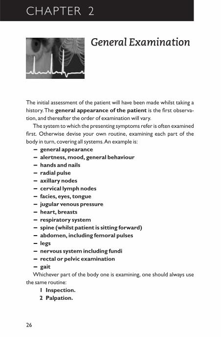
General Examination
The initial assessment of the patient will have been made whilst taking ahistory.The general appearance of the patient is the first observa-tion, and thereafter the order of examination will vary.
The system to which the presenting symptoms refer is often examinedfirst. Otherwise devise your own routine, examining each part of thebody in turn, covering all systems.An example is:
– general appearance– alertness, mood, general behaviour– hands and nails– radial pulse– axillary nodes– cervical lymph nodes– facies, eyes, tongue– jugular venous pressure– heart, breasts– respiratory system– spine (whilst patient is sitting forward)– abdomen, including femoral pulses– legs– nervous system including fundi– rectal or pelvic examination– gaitWhichever part of the body one is examining, one should always use
the same routine:1 Inspection.2 Palpation.
26
3 Percussion.4 Auscultation.
General inspection
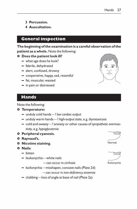
The beginning of the examination is a careful observation of thepatient as a whole. Note the following:
° Does the patient look ill?– what age does he look?– febrile, dehydrated– alert, confused, drowsy– cooperative, happy, sad, resentful– fat, muscular, wasted– in pain or distressed
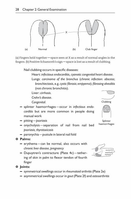
Hands
Note the following:
° Temperature:– unduly cold hands —? low cardiac output– unduly warm hands —? high-output state, e.g. thyrotoxicosis– cold and sweaty —? anxiety or other causes of sympathetic overreac-
tivity, e.g. hypoglycaemia
° Peripheral cyanosis.
° Raynaud’s.
° Nicotine staining.
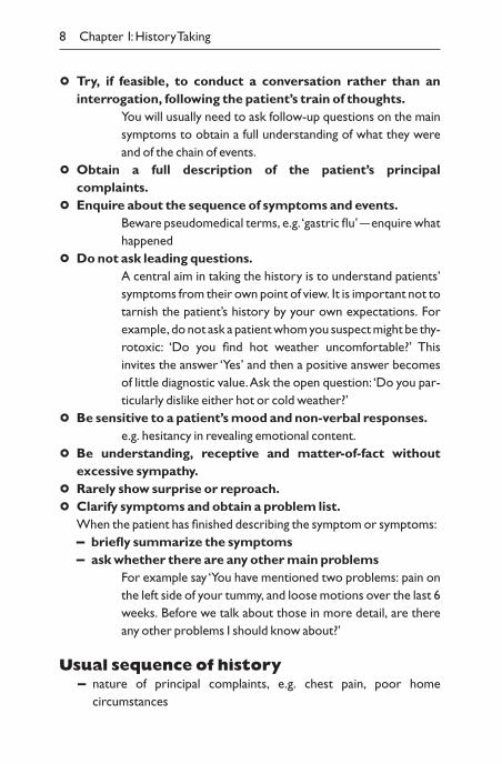
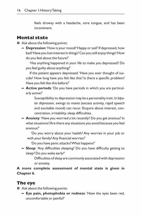
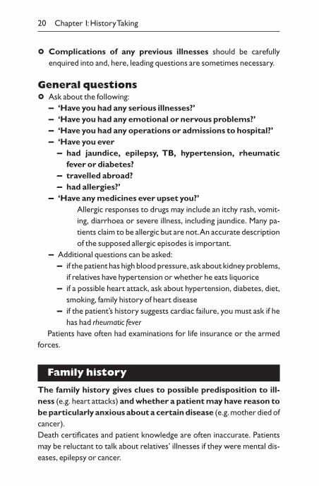
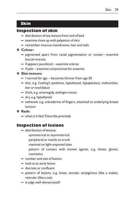
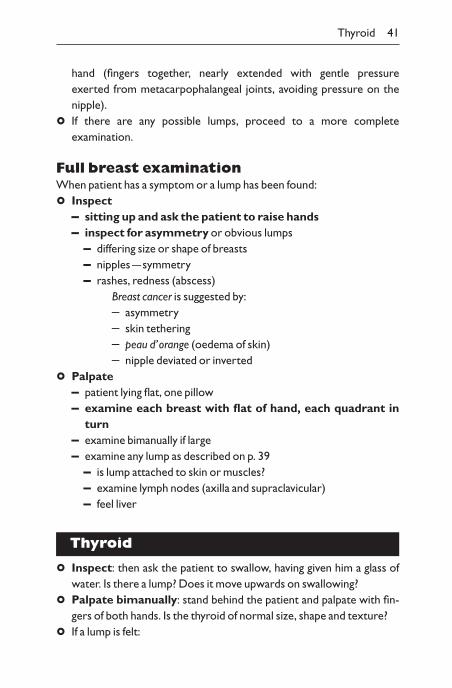
° Nails:– bitten– leukonychia —white nails
—can occur in cirrhosis– koilonychia —misshapen, concave nails (Plate 2d)
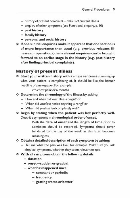
—can occur in iron-deficiency anaemia– clubbing —loss of angle at base of nail (Plate 2a)
Hands 27
Normal
Koilonychia
Nail clubbing occurs in specific diseases:Heart: infectious endocarditis, cyanotic congenital heart disease.Lungs: carcinoma of the bronchus (chronic infection: abscess;
bronchiectasis, e.g. cystic fibrosis; empyema); fibrosing alveolitis(not chronic bronchitis).
Liver: cirrhosis.Crohn’s disease.Congenital.
– splinter haemorrhages —occur in infectious endo-carditis but are more common in people doing manual work
– pitting —psoriasis– onycholysis —separation of nail from nail bed
psoriasis, thyrotoxicosis– paronychia —pustule in lateral nail fold
° Palms:– erythema —can be normal, also occurs with
chronic liver disease, pregnancy– Dupuytren’s contracture (Plate 4c) —tether-
ing of skin in palm to flexor tendon of fourthfinger
° Joints:– symmetrical swellings occur in rheumatoid arthritis (Plate 2e)– asymmetrical swellings occur in gout (Plate 2f) and osteoarthritis
28 Chapter 2:General Examination
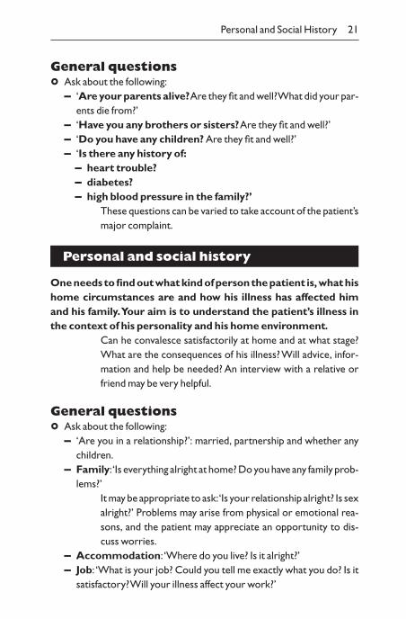
(a) Fingers held together — space seen at X as a result of normal angles in the
fingers. (b) Positive Schamroth’s sign — space is lost as a result of clubbing.
X
Normal Club finger(a) (b)
Clubbing
Splinterhaemorrhages
Skin
Inspection of skin– distribution of any lesions from end of bed– examine close up with palpation of skin– remember mucous membranes, hair and nails
° Colour:– pigmented apart from racial pigmentation or suntan —examine
buccal mucosa– if appears jaundiced —examine sclerae– if pale —examine conjunctivae for anaemia
° Skin texture:– ? normal for age —becomes thinner from age 50– thin, e.g. Cushing’s syndrome, hypothyroid, hypopituitary, malnutrition,
liver or renal failure– thick, e.g. acromegaly, androgen excess– dry, e.g. hypothyroid– tethered, e.g. scleroderma of fingers, attached to underlying breast
tumour
° Rash:– what is it like? Describe precisely
Inspection of lesions– distribution of lesions:
symmetrical or asymmetricalperipheral or mainly on trunkmaximal on light-exposed sitespattern of contact with known agents, e.g. shoes, gloves,cosmetics
– number and size of lesions– look at an early lesion– discrete or confluent– pattern of lesions, e.g. linear, annular, serpiginous (like a snake),
reticular (like a net)– is edge well-demarcated?
Skin 29
– colour– surface, e.g. scaly, shiny
Palpation of lesions– flat, impalpable —macular (Plate 3c)– raised
papular: in skin, localizedplaque: larger, e.g. >0.5cmnodules: deeper in dermis, persisting more than 3 dayswheal: oedema fluid, transient, less than 3 daysvesicles: contain fluid (Plate 3e)bullae: large vesicles, e.g. >0.5cmpustular
– deep in dermis —nodules– temperature– tender?– blanches on pressure —most erythematous lesions, e.g. drug rash,
telangiectasia, dilated capillaries– does not blanch on pressure
Purpura or petechiae are small discrete microhaemorrhagesapproximately 1mm across, red, non-tender macules.If palpable, suggests vasculitis (Plate 3d).Senile purpura local haemorrhages are from minor traumas inthin skin of hands or forearms. Flat purple/brown lesions.
– hard– sclerosis, e.g. scleroderma of fingers (Plate 4b)– infiltration, e.g. lymphoma or cancer– scars
Enquire about the time course of anylesion
– ‘How long has it been there?’– ‘Is it fixed in size and position? Does it come and go?’– ‘Is it itchy, sore, tender or anaesthetic?’
Knowledge of the differential diagnosis will indicate other questions:dermatitis of hand —contact with chemicals or plants, wear andtear;
30 Chapter 2:General Examination
ulcer of toe —arterial disease, diabetes mellitus, neuropathy;pigmentation and ulcer of lower medial leg —varicose veins.
Common diseasesAcne Pilar-sebaceous follicular inflammation —
papules and pustules on face and uppertrunk, blackheads (comedones), cysts.
Basal cell carcinoma Shiny papule with rolled border and(rodent ulcer) (Plate 5e) capillaries on surface. Can have a depressed
centre or ulcerate.Bullae Blisters due to burns, infection of the skin,
allergy or, rarely, autoimmune diseases affecting adhesion within epidermis (pemphigus) or at the epidermal–dermaljunction (pemphigoid).
Café-au-lait patches Permanent discrete brown macules of varying size and shape. If large and numerous, suggests neurofibromatosis.
Drug eruptions (Plate 3c) Usually macular, symmetrical distribution.Can be urticaria, eczematous and variousforms, including erythema multiforme orerythema nodosum (see below).
Eczema (Plate 3b) Atopic dermatitis: dry skin, red, plaques,commonly on the face, antecubital andpopliteal fossae, with fine scales, vesicles and scratch marks secondary to pruritus(itching). Often associated with asthmaand hayfever. Family history of atopy.Contact dermatitis: may be irritant or allergic.Red, scaly plaques with vesicles in acutestages.
Erythema multiforme Symmetrical, widespread inflammatory0.5–1cm macules/papules, often with centralblister. Can be confluent. Usually on handsand feet:
drug reactions
Skin 31
viral infectionsno apparent causeStevens–Johnson syndrome —with mucosaldesquamation involving genitalia, mouthand conjunctivae, with fever.
Erythema nodosum Tender, localized, red, diffusely raised,(Plate 3f) 2–4cm nodules in anterior shins. Due to:
streptococcal infection, e.g. with rheumaticfeverprimary tuberculosis and other infectionssarcoidinflammatory bowel diseasedrug reactionsno apparent cause
Fungus Red, annular, scaly area of skin.When involving the nails, they become thickenedwith loss of compact structure.
Herpes infection Clusters of vesicopustules which crust,(Plate 6f) recurs at the same site, e.g. lips, buttocks.Impetigo Spreading pustules and yellow crusts from
staphylococcal infection.Malignant melanoma Usually irregular pigmented, papule or
plaque, superficial or thick with irregularedge, enlarging with tendency to bleed.
Psoriasis (Plate 3a) Symmetrical eruption: chronic, discrete, redplaques with silvery scales. Gentle scrapingeasily induces bleeding. Often affects scalp,elbows and knees. Nails may be pitted.Familial and precipitated by streptococcalsore throats or skin trauma.
Scabies Mite infection: itching with 2–4mm tunnelsin epidermis, e.g. in webs of fingers, wrists,genitalia.
Squamous cell carcinoma Warty localized thickening, may ulcerate.Urticaria Transient wheal with surrounding erythema.
Lasts around 24 hours. Usually allergic to
32 Chapter 2:General Examination
drugs, e.g. aspirin, or physical, e.g.dermographism, cold.
Vitiligo Permanent demarcated, depigmented whitepatches due to autoimmune disease.
Mouth
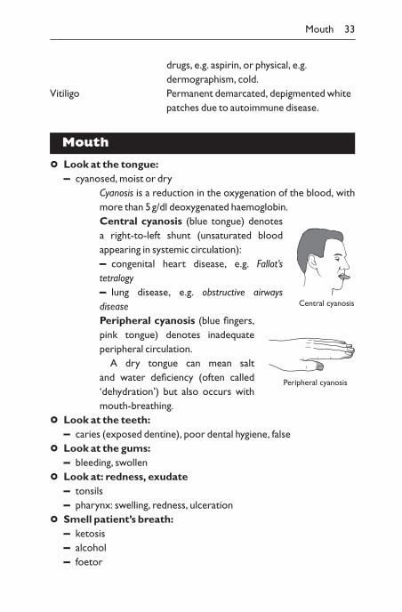
° Look at the tongue:– cyanosed, moist or dry
Cyanosis is a reduction in the oxygenation of the blood, withmore than 5g/dl deoxygenated haemoglobin.Central cyanosis (blue tongue) denotes a right-to-left shunt (unsaturated blood appearing in systemic circulation):– congenital heart disease, e.g. Fallot’s tetralogy– lung disease, e.g. obstructive airways diseasePeripheral cyanosis (blue fingers,pink tongue) denotes inadequate peripheral circulation.
A dry tongue can mean salt and water deficiency (often called‘dehydration’) but also occurs withmouth-breathing.
° Look at the teeth:– caries (exposed dentine), poor dental hygiene, false
° Look at the gums:– bleeding, swollen
° Look at: redness, exudate– tonsils– pharynx: swelling, redness, ulceration
° Smell patient’s breath:– ketosis– alcohol– foetor
Mouth 33
Central cyanosis
Peripheral cyanosis
constipation, appendicitismusty in liver failure
Ketosis is a sweet-smelling breath occurring with starvation orsevere diabetes.Hepatic foetor is a musty smell in liver failure.
Eyes
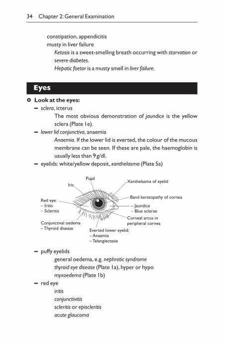
° Look at the eyes:– sclera, icterus
The most obvious demonstration of jaundice is the yellowsclera (Plate 1e).
– lower lid conjunctiva, anaemiaAnaemia. If the lower lid is everted, the colour of the mucousmembrane can be seen. If these are pale, the haemoglobin isusually less than 9g/dl.
– eyelids: white/yellow deposit, xanthelasma (Plate 5a)
34 Chapter 2:General Examination
Everted lower eyelid:– Anaemia– Telangiectasia
Corneal arcus inperipheral cornea
– Jaundice– Blue sclerae
Band keratopathy of cornea
Xanthelasma of eyelid
Red eye:– Iritis– Scleritis
IrisPupil
Conjunctival oedema– Thyroid disease
– puffy eyelidsgeneral oedema, e.g. nephrotic syndromethyroid eye disease (Plate 1a), hyper or hypomyxoedema (Plate 1b)
– red eyeiritisconjunctivitisscleritis or episcleritisacute glaucoma
– white line around cornea, arcus seniliscommon and of little significance in the elderlysuggests hyperlipidaemia in younger patients (Plate 5b)
– white-band keratopathy-hypercalcaemiasarcoidparathyroid tumour or hyperplasialung oat-cell tumourbone secondariesvitamin D excess intakeHypercalcaemia may give a horizontal band across exposed medial and lateral parts of cornea.
Examine the fundiThis is often done as part of the neurological system, when examining thecranial nerves. It is placed here as features cover general medicine.
° Use ophthalmoscope– The patient should be sitting. Start examination at 1m from the pa-
tient, identify red reflex and approach the patient at an angle of 15°to the patient. Approach on the same horizontal plane as patient’sequator of their eye. This will bring the observer straight to theoptic disc. After observing the disc examine the peripheral retinafully by following the blood vessels to and back from the four mainquadrants.– Use your right eye for patient’s right eye, left eye for patient’s left
eye.
° Look at optic disc– normally pink rim with white ‘cup’ below surface of disc– optic atrophy
– disc pale: rim no longer pinkmultiple sclerosisafter optic neuritisoptic nerve compression, e.g. tumour
– papilloedema– disc pink, indistinct margin– cup disappears– dilated retinal veins:
Eyes 35
increased cerebral pressure, e.g. tumouraccelerated hypertensionoptic neuritis, acute stage
– glaucoma —enlarged cup, diminished rim– new vessels —new fronds of vessels coming forward from disc;
ischaemic diabetic retinopathy
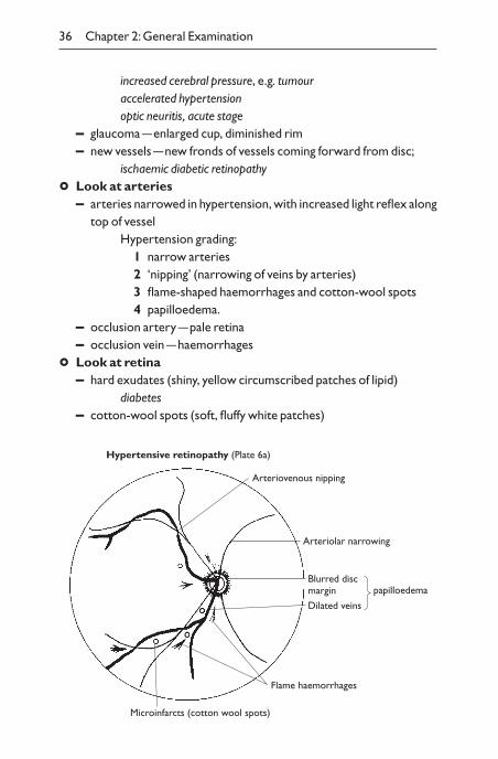
° Look at arteries– arteries narrowed in hypertension, with increased light reflex along
top of vesselHypertension grading:
1 narrow arteries2 ‘nipping’ (narrowing of veins by arteries)3 flame-shaped haemorrhages and cotton-wool spots4 papilloedema.
– occlusion artery —pale retina– occlusion vein —haemorrhages
° Look at retina– hard exudates (shiny, yellow circumscribed patches of lipid)
diabetes– cotton-wool spots (soft, fluffy white patches)
36 Chapter 2:General Examination
Arteriovenous nipping
Arteriolar narrowing
Blurred discmargin
Flame haemorrhages
Microinfarcts (cotton wool spots)
Hypertensive retinopathy (Plate 6a)
Dilated veins
papilloedema
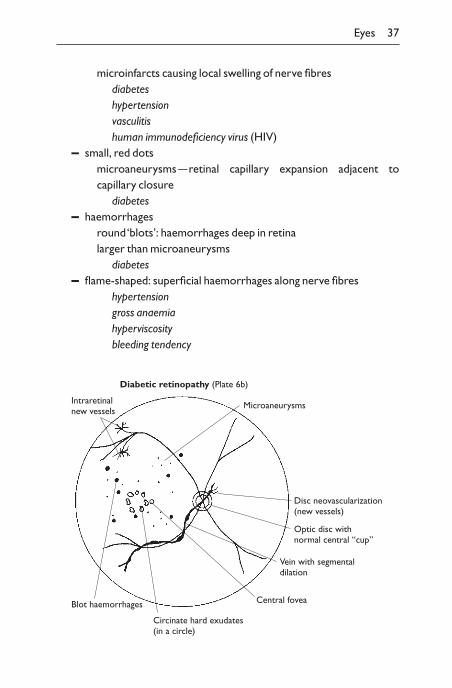
microinfarcts causing local swelling of nerve fibresdiabeteshypertensionvasculitishuman immunodeficiency virus (HIV)
– small, red dotsmicroaneurysms —retinal capillary expansion adjacent to capillary closure
diabetes– haemorrhages
round ‘blots’: haemorrhages deep in retinalarger than microaneurysms
diabetes– flame-shaped: superficial haemorrhages along nerve fibres
hypertensiongross anaemiahyperviscositybleeding tendency
Eyes 37
Microaneurysms
Disc neovascularization(new vessels)
Central fovea
Circinate hard exudates(in a circle)
Blot haemorrhages
Diabetic retinopathy (Plate 6b)
Intraretinalnew vessels
Optic disc withnormal central “cup”
Vein with segmentaldilation
– Roth’s spots (white-centred haemorrhages)microembolic disordersubacute bacterial endocarditis
– pigmentationwidespread
retinitis pigmentosalocalized
choroiditis (clumping of pigment into patches)drug toxicity, e.g. chloroquine
tigroid or tabby fundus: normal variant in choroid beneath retina– peripheral new vessels
ischaemic diabetic retinopathyretinal vein occlusion
– medullated nerve fibres —normal variant, areas of white nerves radiating from optic disc
Examine for palpable lymph nodes
° In the neck:– above clavicle (posterior triangle)– medial to sternomastoid area (anterior triangle)– submandibular (can palpate submandibular gland)– occipital
These glands are best felt by sitting the patient up and examin-ing from behind. A left supraclavicular node can occur fromthe spread of a gastrointestinal malignancy (Virchow’s node).
° In the axillae:– abduct arm, insert your hand along lateral side of axilla, and adduct
arm, thus placing your fingertips in the apex of the axilla. Palpategently
° In the epitrochlear region:– medial to and above elbow
° In the groins:– over inguinal ligament
38 Chapter 2:General Examination
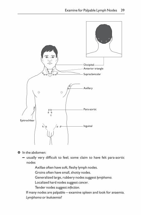
Examine for Palpable Lymph Nodes 39
Epitrochlear
Para-aortic
Inguinal
Axillary
Supraclavicular
Anterior triangleOccipital
° In the abdomen:– usually very difficult to feel; some claim to have felt para-aortic
nodesAxillae often have soft, fleshy lymph nodes.Groins often have small, shotty nodes.Generalized large, rubbery nodes suggest lymphoma.Localized hard nodes suggest cancer.Tender nodes suggest infection.
If many nodes are palpable —examine spleen and look for anaemia.Lymphoma or leukaemia?
Lumps
° If there is an unusual lump, inspect first and palpate later:– site– size (measure in centimetres)– shape– surface, edge– surroundings– fixed or mobile– consistency, e.g. cystic or solid, soft or hard, fluctuance– tender– pulsatile– auscultation– transillumination
A cancer is usually hard, non-tender, irregular, fixed to neigh-bouring tissues, and possibly ulcerating skin.
A cyst may have:– fluctuance: pressure across cyst will cause it to bulge in
another plane– transillumination: a light can be seen through it (usually
only if room is darkened)
° Look at neighbouring lymph nodes. May find:– spread from cancer– inflamed lymph nodes from infection
Breasts
When appropriate, arrange a female chaperone, particularlywhen the patient is a young adult, shy or nervous.
Routine examination
° Examine the female breasts when you examine the precordium.
° Inspect for asymmetry, obvious lumps, inverted nipples, skinchanges.
° Palpate each quadrant of both breasts with the flat of the
40 Chapter 2:General Examination
hand (fingers together, nearly extended with gentle pressure exerted from metacarpophalangeal joints, avoiding pressure on thenipple).
° If there are any possible lumps, proceed to a more complete examination.
Full breast examinationWhen patient has a symptom or a lump has been found:
° Inspect– sitting up and ask the patient to raise hands– inspect for asymmetry or obvious lumps
– differing size or shape of breasts– nipples —symmetry– rashes, redness (abscess)
Breast cancer is suggested by:– asymmetry– skin tethering– peau d’orange (oedema of skin)– nipple deviated or inverted
° Palpate– patient lying flat, one pillow– examine each breast with flat of hand, each quadrant in
turn– examine bimanually if large– examine any lump as described on p. 39
– is lump attached to skin or muscles?– examine lymph nodes (axilla and supraclavicular)– feel liver
Thyroid
° Inspect: then ask the patient to swallow, having given him a glass ofwater. Is there a lump? Does it move upwards on swallowing?
° Palpate bimanually: stand behind the patient and palpate with fin-gers of both hands. Is the thyroid of normal size, shape and texture?
° If a lump is felt:
Thyroid 41
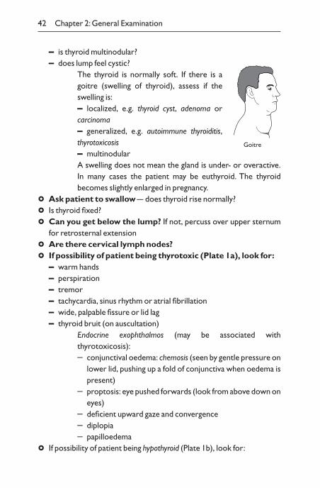
– is thyroid multinodular?– does lump feel cystic?
The thyroid is normally soft. If there is agoitre (swelling of thyroid), assess if theswelling is:– localized, e.g. thyroid cyst, adenoma or carcinoma– generalized, e.g. autoimmune thyroiditis,thyrotoxicosis– multinodularA swelling does not mean the gland is under- or overactive.In many cases the patient may be euthyroid. The thyroid becomes slightly enlarged in pregnancy.
° Ask patient to swallow — does thyroid rise normally?
° Is thyroid fixed?
° Can you get below the lump? If not, percuss over upper sternumfor retrosternal extension
° Are there cervical lymph nodes?
° If possibility of patient being thyrotoxic (Plate 1a), look for:– warm hands– perspiration– tremor– tachycardia, sinus rhythm or atrial fibrillation– wide, palpable fissure or lid lag– thyroid bruit (on auscultation)
Endocrine exophthalmos (may be associated with thyrotoxicosis):– conjunctival oedema: chemosis (seen by gentle pressure on
lower lid, pushing up a fold of conjunctiva when oedema ispresent)
– proptosis: eye pushed forwards (look from above down oneyes)
– deficient upward gaze and convergence– diplopia– papilloedema
° If possibility of patient being hypothyroid (Plate 1b), look for:
42 Chapter 2:General Examination
Goitre
– dry hair and skin– xanthelasma– puffy face– croaky voice– delayed relaxation of supinator or ankle jerks
Other endocrine diseases
Acromegaly (Plate 1c)– enlarged soft tissue of hands, feet, face– coarse features, thick, greasy skin, large tongue (and other organs,
e.g. thyroid)– bitemporal hemianopia (from tumour pressing on optic chiasma)
Hypopituitary– no skin pigmentation– thin skin– decreased secondary sexual hair or delayed puberty– short stature (and on X-ray, delayed fusion of epiphyses)– bitemporal hemianopia if pituitary tumour
Addison’s disease– increased skin pigmentation, including non-exposed areas, e.g.
buccal pigmentation– postural hypotension– if female, decreased body hair
Cushing’s syndrome (Plate 1d)– truncal obesity, round, red face with hirsutism– thin skin and bruising, pink striae, hypertension– proximal muscle weakness
DiabetesDiabetic complications include:
– skin lesionsNecrobiosis lipoidica —ischaemia in skin, usually on shins,
Other Endocrine Diseases 43
leading to fatty replacement of dermis, covered by thin skin.
– ischaemic legs (Plate 4e)– diminished foot pulses– skin shiny blue, white or black– no hairs, thick nails– ulcers (Plate 4f )
– peripheral neuropathy– absent leg reflexes– diminished sensation– thick skin over unusual pressure points from dropped arch
– autonomic neuropathy– dry skin
– mononeuropathy– lateral popliteal nerve —footdrop– III or VI —diplopia– asymmetrical muscle-wasting of the upper leg
– retinopathy (Plate 6b)
Locomotor system
Normally one examines joints briefly when examining neighbouring systems. If a patient specifically complains of joint symptoms or an abnormal posture or joint is noted, a more detailed examination is needed.
General habitus
° Note the following:– is the patient unduly tall or short? Measure height and span– are all limbs, spine and skull of normal size and shape?
– normal person:height = spancrown to pubis = pubis to heel
– long limbs:Marfan’s syndromeeunuchoid during growth
44 Chapter 2:General Examination
– collapsed vertebrae:span > heightpubis to heel > crown to pubis
– is the posture normal?– curvature of the spine:
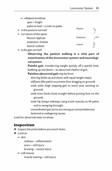
flexion: kyphosisextension: lordosislateral: scoliosis
– is the gait normal?Observing the patient walking is a vital part of examination of the locomotor system and neurologi-cal system.Painful gait, transferring weight quickly off a painful limb,bobbing up and down —an abnormal rhythm of gait.Painless abnormal gait may be from:
short leg (bobs up and down with equal-length steps)stiff joint (lifts pelvis to prevent foot dragging on ground)weak ankle (high stepping gait to avoid toes catching on
ground)weak knee (locks knee straight before putting foot on the
ground)weak hip (sways sideways using trunk muscles to lift pelvis
and to swing leg through)uncoordinated gait (arms are swung as counterbalances)hysterical or malingering causes
Look for abnormal wear on shoes.
Inspection
° Inspect the joints before you touch them.
° Look at:– skin
redness —inflammationscars —old injurybruising —recent injury
– soft tissuesmuscle wasting —old injury
Locomotor System 45
Flexion
Extension
Lateral
swelling —injury/inflammation– bones
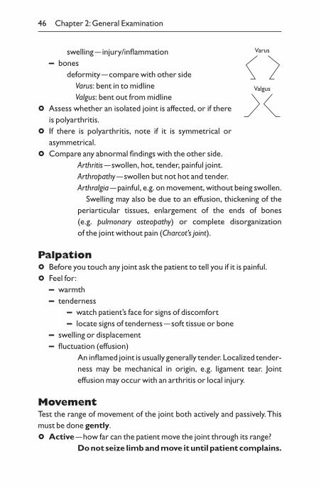
deformity —compare with other sideVarus: bent in to midlineValgus: bent out from midline
° Assess whether an isolated joint is affected, or if thereis polyarthritis.
° If there is polyarthritis, note if it is symmetrical or asymmetrical.
° Compare any abnormal findings with the other side.Arthritis —swollen, hot, tender, painful joint.Arthropathy —swollen but not hot and tender.Arthralgia —painful, e.g. on movement, without being swollen.
Swelling may also be due to an effusion, thickening of theperiarticular tissues, enlargement of the ends of bones (e.g. pulmonary osteopathy) or complete disorganization of the joint without pain (Charcot’s joint).
Palpation
° Before you touch any joint ask the patient to tell you if it is painful.
° Feel for:– warmth– tenderness
– watch patient’s face for signs of discomfort– locate signs of tenderness —soft tissue or bone
– swelling or displacement– fluctuation (effusion)
An inflamed joint is usually generally tender. Localized tender-ness may be mechanical in origin, e.g. ligament tear. Joint effusion may occur with an arthritis or local injury.
MovementTest the range of movement of the joint both actively and passively.Thismust be done gently.
° Active —how far can the patient move the joint through its range?Do not seize limb and move it until patient complains.
46 Chapter 2:General Examination
Valgus
Varus
° Passive —if range is limited, can you further increase the range ofmovement?
Abduction: movement from central axis.Adduction: movement to central axis.
– Is the passive range of movement similar to the active range?Limitation of the range of movement of a joint may be due topain, muscle spasms, contracture, inflammation or thickeningof the capsules or periarticular structures, effusions into thejoint space, bony or cartilaginous outgrowths or painful con-ditions not connected with the joint.
° Resisted movement —ask patient to bend joint while you resistmovement. How much force can be developed?
° Hold your hand round the joint whilst it is moving. A grating orcreaking sensation (crepitus) may be felt.
Crepitus is usually associated with osteoarthritis.
Summary of signs of common illnesses
Osteoarthritis– ‘wear and tear’ of a specific joint —usually large joints– common in elderly or after trauma to joint– often involves joints of the lower limbs and is asymmetrical– often in the lumbar or cervical spine– aches after use, with deep, boring pain at night– Heberden’s nodes —osteophytes on terminal interphalangeal joints
Rheumatoid arthritis (Plate 2e)Characteristically:
– a polyarthritis– symmetrical, inflamed if active– involves proximal interphalangeal and metacarpophalangeal joints
of hands with ulnar deviation of fingers– involves any large joint– muscle wasting from disuse atrophy– rheumatoid nodules on extensor surface of elbows– may include other signs, e.g. with splenomegaly it is Felty’s syndrome
Locomotor System 47
Gout (Plate 2f )Characteristically:
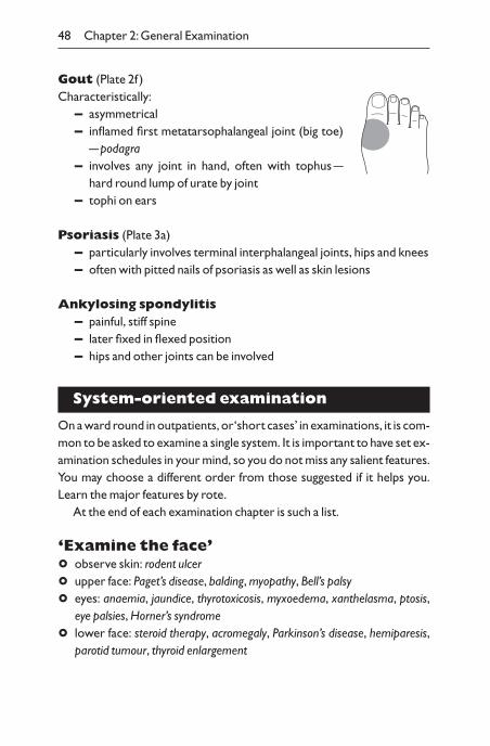
– asymmetrical– inflamed first metatarsophalangeal joint (big toe)
—podagra– involves any joint in hand, often with tophus —
hard round lump of urate by joint– tophi on ears
Psoriasis (Plate 3a)– particularly involves terminal interphalangeal joints, hips and knees– often with pitted nails of psoriasis as well as skin lesions
Ankylosing spondylitis– painful, stiff spine– later fixed in flexed position– hips and other joints can be involved
System-oriented examination
On a ward round in outpatients, or ‘short cases’ in examinations, it is com-mon to be asked to examine a single system. It is important to have set ex-amination schedules in your mind, so you do not miss any salient features.You may choose a different order from those suggested if it helps you.Learn the major features by rote.
At the end of each examination chapter is such a list.
‘Examine the face’
° observe skin: rodent ulcer
° upper face: Paget’s disease, balding, myopathy, Bell’s palsy
° eyes: anaemia, jaundice, thyrotoxicosis, myxoedema, xanthelasma, ptosis,eye palsies, Horner’s syndrome
° lower face: steroid therapy, acromegaly, Parkinson’s disease, hemiparesis,parotid tumour, thyroid enlargement
48 Chapter 2:General Examination
‘Examine the eyes’
° observe: jaundice, anaemia, arcus, ptosis, Horner’s syndrome
° examine:– check if patient is blind —beware of glass eye– movements of the eyes
– amblyopia or palsy– diplopia, nystagmus/false image
– visual acuity– visual fields– pupils: light and accommodation reflexes– fundi: disc, arteries and veins, retina, particularly fovea
‘Examine the neck’
° inspect from front and side– thyroid (ask patient to swallow)– lymph nodes– raised jugular venous pressure– lymph glands– other swellings
° inspect from front– examine neck veins– feel carotid arteries– auscultate bruits over thyroid and carotid arteries– check trachea is central
System-oriented Examination 49
CHAPTER 3
Examination of theCardiovascularSystem
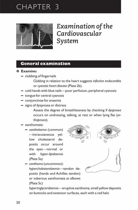
General examination
° Examine:– clubbing of fingernails
Clubbing in relation to the heart suggests infective endocarditisor cyanotic heart disease (Plate 2b).
– cold hands with blue nails —poor perfusion, peripheral cyanosis– tongue for central cyanosis– conjunctivae for anaemia– signs of dyspnoea or distress
Assess the degree of breathlessness by checking if dyspnoeaoccurs on undressing, talking, at rest or when lying flat (or-thopnoea).
– xanthomata:– xanthelasma (common)
—intracutaneous yel-low cholesterol de-posits occur aroundthe eyes —normal orwith hyper-lipidaemia(Plate 5a)
– xanthoma (uncommon):hypercholesterolaemia —tendon de-posits (hands and Achilles tendon)or tuberous xanthomata at elbows(Plate 5c)hypertriglyceridaemia —eruptive xanthoma, small yellow depositson buttocks and extensor surfaces, each with a red halo
50
Palpate the Radial Pulse 51
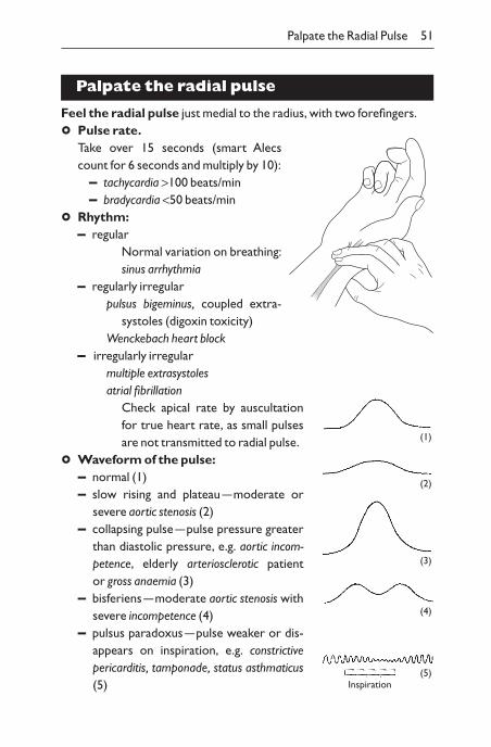
Palpate the radial pulse
Feel the radial pulse just medial to the radius, with two forefingers.
° Pulse rate.Take over 15 seconds (smart Alecscount for 6 seconds and multiply by 10):
– tachycardia >100 beats/min– bradycardia <50 beats/min
° Rhythm:– regular
Normal variation on breathing:sinus arrhythmia
– regularly irregularpulsus bigeminus, coupled extra-
systoles (digoxin toxicity)Wenckebach heart block
– irregularly irregularmultiple extrasystolesatrial fibrillation
Check apical rate by auscultationfor true heart rate, as small pulsesare not transmitted to radial pulse.
° Waveform of the pulse:– normal (1)– slow rising and plateau —moderate or
severe aortic stenosis (2)– collapsing pulse —pulse pressure greater
than diastolic pressure, e.g. aortic incom-petence, elderly arteriosclerotic patient or gross anaemia (3)
– bisferiens —moderate aortic stenosis withsevere incompetence (4)
– pulsus paradoxus —pulse weaker or dis-appears on inspiration, e.g. constrictivepericarditis, tamponade, status asthmaticus(5)
(1)
(2)
(3)
(4)
(5)Inspiration
° Volume:– small volume —low cardiac output– large volume
carbon dioxide retentionthyrotoxicosis
° Stiffness of the vessel wall:– in the elderly, a stiff, strongly pulsating, palpable 5–6cm radial ar-
tery indicates arteriosclerosis, a hardening of the walls of the arterythat is common with aging
is not atheromais associated with systolic hypertension
° Pulsus alternans.A difference of 20mmHg systolic blood pressure betweenconsecutive beats signifies poor left ventricular function.Thisneeds to be measured with a sphygmomanometer.
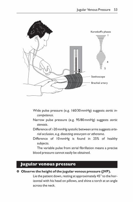
Take the blood pressure
– Wrap the cuff neatly and tightly around either upper arm.The pa-tient should be seated with the arm at the level of the heart.
– Gently inflate the cuff until the radial artery is no longer palpable.
– Using the stethoscope, listen over the brachial artery for the pulseto appear as you drop the pressure slowly (3–4mm/s).
° Systolic blood pressure: appearance of sounds– Korotkoff phase 1
° Diastolic blood pressure: disappearance of sounds– Korotkoff phase 5
Use large cuff for fat arms (circumference >30cm) so that inflatablecuff >1/2 arm circumference.
Beware auscultatory gap with sounds disappearing mid-systole. Ifsounds go to zero, use Korotkoff phase 4.
In adults, ~>140/85 is the current guideline in non-diabetic and ~>130/80 in diabetic patients. The patient may be nervous when first examined and the blood pressure may be falsely high. Take it again at the end of the examination.
52 Chapter 3: Examination of the Cardiovascular System
Jugular Venous Pressure 53
Wide pulse pressure (e.g. 160/30mmHg) suggests aortic in-competence.
Narrow pulse pressure (e.g. 95/80mmHg) suggests aorticstenosis.
Difference of >20mmHg systolic between arms suggests arte-rial occlusion, e.g. dissecting aneurysm or atheroma.
Difference of 10mmHg is found in 25% of healthy subjects.The variable pulse from atrial fibrillation means a precise
blood pressure cannot easily be obtained.
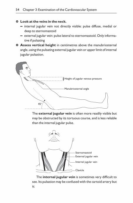
Jugular venous pressure
° Observe the height of the jugular venous pressure (JVP).Lie the patient down, resting at approximately 45° to the hor-izontal with his head on pillows, and shine a torch at an angleacross the neck.
Stethoscope
Brachial artery
Korotkoff’s phases1
4
5
54 Chapter 3: Examination of the Cardiovascular System
° Look at the veins in the neck.– internal jugular vein not directly visible: pulse diffuse, medial or
deep to sternomastoid– external jugular vein: pulse lateral to sternomastoid. Only informa-
tive if pulsating
° Assess vertical height in centimetres above the manubriosternalangle, using the pulsating external jugular vein or upper limit of internaljugular pulsation.
Height of jugular venous pressure
Manubriosternal angle
45∞
The external jugular vein is often more readily visible butmay be obstructed by its tortuous course, and is less reliablethan the internal jugular pulse.
SternomastoidExternal jugular vein
Internal jugular vein
Clavicle
The internal jugular vein is sometimes very difficult tosee. Its pulsation may be confused with the cartoid artery butit:
Jugular Venous Pressure 55
– has a complex pulsation– moves on respiration and decreases on inspiration except in tam-
ponade– cannot be palpated– can be obliterated by pressure on base of neck
The hepatojugular reflux is checked by firm pressurewith the flat of the right hand over the liver, while watching theJVP.
Compression on the dilated hepatic veins increases theJVP by 2cm.
If the JVP is found to be raised above the manubriosternalangle and pulsating, it implies right heart failure. Look for theother signs, i.e. pitting oedema and large tender liver. Some-times the JVP is so raised it can be missed, except that the earswaggle.
Dilated neck veins with no pulsation suggest non-cardiac ob-struction (e.g. carcinoma bronchus causing superior caval ob-struction or a kinked external jugular vein).
If the venous pressure rises on inspiration (it normallyfalls), constrictive pericarditis or pericardial effusion causing tamponade must be considered.
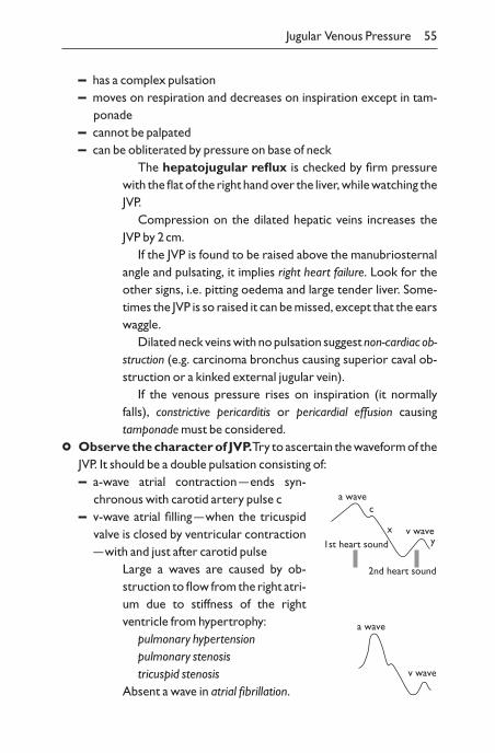
° Observe the character of JVP.Try to ascertain the waveform of theJVP. It should be a double pulsation consisting of:– a-wave atrial contraction —ends syn-
chronous with carotid artery pulse c– v-wave atrial filling —when the tricuspid
valve is closed by ventricular contraction—with and just after carotid pulse
Large a waves are caused by ob-struction to flow from the right atri-um due to stiffness of the rightventricle from hypertrophy:
pulmonary hypertensionpulmonary stenosistricuspid stenosis
Absent a wave in atrial fibrillation.
a wave
v wave
a wave
v wave
c
xy1st heart sound
2nd heart sound
56 Chapter 3: Examination of the Cardiovascular System
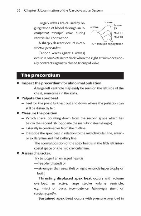
Large v waves are caused by re-gurgitation of blood through an in-competent tricuspid valve duringventricular contraction.
A sharp y descent occurs in con-strictive pericarditis.
Cannon waves (giant a waves)occur in complete heart block when the right atrium occasion-ally contracts against a closed tricuspid valve.
The precordium
° Inspect the precordium for abnormal pulsation.A large left ventricle may easily be seen on the left side of thechest, sometimes in the axilla.
° Palpate the apex beat.– Feel for the point furthest out and down where the pulsation can
still be distinctly felt.
° Measure the position.– Which space, counting down from the second space which lies
below the second rib (opposite the manubriosternal angle).– Laterally in centimetres from the midline.– Describe the apex beat in relation to the mid clavicular line, anteri-
or axillary line and mid axillary line.The normal position of the apex beat is in the fifth left inter-costal space on the mid clavicular line.
° Assess character.Try to judge if an enlarged heart is — feeble (dilated) or — stronger than usual (left or right ventricle hypertrophy or
both)Thrusting displaced apex beat occurs with volume
overload: an active, large stroke volume ventricle,e.g. mitral or aortic incompetence, left-to-right shunt orcardiomyopathy.
Sustained apex beat occurs with pressure overload in
a wave
v waveSevereTR
Mod TRMild TR
TR = tricuspid regurgitation
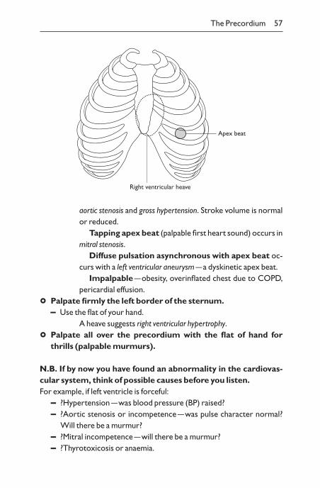
The Precordium 57
aortic stenosis and gross hypertension. Stroke volume is normalor reduced.
Tapping apex beat (palpable first heart sound) occurs inmitral stenosis.
Diffuse pulsation asynchronous with apex beat oc-curs with a left ventricular aneurysm —a dyskinetic apex beat.
Impalpable —obesity, overinflated chest due to COPD,pericardial effusion.
° Palpate firmly the left border of the sternum.– Use the flat of your hand.
A heave suggests right ventricular hypertrophy.
° Palpate all over the precordium with the flat of hand forthrills (palpable murmurs).
N.B. If by now you have found an abnormality in the cardiovas-cular system, think of possible causes before you listen.For example, if left ventricle is forceful:
– ?Hypertension —was blood pressure (BP) raised?– ?Aortic stenosis or incompetence —was pulse character normal?
Will there be a murmur?– ?Mitral incompetence —will there be a murmur?– ?Thyrotoxicosis or anaemia.
Apex beat
Right ventricular heave
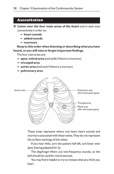
58 Chapter 3: Examination of the Cardiovascular System
Auscultation
° Listen over the four main areas of the heart and in each areaconcentrate in order on:
– heart sounds– added sounds– murmurs
Keep to this order when listening or describing what you haveheard, or you will miss or forget important findings.
The four main areas are:– apex, mitral area (and axilla if there is a murmur)– tricuspid area– aortic area (and neck if there is a murmur)– pulmonary area
Mitral area(5th intercostal space)
Tricuspid area
Pulmonary area(2nd intercostal space)
Aortic area
These areas represent where one hears heart sounds andmurmurs associated with these valves.They do not representthe surface markings of the valves.
If you hear little, turn the patient half left, and listen overapex (having palpated for it).
The diaphragm filters out low-frequency sounds, so thebell should be used for mitral stenosis.
You may find it helpful to try to imitate what you think youhear!
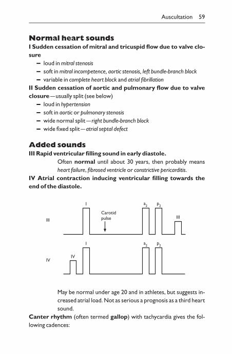
Auscultation 59
Normal heart soundsI Sudden cessation of mitral and tricuspid flow due to valve clo-sure
– loud in mitral stenosis– soft in mitral incompetence, aortic stenosis, left bundle-branch block– variable in complete heart block and atrial fibrillation
II Sudden cessation of aortic and pulmonary flow due to valveclosure —usually split (see below)
– loud in hypertension– soft in aortic or pulmonary stenosis– wide normal split — right bundle-branch block– wide fixed split —atrial septal defect
Added soundsIII Rapid ventricular filling sound in early diastole.
Often normal until about 30 years, then probably meansheart failure, fibrosed ventricle or constrictive pericarditis.
IV Atrial contraction inducing ventricular filling towards theend of the diastole.
III
IVIV
I
I
a2 p2
a2 p2
IIICarotidpulse
May be normal under age 20 and in athletes, but suggests in-creased atrial load. Not as serious a prognosis as a third heartsound.
Canter rhythm (often termed gallop) with tachycardia gives the fol-lowing cadences:
60 Chapter 3: Examination of the Cardiovascular System
III: Tum——te —tum or Ken——tucky (k = first heart sound)IV: te–Tum——te or Tenne——ssee (n = first heart sound)
Opening snap– Mitral valve normally opens silently after second heart sound.– In mitral stenosis, sudden movement of rigid valve makes a click, after
second heart sound (Fig. 3.1).
Ejection click– Aortic valve normally opens silently.– In aortic stenosis or sclerosis, can open with a click after first heart
sound.
Splitting of second heart soundAsk patients to take deep breaths in and out. Blood is drawn into the tho-rax during inspiration and then on to the right ventricle. There is tem-porarily more blood in the right ventricle than the left ventricle, and theright ventricle takes fractionally longer to empty.
Splitting is best heard during first two or three beats of inspiration. Donot ask patient to hold breath in or out when assessing splitting.
Paradoxical splitting occurs in aortic stenosis and left bundle-branchblock.
In both these conditions (Fig. 3.2) the left ventricle takes longer toempty, thus delaying a2 until after p2. During inspiration p2 occurs later andthe sounds draw closer together.
MurmursUse the diaphragm of the stethoscope for most high-pitched sounds ormurmurs (e.g. aortic incompetence) and the bell for low-pitched mur-murs (e.g. mitral stenosis). Note the following:
° Timing systolic or diastolic (compare with finger on carotid pulse)(Fig. 3.1).
° Site and radiation, e.g.:– mitral incompetence Æ axilla– aortic stenosis Æ carotids and apex– aortic incompetence Æ sternum
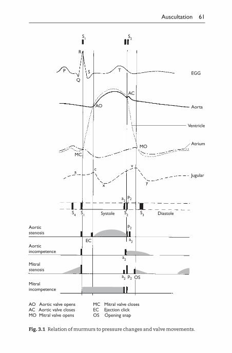
Auscultation 61
Aorticstenosis
Aorticincompetence
Mitralincompetence
Mitralstenosis
S4 S1
EC
S2 S3
a2p2
p2
a2
a2
a2p2 OS
Systole Diastole
ac
x
v
y
MO
MC
AO
AC
Aorta
Ventricle
Atrium
Jugular
EGGTS
Q
P
R
S2S1
AO Aortic valve opensAC Aortic valve closesMO Mitral valve opens
MC Mitral valve closesEC Ejection clickOS Opening snap
Fig. 3.1 Relation of murmurs to pressure changes and valve movements.
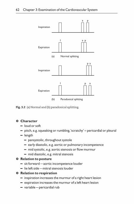
62 Chapter 3: Examination of the Cardiovascular System
° Character– loud or soft– pitch, e.g. squeaking or rumbling, ‘scratchy’ = pericardial or pleural– length
– pansystolic, throughout systole– early diastolic, e.g. aortic or pulmonary incompetence– mid systolic, e.g. aortic stenosis or flow murmur– mid diastolic, e.g. mitral stenosis
° Relation to posture– sit forward —aortic incompetence louder– lie left side —mitral stenosis louder
° Relation to respiration– inspiration increases the murmur of a right heart lesion– expiration increases the murmur of a left heart lesion– variable —pericardial rub
(a)
Inspiration
a p
I a p
(b)
ap
I ap
Expiration
Inspiration
Expiration
Normal splitting
Paradoxical splitting
Fig. 3.2 (a) Normal and (b) paradoxical splitting.
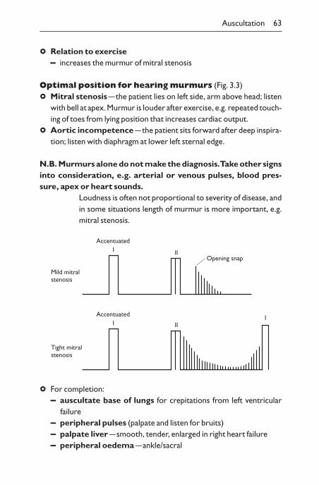
Auscultation 63
° Relation to exercise– increases the murmur of mitral stenosis
Optimal position for hearing murmurs (Fig. 3.3)
° Mitral stenosis —the patient lies on left side, arm above head; listenwith bell at apex. Murmur is louder after exercise, e.g. repeated touch-ing of toes from lying position that increases cardiac output.
° Aortic incompetence —the patient sits forward after deep inspira-tion; listen with diaphragm at lower left sternal edge.
N.B. Murmurs alone do not make the diagnosis.Take other signsinto consideration, e.g. arterial or venous pulses, blood pres-sure, apex or heart sounds.
Loudness is often not proportional to severity of disease, andin some situations length of murmur is more important, e.g.mitral stenosis.
Mild mitralstenosis
I IIOpening snap
Accentuated
Tight mitralstenosis
I IIIAccentuated
° For completion:– auscultate base of lungs for crepitations from left ventricular
failure– peripheral pulses (palpate and listen for bruits)– palpate liver —smooth, tender, enlarged in right heart failure– peripheral oedema —ankle/sacral
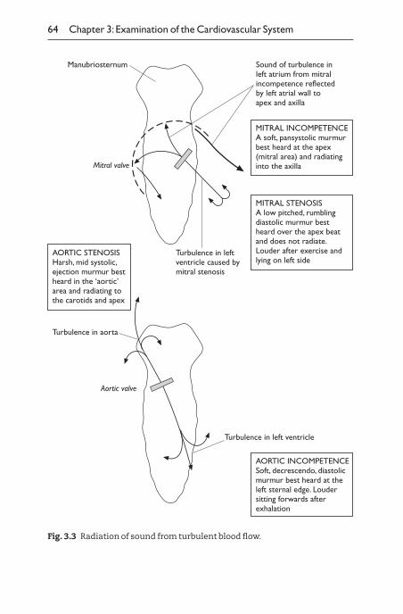
64 Chapter 3: Examination of the Cardiovascular System
Manubriosternum
Mitral valve
Sound of turbulence inleft atrium from mitralincompetence reflectedby left atrial wall toapex and axilla
Turbulence in leftventricle caused bymitral stenosis
MITRAL INCOMPETENCEA soft, pansystolic murmurbest heard at the apex(mitral area) and radiatinginto the axilla
MITRAL STENOSISA low pitched, rumblingdiastolic murmur bestheard over the apex beatand does not radiate.Louder after exercise andlying on left side
AORTIC STENOSISHarsh, mid systolic,ejection murmur bestheard in the ‘aortic’area and radiating tothe carotids and apex
Aortic valve
Turbulence in aorta
Turbulence in left ventricle
AORTIC INCOMPETENCESoft, decrescendo, diastolicmurmur best heard at theleft sternal edge. Loudersitting forwards afterexhalation
Fig. 3.3 Radiation of sound from turbulent blood flow.
Signs of Left and Right Ventricular Failure 65
Summary of timing of murmurs
Ejection systolic murmuraortic stenosis or sclerosis (same murmur, due to stiffness of valve cuspsand aortic walls, with normal pulse pressure)
aortic sclerosis is present in 50% of 50-year-oldspulmonary stenosisatrial septal defectFallot’s syndrome —right outflow tract obstruction
Pansystolic murmurmitral regurgitationtricuspid regurgitationventricular septal defect
Late systolic murmurmitral valve prolapse (click–murmur syndrome)hypertrophic cardiomyopathycoarction aorta (extending in diastole to a ‘machinery murmur’)
Early diastolic murmuraortic regurgitationpulmonary regurgitationGraham Steell murmur in pulmonary hypertension (see p. 70)
Mid–late diastolic murmurmitral stenosistricuspid stenosisAustin Flint murmur in aortic incompetence (see p. 69)left atrial myxoma (variable —can also give other murmurs)
Signs of left and right ventricular failure
Left heart failure– Dyspnoea.– Basal crepitations.
66 Chapter 3: Examination of the Cardiovascular System
– Fourth heart sound, or third in older patients.
° Sit the patient forward and listen at the bases of the lungs with the di-aphragm of the stethoscope for fine crepitations.
Fine crepitations are caused by alveoli opening on inspiration.When a patient has been recumbent for a while, alveoli tendto collapse in the normal lung. On taking a deep breath crepi-tations will be heard but do not mean pulmonary oedema.Askthe patient to cough. If crepitations continue after this, pul-monary oedema may be present.
Right heart failure– Raised JVP.– Enlarged tender liver (see later).– Pitting oedema.
° With the patient sitting forward, look for swelling over the sacral area.If there is, push your thumb into the swelling and see if you leave an in-dentation. If you do, this is called pitting oedema.
° Check both ankles for pitting oedema.Oedema (fluid) collects at the most dependent part of thebody. A patient who is mostly sitting will have ankle oedemawhile a patient who is lying will have predominantly sacraloedema.
Functional result
° Having ascertained the basic pathology (e.g. myocardial infarction, aorticstenosis, pericarditis), make an assessment of the functional result.– History. How far can the patient walk, etc.– Examination. Evidence of:
– cardiac enlargement (hypertrophy or dilatation)– heart failure– arrhythmias– pulmonary hypertension– cyanosis– endocarditis
– Investigations. For example:
Summary of Common Illnesses 67
– chest X-ray– electrocardiogram (ECG)– treadmill exercise test with ECG for ischaemia– echocardiograph —sonar ‘radar’ of heart, for muscle and ven-
tricle size, muscle contractility and ejection fraction, valvefunction
– 24-hour ECG tape for arrhythmias– cardiac catheterization for pressure measurements, blood
oxygenation and angiogram– radioactive scan —to image live, ischaemic or dead cardiac
muscle
Summary of common illnesses
Mitral stenosis– small pulse —fibrillating?– JVP only raised if heart failure– RV++ LVo tapping apex– loud I. Loud p2 if pulmonary hypertension– opening snap (os)– mid diastolic murmur at apex only (low-pitched rumbling)
– severity indicated by early opening snap and long murmur– best heard with the patient in left lateral position, in expira-
tion with the stethoscope bell, particularly after exercise hasincreased cardiac output
– presystolic accentuation of murmur (absent if atrial fibrilla-tion and stiff cusps)
– sounds ‘ta ta rooofoo T’from II os murmur I
Mitral incompetence– fibrillating?– JVP only raised if heart failure– RV+ LV++ systolic thrill– soft I. Loud p2 if pulmonary hypertension– pansystolic murmur apex Æ axilla
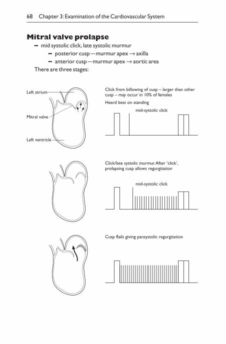
68 Chapter 3: Examination of the Cardiovascular System
Mitral valve prolapse– mid systolic click, late systolic murmur
– posterior cusp —murmur apex Æ axilla– anterior cusp —murmur apex Æ aortic area
There are three stages:
Click/late systolic murmur. After ‘click’,prolapsing cusp allows regurgitation
mid-systolic click
Cusp flails giving pansystolic regurgitation
Left atrium
Mitral valve
Left ventricle
Click from billowing of cusp – larger than othercusp – may occur in 10% of females
mid-systolic click
Heard best on standing
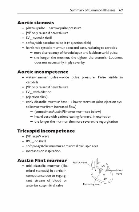
Summary of Common Illnesses 69
Aortic stenosis– plateau pulse —narrow pulse pressure– JVP only raised if heart failure– LV++ systolic thrill– soft a2 with paradoxical split (± ejection click)– harsh mid systolic murmur, apex and base, radiating to carotids
– note discrepancy of forceful apex and feeble arterial pulse– the longer the murmur, the tighter the stenosis. Loudness
does not necessarily imply severity
Aortic incompetence– water-hammer pulse —wide pulse pressure. Pulse visible in
carotids– JVP only raised if heart failure– LV++ with dilation– (ejection click)– early diastolic murmur base Æ lower sternum (also ejection sys-
tolic murmur from increased flow)– (sometimes Austin Flint murmur —see below)– heard best with patient leaning forward, in expiration– the longer the murmur, the more severe the regurgitation
Tricuspid incompetence– JVP large V wave– RV++, no thrill– soft pansystolic murmur at maximal tricuspid area– increases on inspiration
Austin Flint murmur– mid diastolic murmur (like
mitral stenosis) in aortic in-competence due to regurgi-tant stream of blood onanterior cusp mitral valve
Mitralvalve
LA
LV
Aortic valve
Fluttering cusp
70 Chapter 3: Examination of the Cardiovascular System
Graham Steell murmur– pulmonary early diastolic murmur (functional pulmonary
incompetence) in mitral stenosis or other causes of pulmonary hypertension
Atrial septal defect– JVP only raised if failure or tricuspid incompetence– RV++ LVo– widely fixed split-second sound– pulmonary systolic murmur (tricuspid diastolic flow murmur)
Ventricular septal defect– RV+ LV+– Pansystolic murmur on left sternal edge (loud if small defect!)
Patent ductus arteriosus– systolic Æ diastolic ‘machinery’ or continuous murmur below left
clavicle
Metal prosthetic valves– loud clicks with short flow murmur
– aortic systolic– mitral diastolic
– need anticoagulation
Tissue prosthetic valves– porcine xenograft or human homograft– tend to fibrose after 7–10 years, leading to stenosis and
incompetence– may not require anticoagulation
Pericardial rub– scratchy, superficial noise heard in systole and diastole– brought out by stethoscope pressure, and sometimes variable with
respiration
Summary of Common Illnesses 71
Infectious endocarditis (diagnosis made from bloodcultures)
– febrile, unwell, anaemia– clubbing– splinter haemorrhages– Osler’s nodes– cardiac murmur– splenomegaly– haematuria
Rheumatic fever– flitting arthralgia– erythema nodosum or erythema marginatum– tachycardia– murmurs– Sydenham’s chorea (irregular, uncontrollable jerks of limbs,
tongue)
Clues to diagnosis from facial appearance– Down’s syndrome from 21 trisomy – ventricular septal defect
– patent ductus arteriosus– thyrotoxicosis —atrial fibrillation– myxoedema from hypothyroid —cardiomyopathy– dusky, congested face — superior vena cava obstruction– red cheeks in infra-orbital region in mitral facies from mitral
stenosis
Clues to diagnosis from generalappearance
– Turner’s syndrome from sex chromosomes X0– female, short stature, web of neck– coarctation of aorta
– Marfan’s syndrome– tall patient with long, thin fingers– aortic regurgitation
72 Chapter 3: Examination of the Cardiovascular System
Peripheral arteries
° Feel all peripheral pulses (Fig. 3.4). Lower-limb pulses are usuallyfelt after examining the abdomen.
Diminished or absent pulses suggest arterial stenosis or occlusion.
The lower-limb pulses are particularly important if there isa history of intermittent claudication.
Auscultation of the carotid and femoral vessels is useful ifthere is a suspicion these arteries are stenosed. A bruit isheard if the stenosis causes turbulent flow.
Coarctation of the aorta delays the femoral pulse after theradial pulse.
Peripheral vascular disease– white or blue discoloration– ulcers with little granulation tissue and slow healing– shiny skin, loss of hairs, thickened dystrophic nails– absent pulses– Buerger’s test of severity of arterial insufficiency
– loss of autoregulation of blood flow– patient lying supine, lift leg up to 45° —positive test: pallor of
foot; venous guttering– hang legs over side of bed: note time to capillary and venous
filling; reactive hyperaemia; subsequent cyanosisDiabetes, when present, also signs from neuropathy:
– dry skin with thickened epidermis– callus from increased foot pressure over abnormal sites, e.g.
under tarsal heads in mid-foot, secondary to motor neuropathyand change in distribution of weight (Plate 4f)
– absent ankle reflexes– decreased sensation
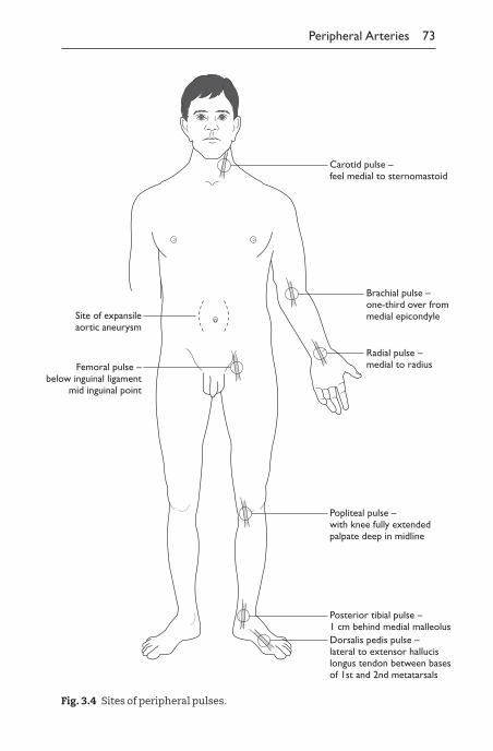
Peripheral Arteries 73
Carotid pulse –feel medial to sternomastoid
Brachial pulse –one-third over frommedial epicondyleSite of expansile
aortic aneurysm
Femoral pulse –below inguinal ligament
mid inguinal point
Popliteal pulse –with knee fully extendedpalpate deep in midline
Dorsalis pedis pulse –lateral to extensor hallucislongus tendon between basesof 1st and 2nd metatarsals
Posterior tibial pulse –1 cm behind medial malleolus
Radial pulse –medial to radius
Fig. 3.4 Sites of peripheral pulses.
74 Chapter 3: Examination of the Cardiovascular System
Aortic aneurysm– central abdominal pulsation visible or palpable– need to distinguish from normal, palpable aorta in midline in thin
people– aortic aneurysm is expansible to each side as well as forwards– a bruit may be audible– associated with femoral and popliteal artery aneurysms
Varicose veins
° Varicose veins and herniae (see p. 95) are examined when the pa-tient is standing, possibly at the end of the whole examination at thesame time as the gait (see p. 45).
Majority are associated with incompetent valves in the longsaphenous vein or short saphenous vein.
Long saphenous —from femoral vein in groin to medialside of lower leg.
Short saphenous —from popliteal fossa to back of calf andlateral malleolus.
° Observe:– swelling– pigmentation– eczema } indicate chronic venous insufficiency
– inflammation —suggests thrombophlebitis
° Palpate:– soft or hard (thrombosed)– tender —thrombophlebitis– cough impulse —implies incompetent valves
Incompetent valves can be confirmed by the Trendelenburg test:
– Elevate leg to empty veins.– Occlude long saphenous vein with a tourniquet around
upper thigh.– Stand patient up.– If veins fill rapidly, this indicates incompetent thigh perfora-
tors below the tourniquet.
System-oriented Examination 75
– If, after release of tourniquet, veins fill rapidly, this indicatesincompetent saphenofemoral junction.
If veins fill immediately on standing, then incompetent valves are inthigh or calf, so do the Perthes test:
– As for Trendelenburg, but on standing let some bloodenter veins by temporary release of groin pressure.
– Ask patient to stand up and down on toes.– Veins become less tense if:
– muscle pump is satisfactory– perforating calf veins are patent with competent valves
System-oriented examination
‘Examine the cardiovascular system’
° hands —splinter haemorrhages
° radial pulse —rate, rhythm, waveform, volume, state of artery
° waveform and volume best examined at the brachial or carotid artery
° ‘I would normally measure the blood pressure now; would you like me to doso?’
° eyes —anaemia
° tongue —central cyanosis
° JVP —height, waveform
° apex beat —site, character
° auscultate —at apex (with thumb on carotid artery for timing)– heart sounds– added sounds– murmurs
– in neck over carotid artery —each area of precordium with di-aphragm
– aortic incompetence —lean forward in full expiration withdiaphragm
– mitral stenosis —lie patient on left side and listen at apexwith bell
° ‘I would normally now listen to the bases for crepitations, examine for he-patomegaly, peripheral oedema and peripheral pulses.Would you like me todo so?’
CHAPTER 4
Examination of the Chest
General inspection
° Examine the patient for:– end of bed for signs of respiratory distress —use of accessory
muscles, on oxygen therapy, inspect sputum pot– nicotine on fingers– clubbing: respiratory causes include:
carcinoma of bronchusmesotheliomabronchiectasislung abscessempyemafibrosing alveolitis
– evidence of respiratory failure:– hypoxia: central cyanosis– hypercapnia: drowsiness, confusion, papilloedema, warm
hands, bounding pulse, dilated veins, coarse tremor/flap– respiratory rate: count per minute– pattern of respiration: Cheyne–Stokes:
– alternating hyperventilation and apnoea– severe increased intracranial pressure– left ventricular failure– high altitude
– obstructive airways disease:– pursed-lip breathing:
– expiration against partially closed lips– chronic obstructive airways disease to delayed closure
of bronchioles
76
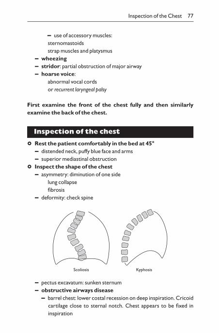
Inspection of the Chest 77
– use of accessory muscles:sternomastoidsstrap muscles and platysmus
– wheezing– stridor: partial obstruction of major airway– hoarse voice:
abnormal vocal cordsor recurrent laryngeal palsy
First examine the front of the chest fully and then similarly examine the back of the chest.
Inspection of the chest
° Rest the patient comfortably in the bed at 45°– distended neck, puffy blue face and arms– superior mediastinal obstruction
° Inspect the shape of the chest– asymmetry: diminution of one side
lung collapsefibrosis
– deformity: check spine
Scoliosis Kyphosis
– pectus excavatum: sunken sternum– obstructive airways disease
– barrel chest: lower costal recession on deep inspiration. Cricoidcartilage close to sternal notch. Chest appears to be fixed in inspiration
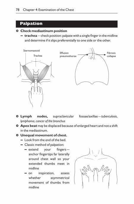
78 Chapter 4: Examination of the Chest
Palpation
° Check mediastinum position– trachea —check position: palpate with a single finger in the midline
and determine if it slips preferentially to one side or the other.
Fibrosiscollapse
Effusionpneumothorax
Sternomastoid
Trachea
° Lymph nodes, supraclavicular fossae/axillae — tuberculosis,lymphoma, cancer of the bronchus
° Apex beat may be displaced because of enlarged heart and not a shiftin the mediastinum.
° Unequal movement of chest.– Look from the end of the bed.– Classic method of palpation:
– extend your fingers —anchor fingertips far laterally around chest wall so your extended thumbs meet inmidline
– on inspiration, assesswhether asymmetricalmovement of thumbs frommidline
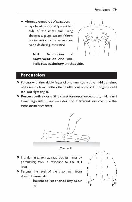
Percussion 79
– Alternative method of palpation:– lay a hand comfortably on either
side of the chest and, usingthese as a gauge, assess if thereis diminution of movement onone side during inspiration
N.B. Diminution ofmovement on one sideindicates pathology on that side.
Percussion
° Percuss with the middle finger of one hand against the middle phalanxof the middle finger of the other, laid flat on the chest.The finger shouldstrike at right angles.
° Percuss both sides of the chest for resonance, at top, middle andlower segments. Compare sides, and if different also compare thefront and back of chest.
Chest wall
° If a dull area exists, map out its limits by percussing from a resonant to the dull area.
° Percuss the level of the diaphragm fromabove downwards.
Increased resonance may occurin:
7
1
3
5
8
2
4
6
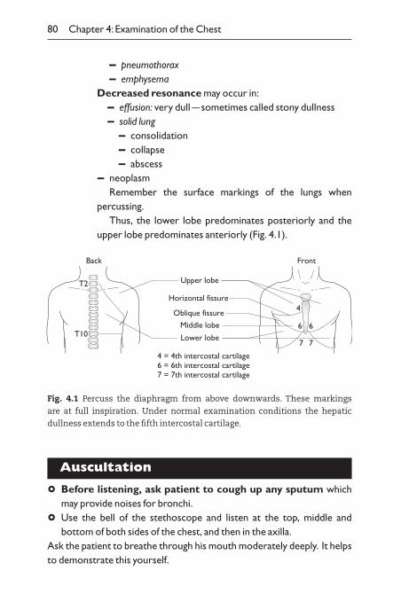
80 Chapter 4: Examination of the Chest
– pneumothorax– emphysema
Decreased resonance may occur in:– effusion: very dull —sometimes called stony dullness– solid lung
– consolidation– collapse– abscess
– neoplasmRemember the surface markings of the lungs when
percussing.Thus, the lower lobe predominates posteriorly and the
upper lobe predominates anteriorly (Fig. 4.1).
Horizontal fissure
Upper lobe
Oblique fissure
Middle lobe
Lower lobe
T2
T10
4 = 4th intercostal cartilage6 = 6th intercostal cartilage7 = 7th intercostal cartilage
4
6
7
6
7
Back Front
Fig. 4.1 Percuss the diaphragm from above downwards. These markings
are at full inspiration. Under normal examination conditions the hepatic
dullness extends to the fifth intercostal cartilage.
Auscultation
° Before listening, ask patient to cough up any sputum whichmay provide noises for bronchi.
° Use the bell of the stethoscope and listen at the top, middle and bottom of both sides of the chest, and then in the axilla.
Ask the patient to breathe through his mouth moderately deeply. It helpsto demonstrate this yourself.
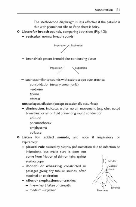
Auscultation 81
The stethoscope diaphragm is less effective if the patient isthin with prominent ribs or if the chest is hairy.
° Listen for breath sounds, comparing both sides (Fig. 4.2):– vesicular: normal breath sounds
Inspiration Expiration
– bronchial: patent bronchi plus conducting tissue
Inspiration Expiration
– sounds similar to sounds with stethoscope over tracheaconsolidation (usually pneumonia)neoplasmfibrosisabscess
not collapse, effusion (except occasionally at surface)– diminution: indicates either no air movement (e.g. obstructed
bronchus) or air or fluid preventing sound conductioneffusionpneumothoraxemphysemacollapse
° Listen for added sounds, and note if inspiratory or expiratory:– pleural rub: caused by pleurisy (inflammation due to infection or
infarction), but make sure it does not come from friction of skin or hairs againststethoscope
– rhonchi or wheezing: constricted airpassages giving dry tubular sounds, oftenmaximal on expiration
– râles or crepitations or crackles:– fine —heart failure or alveolitis– medium — infection
Stridor
Rhonchi
Coarserales
Fine rales
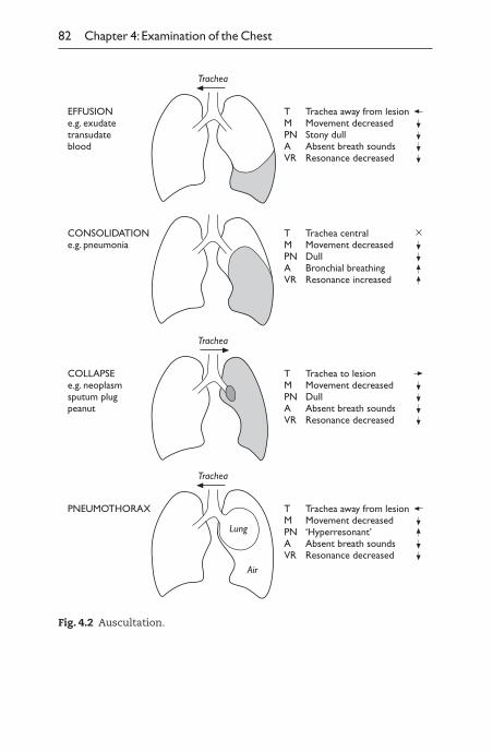
82 Chapter 4: Examination of the Chest
Trachea
TMPNAVR
Trachea away from lesionMovement decreasedStony dullAbsent breath soundsResonance decreased
EFFUSIONe.g. exudatetransudateblood
TMPNAVR
Trachea centralMovement decreasedDullBronchial breathingResonance increased
CONSOLIDATIONe.g. pneumonia
Trachea
TMPNAVR
Trachea to lesionMovement decreasedDullAbsent breath soundsResonance decreased
COLLAPSEe.g. neoplasmsputum plugpeanut
Trachea
TMPNAVR
Trachea away from lesionMovement decreased‘Hyperresonant’Absent breath soundsResonance decreased
PNEUMOTHORAX
Lung
Air
Fig. 4.2 Auscultation.

Sputum 83
– coarse —air bubbling through fluid in larger bronchioles, e.g.bronchiectasis. If relieved by coughing, suggests from bronchioles
Vocal resonance
Normally only done if pathology is suspected, but you mustpractise to become familiar with normal resonance.
° Ask the patient to repeat ‘99’ whilst listening to chest in the sameareas as auscultation.The sounds are louder over areas of consolida-tion. Compare both sides.
At the surface of an effusion the words ‘99’ take on a bleatingcharacter like a goat, which is called aegophony. If vocal resonance is gross, whispering pectoriloquy can be elicited by asking the patient to whisper:‘1, 2, 3, 4’.
N.B. Vocal fremitus, breath sounds and vocal resonance all depend on the same criteria and vary together.
To determine further clues check:– chest movement asymmetry– mediastinum displacement– percussion
Sputum
Examination of the sputum is unpleasant but important.
° Look for:– quantity (increased grossly in bronchiectasis)– consistency (if all mucus it may be saliva)– colour (in yellow or green it may be infected)– blood – cancer, tuberculosis, embolus
Ideally the sputum should be examined under the microscopefor:– bacteria– pus cells– eosinophils– plugs– asbestos
84 Chapter 4: Examination of the Chest
Functional result
° Make an assessment of the functional result:– History. How far can the patient walk, etc.– Examination:
– PO2Ø: central cyanosisconfusion
– PCO2≠: peripheral signs – warm periphery– dilated veins– bounding pulse– flapping tremor
central signs – drowsy– papilloedema– small pupils
– check by arterial blood gases– Tests (usually of obstructive airways disease):
– At the bedside: blowing out a lighted match about 15cm from the mouth and with the mouth wide open is easy as long as your peak flow is above approximately 80 l/min (normal300–500 l/min).
– Expiration time: an assessment of airways obstruction can be made by timing the period of full expiration through wide-open mouth following a deep breath. This should be less than 2 seconds when normal.
– Chest expansion: expansion from full inspiration to full expi-ration should be more than 5cm. Reduced if hyperinflation of thechest is due to chronic obstructive airways disease.
– Peak flow meter: a measure of airways obstruction is the peakrate of flow of air out of the lungs. A record is made using a machine. Normal 300–500l/min.
Summary of common illnesses
Asthma– patient distressed, tachypnoeic, unable to talk easily– wheeze on expiration audible or by auscultation
Summary of Common Illnesses 85
– overinflated chest with hyperresonance– if central cyanosis: critically ill, artificial ventilation?– pulsus paradoxus (may be normal between attacks)– often due to atopy
– enquire about exposure to antigens:– house dust mite– cats or dogs
Obstructive airways disease (chronic)– barrel chest– accessory muscles of respiration in use– hyperresonance– depressed diaphragm —indrawing lower costal margin on
inspiration– diminished breath sounds:
– blue bloater:central cyanosissigns of carbon dioxide retentionobesenot dyspnoeicankle oedema: may or may not have right heart failure
– pink puffer:not cyanosedno carbon dioxide retentionthindyspnoeicno oedema
Bronchiectasis– clubbing– constant green/yellow phlegm– coarse râles over affected area
Allergic alveolitis– clubbing– fine, unexplained râles, widespread over bases
86 Chapter 4: Examination of the Chest
System-oriented examination
‘Examine the respiration system’
° hands: clubbing, signs of increased carbon dioxide (warm hands,bounding pulse, coarse tremor)
° tongue: central cyanosis
° trachea
° supraclavicular nodes
° inspection– shape of chest– chest movements– respiration rate/distress
° palpation: unequal movement of chest using hands
° percussion: upper segments (L, R), middle (L, R) and lower segments(L, R)
° auscultation:– breath sounds– added sounds: crepitations, bronchospasm, pleural rub, stridor
(vocal fremitus)
° if obstructive airways disease:– expiration time (see p. 84)
CHAPTER 5
Examination of the Abdomen
General inspection
° Look for signs of:– chronic liver disease:
– clubbing– leukonychia– palmar erythema– telangiectasia on face– icterus (Plate 1e)– spider naevi (Plate 1f)– gynaecomastia– alcohol abuse
– Dupuytren’s contracture (Plate 4c)– parotid enlargement– testicular atrophy
– liver failure:– liver flap– foetor hepaticus– confusion
Signs of chronic liver disease are usually obvious, but we are allallowed up to six spider naevi (particularly if pregnant!).
– anaemia —look at conjunctiva, tongue– iron deficiency:
– koilonychia (Plate 2d)– smooth tongue– angular stomatitis —can be from ill-fitting dentures or edentu-
lous state– B12 or folate deficiency —‘beef steak’ or smooth tongue
87
Spider naevus:a small collection ofcapillaries fed by acentral arteriole
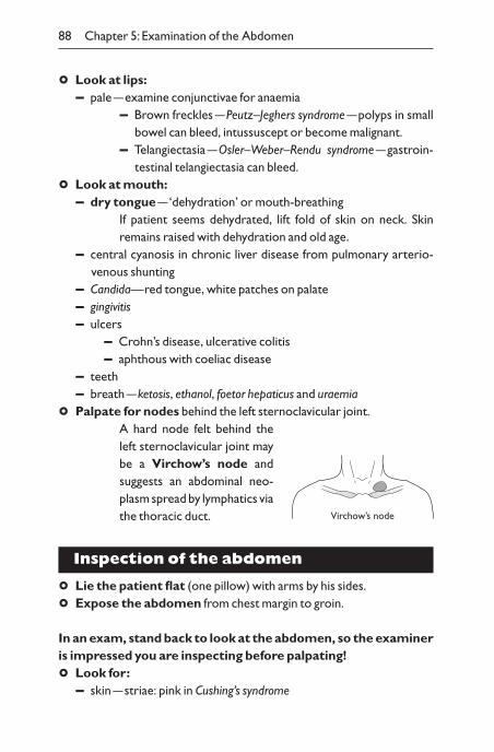
88 Chapter 5: Examination of the Abdomen
° Look at lips:– pale —examine conjunctivae for anaemia
– Brown freckles —Peutz–Jeghers syndrome —polyps in smallbowel can bleed, intussuscept or become malignant.
– Telangiectasia —Osler–Weber–Rendu syndrome —gastroin-testinal telangiectasia can bleed.
° Look at mouth:– dry tongue —‘dehydration’ or mouth-breathing
If patient seems dehydrated, lift fold of skin on neck. Skin remains raised with dehydration and old age.
– central cyanosis in chronic liver disease from pulmonary arterio-venous shunting
– Candida—red tongue, white patches on palate– gingivitis– ulcers
– Crohn’s disease, ulcerative colitis– aphthous with coeliac disease
– teeth– breath —ketosis, ethanol, foetor hepaticus and uraemia
° Palpate for nodes behind the left sternoclavicular joint.A hard node felt behind theleft sternoclavicular joint maybe a Virchow’s node andsuggests an abdominal neo-plasm spread by lymphatics viathe thoracic duct.
Inspection of the abdomen
° Lie the patient flat (one pillow) with arms by his sides.
° Expose the abdomen from chest margin to groin.
In an exam, stand back to look at the abdomen, so the examineris impressed you are inspecting before palpating!
° Look for:– skin —striae: pink in Cushing’s syndrome
Virchow’s node
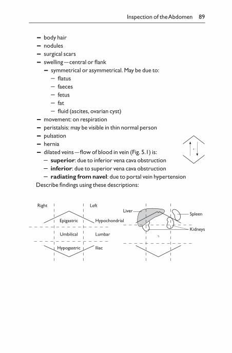
Inspection of the Abdomen 89
– body hair– nodules– surgical scars– swelling —central or flank
– symmetrical or asymmetrical. May be due to:– flatus– faeces– fetus– fat– fluid (ascites, ovarian cyst)
– movement: on respiration– peristalsis: may be visible in thin normal person– pulsation– hernia– dilated veins —flow of blood in vein (Fig. 5.1) is:
– superior: due to inferior vena cava obstruction– inferior: due to superior vena cava obstruction– radiating from navel: due to portal vein hypertension
Describe findings using these descriptions:
Spleen
Kidneys
LiverLeft
Hypochondrial
Lumbar
Iliac
Epigastric
Umbilical
Hypogastric
Right
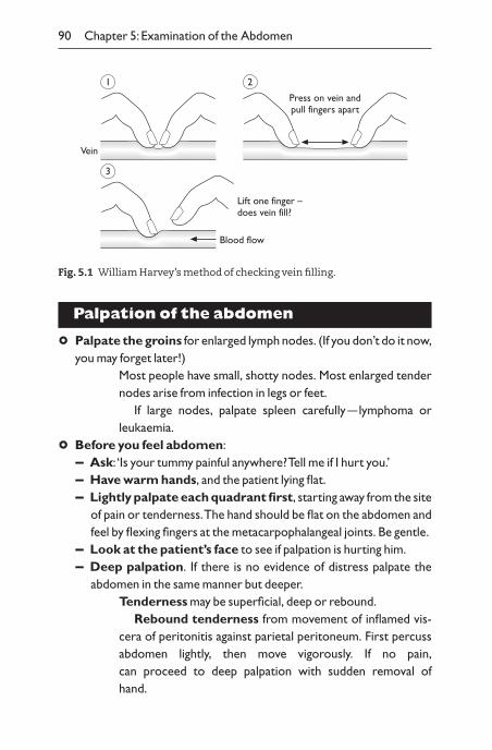
90 Chapter 5: Examination of the Abdomen
Palpation of the abdomen
° Palpate the groins for enlarged lymph nodes. (If you don’t do it now,you may forget later!)
Most people have small, shotty nodes. Most enlarged tendernodes arise from infection in legs or feet.
If large nodes, palpate spleen carefully —lymphoma orleukaemia.
° Before you feel abdomen:– Ask: ‘Is your tummy painful anywhere? Tell me if I hurt you.’– Have warm hands, and the patient lying flat.– Lightly palpate each quadrant first, starting away from the site
of pain or tenderness.The hand should be flat on the abdomen andfeel by flexing fingers at the metacarpophalangeal joints. Be gentle.
– Look at the patient’s face to see if palpation is hurting him.– Deep palpation. If there is no evidence of distress palpate the
abdomen in the same manner but deeper.Tenderness may be superficial, deep or rebound.
Rebound tenderness from movement of inflamed vis-cera of peritonitis against parietal peritoneum. First percussabdomen lightly, then move vigorously. If no pain,can proceed to deep palpation with sudden removal of hand.
Vein
1 2
Press on vein andpull fingers apart
Blood flow
3
Lift one finger –does vein fill?
Fig. 5.1 William Harvey’s method of checking vein filling.
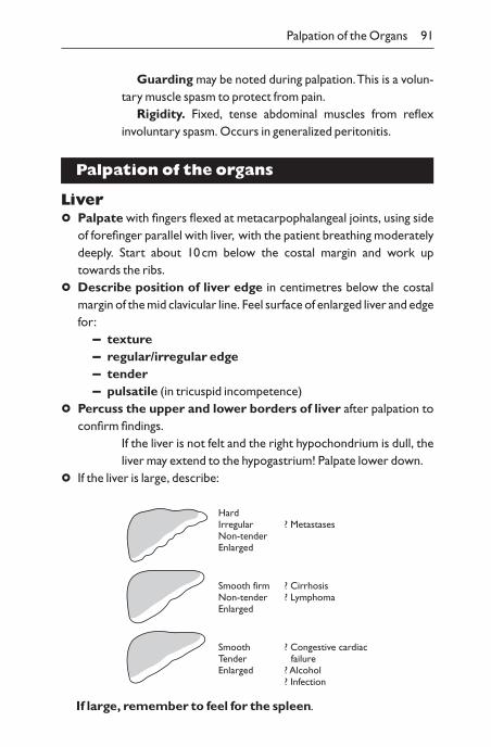
Palpation of the Organs 91
Guarding may be noted during palpation.This is a volun-tary muscle spasm to protect from pain.
Rigidity. Fixed, tense abdominal muscles from reflex involuntary spasm. Occurs in generalized peritonitis.
Palpation of the organs
Liver
° Palpate with fingers flexed at metacarpophalangeal joints, using sideof forefinger parallel with liver, with the patient breathing moderatelydeeply. Start about 10cm below the costal margin and work up towards the ribs.
° Describe position of liver edge in centimetres below the costalmargin of the mid clavicular line. Feel surface of enlarged liver and edgefor:
– texture– regular/irregular edge– tender– pulsatile (in tricuspid incompetence)
° Percuss the upper and lower borders of liver after palpation toconfirm findings.
If the liver is not felt and the right hypochondrium is dull, theliver may extend to the hypogastrium! Palpate lower down.
° If the liver is large, describe:
HardIrregularNon-tenderEnlarged
? Metastases
Smooth firmNon-tenderEnlarged
? Cirrhosis? Lymphoma
SmoothTenderEnlarged
? Congestive cardiac failure? Alcohol? Infection
If large, remember to feel for the spleen.
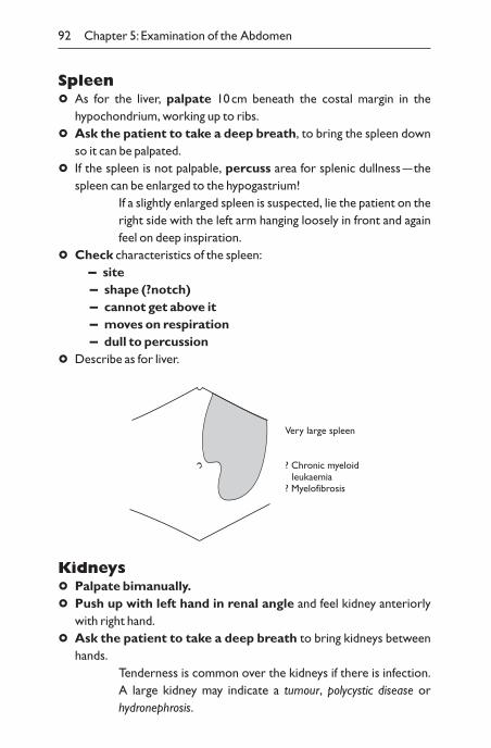
92 Chapter 5: Examination of the Abdomen
Spleen
° As for the liver, palpate 10cm beneath the costal margin in thehypochondrium, working up to ribs.
° Ask the patient to take a deep breath, to bring the spleen downso it can be palpated.
° If the spleen is not palpable, percuss area for splenic dullness —thespleen can be enlarged to the hypogastrium!
If a slightly enlarged spleen is suspected, lie the patient on theright side with the left arm hanging loosely in front and againfeel on deep inspiration.
° Check characteristics of the spleen:– site– shape (?notch)– cannot get above it– moves on respiration– dull to percussion
° Describe as for liver.
Kidneys
° Palpate bimanually.
° Push up with left hand in renal angle and feel kidney anteriorlywith right hand.
° Ask the patient to take a deep breath to bring kidneys betweenhands.
Tenderness is common over the kidneys if there is infection.A large kidney may indicate a tumour, polycystic disease or hydronephrosis.
Very large spleen
? Chronic myeloid leukaemia? Myelofibrosis
Percussion 93
Masses
° Carefully palpate the whole of the abdomen. If a mass is found,describe:
– site– size– shape– consistency —faeces may be indented by pressure– fixation or mobility —does it move on respiration?– tender– pulsatile —transmitted pulsation from aorta or pulsatile
swelling– dull to percussion —particularly important to determine if
bowel is in front of mass– does it alter after defaecation or micturition?
Aorta
° Palpate in the midline above the umbilicus for a pulsatile mass. Ifeasily palpated, suspect aortic aneurysm and proceed to ultrasonogra-phy in males over 50 and women over 60 years.
– may be normal aorta in a thin person– unfolded aorta– aneurysm
Percussion
Dullness on percussion:– ascites —free fluid– an organ, e.g. liver, spleen– tumour, e.g. large ovarian cyst
° Percuss liver, speen and kidneys after palpation of eachorgan.
° Percuss any suspected mass.The midline of the abdomen should be resonant —if not,think of gastric neoplasm, omental secondaries, enlarged bladder,ovarian cyst, pregnancy.
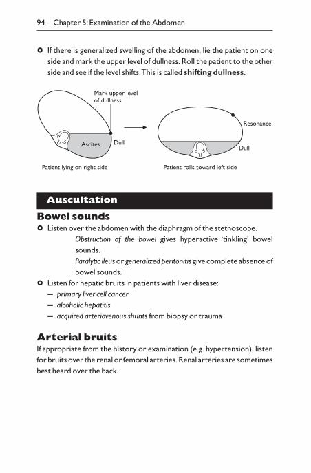
94 Chapter 5: Examination of the Abdomen
° If there is generalized swelling of the abdomen, lie the patient on oneside and mark the upper level of dullness. Roll the patient to the otherside and see if the level shifts.This is called shifting dullness.
Dull
Patient lying on right side Patient rolls toward left side
Mark upper levelof dullness
AscitesDull
Resonance
Auscultation
Bowel sounds
° Listen over the abdomen with the diaphragm of the stethoscope.Obstruction of the bowel gives hyperactive ‘tinkling’ bowelsounds.Paralytic ileus or generalized peritonitis give complete absence ofbowel sounds.
° Listen for hepatic bruits in patients with liver disease:– primary liver cell cancer– alcoholic hepatitis– acquired arteriovenous shunts from biopsy or trauma
Arterial bruitsIf appropriate from the history or examination (e.g. hypertension), listenfor bruits over the renal or femoral arteries. Renal arteries are sometimesbest heard over the back.
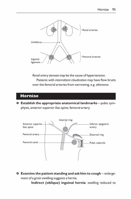
Herniae 95
Renal artery stenosis may be the cause of hypertension.Patients with intermittent claudication may have flow bruits
over the femoral arteries from narrowing, e.g. atheroma.
Herniae
° Establish the appropriate anatomical landmarks —pubic sym-physis, anterior superior iliac spine, femoral artery.
Renal arteries
Femoral arteries
Umbilicus
Inguinalligament
Anterior superioriliac spine
Femoral artery
Femoral canal
Inferior epigastricartery
Internal ring
External ring
Pubic tubercle
° Examine the patient standing and ask him to cough —enlarge-ment of a groin swelling suggests a hernia.
Indirect (oblique) inguinal hernia: swelling reduced to
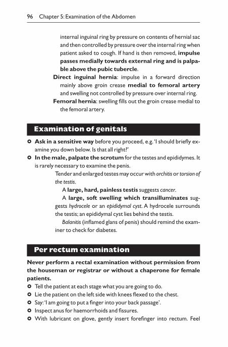
96 Chapter 5: Examination of the Abdomen
internal inguinal ring by pressure on contents of hernial sacand then controlled by pressure over the internal ring whenpatient asked to cough. If hand is then removed, impulsepasses medially towards external ring and is palpa-ble above the pubic tubercle.
Direct inguinal hernia: impulse in a forward direction mainly above groin crease medial to femoral arteryand swelling not controlled by pressure over internal ring.
Femoral hernia: swelling fills out the groin crease medial tothe femoral artery.
Examination of genitals
° Ask in a sensitive way before you proceed, e.g. ‘I should briefly ex-amine you down below. Is that all right?’
° In the male, palpate the scrotum for the testes and epididymes. Itis rarely necessary to examine the penis.
Tender and enlarged testes may occur with orchitis or torsion ofthe testis.
A large, hard, painless testis suggests cancer.A large, soft swelling which transilluminates sug-
gests hydrocele or an epididymal cyst. A hydrocele surroundsthe testis; an epididymal cyst lies behind the testis.
Balanitis (inflamed glans of penis) should remind the exam-iner to check for diabetes.
Per rectum examination
Never perform a rectal examination without permission fromthe houseman or registrar or without a chaperone for femalepatients.
° Tell the patient at each stage what you are going to do.
° Lie the patient on the left side with knees flexed to the chest.
° Say: ‘I am going to put a finger into your back passage’.
° Inspect anus for haemorrhoids and fissures.
° With lubricant on glove, gently insert forefinger into rectum. Feel
Summary of Common Illnesses 97
the tone of the sphincter, size and character of the prostate and any lateral masses. If appropriate, proceed to proctoscopy.
° Test stool on your glove for occult blood.
Per vaginam examination
Never perform a vaginal examination without a chaperone,female if possible, and only on the direction of a qualified instructor.
° Tell the patient at each stage what you are going to do.
° Lie the patient on her left side as for per rectum examination (althoughsome physicians prefer patient lying on her back with hips flexed andabducted).
° Inspect the external genitalia.
° With lubricant on glove insert one finger into vagina and then a secondfinger if there is room.
° Palpate the cervix.
° Examine for position and enlargement of uterus, tenderness of appendages and masses.
° Check for discharge by observing glove.
Summary of common illnesses
Cirrhosis– white nails– clubbing– liver palms– spider naevi– jaundice– firm liver
Portal hypertension– splenomegaly– ascites– caput medusa
98 Chapter 5: Examination of the Abdomen
Hepatic encephalopathy– liver flap– drowsy– constructional apraxia (cannot draw five-pointed star)– musty foetor
‘Dehydration’ (water and salt loss)– dry skin– veins collapsed– diminished skin turgor —pinched fold of skin remains raised– tongue dry– eyes sunken– blood pressure low with postural drop
Intestinal obstruction– patient ‘dehydrated’ if he has been vomiting– abdomen centrally swelling– visible peristalsis– not tender (unless inflammation, or some other pathology)– resonant to percussion– loud ‘tinkling’ bowel sounds
Pyloric stenosis– upper abdomen swelling– may have ‘succussion splash’ on shaking abdomen– otherwise like intestinal obstruction
Appendicitis– slight fever– deep tenderness right iliac fossa or per rectum– otherwise little to find unless has spread to peritonitis
Peritonitis– lies still– abdomen
– does not move on respiration
– rigid on palpation (guarding)– tender, particularly on removing fingers rapidly (rebound
tenderness)– absent bowel sounds
Cholecystitis– tender right hypochondrium, particularly on breathing in (Murphy’s
sign —tender gallbladder descends on inspiration to touch yourpalpating hand)
Jaundice and palpable gallbladder– obstruction is not due to gallstones, but from another obstruc-
tion such as a neoplasm of the pancreas (Courvoisier’s law).Gallstones have usually caused a fibrosed gallbladder which cannot dilate from back-pressure from gallstones in common bileduct
Enlarged spleen– infective, e.g. septicaemia or subacute bacterial endocarditis– portal hypertension, e.g. cirrhosis– lymphoma, leukaemia and other haematological diseases– autoimmune, e.g. systemic lupus, Felty’s syndrome
System-oriented examination
‘Examine the abdomen’
° hands: clubbing, liver flap, Dupuytren’s contracture
° eyes: jaundice, anaemia
° tongue: foetor, smooth
° neck:Virchow’s lymph node
° chest: spider naevi, gynaecomastia
° palpate inguinal lymph nodes briefly
° inspect abdomen asymmetry, movement, pulsation, swelling
° enquire whether pain or tenderness
° palpate four quadrants for masses: note abdominal tenderness, guard-ing, rigidity
System-oriented Examination 99
100 Chapter 5: Examination of the Abdomen
° palpate liver, kidneys, spleen, aortic aneurysm
° ascites: test for shifting dullness
° auscultate bowel sounds, arterial or liver bruits
° examine for hernia: ask patient to cough. Stand patient up if a hernia isa possibility
° enquire whether appropriate:– to examine vulva/testes– to do rectal examination
CHAPTER 6
Examination of the Mental State
Introduction
Examination of the mental state is necessary in all patients, not just thoseseen in psychiatric settings.The main headings are:
– appearance and behaviour– mood– speech —rate, form, content– thinking —form, content– abnormal beliefs —odd ideas and delusions– abnormal perceptions —hallucinations and illusions– cognitive function —concentration, orientation, memory,
reasoning– understanding of condition
The distinction between history and examination becomes blurredwhen examining disordered mental states.
Much of the examination is done by careful observation whilst takingthe history, and then supplemented with additional questions afterwards(see pp. 16–17 for mental state history, pp. 112–117 for examination).
If there is difficulty obtaining a clear history or if the patientappears distressed, it is particularly important to examine themental state.
General rules
° Be non-judgemental.
° Be alert to phenomenon that are observed.
° Do not jump to conclusions about what the patient is saying.
° Clarify with gentle enquiry:– ‘Can you tell me more about that?’
101
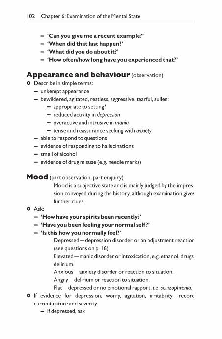
102 Chapter 6: Examination of the Mental State
– ‘Can you give me a recent example?’– ‘When did that last happen?’– ‘What did you do about it?’– ‘How often/how long have you experienced that?’
Appearance and behaviour (observation)
° Describe in simple terms:– unkempt appearance– bewildered, agitated, restless, aggressive, tearful, sullen:
– appropriate to setting?– reduced activity in depression– overactive and intrusive in mania– tense and reassurance seeking with anxiety
– able to respond to questions– evidence of responding to hallucinations– smell of alcohol– evidence of drug misuse (e.g. needle marks)
Mood (part observation, part enquiry)Mood is a subjective state and is mainly judged by the impres-sion conveyed during the history, although examination givesfurther clues.
° Ask:– ‘How have your spirits been recently?’– ‘Have you been feeling your normal self ?’– ‘Is this how you normally feel?’
Depressed —depression disorder or an adjustment reaction(see questions on p. 16)Elevated —manic disorder or intoxication, e.g. ethanol, drugs,delirium.Anxious —anxiety disorder or reaction to situation.Angry —delirium or reaction to situation.Flat —depressed or no emotional rapport, i.e. schizophrenia.
° If evidence for depression, worry, agitation, irritability —record current nature and severity.
– if depressed, ask
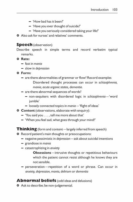
Introduction 103
– ‘How bad has it been?’– ‘Have you ever thought of suicide?’– ‘Have you seriously considered taking your life?’
° Also ask for nurses’ and relatives’ comments.
Speech (observation)Describe speech in simple terms and record verbatim typical
remarks.
° Rate:– fast in mania– slow in depression
° Form:– are there abnormalities of grammar or flow? Record examples
Disordered thought processes can occur in schizophrenia,mania, acute organic states, dementia.
– are there abnormal sequences of words?– non-sequiters with disordered logic in schizophrenia —‘word
jumble’– loosely connected topics in mania —‘flight of ideas’
° Content (observations, elaborate with enquiry):– ‘You said you . . . , tell me more about that’– ‘When you feel sad, what goes through your mind?’
Thinking (form and content —largely inferred from speech)
° Record patient’s main thoughts or preoccupations:– negative pessimistic in depression —ask about suicidal intentions– grandiose in mania– catastrophizing in anxiety
Obsessions —intrusive thoughts or repetitious behaviourswhich the patient cannot resist although he knows they arenot sensible.
– perseveration —repetition of a word or phrase. Can occur in anxiety, depression, mania, delirium or dementia
Abnormal beliefs (odd ideas and delusions)
° Ask to describe; be non-judgemental.
104 Chapter 6: Examination of the Mental State
° Ask why he thinks that —may reveal psychotic thoughts or hallucinations.
Delusions are fixed, false beliefs without reasonable evidence, e.g. I’ve got AIDS/cancer.
– ‘Did it ever seem to you that people were talking about you?’– ‘Have you ever received special messages from the television, radio
or newspaper?’– ‘Do people seem to be going out of their way to get at you?’– ‘Have you ever felt that you were especially important in some way
or that you had special powers?’– ‘Do you ever feel you have committed a crime or done something
terrible for which you should be punished?’
Abnormal perceptions (hallucinations and illusions —usually apparent from history)
° Ask —‘Have you had any unusual experiences recently?’– ‘Do they seem as if they are in the real world or as if they are ‘inside’
your head?’Hallucinations are false perceptions without a stimulus (e.g.pink elephants —experienced as real).
– They can occur in any sensory modality.– Visual hallucinations are suggestive of an organic
state.– Third person (‘he’ or ‘she’) auditory hallucinations are
suggestive of schizophrenia.– ‘Do you ever hear things that other people can’t
hear such as the voices of people talking?’– ‘Do you ever have visions or see things that other
people can’t see?’– ‘Do you ever have strange sensations in your body
or skin?’Illusions are misinterpreted perceptions (e.g. he thinks youare a policeman). They are common in acute organic states(psychosis).
General History and Examination 105
Cognitive functioning (observations supplemented byspecific enquiry)
° Impairment of concentration can occur in:– depression– anxiety states– dementia– confusional state
° Orientation, thought processes, memory and logic. These aspects must be tested as part of examination of the mental state (see Examination of the nervous system, p. 112).
Understanding of condition
° ‘What do you think is wrong with you?’– ‘Is there any illness that you are particularly worried
about?’– ‘What treatment do you feel is appropriate?’– ‘Are there any treatments you are frightened of?’
It is important to ask all patients these questions. If the patientlacks insight into abnormal beliefs or behaviour, this suggestsa psychotic illness.
General history and examination
Mental illness can be the presentation of a physical illness and a full historyand examination should be done in all patients.
Physical illnesses that masquerade as mental illnesses include:– hypothyroid, hyperthyroid– hypercalcaemia, hypokalaemia, hypomagnesaemia or
hyponatraemia– cerebral tumour– other causes of increased intracranial pressure– chronic, occult infection– drugs– porphyria
It is arguable that all mental illness arises from a physical imbalance oftransmitters/receptor function in the brain, and the division of illness intophysical and mental is spurious. In any case, all patients, whatever the
106 Chapter 6: Examination of the Mental State
nature of their illness, should be treated non-judgementally and with respect.
Problem patients
Angry patients– Inordinate anger is often symptomatic of another problem.– Assess whether the grievance is justified and whether it can be
resolved.– ‘Is there anything else that is upsetting you?’– If the antagonism is directed against you, enquire whether the
patient would prefer to see somebody else.
Aggressive patients– Do not take risks (have help nearby).– Ensure patient does not have a weapon.– Determine orientation and whether intoxicated or deluded.– Fear often underlines aggression —what is the fear?
Tearful patients– If a patient starts to weep, be calm and gently sympathetic.– When less tearful, enquire why they are upset. If you can find out
reason:– it may help rapport– it may allow resolution of the problem
Suicidal patients– Assess intent of recent attempt (if any):
– planning and likelihood of discovery– perceived dangerousness of method– intention at time
– Assess current intent:– how likely to attempt suicide?– what does he want to happen?– what would increase/decrease risk?
Summary of Common Illnesses 107
Embarrassed patients– Patients may not wish to talk about a distressing situation.– It may help to reassure about confidentiality.– If you think you know the problem, you can bring it up:
– ‘Is there a problem with money/sex/children, etc?’– If the patient really does not want to talk about a problem, it is
better to leave it and broach it later, possibly at another interview.
Talkative patientsSome patients go into irrelevant detail, digress and repeat themselves.
– Remind patient of time left and need to cover main points. Checkwhat they are and proceed.
– Politely say ‘Thank you. Please help me with some specific ques-tions . . . ’ and proceed to specific questions that require answers.
Nonsense history– Occasionally you get nowhere —contradictory remarks, descrip-
tion of improbable events, perseveration or just silence and monosyllables.
– Change to other aspects of history, e.g. personal or family history,social circumstances.
– If these also do not help, proceed if feasible to examination.You mayidentify dementia, evidence of drug abuse, hysteria or some other illness which explains the problem.
– Essential to interview other informants (e.g. telephone a relative).
Summary of common illnesses
Depression– low mood, tearfulness (not always present)– lack of interest and self-care– poor concentration– negative thought content– low self-esteem– wakes up early– depressed facies
108 Chapter 6: Examination of the Mental State
– slow movements and speech– weight loss– negative speech content
Anxiety– generally worried– thought focuses on catastrophies– cannot get to sleep– tense lined face, furrowed brow– sweaty palms– shaky– hyperventilation– tachycardia
Anorexia nervosa– thin, little body fat– increased, fine body hair– sees self as fat even if thin– thoughts dominated by food
Bulimia nervosa– often normal weight– binges followed by self-induced vomiting– thoughts dominated by food– erosion of teeth from vomiting
Acute psychosis (schizophrenia, mania or depressive psychosis)– alert and oriented– normal activities disrupted– unpredictable behaviour– reports or behaves as if responding to hallucinations– reports delusional beliefs
Schizophrenic psychosis– illogical thought, even a disjointed ‘word salad’– auditory hallucinations (third person)
Summary of Common Illnesses 109
– delusions (especially concerning thoughts, e.g. broadcast)– activity may be responding to hallucinations and delusions
Manic psychosis– rapid speech with ‘flight of ideas’– overactive, cannot keep still– normal activities disrupted– overly cheerful or irritable– stands close and is argumentative
Depressive psychosis– depressed affect– slow movements and speech– negative thoughts and delusions, e.g. brain is rotting– suicidal thoughts
Chronic schizophrenia– alert and oriented– unkempt– odd rambling speech– elaborate delusions– mannerisms and odd gestures– look for tardive dyskinesia (parkinsonian features from long-term
neuroleptic treatment)
Delirium– fluctuating level of concentration and orientation —worse at night– fleeting delusions, often persecutory in nature– evidence of toxicity (fever, etc.)
Intoxication (a type of delirium)– smells of alcohol or glue– needle marks– impaired alertness and drowsy– visual hallucinations
110 Chapter 6: Examination of the Mental State
Dementia– alert (unless also delirious)– may be unkempt– poor orientation in time and space– cognitive function subnormal
– poor short-term memory– cannot remember recent events– cannot remember and repeat series of numbers or an address– cannot explain proverbs
– paucity of thought and speech
Bereavement– low mood and tearfulness when thinking of lost person– may have somatic symptoms– assess suicide risk (to join lost person)– if excessively severe or prolonged (more than 6 months), may be
considered ‘pathological’
Somatization/hypochondriasis– somatic symptoms (often pain, fatigue) with no organic disease– assess for sign of depression– determine patient’s illness, fears or beliefs– symptoms are main concern in somatization, fear of illness in
hypochondriasis
CHAPTER 7
Examination of theNervous System
Introduction
The goal of the examination is to answer three questions: (1) Does the patient have a neurological illness? (2) Where in the nervous system is the pathology located? (3) What is the pathology?
As always, the history is critical.The following features in the historycan be informative:
– onset– abrupt —vascular, mechanical– seconds — seizure– minutes —migraine– hours — infective, inflammatory– days/weeks —neoplasm or degenerative disorder
– duration– brief episodes with recovery, e.g. TIA, epilepsy, migraine,syncope– longer episodes with recovery —mechanical, obstruction orpressure– demyelination, e.g. multiple scelorsis
– frequency– witness description —particularly if the patient has episodic loss
of consciousness or is confused.The examination of the nervous system can be elaborated almost indefinitely. Of far greater importance is to acquire the ability to conduct a thorough but comparatively rapid examination withconfidence in the findings. It is best to develop your own basic systemfor doing the examination and to perform it consistently. This will avoidomissions.
111
° Adapt your examination to the situation.The routine examina-tion must be mastered but can be altered to fit the situation.The examination of the nervous system is approached under the fol-
lowing headings:
° Higher cerebral function.
° Cranial nerves.
° Motor.
° Coordination.
° Reflexes.
° Sensation.
° Gait and station.The nervous system cannot be examined in isolation.
Other points of relevance may include:– configuration of the skull and spine– neck stiffness– ear drums for otitis media– blood pressure– heart, e.g. arrhythmia, mitral stenosis– carotid arteries —palpation and bruit– neoplasms —breast, lung, abdominal– jaundice
Higher cerebral functions
Higher mental functions can only be properly assessed in an alert, awakeand cooperative patient. From the history it is usually obvious whether it is necessary to examine the higher cerebral functions in detail. The ability to give a coherent history suggests normal higher mental function.A patient with sciatica would rightly be dismayed by an examination be-ginning with asking the patient to name the parts of a watch. However, ifthe patient is unable to give a coherent history, then cognitive testing isnecessary.
General observation
° Appearance, e.g. unkempt.
° Behaviour, e.g. bewildered, restless, agitated.
112 Chapter 7: Examination of the Nervous System
° Emotional state, e.g. depressed, euphoric, hostile.Observe, and ask for nurses’ and relatives’ comments.
Conscious levelIf the patient is not fully conscious shake him gently or speak to him loudly.
The Glasgow Coma ScaleThe GCS provides a rapid, widely used assessment of level of conscious-ness. Monitor responses to verbal command or, if no response to painfulstimulus, e.g. supraorbital pressure (with thumb nail in supraorbitalgroove), sternal rub (with knuckles over ribs), nailbed pressure (withthumb nail), twist fold of skin (but do not leave a bruise). It is helpful torecord a description of the patient as well as the GCS score.
Add up the total from A, B, C below.
ScoreA Eye opening 1 Eyes remain closed
2 Eyes open to pain3 Eyes open to command4 Spontaneous with normal blinking
B Verbal response 1 No response2 Incomprehensible, moaning sounds only3 Inappropriate —words spoken but no
conversation4 Confused speech5 Normal speech
C Motor response 1 No response2 Extensor reflex response to pain —adduction
and internal rotation at shoulder, extension at elbows, pronation of forearms
3 Flexor reflex response to pain4 Withdrawal of limb5 Localizing: attempts to protect site of pain6 Voluntary: responds normally to commands
Higher Cerebral Functions 113
ConfusionIf a patient appears confused, move on to assess cognitive state, includingdisorientation (p. 115). Make sure the patient does not have a receptiveaphasia first (see below).
Language/speechAssess from conversation:
° Is there difficulty in articulation?If necessary, ask patient to say ‘British Constitution’, ‘West RegisterStreet’.– Dysarthria
– cerebellar —scanning or staccato– lower motor neuron– palatal palsy —nasal– upper motor neuron —slow, ‘spastic’, seen in pseudobulbar
palsy– acute alcohol poisoning
° Is there altered voice tone?– extrapyramidal (monotonous and slow)
– Dysphonia– cord lesion —hoarse– hysterical
° Is there difficulty in finding the right word?– Dysphasia or aphasia —disorder of use of words as symbols in
speech, writing and understanding. Nearly always due to lefthemiphere lesion.N.B. Right- or left-handed? May be right hemisphere lesionif left-handed.
– Expressive dysphasia —difficulty finding words; speech slow andhesitant, may use circumlocutions; due to a lesion in Broca’s area.Test for by asking patient to name objects you point to, e.g. wrist-watch, pen, tie. Comprehension should be intact.
– Receptive dysphasia —speech fluent, but comprehension poor;patient may seem ‘confused’. Test for by asking patient to followcommands —a three-step command is a good screening test (e.g.‘please pick up the glass, but first point to the curtain and then thedoor’). Due to a lesion in Wernicke’s area.
114 Chapter 7: Examination of the Nervous System
– Mixed dysphasia —most common; spontaneous speech scanty,small vocabulary, often with wrong words used, comprehension impaired.There are also other dysphasias produced by interruptionof the connecting pathways between the speech centres.
– Mutism —no speech at all. This may be due to aphasia, anarthria,psychiatric disease or occasionally diffuse cerebral pathology.
Other defects occurring in absence ofmotor or sensory dysfunction
– Dyslexia —inappropriate difficulty with reading. Read few linesfrom newspaper (having established that comprehension and ex-pressive speech are intact).
– Dysgraphia —loss of ability to write.– Acalculia —loss of ability to do mental and written sums.– Apraxia —inability to perform a purposeful task when no motor
or sensory loss, e.g. opening matchbox, waving goodbye. Apraxiafor dressing is common in diffuse brain disease. Inability to draw five-pointed star occurs in hepatic pre-coma.
– Agnosia —inability to recognize objects (e.g. a key or coin whenplaced in hand.Tactile agnosia = astereognosis).
– Parietal lobe lesions —especially right, cause spatial difficulties;getting lost in familiar places, inability to lay table or draw or makepatterns with matches, neglect of left side of space, or of half ofbody.
Cognitive functionTake account of any evidence you have about the patient’s intelligence,education and interests.
‘Cognitive’ is a term that covers orientation, thought processesand logic. Regular use of a simple mental state test issuggested —examples are shown in Appendix 3.The section belowdescribes some aspects of cognitive testing in more detail.
Orientation
° Check awareness of:– time: ‘What day is it?’ (time, month, year)– place: ‘Where are you?’
Higher Cerebral Functions 115
– person: ‘What is your name?’Disorientation suggests acute organic state or dementia. De-pressed patients may be unwilling to reply although they knowthe answers.
Attention and calculation
° Tests of concentration include asking the patient to take away 7 from100, 7 from 93, etc.; 20 minus 3 in another simpler version of this test;or by asking for the months of the year backwards, or by spelling theword ‘world’ backwards.
Concentration may be impaired with many cerebral abnor-malities especially delirium, depression and anxiety.
MemoryImmediate recall —digit span
° Repeat digits spoken slowly. Start with easy short sequence and thenincrease the numbers. Most people manage seven digits forwards, fivebackwards.
Short-term memory
° Ask patient to tell you:– what he had for breakfast– what he did the night before– what he has read in today’s paper– recent topical news items. This should be geared to the patient’s
interests, e.g. football results for an avid football fanDemented patients will be unable to do this. They may con-fabulate (make up impressive stories) to cover their igno-rance (particularly likely in alcohol-related dementia).
New memory
° Give a name and address, make sure the patient has learnt it, and thentest recall at 5 minutes.
Longer-term memory
° Ask patient:
116 Chapter 7: Examination of the Nervous System
– for events before illness, e.g. last year, or during last week– ‘What is your address?’
General knowledge
° Assess in relation to anticipated performance from history.– What is the name of the Queen/President/Prime Minister?– Name six capital cities.– What were the dates of the last war?
In acute organic states and dementia, new learning, recentmemory and reasoning appear to be more impaired than re-mote memory.Vocabulary is usually well-preserved in demen-tia. In depression, patients may be unwilling to reply, and appeardemented.
– A history from a relative or employer is very important in early dementia, particularly for ability to function. Demented patientstend not to be able to work appropriately or drive safely; anxiousand depressed patients usually can.
Reasoning (abstract thought)
° What would you do if you found a stamped addressed envelope on theground?
Skull and spine
° Inspect and palpate skull if there is any possibility of a head injury.
° Check neck stiffness —meningeal irritation (see p. 147)
° Inspect spine —usually when examining back of chest.
° If there is any possibility of pathology, stand patient and check all move-ments of spine.
Cranial nerves
Examine cranial nerves and upper limbs with patient sitting up,preferably on side of bed or on a chair.
Cranial Nerves 117
I OlfactoryNot normally tested. Occasionally can be useful if there are other neurological defects, including papilloedema, undiagnosed headache orhead injury.
Oil of cloves, peppermint, coffee, etc. —each nostril in turn. Itis normal not to be able to name smells, but one smell shouldbe distinguished from another.
Pungent or noxious smells such as ammonia should not beused.
Abnormal:– rhinitis– head injury– olfactory groove meningioma– smoking
II Optic
Visual acuity
° Test each eye separately.
° Ask patient to read small newspaper print with each eye sepa-rately, with reading glasses if used.
° If sight poor, test formally:– near vision —newsprint or Jaeger type (each eye in turn) (see
Appendix 1).– distant vision —Snellen type (more precise method) (see
Appendix 2).Stand patient at 6m from Snellen’s card (each eye in turn).Results expressed as a ratio:
– 6 —distance of person from card– x —distance at which patient should be able to read type
i.e. 6/6 is good vision, 6/60 means the smallest type the patientcan read is large enough to be normally read at 60m.
If the patient cannot read 6/6, try after correctionwith glasses or pinhole. Looking through a pinhole in a cardobviates refractive errors, analogous to a pinhole camera. If
118 Chapter 7: Examination of the Nervous System
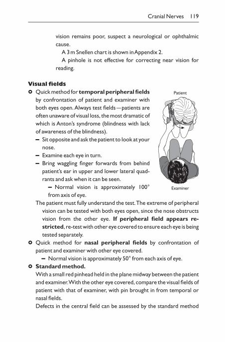
vision remains poor, suspect a neurological or ophthalmiccause.
A 3m Snellen chart is shown in Appendix 2.A pinhole is not effective for correcting near vision for
reading.
Visual fields
° Quick method for temporal peripheral fieldsby confrontation of patient and examiner withboth eyes open. Always test fields —patients areoften unaware of visual loss, the most dramatic ofwhich is Anton’s syndrome (blindness with lack of awareness of the blindness).– Sit opposite and ask the patient to look at your
nose.– Examine each eye in turn.– Bring waggling finger forwards from behind
patient’s ear in upper and lower lateral quad-rants and ask when it can be seen.
– Normal vision is approximately 100°from axis of eye.
The patient must fully understand the test.The extreme of peripheralvision can be tested with both eyes open, since the nose obstructsvision from the other eye. If peripheral field appears re-stricted, re-test with other eye covered to ensure each eye is beingtested separately.
° Quick method for nasal peripheral fields by confrontation of patient and examiner with other eye covered.
– Normal vision is approximately 50° from each axis of eye.
° Standard method.With a small red pinhead held in the plane midway between the patientand examiner.With the other eye covered, compare the visual fields ofpatient with that of examiner, with pin brought in from temporal ornasal fields.Defects in the central field can be assessed by the standard method
Cranial Nerves 119
Patient
Examiner
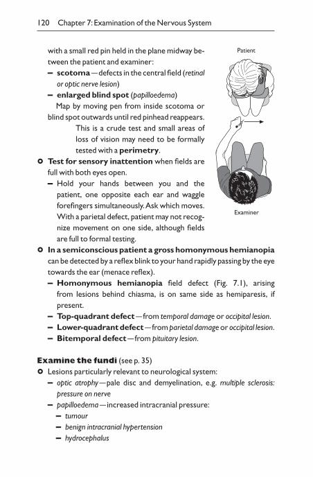
with a small red pin held in the plane midway be-tween the patient and examiner:– scotoma —defects in the central field (retinal
or optic nerve lesion)– enlarged blind spot (papilloedema)
Map by moving pen from inside scotoma orblind spot outwards until red pinhead reappears.
This is a crude test and small areas ofloss of vision may need to be formallytested with a perimetry.
° Test for sensory inattention when fields arefull with both eyes open.– Hold your hands between you and the
patient, one opposite each ear and waggleforefingers simultaneously.Ask which moves.With a parietal defect, patient may not recog-nize movement on one side, although fieldsare full to formal testing.
° In a semiconscious patient a gross homonymous hemianopiacan be detected by a reflex blink to your hand rapidly passing by the eyetowards the ear (menace reflex).– Homonymous hemianopia field defect (Fig. 7.1), arising
from lesions behind chiasma, is on same side as hemiparesis, if present.
– Top-quadrant defect —from temporal damage or occipital lesion.– Lower-quadrant defect —from parietal damage or occipital lesion.– Bitemporal defect —from pituitary lesion.
Examine the fundi (see p. 35)
° Lesions particularly relevant to neurological system:– optic atrophy —pale disc and demyelination, e.g. multiple sclerosis:
pressure on nerve– papilloedema —increased intracranial pressure:
– tumour– benign intracranial hypertension– hydrocephalus
120 Chapter 7: Examination of the Nervous System
Patient
Examiner
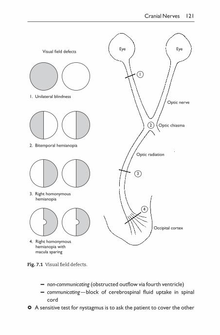
Cranial Nerves 121
1. Unilateral blindness
2. Bitemporal hemianopia
3. Right homonymous hemianopia
4. Right homonymoushemianopia withmacula sparing
Eye Eye
1
2
4
3
Optic radiation
Optic chiasma
Optic nerve
Occipital cortex
Visual field defects
Fig. 7.1 Visual field defects.
– non-communicating (obstructed outflow via fourth ventricle)– communicating —block of cerebrospinal fluid uptake in spinal
cord
° A sensitive test for nystagmus is to ask the patient to cover the other
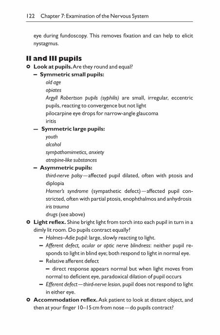
eye during fundoscopy. This removes fixation and can help to elicit nystagmus.
II and III pupils
° Look at pupils.Are they round and equal?– Symmetric small pupils:
old ageopiatesArgyll Robertson pupils (syphilis) are small, irregular, eccentricpupils, reacting to convergence but not lightpilocarpine eye drops for narrow-angle glaucomairitis
— Symmetric large pupils:youthalcoholsympathomimetics, anxietyatropine-like substances
– Asymmetric pupils:third-nerve palsy —affected pupil dilated, often with ptosis anddiplopiaHorner’s syndrome (sympathetic defect) —affected pupil con-stricted, often with partial ptosis, enophthalmos and anhydrosisiris traumadrugs (see above)
° Light reflex. Shine bright light from torch into each pupil in turn in adimly lit room. Do pupils contract equally?
– Holmes–Adie pupil: large, slowly reacting to light.– Afferent defect, ocular or optic nerve blindness: neither pupil re-
sponds to light in blind eye; both respond to light in normal eye.– Relative afferent defect
– direct response appears normal but when light moves fromnormal to deficient eye, paradoxical dilation of pupil occurs
– Efferent defect — third-nerve lesion, pupil does not respond to lightin either eye.
° Accommodation reflex.Ask patient to look at distant object, andthen at your finger 10–15cm from nose —do pupils contract?
122 Chapter 7: Examination of the Nervous System
– Response to accommodation but not light:Argyll RobertsonHolmes–Adieoccular blindnessmidbrain lesionsome recovering third-nerve lesions
III Oculomotor
IV Trochlear
VI Abducens
External ocular movements
° Test the eye movements in the four cardinal directions (left,right, up, down) and convergence using your finger at 1m distance.Look for abnormal eye movements.
° Ask:‘Tell me if you see double’.Upward gaze and convergence are often reduced in unco-operative patients.
° To detect minor lesions:– Find direction of gaze with maximum separation of
images.– Cover one eye and ask which image has gone.
Peripheral image is seen by the eye that is not moving fully.Peripheral image is displaced in direction of action ofweak muscle, e.g. maximum diplopia on gaze to left. Left eye
Cranial Nerves 123
Distance vision
Sudden close vision
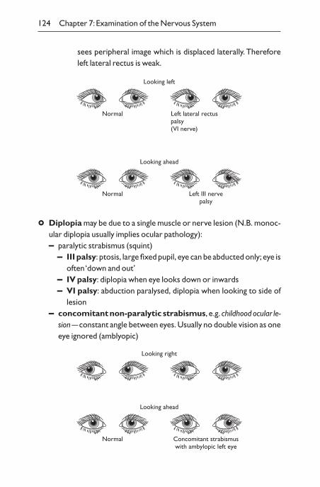
° Diplopia may be due to a single muscle or nerve lesion (N.B. monoc-ular diplopia usually implies ocular pathology):– paralytic strabismus (squint)
– III palsy: ptosis, large fixed pupil, eye can be abducted only; eye isoften ‘down and out’
– IV palsy: diplopia when eye looks down or inwards– VI palsy: abduction paralysed, diplopia when looking to side of
lesion– concomitant non-paralytic strabismus, e.g. childhood ocular le-
sion —constant angle between eyes. Usually no double vision as oneeye ignored (amblyopic)
124 Chapter 7: Examination of the Nervous System
sees peripheral image which is displaced laterally. Thereforeleft lateral rectus is weak.
Normal Left lateral rectuspalsy(VI nerve)
Looking left
Normal Left III nervepalsy
Looking ahead
Looking right
Normal Concomitant strabismuswith ambylopic left eye
Looking ahead
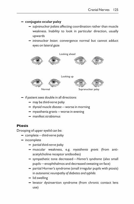
– conjugate ocular palsy– supranuclear palsies affecting coordination rather than muscle
weakness. Inability to look in particular direction, usually upwards
– intranuclear lesion: convergence normal but cannot adducteyes on lateral gaze
Cranial Nerves 125
Looking ahead
Normal
Looking up
Supranuclear palsy
– if patient sees double in all directions– may be third-nerve palsy– thyroid muscle disease —worse in morning– myasthenia gravis —worse in evening– manifest strabismus
PtosisDrooping of upper eyelid can be:
– complete — third-nerve palsy– incomplete
– partial third-nerve palsy– muscular weakness, e.g. myasthenia gravis (from anti-
acetylcholine receptor antibodies)– sympathetic tone decreased —Horner’s syndrome (also small
pupils —enophthalmos and decreased sweating on face)– partial Horner’s syndrome (small irregular pupils with ptosis)
in autonomic neuropathy of diabetes and syphilis– lid swelling– levator dysinsertion syndrome (from chronic contact lens
use)
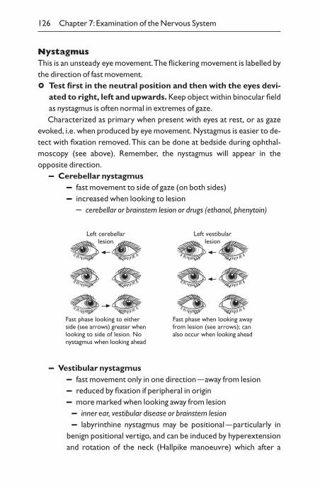
NystagmusThis is an unsteady eye movement.The flickering movement is labelled bythe direction of fast movement.
° Test first in the neutral position and then with the eyes devi-ated to right, left and upwards. Keep object within binocular fieldas nystagmus is often normal in extremes of gaze.Characterized as primary when present with eyes at rest, or as gaze
evoked, i.e. when produced by eye movement. Nystagmus is easier to de-tect with fixation removed.This can be done at bedside during ophthal-moscopy (see above). Remember, the nystagmus will appear in theopposite direction.
– Cerebellar nystagmus– fast movement to side of gaze (on both sides)– increased when looking to lesion
– cerebellar or brainstem lesion or drugs (ethanol, phenytoin)
126 Chapter 7: Examination of the Nervous System
Left cerebellarlesion
Fast phase looking to eitherside (see arrows) greater whenlooking to side of lesion. Nonystagmus when looking ahead
Left vestibularlesion
Fast phase when looking awayfrom lesion (see arrows); canalso occur when looking ahead
– Vestibular nystagmus– fast movement only in one direction —away from lesion– reduced by fixation if peripheral in origin– more marked when looking away from lesion– inner ear, vestibular disease or brainstem lesion– labyrinthine nystagmus may be positional —particularly in
benign positional vertigo, and can be induced by hyperextensionand rotation of the neck (Hallpike manoeuvre) which after a
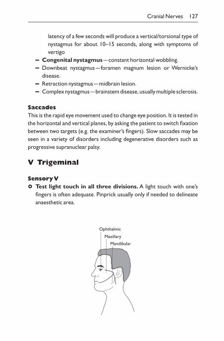
latency of a few seconds will produce a vertical/torsional type ofnystagmus for about 10–15 seconds, along with symptoms ofvertigo
– Congenital nystagmus —constant horizontal wobbling.– Downbeat nystagmus —foramen magnum lesion or Wernicke’s
disease.– Retraction nystagmus —midbrain lesion.– Complex nystagmus —brainstem disease, usually multiple sclerosis.
SaccadesThis is the rapid eye movement used to change eye position. It is tested inthe horizontal and vertical planes, by asking the patient to switch fixationbetween two targets (e.g. the examiner’s fingers). Slow saccades may beseen in a variety of disorders including degenerative disorders such asprogressive supranuclear palsy.
V Trigeminal
Sensory V
° Test light touch in all three divisions. A light touch with one’s fingers is often adequate. Pinprick usually only if needed to delineateanaesthetic area.
Cranial Nerves 127
Ophthalmic
Maxillary
Mandibular
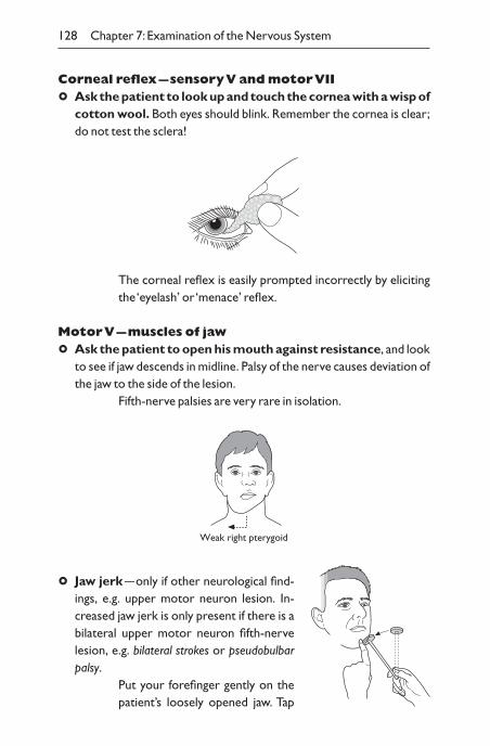
Corneal reflex —sensory V and motor VII
° Ask the patient to look up and touch the cornea with a wisp ofcotton wool. Both eyes should blink. Remember the cornea is clear;do not test the sclera!
128 Chapter 7: Examination of the Nervous System
The corneal reflex is easily prompted incorrectly by elicitingthe ‘eyelash’ or ‘menace’ reflex.
Motor V —muscles of jaw
° Ask the patient to open his mouth against resistance, and lookto see if jaw descends in midline. Palsy of the nerve causes deviation ofthe jaw to the side of the lesion.
Fifth-nerve palsies are very rare in isolation.
Weak right pterygoid
° Jaw jerk —only if other neurological find-ings, e.g. upper motor neuron lesion. In-creased jaw jerk is only present if there is abilateral upper motor neuron fifth-nervelesion, e.g. bilateral strokes or pseudobulbarpalsy.
Put your forefinger gently on thepatient’s loosely opened jaw. Tap
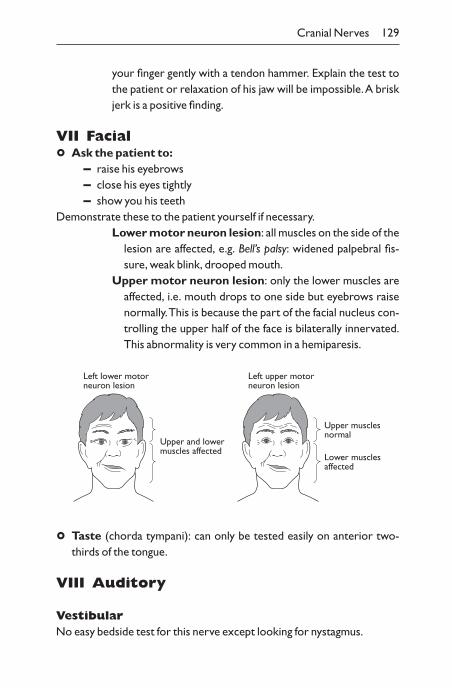
your finger gently with a tendon hammer. Explain the test tothe patient or relaxation of his jaw will be impossible.A briskjerk is a positive finding.
VII Facial
° Ask the patient to:– raise his eyebrows– close his eyes tightly– show you his teeth
Demonstrate these to the patient yourself if necessary.Lower motor neuron lesion: all muscles on the side of the
lesion are affected, e.g. Bell’s palsy: widened palpebral fis-sure, weak blink, drooped mouth.
Upper motor neuron lesion: only the lower muscles areaffected, i.e. mouth drops to one side but eyebrows raisenormally.This is because the part of the facial nucleus con-trolling the upper half of the face is bilaterally innervated.This abnormality is very common in a hemiparesis.
Cranial Nerves 129
Upper and lowermuscles affected
Left lower motorneuron lesion
Upper musclesnormal
Lower musclesaffected
Left upper motorneuron lesion
° Taste (chorda tympani): can only be tested easily on anterior two-thirds of the tongue.
VIII Auditory
VestibularNo easy bedside test for this nerve except looking for nystagmus.
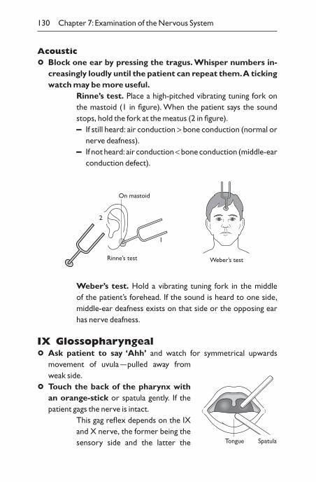
Acoustic
° Block one ear by pressing the tragus. Whisper numbers in-creasingly loudly until the patient can repeat them.A tickingwatch may be more useful.
Rinne’s test. Place a high-pitched vibrating tuning fork onthe mastoid (1 in figure). When the patient says the soundstops, hold the fork at the meatus (2 in figure).– If still heard: air conduction > bone conduction (normal or
nerve deafness).– If not heard: air conduction < bone conduction (middle-ear
conduction defect).
130 Chapter 7: Examination of the Nervous System
Weber’s test
On mastoid
2
1
Rinne’s test
Weber’s test. Hold a vibrating tuning fork in the middle of the patient’s forehead. If the sound is heard to one side,middle-ear deafness exists on that side or the opposing earhas nerve deafness.
IX Glossopharyngeal
° Ask patient to say ‘Ahh’ and watch for symmetrical upwards movement of uvula —pulled away fromweak side.
° Touch the back of the pharynx with an orange-stick or spatula gently. If thepatient gags the nerve is intact.
This gag reflex depends on the IXand X nerve, the former being thesensory side and the latter the Tongue Spatula
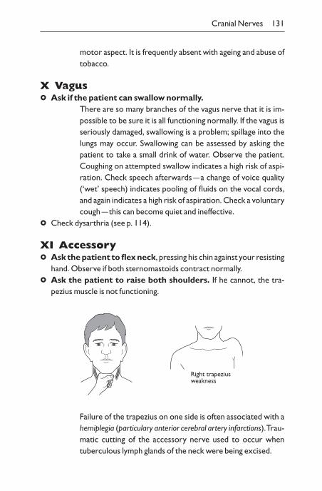
motor aspect. It is frequently absent with ageing and abuse oftobacco.
X Vagus
° Ask if the patient can swallow normally.There are so many branches of the vagus nerve that it is im-possible to be sure it is all functioning normally. If the vagus isseriously damaged, swallowing is a problem; spillage into thelungs may occur. Swallowing can be assessed by asking the patient to take a small drink of water. Observe the patient.Coughing on attempted swallow indicates a high risk of aspi-ration. Check speech afterwards —a change of voice quality(‘wet’ speech) indicates pooling of fluids on the vocal cords,and again indicates a high risk of aspiration. Check a voluntarycough —this can become quiet and ineffective.
° Check dysarthria (see p. 114).
XI Accessory
° Ask the patient to flex neck, pressing his chin against your resistinghand. Observe if both sternomastoids contract normally.
° Ask the patient to raise both shoulders. If he cannot, the tra-pezius muscle is not functioning.
Cranial Nerves 131
Right trapeziusweakness
Failure of the trapezius on one side is often associated with ahemiplegia (particulary anterior cerebral artery infarctions).Trau-matic cutting of the accessory nerve used to occur when tuberculous lymph glands of the neck were being excised.
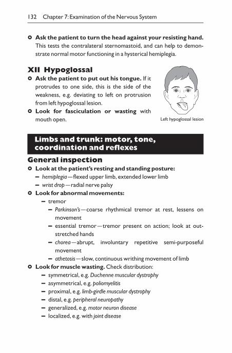
° Ask the patient to turn the head against your resisting hand.This tests the contralateral sternomastoid, and can help to demon-strate normal motor functioning in a hysterical hemiplegia.
XII Hypoglossal
° Ask the patient to put out his tongue. If itprotrudes to one side, this is the side of theweakness, e.g. deviating to left on protrusionfrom left hypoglossal lesion.
° Look for fasciculation or wasting withmouth open.
Limbs and trunk: motor, tone,coordination and reflexes
General inspection
° Look at the patient’s resting and standing posture:– hemiplegia —flexed upper limb, extended lower limb– wrist drop —radial nerve palsy
° Look for abnormal movements:– tremor
– Parkinson’s —coarse rhythmical tremor at rest, lessens on movement
– essential tremor —tremor present on action; look at out-stretched hands
– chorea —abrupt, involuntary repetitive semi-purposefulmovement
– athetosis —slow, continuous writhing movement of limb
° Look for muscle wasting. Check distribution:– symmetrical, e.g. Duchenne muscular dystrophy– asymmetrical, e.g. poliomyelitis– proximal, e.g. limb-girdle muscular dystrophy– distal, e.g. peripheral neuropathy– generalized, e.g. motor neuron disease– localized, e.g. with joint disease
132 Chapter 7: Examination of the Nervous System
Left hypoglossal lesion
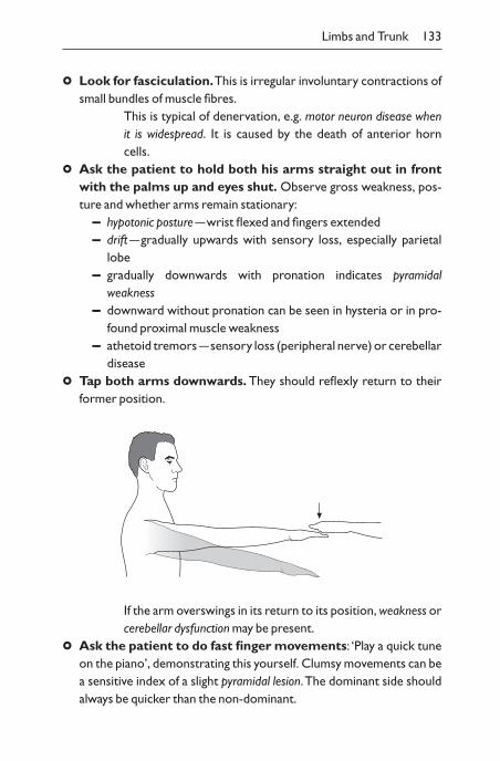
Limbs and Trunk 133
° Look for fasciculation.This is irregular involuntary contractions ofsmall bundles of muscle fibres.
This is typical of denervation, e.g. motor neuron disease when it is widespread. It is caused by the death of anterior horn cells.
° Ask the patient to hold both his arms straight out in frontwith the palms up and eyes shut. Observe gross weakness, pos-ture and whether arms remain stationary:
– hypotonic posture —wrist flexed and fingers extended– drift —gradually upwards with sensory loss, especially parietal
lobe– gradually downwards with pronation indicates pyramidal
weakness– downward without pronation can be seen in hysteria or in pro-
found proximal muscle weakness– athetoid tremors —sensory loss (peripheral nerve) or cerebellar
disease
° Tap both arms downwards. They should reflexly return to theirformer position.
If the arm overswings in its return to its position, weakness orcerebellar dysfunction may be present.
° Ask the patient to do fast finger movements: ‘Play a quick tuneon the piano’, demonstrating this yourself. Clumsy movements can bea sensitive index of a slight pyramidal lesion.The dominant side shouldalways be quicker than the non-dominant.
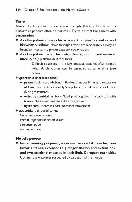
ToneAlways check tone before you assess strength. This is a difficult test to perform as patients often do not relax. Try to distract the patient withconversation.
° Ask the patient to relax his arm and then you flex and extendhis wrist or elbow. Move through a wide arc moderately slowly, at irregular intervals to prevent patient cooperation.
° Ask the patient to let the limb go loose, lift it up and move atknee joint (hip and ankle if required).
Difficult to assess in the legs because patients often cannotrelax. Ankle clonus can be assessed at same time (see below).
Hypertonia (increased tone):– pyramidal: more obvious in flexion of upper limbs and extension
of lower limbs. Occasionally ‘clasp knife’, i.e. diminution of tone during movement
– extrapyramidal: uniform ‘lead pipe’ rigidity. If associated withtremor the movement feels like a ‘cog wheel’
– hysterical: increases with increased movementHypotonia (decreased tone):
lower motor neuron lesionrecent upper motor neuron lesioncerebellar lesionunconsciousness
Muscle power
° For screening purposes, examine two distal muscles, oneflexor and one extensor (e.g. finger flexion and extension),and two proximal muscles in each limb. Compare each side.Confirm the weakness suspected by palpation of the muscle.
134 Chapter 7: Examination of the Nervous System
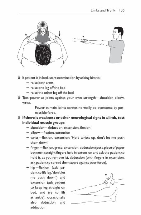
° If patient is in bed, start examination by asking him to:– raise both arms– raise one leg off the bed– raise the other leg off the bed
° Test power at joints against your own strength —shoulder, elbow,wrist.
Power at main joints cannot normally be overcome by per-missible force.
° If there is weakness or other neurological signs in a limb, testindividual muscle groups:
– shoulder —abduction, extension, flexion– elbow —flexion, extension– wrist —flexion, extension: ‘Hold wrists up, don’t let me push
them down’– finger —flexion, grasp, extension, adduction (put a piece of paper
between straight fingers held in extension and ask the patient tohold it, as you remove it), abduction (with fingers in extension,ask patient to spread them apart against your force).
– hip —flexion (ask pa-tient to lift leg, ‘don’t letme push down’) and extension (ask patientto keep leg straight onbed, and try to lift at ankle); occasionallyalso abduction and adduction
Limbs and Trunk 135
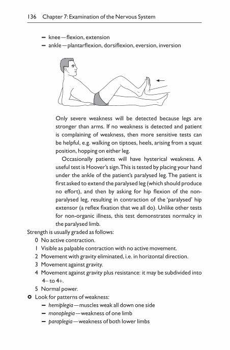
– knee —flexion, extension– ankle —plantarflexion, dorsiflexion, eversion, inversion
136 Chapter 7: Examination of the Nervous System
Only severe weakness will be detected because legs arestronger than arms. If no weakness is detected and patient is complaining of weakness, then more sensitive tests can be helpful, e.g. walking on tiptoes, heels, arising from a squatposition, hopping on either leg.
Occasionally patients will have hysterical weakness. A useful test is Hoover’s sign.This is tested by placing your handunder the ankle of the patient’s paralysed leg. The patient isfirst asked to extend the paralysed leg (which should produceno effort), and then by asking for hip flexion of the non-paralysed leg, resulting in contraction of the ‘paralysed’ hip extensor (a reflex fixation that we all do). Unlike other testsfor non-organic illness, this test demonstrates normalcy inthe paralysed limb.
Strength is usually graded as follows:0 No active contraction.1 Visible as palpable contraction with no active movement.2 Movement with gravity eliminated, i.e. in horizontal direction.3 Movement against gravity.4 Movement against gravity plus resistance: it may be subdivided into
4- to 4+.5 Normal power.
° Look for patterns of weakness:– hemiplegia —muscles weak all down one side– monoplegia —weakness of one limb– paraplegia —weakness of both lower limbs
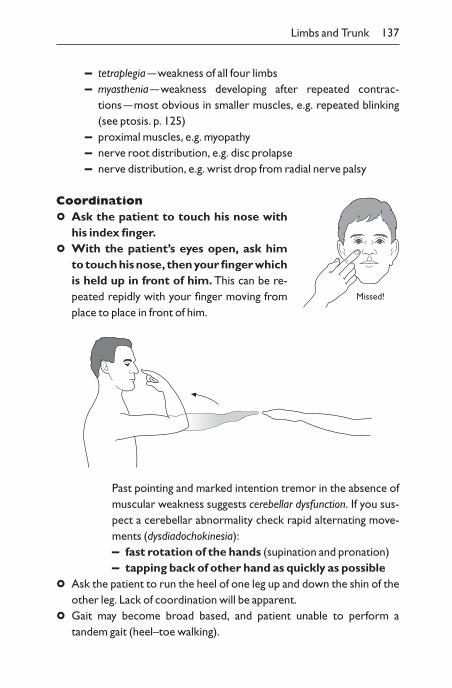
– tetraplegia —weakness of all four limbs– myasthenia —weakness developing after repeated contrac-
tions —most obvious in smaller muscles, e.g. repeated blinking(see ptosis. p. 125)
– proximal muscles, e.g. myopathy– nerve root distribution, e.g. disc prolapse– nerve distribution, e.g. wrist drop from radial nerve palsy
Coordination
° Ask the patient to touch his nose withhis index finger.
° With the patient’s eyes open, ask him to touch his nose, then your finger whichis held up in front of him. This can be re-peated repidly with your finger moving fromplace to place in front of him.
Limbs and Trunk 137
Missed!
Past pointing and marked intention tremor in the absence ofmuscular weakness suggests cerebellar dysfunction. If you sus-pect a cerebellar abnormality check rapid alternating move-ments (dysdiadochokinesia):– fast rotation of the hands (supination and pronation)– tapping back of other hand as quickly as possible
° Ask the patient to run the heel of one leg up and down the shin of theother leg. Lack of coordination will be apparent.
° Gait may become broad based, and patient unable to perform a tandem gait (heel–toe walking).

Tendon reflexes
° Place arms comfortably by side with elbows flexed and hand on upperabdomen.Tell the patient to relax.
° Supinator reflex: tap the distal end of the radius with a tendon hammer.
° Biceps reflex: tap your forefinger or thumb over biceps tendon.
° Triceps reflex: hold arm across chest to tap the triceps tendon.
° Knee reflexes: by passing left forearm behind both knees, supportingthem partly flexed.Ask the patient to let leg go loose and tap the ten-dons below patella.
138 Chapter 7: Examination of the Nervous System
Heel-on-shin test
Knee L3
Testing the knee reflexes
° Ankle reflex: by flexing the knee and abducting the leg. Apply gentle pressure to the ball of the foot, with it at a right angle and tap thetendon.
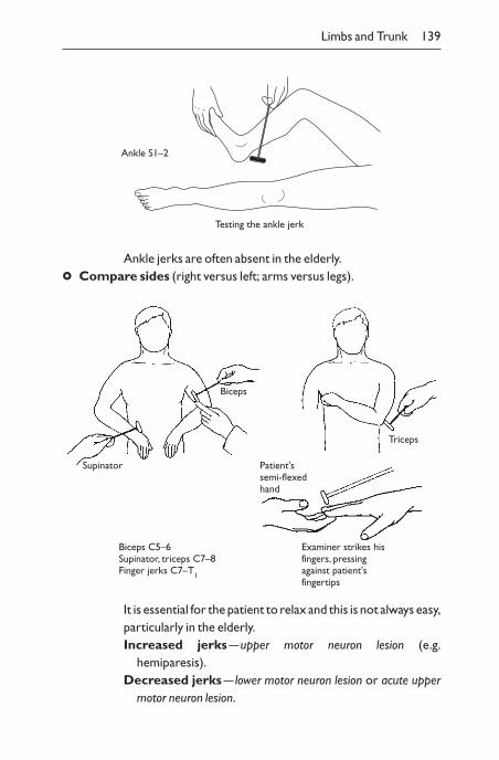
Ankle jerks are often absent in the elderly.
° Compare sides (right versus left; arms versus legs).
Limbs and Trunk 139
Testing the ankle jerk
Ankle S1–2
Biceps
Supinator
Triceps
Patient'ssemi-flexedhand
Examiner strikes hisfingers, pressingagainst patient'sfingertips
Biceps C5–6Supinator, triceps C7–8Finger jerks C7–T1
It is essential for the patient to relax and this is not always easy,particularly in the elderly.Increased jerks —upper motor neuron lesion (e.g.
hemiparesis).Decreased jerks — lower motor neuron lesion or acute upper
motor neuron lesion.
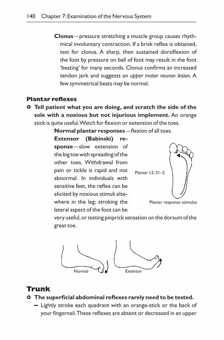
Clonus —pressure stretching a muscle group causes rhyth-mical involuntary contraction. If a brisk reflex is obtained,test for clonus. A sharp, then sustained dorsiflexion of the foot by pressure on ball of foot may result in the foot‘beating’ for many seconds. Clonus confirms an increasedtendon jerk and suggests an upper motor neuron lesion. A few symmetrical beats may be normal.
Plantar reflexes
° Tell patient what you are doing, and scratch the side of thesole with a noxious but not injurious implement. An orangestick is quite useful.Watch for flexion or extension of the toes.
Normal plantar responses —flexion of all toes.Extensor (Babinski) re-sponse —slow extension ofthe big toe with spreading of theother toes. Withdrawal frompain or tickle is rapid and notabnormal. In individuals withsensitive feet, the reflex can beelicited by noxious stimuli else-where in the leg; stroking thelateral aspect of the foot can bevery useful, or testing pinprick sensation on the dorsum of thegreat toe.
140 Chapter 7: Examination of the Nervous System
Plantar response stimulus
Plantar L5, S1–2
ExtensorNormal
Trunk
° The superficial abdominal reflexes rarely need to be tested.– Lightly stroke each quadrant with an orange-stick or the back of
your fingernail.These reflexes are absent or decreased in an upper
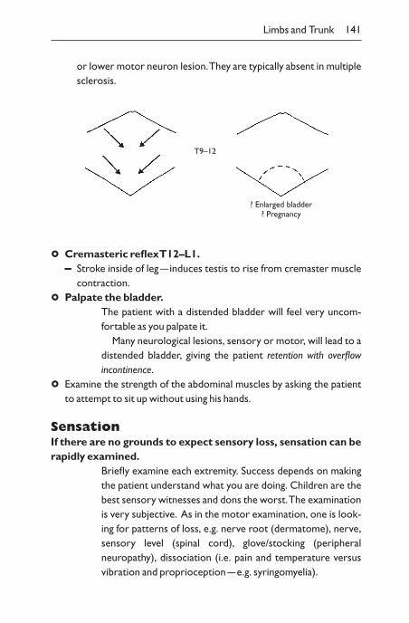
or lower motor neuron lesion.They are typically absent in multiplesclerosis.
Limbs and Trunk 141
T9–12
? Enlarged bladder? Pregnancy
° Cremasteric reflex T12–L1.– Stroke inside of leg —induces testis to rise from cremaster muscle
contraction.
° Palpate the bladder.The patient with a distended bladder will feel very uncom-fortable as you palpate it.
Many neurological lesions, sensory or motor, will lead to adistended bladder, giving the patient retention with overflow incontinence.
° Examine the strength of the abdominal muscles by asking the patient to attempt to sit up without using his hands.
SensationIf there are no grounds to expect sensory loss, sensation can berapidly examined.
Briefly examine each extremity. Success depends on makingthe patient understand what you are doing. Children are thebest sensory witnesses and dons the worst.The examinationis very subjective. As in the motor examination, one is look-ing for patterns of loss, e.g. nerve root (dermatome), nerve,sensory level (spinal cord), glove/stocking (peripheral neuropathy), dissociation (i.e. pain and temperature versus vibration and proprioception —e.g. syringomyelia).
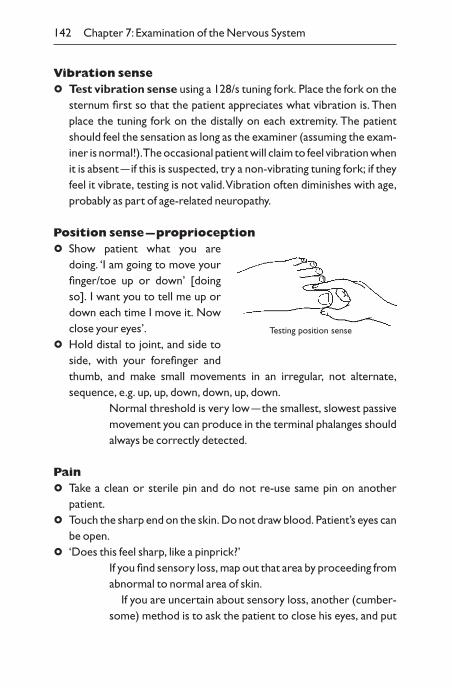
Vibration sense
° Test vibration sense using a 128/s tuning fork. Place the fork on thesternum first so that the patient appreciates what vibration is. Thenplace the tuning fork on the distally on each extremity. The patientshould feel the sensation as long as the examiner (assuming the exam-iner is normal!).The occasional patient will claim to feel vibration whenit is absent —if this is suspected, try a non-vibrating tuning fork; if theyfeel it vibrate, testing is not valid.Vibration often diminishes with age,probably as part of age-related neuropathy.
Position sense —proprioception
° Show patient what you aredoing. ‘I am going to move yourfinger/toe up or down’ [doingso]. I want you to tell me up ordown each time I move it. Nowclose your eyes’.
° Hold distal to joint, and side toside, with your forefinger andthumb, and make small movements in an irregular, not alternate,sequence, e.g. up, up, down, down, up, down.
Normal threshold is very low —the smallest, slowest passivemovement you can produce in the terminal phalanges shouldalways be correctly detected.
Pain
° Take a clean or sterile pin and do not re-use same pin on another patient.
° Touch the sharp end on the skin. Do not draw blood. Patient’s eyes canbe open.
° ‘Does this feel sharp, like a pinprick?’If you find sensory loss, map out that area by proceeding fromabnormal to normal area of skin.
If you are uncertain about sensory loss, another (cumber-some) method is to ask the patient to close his eyes, and put
142 Chapter 7: Examination of the Nervous System
Testing position sense
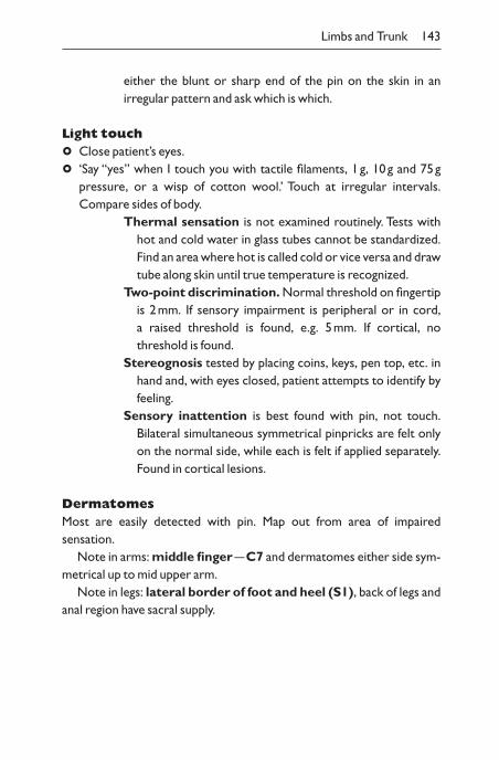
either the blunt or sharp end of the pin on the skin in an irregular pattern and ask which is which.
Light touch
° Close patient’s eyes.
° ‘Say “yes” when I touch you with tactile filaments, 1 g, 10g and 75gpressure, or a wisp of cotton wool.’ Touch at irregular intervals.Compare sides of body.
Thermal sensation is not examined routinely. Tests withhot and cold water in glass tubes cannot be standardized.Find an area where hot is called cold or vice versa and drawtube along skin until true temperature is recognized.
Two-point discrimination. Normal threshold on fingertipis 2mm. If sensory impairment is peripheral or in cord,a raised threshold is found, e.g. 5mm. If cortical, no threshold is found.
Stereognosis tested by placing coins, keys, pen top, etc. inhand and, with eyes closed, patient attempts to identify byfeeling.
Sensory inattention is best found with pin, not touch.Bilateral simultaneous symmetrical pinpricks are felt onlyon the normal side, while each is felt if applied separately.Found in cortical lesions.
DermatomesMost are easily detected with pin. Map out from area of impaired sensation.
Note in arms: middle finger —C7 and dermatomes either side sym-metrical up to mid upper arm.
Note in legs: lateral border of foot and heel (S1), back of legs andanal region have sacral supply.
Limbs and Trunk 143
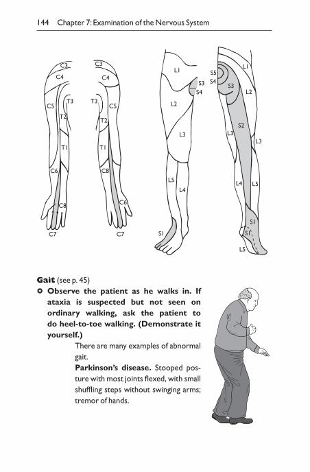
Gait (see p. 45)
° Observe the patient as he walks in. If ataxia is suspected but not seen on ordinary walking, ask the patient to do heel-to-toe walking. (Demonstrate ityourself.)
There are many examples of abnormalgait.Parkinson’s disease. Stooped pos-ture with most joints flexed, with smallshuffling steps without swinging arms;tremor of hands.
144 Chapter 7: Examination of the Nervous System
C3C3
C4C4
C5 C5T3T3
T2T2
T1T1
C8C6
C6C8
C7 C7
L1
L2
L3
L4
L5
S1
S3S4
S5S4
S3
S2
S1
S1
L4 L5
L3L3
L2
L1
L5
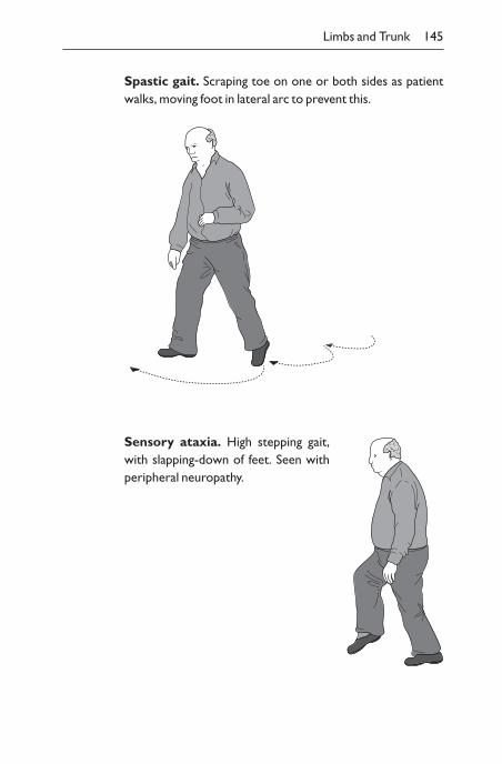
Spastic gait. Scraping toe on one or both sides as patientwalks, moving foot in lateral arc to prevent this.
Limbs and Trunk 145
Sensory ataxia. High stepping gait,with slapping-down of feet. Seen withperipheral neuropathy.
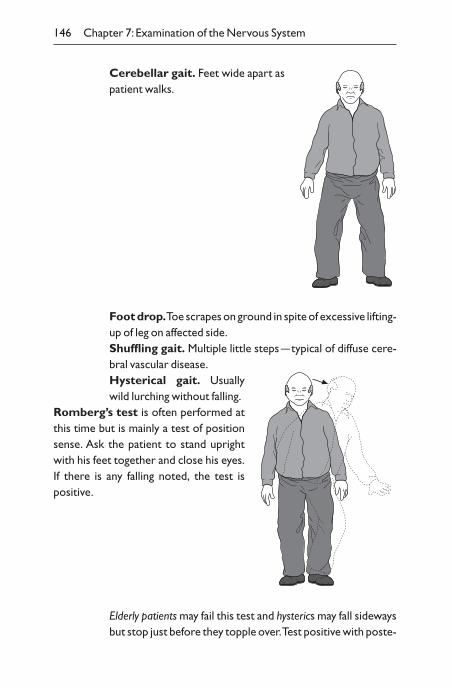
Cerebellar gait. Feet wide apart aspatient walks.
146 Chapter 7: Examination of the Nervous System
Foot drop.Toe scrapes on ground in spite of excessive lifting-up of leg on affected side.Shuffling gait. Multiple little steps —typical of diffuse cere-bral vascular disease.Hysterical gait. Usuallywild lurching without falling.
Romberg’s test is often performed atthis time but is mainly a test of positionsense. Ask the patient to stand uprightwith his feet together and close his eyes.If there is any falling noted, the test is positive.
Elderly patients may fail this test and hysterics may fall sidewaysbut stop just before they topple over.Test positive with poste-
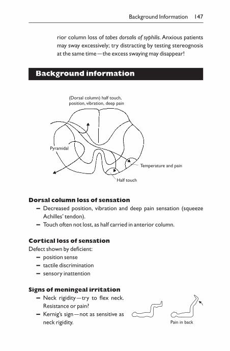
rior column loss of tabes dorsalis of syphilis. Anxious patientsmay sway excessively; try distracting by testing stereognosisat the same time —the excess swaying may disappear!
Background information
Background Information 147
Pyramidal
Temperature and pain
Half touch
(Dorsal column) half touch,position, vibration, deep pain
Dorsal column loss of sensation– Decreased position, vibration and deep pain sensation (squeeze
Achilles’ tendon).– Touch often not lost, as half carried in anterior column.
Cortical loss of sensationDefect shown by deficient:
– position sense– tactile discrimination– sensory inattention
Signs of meningeal irritation– Neck rigidity —try to flex neck.
Resistance or pain?– Kernig’s sign —not as sensitive as
neck rigidity. Pain in back
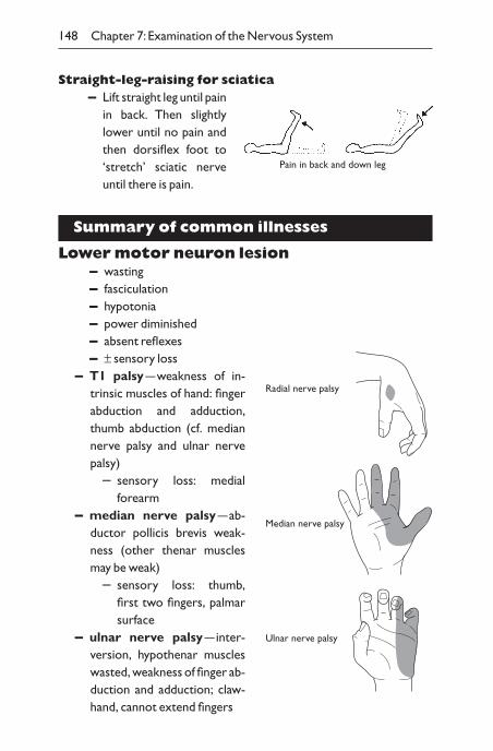
Straight-leg-raising for sciatica– Lift straight leg until pain
in back. Then slightlylower until no pain andthen dorsiflex foot to‘stretch’ sciatic nerveuntil there is pain.
Summary of common illnesses
Lower motor neuron lesion– wasting– fasciculation– hypotonia– power diminished– absent reflexes– ± sensory loss
– T1 palsy —weakness of in-trinsic muscles of hand: fingerabduction and adduction,thumb abduction (cf. mediannerve palsy and ulnar nervepalsy)
– sensory loss: medialforearm
– median nerve palsy —ab-ductor pollicis brevis weak-ness (other thenar musclesmay be weak)
– sensory loss: thumb,first two fingers, palmarsurface
– ulnar nerve palsy —inter-version, hypothenar muscleswasted, weakness of finger ab-duction and adduction; claw-hand, cannot extend fingers
148 Chapter 7: Examination of the Nervous System
Pain in back and down leg
Radial nerve palsy
Median nerve palsy
Ulnar nerve palsy
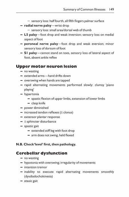
– sensory loss: half fourth, all fifth fingers palmar surface– radial nerve palsy —wrist drop
– sensory loss: small area/dorsal web of thumb– L5 palsy —foot drop and weak inversion; sensory loss on medial
aspect of foot– peroneal nerve palsy —foot drop and weak eversion; minor
sensory loss of dorsum of foot– S1 palsy —cannot stand on toes, sensory loss of lateral aspect of
foot, absent ankle reflex
Upper motor neuron lesion– no wasting– extended arms —hand drifts down– overswing when hands are tapped– rapid alternating movements performed slowly: clumsy ‘piano
playing’– hypertonia
– spastic flexion of upper limbs, extension of lower limbs– clasp knife
– power diminished– increased tendon reflexes (± clonus)– extensor plantar response– ± sphincter disturbance– spastic gait
– extended stiff leg with foot drop– arm does not swing, held flexed
N.B. Check ‘level’ first, then pathology.
Cerebellar dysfunction– no wasting– hypotonia with overswing; irregularity of movements– intention tremor– inability to execute rapid alternating movements smoothly
(dysdiadochokinesia)– ataxic gait
Summary of Common Illnesses 149
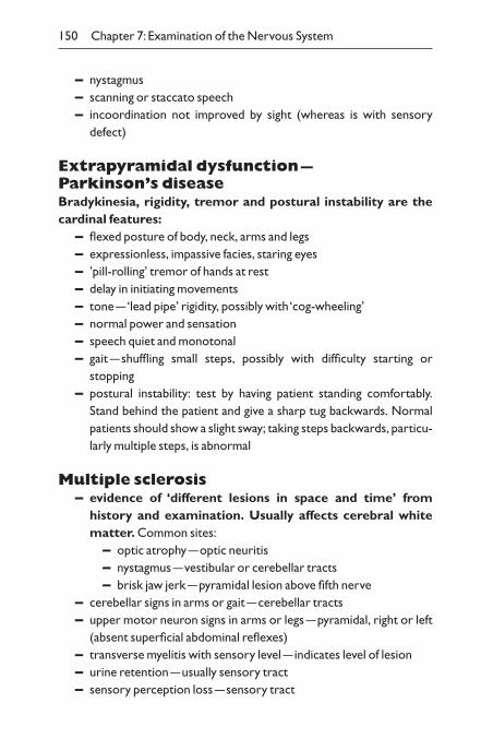
– nystagmus– scanning or staccato speech– incoordination not improved by sight (whereas is with sensory
defect)
Extrapyramidal dysfunction —Parkinson’s diseaseBradykinesia, rigidity, tremor and postural instability are thecardinal features:
– flexed posture of body, neck, arms and legs– expressionless, impassive facies, staring eyes– ’pill-rolling’ tremor of hands at rest– delay in initiating movements– tone —‘lead pipe’ rigidity, possibly with ‘cog-wheeling’– normal power and sensation– speech quiet and monotonal– gait —shuffling small steps, possibly with difficulty starting or
stopping– postural instability: test by having patient standing comfortably.
Stand behind the patient and give a sharp tug backwards. Normalpatients should show a slight sway; taking steps backwards, particu-larly multiple steps, is abnormal
Multiple sclerosis– evidence of ‘different lesions in space and time’ from
history and examination. Usually affects cerebral whitematter. Common sites:
– optic atrophy —optic neuritis– nystagmus —vestibular or cerebellar tracts– brisk jaw jerk —pyramidal lesion above fifth nerve
– cerebellar signs in arms or gait —cerebellar tracts– upper motor neuron signs in arms or legs —pyramidal, right or left
(absent superficial abdominal reflexes)– transverse myelitis with sensory level —indicates level of lesion– urine retention —usually sensory tract– sensory perception loss —sensory tract
150 Chapter 7: Examination of the Nervous System
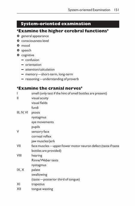
System-oriented examination
‘Examine the higher cerebral functions’
° general appearance
° consciousness level
° mood
° speech
° cognitive– confusion– orientation– attention/calculation– memory —short-term, long-term– reasoning —understanding of proverb
‘Examine the cranial nerves’I smell (only test if the hint of smell bottles are present)II visual acuity
visual fieldsfundi
III, IV, VI ptosisnystagmuseye movementspupils
V sensory facecorneal reflexjaw muscles/jerk
VII face muscles —upper/lower motor neuron defect (taste if tastebottles are provided)
VIII hearingRinne/Weber testsnystagmus
IX, X palateswallowing(taste —posterior third of tongue)
XI trapeziusXII tongue wasting
System-oriented Examination 151
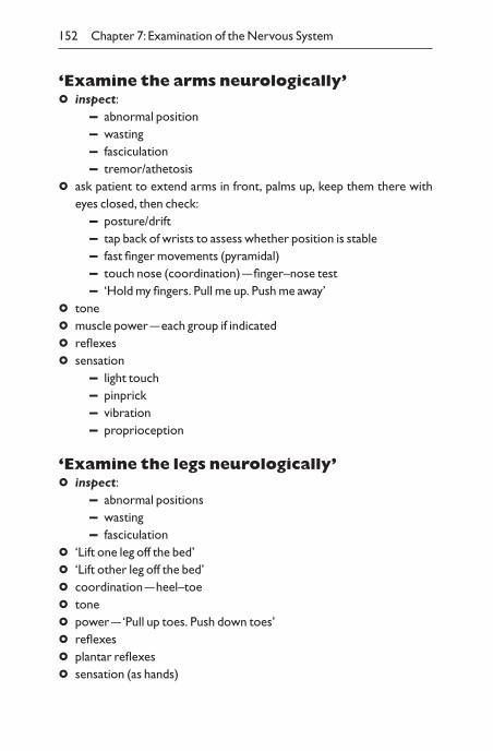
‘Examine the arms neurologically’
° inspect:– abnormal position– wasting– fasciculation– tremor/athetosis
° ask patient to extend arms in front, palms up, keep them there witheyes closed, then check:
– posture/drift– tap back of wrists to assess whether position is stable– fast finger movements (pyramidal)– touch nose (coordination) —finger–nose test– ‘Hold my fingers. Pull me up. Push me away’
° tone
° muscle power —each group if indicated
° reflexes
° sensation– light touch– pinprick– vibration– proprioception
‘Examine the legs neurologically’
° inspect:– abnormal positions– wasting– fasciculation
° ‘Lift one leg off the bed’
° ‘Lift other leg off the bed’
° coordination —heel–toe
° tone
° power —‘Pull up toes. Push down toes’
° reflexes
° plantar reflexes
° sensation (as hands)
152 Chapter 7: Examination of the Nervous System
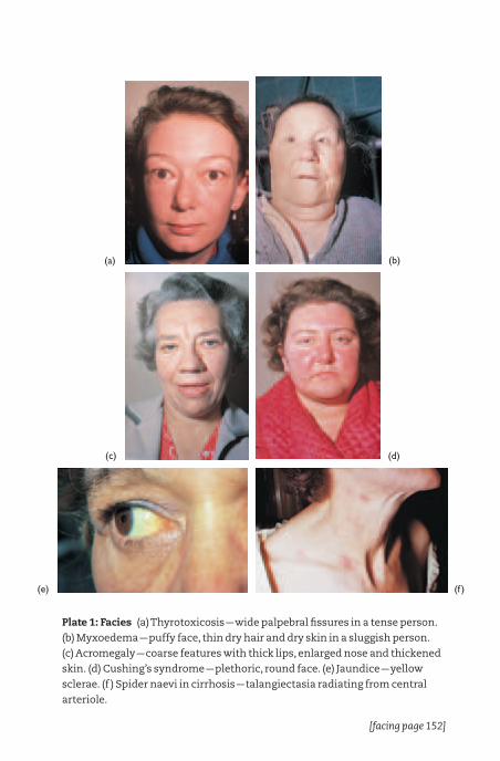
Plate 1: Facies (a) Thyrotoxicosis —wide palpebral fissures in a tense person.
(b) Myxoedema —puffy face, thin dry hair and dry skin in a sluggish person.
(c) Acromegaly —coarse features with thick lips, enlarged nose and thickened
skin. (d) Cushing’s syndrome —plethoric, round face. (e) Jaundice —yellow
sclerae. (f ) Spider naevi in cirrhosis —talangiectasia radiating from central
arteriole.
(a)
(c)
(b)
(d)
(e) (f )
[facing page 152]
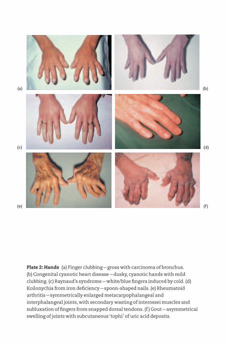
(a)
(c)
(b)
(d)
(e) (f )
Plate 2: Hands (a) Finger clubbing —gross with carcinoma of bronchus.
(b) Congenital cyanotic heart disease —dusky, cyanotic hands with mild
clubbing. (c) Raynaud’s syndrome —white/blue fingers induced by cold. (d)
Koilonychia from iron deficiency —spoon-shaped nails. (e) Rheumatoid
arthritis —symmetrically enlarged metacarpophalangeal and
interphalangeal joints, with secondary wasting of interossei muscles and
subluxation of fingers from snapped dorsal tendons. (f ) Gout —asymmetrical
swelling of joints with subcutaneous ‘tophi’ of uric acid deposits.
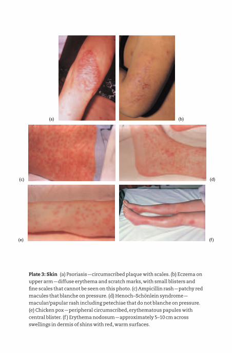
(a)
(c)
(b)
(d)
(e) (f )
Plate 3: Skin (a) Psoriasis —circumscribed plaque with scales. (b) Eczema on
upper arm —diffuse erythema and scratch marks, with small blisters and
fine scales that cannot be seen on this photo. (c) Ampicillin rash —patchy red
macules that blanche on pressure. (d) Henoch–Schönlein syndrome —
macular/papular rash including petechiae that do not blanche on pressure.
(e) Chicken pox —peripheral circumscribed, erythematous papules with
central blister. (f ) Erythema nodosum —approximately 5–10 cm across
swellings in dermis of shins with red, warm surfaces.
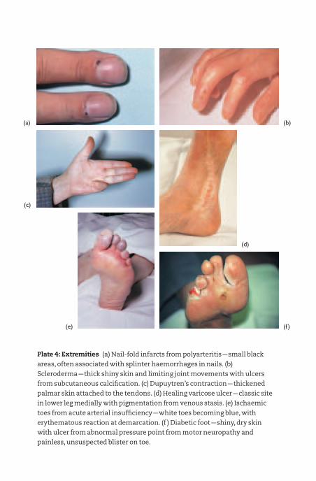
(a)
(c)
(b)
(d)
(e) (f )
Plate 4: Extremities (a) Nail-fold infarcts from polyarteritis —small black
areas, often associated with splinter haemorrhages in nails. (b)
Scleroderma —thick shiny skin and limiting joint movements with ulcers
from subcutaneous calcification. (c) Dupuytren’s contraction —thickened
palmar skin attached to the tendons. (d) Healing varicose ulcer —classic site
in lower leg medially with pigmentation from venous stasis. (e) Ischaemic
toes from acute arterial insufficiency —white toes becoming blue, with
erythematous reaction at demarcation. (f ) Diabetic foot —shiny, dry skin
with ulcer from abnormal pressure point from motor neuropathy and
painless, unsuspected blister on toe.
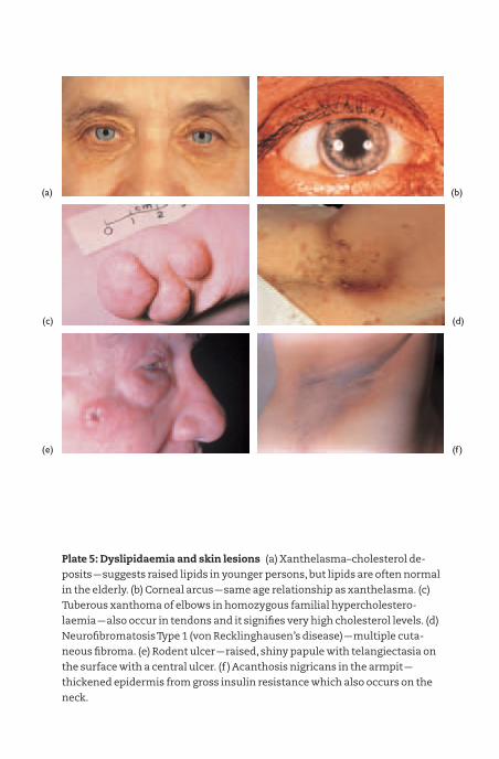
(a)
(c)
(b)
(d)
(e) (f )
Plate 5: Dyslipidaemia and skin lesions (a) Xanthelasma–cholesterol de-
posits —suggests raised lipids in younger persons, but lipids are often normal
in the elderly. (b) Corneal arcus —same age relationship as xanthelasma. (c)
Tuberous xanthoma of elbows in homozygous familial hypercholestero-
laemia —also occur in tendons and it signifies very high cholesterol levels. (d)
Neurofibromatosis Type 1 (von Recklinghausen’s disease) —multiple cuta-
neous fibroma. (e) Rodent ulcer —raised, shiny papule with telangiectasia on
the surface with a central ulcer. (f ) Acanthosis nigricans in the armpit —
thickened epidermis from gross insulin resistance which also occurs on the
neck.
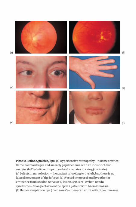
(a)
(c)
(b)
(d)
(e) (f )
Plate 6: Retinae, palsies, lips (a) Hypertensive retinopathy —narrow arteries,
flame haemorrhages and an early papilloedema with an indistinct disc
margin. (b) Diabetic retinopathy —hard exudates in a ring (circinate).
(c) Left sixth nerve lesion —the patient is looking to the left, but there is no
lateral movement of the left eye. (d) Wasted interossei and hypothenar
eminence from an ulna nerve or T1 lesion. (e) Osler–Weber–Rendu
syndrome —telangiectasia on the lip in a patient with haematemasis.
(f ) Herpes simplex on lips (‘cold sores’) —these can erupt with other illnesses.
° Romberg test
° gait and tandem gait
‘Examine the arms or legs’
° inspect:– colour– skin/nail changes– ulcers– wasting (are both arms and legs involved?)– joints
° palpate:– temperature, pulses– lumps (see above)– joints
– active movements– feel for crepitus, e.g. hand over knee during flexion– passive movements (do not hurt patient)
– reflexes– sensation
System-oriented Examination 153
CHAPTER 8
Assessment ofDisability IncludingCare of the Elderly
Introduction
It is important, particularly in the elderly, to assess whether the patient has a disability:
– which interferes with normal life and aspirations– which makes the patient dependent on others
– requires temporary assistance for specific problems– occasional or regular assistance long-term– supervised accommodation– nursing home with 24-hour care
It is necessary to assess the following in a patient:
° ability to do day-to-day functions
° mental ability, including confusion or dementia
° emotional state and driveThe descriptive terms used for disability have specific definitions in a
World Health Organization classification.– Impairment —any loss or abnormality of anatomical, physiologi-
cal or psychological function, i.e. systems or parts of body thatdo not work.
– Disability —any restrictions or lack of ability (due to an impair-ment) to perform an activity within the range considered normal,i.e. activities that cannot be done.
– Handicap —a limitation of normal occupation because of impair-ment or disability, i.e. social consequences.
Thus:– A hemiparesis is an impairment.– An inability to wash or dress is a disability.– An inability to do an occupation is a handicap.
154
The introductory clinical training in the first fewchapters of this book concentrates on evaluation ofimpairments. Disability and handicap are not alwaysgiven due attention and are the practical and socialaspects of the disease process. It is a mistake if thedoctor is preoccupied by impairments, since the patient often perceives disability as the major problem.
The impairments, disability and handicap should have been covered ina normal history and examination, but it can be helpful to bring togetherimportant facts to provide an overall assessment.
A summary description of a patient may include the following.– Aetiology —familial hypercholesterolaemia.– Pathology
– atheroma– right middle cerebral artery thrombosis
– Impairment– left hemiparesis– paralysed left arm, fixed in flexion– upper motor neuron signs in left arm and face
– Disability —difficulty during feeding. Cannot drive his car.– Handicap
– can no longer work as a travelling salesman– embarrassed to socialize
– Social circumstances —partner can cope with day-to-day living, but lack of income from his occupation and withdrawalfrom society present major problems.
Assessment of impairment
The routine history and examination will often reveal impairments.Additional standard clinical measures are often used to assist quantita-tion, e.g.
– treadmill exercise test– peak flowmeter– Medical Research Council scale of muscle power
Assessment of Impairment 155
– making five-pointed star from matches (to detect dyspraxia in hepatic encephalopathy)
Questionnaires can similarly provide a semiquantitative index of important aspects of impairment and give a brief short-hand descriptionof a patient.The role of the questionnaire is in part a checklist to make surethe key questions are asked.
Cognitive functionIn the elderly, impaired cognitive function can be assessed by a standard10-point mental test score introduced by Hodkinson. The test assumes normal communication skills. One mark each is given for correct answers to 10 standard questions (see Appendix 3 for questionnaire):
– age of patient– time (to nearest hour)– address given, for recall at end of test, e.g. 42 West Street or 92
Columbia Road– recognize two people– year (if Januar, the previous year is accepted)– name of place, e.g. hospital or area of town if at home– date of birth of patient– start of First World War– name of monarch in UK, president in USA– count backwards from 20 to 1 (no errors allowed unless
self-corrected)– (check recall of address)This scale is a basic test of gross defects of memory and orientation
and is designed to detect cognitive impairment. It has the advantages of brevity, relative lack of culture-specific knowledge and widespread use. In the elderly, 8–10 is normal, 7 is probably normal, 6 or less is abnormal.
Specific problems, such as confusion or wandering at night, are not included in the mental test score, and indicate that the score is a useful checklist but not a substitute for a clinical assessment.
156 Chapter 8: Assessment of Disability
Affect and driveMotivation is an important determinant of successful rehabilitation.Depression, accompanied by lack of motivation, is a major cause of disability.
Enquire about symptoms of depression (p.16) and relevant examina-tion (pp. 102 and 115), e.g. ‘How is your mood? Have you lost interest inthings?’
Making appropriate lifestyle changes, recruiting help from friends orrelatives, can be key to increasing motivation. Pharmaceutical treatmentof depression can also be helpful.
Assessment of disability
Assessing restrictions to daily activities is often the key to suc-cessful management.
° Make a list of disabilities separate from other problems, e.g.diagnoses, symptoms, impairments, social problems.
This list can assist with setting priorities, including which investiga-tions or therapies are most likely to be of benefit to the patient.
Activities of daily living (ADL)These are key functions which in the elderly affect the degree of independence. Several scales of disability have been used. One of these,the Barthel index of ADL, records the following disabilities that can affect self-care and mobility (see Appendix 4 for questionnaire):
– continence —urinary and faecal– ability to use toilet– grooming– feeding– dressing– bathing– transfer, e.g. chair to bed– walking– using stairs
The assessment denotes the current state and not the underlyingcause or the potential improvement. It does not include cognitive func-
Assessment of Disability 157
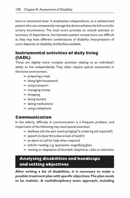
tions or emotional state. It emphasizes independence, so a catheterizedpatient who can competently manage the device achieves the full score forurinary incontinence. The total score provides an overall estimate or summary of dependence, but between-patient comparisons are difficultas they may have different combinations of disability. Interpretation ofscore depends on disability and facilties available.
Instrumental activities of daily living(IADL)These are slightly more complex activities relating to an individual’s ability to live independently. They often require special assessment in the home environment.
– preparing a meal– doing light housework– using transport– managing money– shopping– doing laundry– taking medications– using a telephone
CommunicationIn the elderly, difficulty in communication is a frequent problem, and impairment of the following may need special attention:
– deafness (do the ears need syringing? Is a hearing aid required?)– speech (is dysarthria due to lack of teeth?)– an alarm to call for help when required– aids for reading, e.g. spectacles, magnifying glass– resiting or adaptation of doorbell, telephone, radio or television
Analysing disabilities and handicapsand setting objectives
After writing a list of disabilities, it is necessary to make a possible treatment plan with specific objectives.The plan needsto be realistic. A multidisciplinary team approach, including
158 Chapter 8: Assessment of Disability
social workers, physiotherapists, occupational therapists,nurses and doctors is usually essential in rehabilitation of elderly patients.The overall aims in treating the elderly include the following:
– To make diagnoses, if feasible, particularly to treatable illnesses.– To comfort and alleviate problems and stresses, even if one
cannot cure.– To add life to years, even if one cannot add years to life.
Specific aspects which may need attention include the following:– Alleviate social problems if feasible.– Improve heating, clothing, toilet facilities, cooking facilities.– Arrange support services, e.g. help with shopping, provision of
meals, attendance to day centre.– Arrange regular visits from nurse or other helper.– Make sure family, neighbours and friends understand the
situation.– Treat depression.– Help with sorting out finances.– Provide aids, e.g.
– large-handled implements– walking frame or stick– slip-on shoes– handles by bath or toilet
– Help to keep as mobile as feasible.– Facilitate visits to hearing-aid centre, optician, chiropodist,
dentist.– Ensure medications are kept to a minimum, and the instructions
and packaging are suitable.A major problem is if the disability leads to the patient being unwelcome.This depends on the reactions of others and requires tactful discussionwith all concerned.
Identifying causes for disabilities
Specific disabilities may have specific causes which can be alleviated. In theelderly, common problems include the following.
Identifying Causes for Disabilities 159
ConfusionThis is an impairment. Common causes are:
– infection– drugs– other illnesses, e.g. heart failure– sensory deprivation, e.g. deafness, darkness
Assume all confusion is an acute response to an unidentified cause.Incontinence
– toilet far away, e.g. upstairs– physical restriction of gait– urine infection– faecal impaction– uterine prolapse– diabetes
‘Off legs’– neurological impairment– unsuspected fracture of leg– depressed– general illness, e.g. infection, heart failure, renal failure,
hypothermia, hypothyroid, diabetes, hypokalaemiaFalls
– insecure carpet– dark stairs– poor vision, e.g. cataracts– postural hypotension– cardiac arrhythmias– epilepsy– neurological deficit, e.g. Parkinson’s disease, hemiparesis– cough or micturition syncope– intoxication
160 Chapter 8: Assessment of Disability
CHAPTER 9
Basic Examination,Notes and DiagnosticPrinciples
Basic examination
In practice, one cannot attempt to elicit every single physical sign for eachsystem. Basic signs should be sought on every examination, and if there is any hint of abnormality, additional physical signs can be elicitedto confirm the suspicion. Listed below are the basic examinations of the systems which will enable you to complete a routine examination adequately but not excessively.
° General examination– general appearance– is the patient well or ill?– look at temperature chart or take patient’s temperature– any obvious abnormality?– mental state, mood, behaviour
° General and cardiovascular system– observation —dyspnoea, distress– blood pressure– hands
– temperature– nails, e.g. clubbing, liver palms
– pulse —rate, rhythm, character– axillae —lymph nodes– neck —lymph nodes– face and eyes —anaemia, jaundice– tongue and fauces —central cyanosis– jugular venous pressure ( JVP) —height and waveform– apex beat —position and character– parasternal —heave or thrills
161
– stethoscope– heart sounds, added sounds, murmurs– listen in all four areas with stethoscope diaphragm– lie patient on left side, bell of stethoscope —mitral stenosis
(MS)– sit patient up, lean forward, breathe out —aortic
incompetence (AI)
° Respiratory system– observation– trachea —position– front of chest
– movement– percuss —compare sides– auscultate
– back of chest– movement– percuss —particularly level of bases– auscultate
– examine sputum
° Examine spine.
° Abdomen– lie patient flat– feel femoral pulses and inguinal lymph nodes– herniae– look at abdomen —ask if pain or tenderness– palpate abdomen gently
– generally all over? masses– liver —then percuss– spleen —then percuss– kidneys– (ascites if indicated)
– (auscultate if indicated)– males —genitals– per rectum (PR; only if given permission) —usually at end of
examination– per vaginam (PV) —rarely by student
162 Chapter 9:Basic Examination
° Legs– observation– arterial pulses (joints if indicated)– neurology:
– reflexes – knees toneonly– ankles power } if indicated– plantar responses coordination
– sensation – pinprick positiononly– vibration cotton-wool }
temperatureif indicated
° Arms– posture: outstretched hands, eyes closed, rapid finger movements– finger–nose coordination:
– reflexes – triceps toneonly– biceps power } if indicated– supinator
– sensation – pinprick vibrationposition onlycotton-wool } if indicatedtemperature
° Cranial nerves– I (if indicated)– II: eyes – reading print
– pupils —torch and accommodation– ophthalmoscope —fundi– fields
– III, IV, VI: eye movements —‘Do you see double?’note nystagmus
– V, VII – open mouth– grit teeth —feel masseters– sensation —cotton-wool– (corneal reflex —if indicated)– (taste —if indicated)
– VIII: hearing —watch at each ear– (Rinne,Weber tests if indicated)
– IX, X: fauces movement
Basic Examination 163
– XI: shrug shoulders– XII: put out tongue
° Walk – look at gait.
° Herniae and varicose veins.
Example of notes
Patient’s name: Age: Occupation:State your name and designation, e.g. A. Bloggs SHO medicineDate of admission:Complains of:
– list, in patient’s wordsHistory of present illness:
– detailed description of each symptom (even if appears irrelevant)– last well– chronological order, with both actual date of onset, and time
previous to admission– (may include history from informant —in which case, state this is
so)– then detail other questions which seem relevant to possible
differential diagnoses– then functional enquiry, ‘check’ system for other symptoms– (minimal statement in notes —weight, appetite, digestion, bowels,
micturition, menstruation, if appropriate)Past history:
– chronological orderFamily history:Personal and social history:
– must include details of home circumstances, dependants, patient’soccupation
– effect of illness on life and its relevance to foreseeable discharge ofpatient
– smoking, alcohol, drug misuse, medicationsPhysical examination:
– general appearance, etc.– then record findings according to systems
164 Chapter 9:Basic Examination
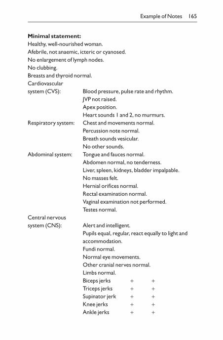
Minimal statement:Healthy, well-nourished woman.Afebrile, not anaemic, icteric or cyanosed.No enlargement of lymph nodes.No clubbing.Breasts and thyroid normal.Cardiovascularsystem (CVS): Blood pressure, pulse rate and rhythm.
JVP not raised.Apex position.Heart sounds 1 and 2, no murmurs.
Respiratory system: Chest and movements normal.Percussion note normal.Breath sounds vesicular.No other sounds.
Abdominal system: Tongue and fauces normal.Abdomen normal, no tenderness.Liver, spleen, kidneys, bladder impalpable.No masses felt.Hernial orifices normal.Rectal examination normal.Vaginal examination not performed.Testes normal.
Central nervoussystem (CNS): Alert and intelligent.
Pupils equal, regular, react equally to light andaccommodation.Fundi normal.Normal eye movements.Other cranial nerves normal.Limbs normal.Biceps jerks + +Triceps jerks + +Supinator jerk + +Knee jerks + +Ankle jerks + +
Example of Notes 165
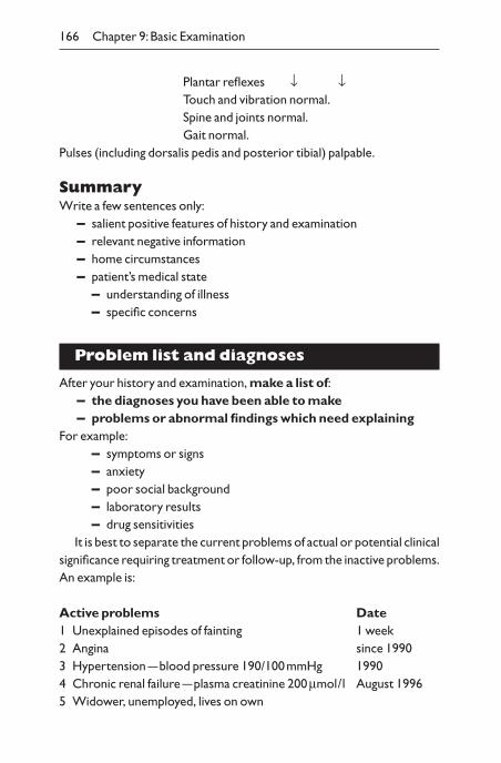
Plantar reflexes Ø ØTouch and vibration normal.Spine and joints normal.Gait normal.
Pulses (including dorsalis pedis and posterior tibial) palpable.
SummaryWrite a few sentences only:
– salient positive features of history and examination– relevant negative information– home circumstances– patient’s medical state
– understanding of illness– specific concerns
Problem list and diagnoses
After your history and examination, make a list of:– the diagnoses you have been able to make– problems or abnormal findings which need explaining
For example:– symptoms or signs– anxiety– poor social background– laboratory results– drug sensitivities
It is best to separate the current problems of actual or potential clinicalsignificance requiring treatment or follow-up, from the inactive problems.An example is:
Active problems Date1 Unexplained episodes of fainting 1 week2 Angina since 19903 Hypertension —blood pressure 190/100mmHg 19904 Chronic renal failure —plasma creatinine 200mmol/l August 19965 Widower, unemployed, lives on own
166 Chapter 9:Basic Examination
Problem List and Diagnoses 167
6 Anxious about possibility of being injured in a fall7 Smokes 40 cigarettes per day
Inactive problems Date1 Thyrotoxicosis treated by partial thyroidectomy 19762 ACE inhibitor-induced cough 1991
At first you will have difficulty knowing which problems to put down separately, and which can be covered under one diagnosis and a singleentry. It is therefore advisable to rewrite the problem list if a problem re-solves or can be explained by a diagnosis. When you have some experi-ence, it will be appropriate to fill out the problems on a complete problemlist at the front of the notes:
Active problems Date Inactive problems DateInclude symptoms, Include major pastsigns, unexplained illness, operation orabnormal investigations, hypersensitivities. Dosocial and psychiatric not include problemsproblems requiring active care
From the problem list, you should be able to make:
° differential diagnoses, including that which you think is most likely.Remember:
– common diseases occur commonly– an unusual manifestation of a common disease is more
likely than an uncommon disease– when you hear hoof beats, think of horses, not zebras
– do not necessarily be put off by some aspect which doesnot fit. (What is the farmer’s friend which has four legs, wags itstail and says ‘cockadoodledoo’? A dog, and the sound was fromanother animal)
° possible diagnostic investigations you feel are appropriate
° management and therapy you think are appropriate
° prognostic implications
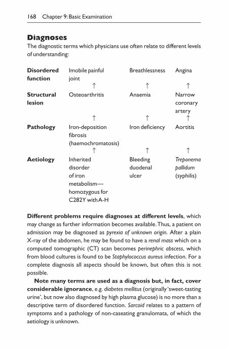
DiagnosesThe diagnostic terms which physicians use often relate to different levelsof understanding:
Disordered Imobile painful Breathlessness Anginafunction joint
≠ ≠ ≠Structural Osteoarthritis Anaemia Narrowlesion coronary
artery≠ ≠ ≠
Pathology Iron-deposition Iron deficiency Aortitisfibrosis(haemochromatosis)
≠ ≠ ≠Aetiology Inherited Bleeding Treponema
disorder duodenal pallidumof iron ulcer (syphilis)metabolism—homozygous forC282Y with A-H
Different problems require diagnoses at different levels, whichmay change as further information becomes available.Thus, a patient onadmission may be diagnosed as pyrexia of unknown origin. After a plain X-ray of the abdomen, he may be found to have a renal mass which on a computed tomographic (CT) scan becomes perinephric abscess, whichfrom blood cultures is found to be Staphylococcus aureus infection. For acomplete diagnosis all aspects should be known, but often this is not possible.
Note many terms are used as a diagnosis but, in fact, coverconsiderable ignorance, e.g. diabetes mellitus (originally ‘sweet-tastingurine’, but now also diagnosed by high plasma glucose) is no more than adescriptive term of disordered function. Sarcoid relates to a pattern ofsymptoms and a pathology of non-caseating granulomata, of which the aetiology is unknown.
168 Chapter 9:Basic Examination
Progress notesWhile the patient is in hospital, full progress notes should be kept to give acomplete picture of:
– how the diagnosis was established– how the patient was treated– the evolution of the illness– any complications that occured
These notes are as important as the account of the original examina-tion. In acute cases, record daily changes in signs and symptoms. In chronic cases, the relevant systems should be re-examined at least once aweek and the findings recorded.
It is useful to separate different aspects of the illness:– symptoms– signs– laboratory investigations– general assessment, e.g. apparent response to therapy– further plans, which would include educating the patient and his
family about the illness.Objective findings such as alterations in weight, improvement in
colour, pulse, character of respirations or fluid intake and output aremore valuable than purely subjective statements such as ‘feeling better’ or ‘slept well’.
When appropriate, daily blood pressure readings or analyses of theurine should be recorded.
An account of all ward procedures such as aspirations of chest shouldbe included.
Specifically record:– the findings and comments of the physician or surgeon in
charge– results of a case conference– an opinion from another department
Problem-oriented recordsDr Larry Weed proposed a system of note-keeping in which the historyand examination constituted a database.All subsequent notes are struc-tured according to the specific numbered problems in a problem list.
Problem List and Diagnoses 169
Problem-oriented records really require a special system of note-keeping.The full system is therefore not often used, but the problem list is an extremely valuable check that all aspects of the patient’s illness are being covered.
Serial investigationsThe results of these should be collected together in a table on a specialsheet. When any large series of investigations is made, e.g. serial bloodcounts, erythrocyte sedimentation rates or multiple biochemical analyses, the results can also be expressed by a graph.
Operation notesIn patients undergoing surgical treatment, an operation note must bewritten immediately after the operation. Do not trust your memory forany length of time as several similar problems may be operated on at onesession. Even if you are distracted by an emergency, the notes must bewritten up the same day as the operation. These notes should contain definite statements on the following facts:
– name of surgeon performing the operation and his assistant– name of anaesthetist and anaesthetic used– type and dimension of incision used– pathological condition found, and mention of anatomical
variations– operative procedures carried out– method of repair of wound and suture materials used– whether drainage used, material used, and whether sutured to
wound– type of dressing used
Postoperative notesWithin the first 2 days after an operation note:
– the general condition of the patient– any complication or troublesome symptom, e.g. pain, haemor-
rhage, vomiting, distension, etc.– any treatment
170 Chapter 9:Basic Examination
Discharge noteA full statement of the patient’s condition on discharge should be written:
– final diagnosis– active problems– medication and other therapies– plan– specific follow-up points, e.g. persistent depressive disorder, blood
pressure monitoring– what the patient has been told– where the patient has gone, and what help is available– when the patient is next being seen– an estimate of the prognosisIf the patient dies, the student must attend a post-mortem and then
complete his note by a short account of the autopsy findings.
Problem List and Diagnoses 171
CHAPTER 10
Presenting Cases and Communication
Presentations to doctors
Medicine is a subject in which you have to be able to talk.The more prac-tice you get, the better you will become and the more confident you willappear in front of doctors, nurses and patients. Confidence displayed bythe doctor is an important aspect of therapy and the value to the patientof a doctor who can speak lucidly is enormous.
Practise talking to yourself in a mirror, avoiding any breaks or interpo-lating the word ‘er’. Open a textbook, find a subject and give a little talk onit to yourself. Even if you do not know anything about the subject, you willbe able to make up a few coherent sentences once you are practised.
A presentation is not the time to demonstrate you have beenthorough and have asked all questions, but is a time to show youcan intelligently assemble the essential facts.
In all presentations, give the salient positive findings and therelevant negative findings.
For example:– In a patient with progressive dyspnoea, state if patient has ever
smoked.– In a patient with icterus, state if patient has not been abroad, has not
had any recent injections or drugs, or contact with other jaundicedpatients.
Three types of presentations are likely to be encountered: presenta-tion of a case to a meeting, presentation of a new case on a ward round anda brief follow-up presentation.
Presentation of a case to a meetingThis must be properly prepared, including visual aids as necessary. The
172
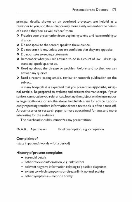
principal details, shown on an overhead projector, are helpful as a reminder to you, and the audience may more easily remember the detailsof a case if they ‘see’ as well as ‘hear’ them.
° Practise your presentation from beginning to end and leave nothing tochance.
° Do not speak to the screen; speak to the audience.
° Do not crack jokes, unless you are confident that they are apposite.
° Do not make sweeping statements.
° Remember what you are advised to do in a court of law —dress up,stand up, speak up, shut up.
° Read up about the disease or problem beforehand so that you can answer any queries.
° Read a recent leading article, review or research publication on thesubject.In many hospitals it is expected that you present an apposite, origi-
nal article. Be prepared to evaluate and criticize the manuscript. If yourseniors cannot give you references, look up the subject on the internet orin large textbooks, or ask the always helpful librarian for advice. Labori-ously repeating standard information from a textbook is often a turn-off.A recent series or research paper is more educational for you, and moreinteresting for the audience.
The overhead should summarizes any presentation:
Mr A.B. Age: x years Brief description, e.g. occupation
Complains of(state in patient’s words —for x period)
History of present complaint– essential details– other relevant information, e.g. risk factors– relevant negative information relating to possible diagnoses– extent to which symptoms or disease limit normal activity– other symptoms —mention briefly
Presentations to Doctors 173
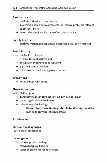
Past history– briefly mention inactive problems– information about active problems, or inactive problems relevant
to present illness– record allergies, including type of reaction to drugs
Family history– brief information about parents, otherwise detail only if relevant
Social history– brief unless relevant– give family social background– occupation and previous occupations– any other special problems– tobacco or ethanol abuse, past or present
Treatment– note all drugs with doses
On examinationGeneral description
– introductory descriptive sentence, e.g. well, obese man– clinical signs relevant to disease– relevant negative findings
Remember these findings should be descriptive datarather than your interpretation.
Problem list
Differential diagnoses(put in order of likelihood)
Investigations– relevant positive findings– relevant negative findings– tables or graphs for repetitive data
174 Chapter 10:Presenting Cases and Communication
– photocopy an electrocardiogram or temperature chart for overhead presentation
Progress report
PlanSubjects which often are discussed after your presentation are:
– other differential diagnoses– other features of presumed diagnosis that might have been present
or require investigation– pathophysiological mechanisms– mechanisms of action of drugs and possible side-effectsAfter clinical discussion, be prepared to present a publication with
essential details on an overhead.
Presentation of a new case on a ward round
° Good written notes are of great assistance. Do not read your notesword for word —use your notes as a reference.
° Highlight, underline or asterisk key features you wish to refer to, orwrite up a separate note-card for reference.
° Talk formally and avoid speaking too quickly or too slowly. Speak to thewhole assembled group rather than a tête-à-tête with the consultant.
° Stand upright —it helps to make you appear confident.
° If you are interrupted by a discussion, note where you are and be readyto resume, repeating the last sentence before proceeding.
HistoryThe format will be similar to that on an overhead, with emphasis on positive findings and relevant negative information. A full description of the initial main symptom is usually required.
ExaminationOnce your history is complete the consultant may ask for the relevantclinical signs only. Still add in relevant negative signs you think are important.
Presentations to Doctors 175
SummaryBe prepared to give a problem list and differential diagnoses.
If you are presenting the patient at the bedside, ensure the patient iscomfortable. If the patient wishes to make an additional point or clarifi-cation, it is best to welcome this. If it is relevant it can be helpful. If irrele-vant, politely say to the patient you will come back to him in a moment,after you have presented the findings. Do not appear to argue with the patient.
Brief follow-up presentationGive a brief, orienting introduction to provide a framework on whichother information can be placed. For example:
A xx-year-old man who was admitted xx days ago.Long-standing problems include xxxxx (list briefly).Presented with xx symptoms for x period.On examination had xx signs.Initial diagnosis of xx was confirmed/supported by/not supported byxx investigations.He was treated by xx.Since then xx progress:
– symptoms– examination
Start with general description and temperature chart and, ifrelevant, investigations.
If there are multiple active problems, describe each separately, e.g.– first in regard to the xxxx– second in regard to the xxxx
The outstanding problems are xxxx.The plan is xxxx.
Aide-mémoiresThese are basic lists that provide brief reminders when presenting patients and diseases. Organizing one’s thoughts along structured lines is helpful.
176 Chapter 10:Presenting Cases and Communication
History– principal symptom(s)– history of present illness: ‘How did your illness begin?’– note chronology– present situation– functional enquiry– past history– family history– personal and social history
Pain or other symptoms– site– radiation– character– severity– onset/duration– frequency/periodicity or constant– precipitating factors– relieving factors– associated symptoms– getting worse or better?
LumpsInspection
– site– size– shape– surface– surroundings
Palpation– soft/solid consistency– surroundings —fixed/mobile– tender– pulsatility– transmission of illumination
Presentations to Doctors 177
Local lymph nodes
‘Tell me about the disease’– incidence– geographical area– gender/age– aetiology– pathology
– macroscopic– microscopic
– pathophysiology– symptoms– signs– therapy– prognosis
Causes of disease
° genetic
° infective– virus– bacterial– fungal– parasitic
° neoplastic– cancer
– primary– secondary
– lymphoma
° vascular– atheroma– hypertension– other, e.g. arteritis
° infiltrative– fibrosis– amyloid– granuloma
178 Chapter 10:Presenting Cases and Communication
° autoimmune
° endocrine
° degenerative
° environmental– trauma– iatrogenic —drug side-effects– poisoning
° malnutrition– general– specific, e.g. vitamin defiency– perinatal with effects on subsequent development
Diagnostic labels
° aetiology, e.g. tuberculosis, geneticØ
° pathology, e.g. sarcoid, amyloidØ
° disordered function, e.g. hypertension, diabetesØ
° symptoms or signs, e.g. jaundice, erythema nodosum
People—including patients
A significant number of disasters, a great deal of irritation and a lot of unpleasantness could be avoided in hospitals by proper communication.The doctor is not the boss but is part of a team, all of whom significantlyhelp the patient. You must be able to communicate properly with the nursing staff, physiotherapists, occupational therapists, administrators,ancillary staff and, above all, patients.
When you first arrive on the wards it is a good idea to go and see theward sister, physiotherapist, etc. and find out what their job is, what theirdifficulties are and how they view the patient, other groups and, most importantly, yourself.
Remember these points.
° Time —when you talk to anyone, try not to appear in a rush or theywill lose concentration and not listen. A little time taken to talk to
People —Including Patients 179
somebody properly will help enormously. One minute spent sittingdown can seem like 5 minutes to the patient; 5 minutes standing up canseem like 1 minute.
° Silence —in normal social interaction we tend to avoid silences. In aconversation, as soon as one person stops talking (or even before) theother person jumps in to say his or her bit.When interviewing patients,it is often useful, if you wish to encourage the patient to talk further, toremain silent a moment longer than would be natural.An encouragingnod of the head, or an echoing of the patient’s last word or two mayalso encourage the patient to talk further.
° Listen —active listening to someone is not easy but is essential forgood communication. Many people stop talking but not all appear tobe listening. Sitting down with the patient is advantageous, both inhelping you to concentrate and in transmitting to the patient that youare willing to listen.
° Smile —grumpiness or irritation is the best way to stop a patient talking. A smile will often encourage a patient to tell you problems he would not normally do. It helps everybody to relax.
° Reassurance —if you appear confident and relaxed this helps othersto feel the same. Being calm without excessive body movements canhelp. Note how a good consultant has a reassuring word for patientsand allows others in the team to feel they are (or are capable of) work-ing effectively.As a student you are not in a position to do this, but youcan contribute by playing your role efficiently and calmly.
° Humility —no one, in particular the patient, is inferior to you.
180 Chapter 10:Presenting Cases and Communication
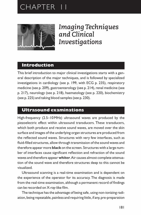
CHAPTER 11
Imaging Techniquesand ClinicalInvestigations
Introduction
This brief introduction to major clinical investigations starts with a gen-eral description of the major techniques, and is followed by specialized investigations in cardiology (see p. 199, with ECG p. 235), respiratorymedicine (see p. 209), gastroenterology (see p. 214), renal medicine (seep. 217), neurology (see p. 218), haematology (see p. 220), biochemistry(see p. 225) and taking blood samples (see p. 230).
Ultrasound examinations
High-frequency (2.5–10MHz) ultrasound waves are produced by thepiezoelectric effect within ultrasound transducers. These transducers,which both produce and receive sound waves, are moved over the skinsurface and images of the underlying organ structures are produced fromthe reflected sound waves. Structures with very few interfaces, such asfluid-filled structures, allow through transmission of the sound waves andtherefore appear more black on the screen. Structures with a large num-ber of interfaces cause significant reflection and refraction of the soundwaves and therefore appear whiter.Air causes almost complete attenua-tion of the sound wave and therefore structures deep to this cannot be visualized.
Ultrasound scanning is a real-time examination and is dependent onthe experience of the operator for its accuracy. The diagnosis is madefrom the real-time examination, although a permanent record of findingscan be recorded on X-ray-like film.
The technique has the advantage of being safe, using non-ionizing radi-ation, being repeatable, painless and requiring little, if any, pre-preparation
181
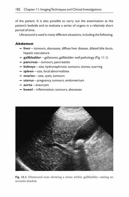
of the patient. It is also possible to carry out the examination at the patient’s bedside and to evaluate a series of organs in a relatively short period of time.
Ultrasound is used in many different situations, including the following.
Abdomen– liver —tumours, abscesses, diffuse liver disease, dilated bile ducts,
hepatic vasculature– gallbladder —gallstones, gallbladder wall pathology (Fig. 11.1)– pancreas —tumours, pancreatitis– kidneys —size, hydronephrosis, tumours, stones, scarring– spleen —size, focal abnormalities– ovaries —size, cysts, tumours– uterus —pregnancy, tumours, endometrium– aorta —aneurysm– bowel —inflammation, tumours, abscesses
182 Chapter 11: Imaging Techniques and Clinical Investigations
Fig. 11.1 Ultrasound scan showing a stone within gallbladder, casting an
acoustic shadow.
Brain– possible in the infant before the anterior fontanelle closes
HeartSee Echocardiography, p. 200.
Pleura– pleural fluid or thickening
Blood vessels– aneurysms, stenoses, clot in veins
Neck– thyroid —characterization of masses
Scrotum– tumours, inflammation
Musculoskeletal– joint effusions, soft-tissue masses
Endoscopy
Internal organs are directly visualized, usually with a flexible fibreoptic endoscope.
GastroscopyA flexible scope is inserted by mouth after intravenous diazepam for direct vision of oesophagus, stomach and duodenum (see p. 214) (also seeEndoscopic retrograde cholangiopancreatography (ERCP), p. 215).
ProctoscopyWith the patient lying in a left lateral position on one side, with knees andhips flexed, a short tube is introduced through the anus with a removableobturator lubricated with a gel.To investigate:
– rectal bleeding —haemorrhoids or anal carcinoma
Endoscopy 183
SigmoidoscopyWith the patient in left lateral position, either a rigid tube with a remov-able obturator or a flexible fibreoptic endoscope is introduced. Bowel iskept patent with air from a hand pump.To investigate:
– bleeding, diarrhoea or constipation —ulcerative colitis, otherinflammatory bowel disease or carcinoma
– inflamed area or lumps can be biopsied
ColonoscopyAfter the bowel is emptied with an oral purgative and a washout if necessary, the whole of the colon and possibly the terminal ileum can beexamined.To investigate:
– bleeding, diarrhoea or constipation — inflammatory bowel dis-ease, polyps or carcinoma
BronchoscopyAfter intravenous diazepam, the major bronchi are observed. To investigate:
– haemoptysis or suspected bronchial obstruction —bronchialcarcinoma and for clearing obstructed bronchi, e.g. peanuts, plug ofmucus
LaparoscopyAfter general anaesthetic, organs can be observed through a small abdominal incision, aspirated for cells or organisms, or biopsied.Laparoscopic surgery includes sterilization, ova collection for in vitrofertilization and laparoscopic cholecystectomy.
CystoscopyAfter local anaesthetic, a cystoscope is inserted into the urethral meatus.To investigate:
– urinary bleeding or poor flow —bladder tumours– under direct vision, catheters can be inserted into ureters for ret-
rograde pyelograms
184 Chapter 11: Imaging Techniques and Clinical Investigations
ColposcopyExamination of cervix, usually to take a cervical smear.To investigate:
– premalignant changes or cancer
Needle biopsy
Core biopsyA small core of tissue (30 ¥ 1mm) is obtained through needle puncture oforgans for histological diagnosis.To investigate:
– liver —cirrhosis, alcoholic liver disease, chronic active hepatitis– kidney —glomerulonephritis, interstitial nephritis– lung —fibrosis, tumours, tuberculosis
Fine-needle aspirationA technique to obtain cells for diagnosis of tumours or for microbiol-ogical diagnosis. The needle position is guided by ultrasound, computedtomographic (CT) scan or magnetic resonance imaging (MRI) scan. For investigation of many unexplained lumps, e.g. pancreas or breast lumps, todiagnose carcinoma.
Radiology
Conventional X-rays visualize only four basic radiographic densities: air,metal, fat and water. Air densities are black; metal densities (the mostcommon of which are calcium and barium) are white with well-definededges; fat and water densities are dark and mid grey.
There can be difficulty in visualizing a three-dimensional structurefrom a two-dimensional film. One helpful rule in deciding where a lesion issituated is to note which, if any, adjacent normal landmarks are obliter-ated. For example, a water density lesion which obliterates the right border of the heart must lie in the right middle lobe and not the lowerlobe.A different view, e.g. lateral chest radiograph, is needed to be certainof the position of densities.
Chest radiographUse a systematic approach.
Radiology 185
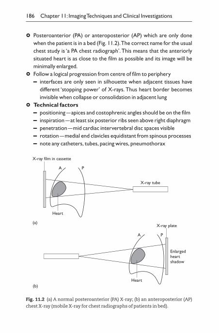
° Posteroanterior (PA) or anteroposterior (AP) which are only donewhen the patient is in a bed (Fig. 11.2).The correct name for the usualchest study is ‘a PA chest radiograph’. This means that the anteriorlysituated heart is as close to the film as possible and its image will beminimally enlarged.
° Follow a logical progression from centre of film to periphery– interfaces are only seen in silhouette when adjacent tissues have
different ‘stopping power’ of X-rays. Thus heart border becomes invisible when collapse or consolidation in adjacent lung
° Technical factors– positioning —apices and costophrenic angles should be on the film– inspiration —at least six posterior ribs seen above right diaphragm– penetration —mid cardiac intervertebral disc spaces visible– rotation —medial end clavicles equidistant from spinous processes– note any catheters, tubes, pacing wires, pneumothorax
186 Chapter 11: Imaging Techniques and Clinical Investigations
A P
X-ray film in cassette
Heart
X-ray tube
(a)
A P
X-ray plate
Heart
Enlargedheartshadow
(b)
Fig. 11.2 (a) A normal posteroanterior (PA) X-ray; (b) an anteroposterior (AP)
chest X-ray (mobile X-ray for chest radiographs of patients in bed).
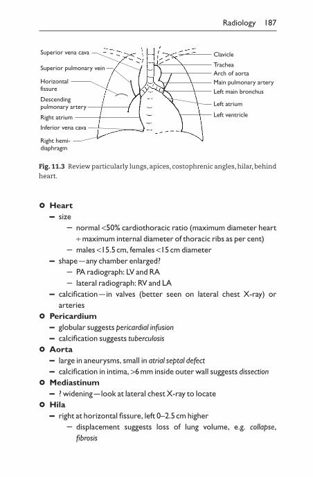
° Heart– size
– normal <50% cardiothoracic ratio (maximum diameter heart∏ maximum internal diameter of thoracic ribs as per cent)
– males <15.5cm, females <15cm diameter– shape —any chamber enlarged?
– PA radiograph: LV and RA– lateral radiograph: RV and LA
– calcification —in valves (better seen on lateral chest X-ray) or arteries
° Pericardium– globular suggests pericardial infusion– calcification suggests tuberculosis
° Aorta– large in aneurysms, small in atrial septal defect– calcification in intima, >6mm inside outer wall suggests dissection
° Mediastinum– ? widening —look at lateral chest X-ray to locate
° Hila– right at horizontal fissure, left 0–2.5cm higher
– displacement suggests loss of lung volume, e.g. collapse,fibrosis
Radiology 187
Clavicle
TracheaArch of aortaMain pulmonary arteryLeft main bronchus
Left atrium
Left ventricle
Superior vena cava
Superior pulmonary vein
Horizontalfissure
Descendingpulmonary artery
Right atrium
Inferior vena cava
Right hemi-diaphragm
Fig. 11.3 Review particularly lungs,apices, costophrenic angles,hilar,behind
heart.
– enlargement– if lobulated —a mass or lymph nodes– ? vascular dilation
– density —? mass projected over hilum
° Pulmonary vessels– large in intracardiac or peripheral shunts —prominent in outer
third (plethora)– large in pulmonary hypertension with small vessels in outer third
(pruning) — shunts, hypoxia, emboli, chronic lung disease– segmental avascularity —pulmonary emboli– small in congenital heart disease, right ventricular/pulmonary artery
atresia
° Lung parenchyma– lungs should be equally transradiant (black)– alveolar shadows —ill-defined or confluent and dense
– air bronchogram —water, pus, blood, tumour around patentbronchi, often seen end on, as a circle, near hila
– nodular shadows, e.g. granuloma, tuberculosis– reticular shadows —fibrotic lung disease
Note uniformity, symmetry, unilateral or bilateral, upper orlower zones.
– masses– define position (ask for lateral chest radiograph), edge, shape,
size– tumour, abscess, embolus, infection
° Pleura– fluid
– homogeneous, opaque shadow, usually with lateral meniscus– if air–fluid interface, empyema or after thoracocentesis
– pneumothorax– peripheral space devoid of markings with edge of lung
visible– look for mediastinal displacement — tension pneumothorax
– masses– lobulated shadows —loculated fluid or tumour
188 Chapter 11: Imaging Techniques and Clinical Investigations
° Skeleton– sclerosis, focal — ?metastases, e.g. breast, prostate, stomach, kidney,
thyroid, lymphoma—myelofibrosis, Paget’s disease
– lytic —?metastases, e.g. lung, colorectal, myeloma– osteopenia (only visible when advanced) —osteoporosis and
osteomalacia cannot be distinguished on radiographs, except Looser’s zones (pseudofracture) in osteomalacia
– look for fractures
° Other areas– hiatus hernia, behind heart– left lower lobe collapse, behind heart– lungs behind dome of diaphragm– gas below diaphragm on erect chest radiograph —perforated viscus,
recent surgery– apices —? lung visible above clavicle
Abdominal radiographyThis is less satisfactory than chest radiography because there are fewercontrasting densities.Air in the gut is helpful, as are the psoas lines.Try tofind as many organ outlines as possible.
– Supine (AP) radiograph —routine.– Erect radiograph
– for air–fluid levels (AFLs)– <5 short AFLs normal– many —obstruction– also in paralytic ileus, coeliac disease, jejunal diverticula
° Visceral organs– liver
Radiology 189
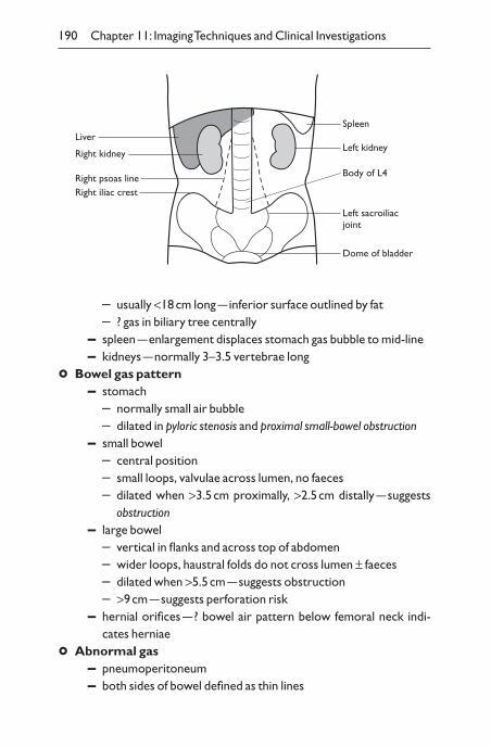
– usually <18cm long —inferior surface outlined by fat– ? gas in biliary tree centrally
– spleen —enlargement displaces stomach gas bubble to mid-line– kidneys —normally 3–3.5 vertebrae long
° Bowel gas pattern– stomach
– normally small air bubble– dilated in pyloric stenosis and proximal small-bowel obstruction
– small bowel– central position– small loops, valvulae across lumen, no faeces– dilated when >3.5cm proximally, >2.5cm distally —suggests
obstruction– large bowel
– vertical in flanks and across top of abdomen– wider loops, haustral folds do not cross lumen ± faeces– dilated when >5.5cm —suggests obstruction– >9cm —suggests perforation risk
– hernial orifices —? bowel air pattern below femoral neck indi-cates herniae
° Abnormal gas– pneumoperitoneum– both sides of bowel defined as thin lines
190 Chapter 11: Imaging Techniques and Clinical Investigations
Spleen
Left kidney
Dome of bladder
Left sacroiliacjoint
Body of L4
Liver
Right kidney
Right psoas lineRight iliac crest
– loss of liver density from gas anteriorly– bowel wall —thin streaks of gas suggest infarction or gas-
producing bacteria
° Abnormal calcification– 30% gallstones are radiopaque —can be anywhere in abdomen– pancreas calcification —follows oblique line of pancreas and sug-
gests chronic pancreatitis– renal stones —usually radiopaque– nephrocalcinosis —medullary sponge kidney or metabolic calcinosis– in phleboliths or foecoliths in diverticulae
° Other soft tissues– psoas lines
– outlined by retroperitoneal fat– absent in 20% of normals– unilateral absence suggests retroperitoneal mass or haematoma
– ascites– uniformly grey appearance– bowel gas ‘floats’ centrally
Computed tomographyA segment of the body is X-rayed at numerous angles as the apparatus rotates through 360°. A computer summarizes the data from multiple pictures to provide a composite picture (Fig. 11.4).Attenuation of X-raysdepends on tissue —water is arbritrary 0, black is -1000 and white is+1000 Hounsfield units. Different ‘windows’ are chosen to display differ-ent characteristics, e.g. soft-tissue window, lung window, bone window.CT can be used:
– for organs and masses in abdomen and thorax– to diagnose tumours, infarcts and bleeds in cerebral hemispheres– for posterior fossa —lesions less easy to visualize because of bony
base of skull– to visualize disc prolapse and neoplasm in spinal cord, but adjacent
bones interfere. Intrathecal contrast medium is often required forcord tumours
Variants of CT:– intravenous contrast
Radiology 191
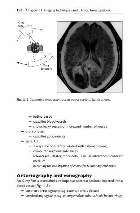
192 Chapter 11: Imaging Techniques and Clinical Investigations
– iodine-based– opacifies blood vessels– shows leaky vessels or increased number of vessels
– oral contrast– opacifies gut contents
– spiral CT– X-ray tube constantly rotated with patient moving– computer segments into slices– advantages —faster, more detail, can use intravenous contrast
medium– becoming the investigation of choice for pulmonary embolism
Arteriography and venographyAn X-ray film is taken after a radiopaque contrast has been injected into ablood vessel (Fig. 11.5):
– coronary arteriography, e.g. coronary artery disease– cerebral angiography, e.g. aneurysm after subarachnoid haemorrhage
X-raytube
X-raydetectors
Fig. 11.4 Computed tomographic scan across cerebral hemispheres.
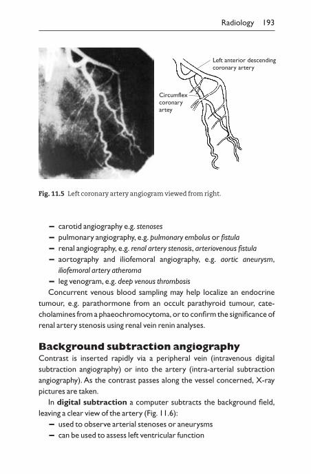
– carotid angiography e.g. stenoses– pulmonary angiography, e.g. pulmonary embolus or fistula– renal angiography, e.g. renal artery stenosis, arteriovenous fistula– aortography and iliofemoral angiography, e.g. aortic aneurysm,
iliofemoral artery atheroma– leg venogram, e.g. deep venous thrombosisConcurrent venous blood sampling may help localize an endocrine
tumour, e.g. parathormone from an occult parathyroid tumour, cate-cholamines from a phaeochromocytoma, or to confirm the significance ofrenal artery stenosis using renal vein renin analyses.
Background subtraction angiographyContrast is inserted rapidly via a peripheral vein (intravenous digital subtraction angiography) or into the artery (intra-arterial subtraction angiography). As the contrast passes along the vessel concerned, X-raypictures are taken.
In digital subtraction a computer subtracts the background field,leaving a clear view of the artery (Fig. 11.6):
– used to observe arterial stenoses or aneurysms– can be used to assess left ventricular function
Radiology 193
Left anterior descendingcoronary artery
Circumflexcoronaryartey
Fig. 11.5 Left coronary artery angiogram viewed from right.
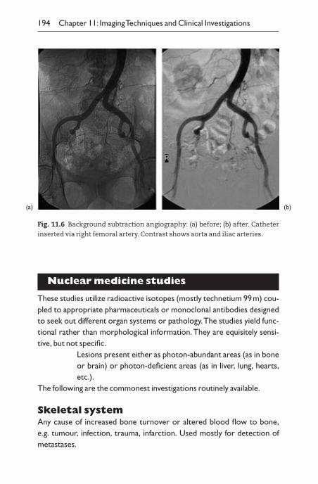
Nuclear medicine studies
These studies utilize radioactive isotopes (mostly technetium 99m) cou-pled to appropriate pharmaceuticals or monoclonal antibodies designedto seek out different organ systems or pathology.The studies yield func-tional rather than morphological information. They are equisitely sensi-tive, but not specific.
Lesions present either as photon-abundant areas (as in boneor brain) or photon-deficient areas (as in liver, lung, hearts,etc.).
The following are the commonest investigations routinely available.
Skeletal systemAny cause of increased bone turnover or altered blood flow to bone,e.g. tumour, infection, trauma, infarction. Used mostly for detection ofmetastases.
194 Chapter 11: Imaging Techniques and Clinical Investigations
Fig. 11.6 Background subtraction angiography: (a) before; (b) after. Catheter
inserted via right femoral artery. Contrast shows aorta and iliac arteries.
(a) (b)

Pulmonary systemThe diagnosis of pulmonary emboli using perfusion scintigraphy, whenemboli cause defects which do not correspond to water densities in thesame position on simultaneous chest radiographs. Usually only indicatedwhen chronic obstructive airways disease is present (see p. 213).
Cardiovascular systemFor the measurement of ventricular function, e.g. ejection fractions, andfor examining myocardial integrity. Ischaemia or scarring causes ‘cold’areas on myocardial scintigrams. Studies are usually carried out at rest andafter exercise (see p. 203).
Urogenital systemRenography (an activity–time curve of the passage of radioactive tracerthrough the kidney) for detecting abnormalities of renal blood flow,parenchymal function and excretion. Renal scintigraphy will detect scar-ring and is used to measure divided renal function. Chromium-51 EDTA(ethylene diamine tetra-acetic acid) clearance measurements yield accu-rate assessment of glomerular filtration rate. Methods are also availablefor detecting testicular torsion.
Cerebral scintigraphyFor the detection of abnormalities associated with certain neuro-psychiatric disorders, notably the dementias, schizophrenia and epilepsy.
Nuclear Medicine Studies 195
Anterior wallleft ventricle
Inferior wallleft ventricle
Fig. 11.7 Thallium 201 study of the heart.
ThyroidFor estimation of the size, shape and position of the gland, detecting thepresence of ‘hot’ thyrotoxic nodules or ‘cold’ nodules caused by adenoma,carcinoma, cysts, haemorrhage or any combination thereof. Iodine up-take can also be estimated simultaneously.
AdrenalsThe detection of autonomously functioning Conn’s tumours (cortex) andphaechromocytoma (medulla).
Reticuloendothelial systemMapping of the bone marrow and lymphatic flow. Occasionally used to visualize the liver and spleen if ultrasound not available.
In addition radiolabelled white cells can be used to search for infectionor inflammation, notably in bone, suspected inflammatory bowel diseaseand after abdominal surgery.
Tracers are also available for detecting certain tumours, notably lymphoma, colonic carcinoma, ovarian carcinoma and malignant mela-noma. Labelled red cells can detect sites of gastrointestinal bleeding.Oesophageal and gastric emptying studies are also available.
Magnetic resonance imaging
Also known as nuclear magnetic resonance (NMR). Provides cross-sectional images (MRI) or spectroscopic information on chemicals in tissues (magnetic resonance spectroscopy, MRS).
A small trolley carries the patient into a super-conducting magnet thatprovides a strong external magnetic field.
196 Chapter 11: Imaging Techniques and Clinical Investigations

The axes of individual hydrogen ions usually lie at random but can belined up at a particular angle by a strong magnetic field (position a).Whensubjected to a second radiofrequency magnetic field the angle is changed(to position b).When the radiowaves cease, position a is restored by thecontinuing magnetic field and a radiowave is emitted and detected.
Hydrogen MRIHydrogen is the most plentiful element in the body. MRI can detect differ-ences between the concentration of hydrogen ions in different tissues,notably fat ( —CH2 —) and water (HOH).
Excellent for the examination of the head and spinal cord:– the brain for demonstrating tumours, multiple areas of demyelina-
tion of white matter in multiple sclerosis (Fig. 11.8), spinal cord lesions, including disc prolapse
– bone and soft-tissue tumoursMRI will show detailed cross-sectional anatomical detail similar to CT
scanning but can also provide coronal and sagittal planes in addition to thestandard axial plane available from CT scanning.
Images can be obtained that accentuate different characteristics:
Magnetic Resonance Imaging 197
Protons
Strong externalmagnetic field
a
Radiowave source
b
a
RF field
b
a
RF signal
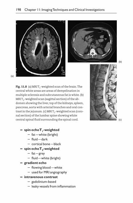
– spin echo T1-weighted– fat —white (bright)– fluid —dark– cortical bone —black
– spin echo T2-weighted– fat —grey– fluid —white (bright)
– gradient echo– flowing blood —white– used for MRI angiography
– intravenous contrast– gadolinium-based– leaky vessels from inflammation
198 Chapter 11: Imaging Techniques and Clinical Investigations
(a)
(b)
(c)
Fig. 11.8 (a) MRI T1-weighted scan of the brain.The
central white areas are areas of demyelination in
multiple sclerosis and subcutaneous fat is white. (b)
MRI T2-weighted scan (sagittal section) of the ab-
domen showing the liver, top of the kidneys, spleen,
pancreas, aorta with arterial branches and oral con-
trast in the jejunum. (c) MRI T2-weighted scan (coro-
nal section) of the lumbar spine showing white
central spinal fluid surrounding the spinal cord.
– increased number of vessels from neoplasm– oral contrast —to label bowel
N.B. Patients with pacemakers should not be subjected to MRI. Patientswith metal implants may not be able to undergo MRI and must be dis-cussed with a radiologist. MRI has an expanding role in many fields of medicine and the indications are likely to increase.
PET scanningPositron emmission tomography (PET) is imaging using 18-F-dioxyglucose (FDG). FDG uptake correlates with glucose metabolism.Malignant tumours actively metabolize glucose making it possible toimage tumours using this technique. PET scanning needs futher evaluationbut is likely to be useful in oncology.
Cardiological investigations
ElectrocardiogramSee Chapter 12, p. 235.
Exercise electrocardiography (stress testing)
– Exercise may reveal cardiac dysfunction not apparent at rest.– Most commonly used in suspected coronary artery disease.
Connected to a 12 lead electrocardiograph (ECG) machine, with resusci-tation equipment available, the patient exercises at an increasing work-load on a treadmill (or bicycle). Bruce protocol: 3-minute stages ofincreasing belt speed and treadmill gradient. Take ECG every minute,blood pressure every 3 minutes.
This assesses:– exercise capacity– haemodynamic response– symptoms– ECG changes
Exercise for as long as possible stopping when there are:– marked symptoms– severe ECG changes
Cardiological Investigations 199
– ventricular arrhythmias– fall in blood pressure
Myocardial ischaemia causes ST segment depression. A high false-positive rate occurs in absence of angina (c. 20%). False-positive incidencedepends on age and sex, with young females having the highest rate, evenin the presence of typical symptoms of angina.
Clinically important abnormalities are:– horizontal or downward sloping ST depression (Fig. 11.9)– deep ST depression– ST changes with typical anginal symptoms
A definitely negative test at a high workload denotes an excellent prognosis.
– Angiography is indicated if only a low workload is achieved before important abnormalities occur
– Medical treatment of angina may be appropriate if three orfour stages are completed.
EchocardiographyThis visualizes structures and function of the heart. Uses ultrasound(2.5–7.5MHz) to reflect from interfaces in the heart, e.g. ventricle andatrial walls, heart, valves, major vessels.The higher frequency gives betterdiscrimination but lower tissue penetration. The time delay betweentransmission and reception indicates depth.
Two-dimensional echocardiography2D echocardiography (Fig. 11.10) uses a scanning beam swept backwardsand forwards across a 45° or 60° arc to construct a picture of the anato-my of the heart.
2D echocardiography is excellent for demonstrating:– valvular anatomy– ventricular function, e.g. poor contraction, low ejection fraction,
akinetic segment, paradoxical motion in aneurysm– structural abnormalities:
– pericardial effusion– ventricular hypertrophy– congenital heart disease
200 Chapter 11: Imaging Techniques and Clinical Investigations
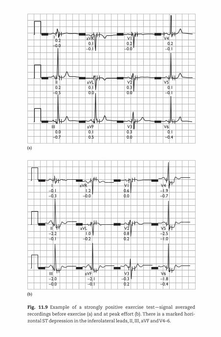
0.2–0.0
I0.1
–0.1
aVR0.2
–0.0
V10.2
–0.1
V4
0.10.0
aVL0.30.0
V20.1
–0.1
V50.2
–0.5
II
0.10.5
aVF0.30.0
V30.1
–0.4
V60.0
–0.7
III
(a)
(b)
–0.1–0.3
I0.60.0
V1–1.9–0.7
V41.2
–0.0
aVR
–2.2–0.1
II0.80.2
V2–2.5–1.0
V51.0
–0.2
aVL
–2.0–0.0
III–0.30.2
V3–1.8–0.4
V6–2.1–0.1
aVF
Fig. 11.9 Example of a strongly positive exercise test —signal averaged
recordings before exercise (a) and at peak effort (b). There is a marked hori-
zontal ST depression in the inferolateral leads, II, III, aVF and V4–6.
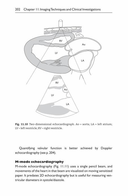
Quantifying valvular function is better achieved by Doppler echocardiography (see p. 204).
M-mode echocardiographyM-mode echocardiography (Fig. 11.11) uses a single pencil beam, andmovements of the heart in that beam are visualized on moving sensitizedpaper. It predates 2D echocardiography but is useful for measuring ven-tricular diameters in systole/diastole.
202 Chapter 11: Imaging Techniques and Clinical Investigations
LV
RVAo
LA
LV
RV
Ao
LA
Fig. 11.10 Two-dimensional echocardiograph. Ao = aorta; LA = left atrium;
LV = left ventricle; RV = right ventricle.
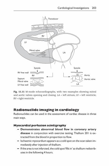
Radionuclide imaging in cardiologyRadionuclides can be used in the assessment of cardiac disease in threemain ways.
Myocardial perfusion scintigraphy– Demonstrates abnormal blood flow in coronary artery
disease in conjunction with exercise testing. Thallium 201 is ex-tracted from the blood in proportion to flow.
– Ischaemic myocardium appears as a cold spot on the scan taken im-mediately after injection of thallium.
– If the area is not infarcted, the cold spot ‘fills in’ as thallium redistrib-utes in the following 4 hours.
Cardiological Investigations 203
LV
RV
LA
Transducer
Mitral valve
Systole
RVRV free wall
SeptumMitral valve
LV free wall
Systole
RV
LA
Aorta
Aortic valve
Fig. 11.11 M-mode echocardiographs, with two examples showing mitral
and aortic valves opening and closing. LA = left atrium; LV = left ventricle;
RV = right ventricle.
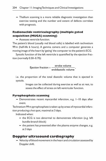
– Thallium scanning is a more reliable diagnostic investigation thanexercise testing and the number and extent of defects correlatewith prognosis.
Radionuclide ventriculography (multiple gatedacquisition (MUGA) scanning)
– Assesses ventricle function.The patient’s blood (usually red blood cells) is labelled with technetium 99m (half-life 6 hours). A gamma camera and a computer generate a moving image of the heart by ‘gating’ the computer to the patient’s ECG.
Systolic function of the left ventricle is quantified by the ejection frac-tion (normally 0.50–0.70):
i.e. the proportion of the total diastolic volume that is ejected in systole.
Images can be collected during exercise as well as at rest, toassess the effect of stress on left ventricular function.
Pyrophosphate scanning– Demonstrates recent myocardial infarction, e.g. 1–10 days after
event.Technetium 99m pyrophosphate is taken up by areas of myocardial infarc-tion producing a hot spot, maximal at 3 days.
Indicated when:– the ECG is too abnormal to demonstrate infarction (e.g. left
bundle-branch block)– the patient has presented after the plasma enzyme changes, e.g.
at 3 days
Doppler ultrasound cardiography– Velocity of blood movement in the heart and circulation assessed by
Doppler shift.
Ejection fractionstroke volume
enddiastolic volume
-
= ,
204 Chapter 11: Imaging Techniques and Clinical Investigations
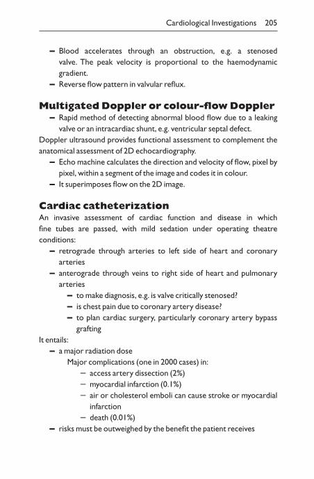
– Blood accelerates through an obstruction, e.g. a stenosed valve. The peak velocity is proportional to the haemodynamic gradient.
– Reverse flow pattern in valvular reflux.
Multigated Doppler or colour-flow Doppler– Rapid method of detecting abnormal blood flow due to a leaking
valve or an intracardiac shunt, e.g. ventricular septal defect.Doppler ultrasound provides functional assessment to complement theanatomical assessment of 2D echocardiography.
– Echo machine calculates the direction and velocity of flow, pixel bypixel, within a segment of the image and codes it in colour.
– It superimposes flow on the 2D image.
Cardiac catheterizationAn invasive assessment of cardiac function and disease in which fine tubes are passed, with mild sedation under operating theatre conditions:
– retrograde through arteries to left side of heart and coronary arteries
– anterograde through veins to right side of heart and pulmonary arteries
– to make diagnosis, e.g. is valve critically stenosed?– is chest pain due to coronary artery disease?– to plan cardiac surgery, particularly coronary artery bypass
graftingIt entails:
– a major radiation doseMajor complications (one in 2000 cases) in:
– access artery dissection (2%)– myocardial infarction (0.1%)– air or cholesterol emboli can cause stroke or myocardial
infarction– death (0.01%)
– risks must be outweighed by the benefit the patient receives
Cardiological Investigations 205
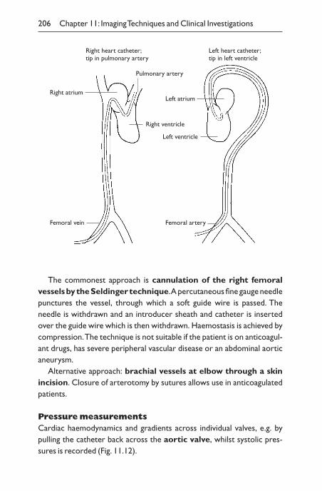
The commonest approach is cannulation of the right femoralvessels by the Seldinger technique.A percutaneous fine gauge needlepunctures the vessel, through which a soft guide wire is passed. The needle is withdrawn and an introducer sheath and catheter is insertedover the guide wire which is then withdrawn. Haemostasis is achieved bycompression.The technique is not suitable if the patient is on anticoagul-ant drugs, has severe peripheral vascular disease or an abdominal aorticaneurysm.
Alternative approach: brachial vessels at elbow through a skin incision. Closure of arterotomy by sutures allows use in anticoagulatedpatients.
Pressure measurementsCardiac haemodynamics and gradients across individual valves, e.g. bypulling the catheter back across the aortic valve, whilst systolic pres-sures is recorded (Fig. 11.12).
206 Chapter 11: Imaging Techniques and Clinical Investigations
Right atrium
Femoral vein
Left atrium
Femoral artery
Right ventricle
Left ventricle
Pulmonary artery
Right heart catheter;tip in pulmonary artery
Left heart catheter;tip in left ventricle
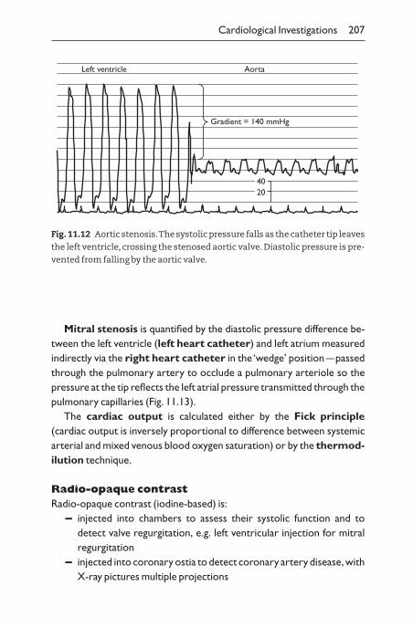
Cardiological Investigations 207
Left ventricle Aorta
Gradient = 140 mmHg
4020
Fig. 11.12 Aortic stenosis.The systolic pressure falls as the catheter tip leaves
the left ventricle, crossing the stenosed aortic valve. Diastolic pressure is pre-
vented from falling by the aortic valve.
Mitral stenosis is quantified by the diastolic pressure difference be-tween the left ventricle (left heart catheter) and left atrium measuredindirectly via the right heart catheter in the ‘wedge’ position —passedthrough the pulmonary artery to occlude a pulmonary arteriole so thepressure at the tip reflects the left atrial pressure transmitted through thepulmonary capillaries (Fig. 11.13).
The cardiac output is calculated either by the Fick principle(cardiac output is inversely proportional to difference between systemicarterial and mixed venous blood oxygen saturation) or by the thermod-ilution technique.
Radio-opaque contrastRadio-opaque contrast (iodine-based) is:
– injected into chambers to assess their systolic function and to detect valve regurgitation, e.g. left ventricular injection for mitralregurgitation
– injected into coronary ostia to detect coronary artery disease, withX-ray pictures multiple projections
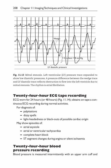
Twenty-four-hour ECG tape recordingECG worn for 24 hours (or 48 hours) (Fig. 11.14); obtains on tape a con-tinuous ECG recording during normal activities.
For diagnosis of:– palpitations– dizzy spells– light-headedness or black-outs of possible cardiac origin
May show episodes of:– atrial asystole– atrial or ventricular tachycardias– complete heart block– ST segment changes during angina or silent ischaemia
Twenty-four-hour blood pressure recordingBlood pressure is measured intermittently with an upper arm cuff and
208 Chapter 11: Imaging Techniques and Clinical Investigations
Fig. 11.13 Mitral stenosis. Left ventricular (LV) pressure trace expanded to
show low diastolic pressures. A pressure difference between the wedge trace
and LV diastolic trace reflects obstruction to flow into the left ventricle due to
mitral stenosis.The rhythm is atrial fibrillation.
WWedgeedgeWedge
LV diastolic pressure
Gradient = 8 mmHgGradient = 8 mmHg

microphone, with recording on a tape. Allows evaluation of blood pres-sure during everyday activities without the ‘white coat’ effect of anxiety atthe doctor’s surgery increasing measured blood pressure. Hypertensionis defined as daytime average >140/>90mmHg. Absence of lower bloodpressure during the night (‘dip’) suggests secondary hypertension.
Respiratory investigations
pH and arterial blood gasesNormal ranges:
– pH 7.35–7.45– PCO2 4.5–6.2pKa
– PO2 > 10.6pKa
– HCO3- 22–26mmol/l
– base excess is the amount of acid required to titrate pH to 7.4In ventilatory failure:
– PO2 low– PCO2 high
In respiratory failure from lung disease often:– PO2 low
Respiratory Investigations 209
Tape recorder
Electrodes
Fig. 11.14 Arrangement for the 24-hour ECG tape recorder.
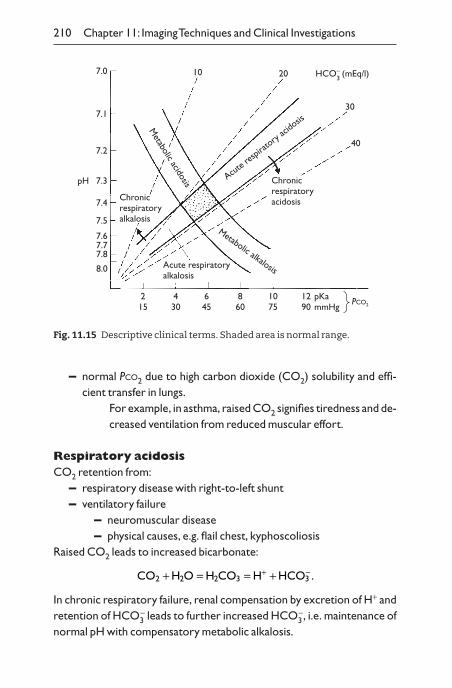
– normal PCO2 due to high carbon dioxide (CO2) solubility and effi-cient transfer in lungs.
For example, in asthma, raised CO2 signifies tiredness and de-creased ventilation from reduced muscular effort.
Respiratory acidosisCO2 retention from:
– respiratory disease with right-to-left shunt– ventilatory failure
– neuromuscular disease– physical causes, e.g. flail chest, kyphoscoliosis
Raised CO2 leads to increased bicarbonate:
In chronic respiratory failure, renal compensation by excretion of H+ andretention of HCO3
- leads to further increased HCO3-, i.e. maintenance of
normal pH with compensatory metabolic alkalosis.
CO H O H CO H HCO2 2 2 3 3+ = = ++ - .
210 Chapter 11: Imaging Techniques and Clinical Investigations
7.0
Chronicrespiratoryalkalosis
7.1
7.2
7.3
7.4
7.5
7.67.77.8
8.0
215
430
645
860
1075
1290
pKammHg
PCO2
10 20
30
40
HCO–3 (mEq/l)
Acute respiratoryalkalosis
Metabolic alkalosis
Chronicrespiratoryacidosis
Metabolic acidosis Acut
e resp
iratory
acido
sis
pH
Fig. 11.15 Descriptive clinical terms. Shaded area is normal range.
Respiratory alkalosisCO2 blown off by hyperventilation due to:
– hysteria– brainstem stimulation (rare)
In respiratory alkalosis:– PO2 normal– PCO2 low
If chronic, compensated by metabolic acidosis with renal retention ofH+ and excretion of HCO3
-.
Metabolic acidosisExcess H+ in blood:
– ketosis —3-OH butyric acid accumulation in diabetes or starvation
– uraemia —lack of renal H+ excretion– renal tubular acidosis —lack of H+ or NH4
- excretion– acid ingestion —aspirin– lactic acid accumulation —shock, hypoxia, exercise, biguanide– formic acid accumulation —methanol intake– loss of base —diarrhoea
Usually compensatory respiratory alkalosis, e.g. Kussmaul respiration ofdiabetic coma (hyerventilation with deep breathing):
– PO2 normal– PCO2 low
– to assist diagnosis, measure anion gap
If anion gap >16mmol/l, unestimated anions are present, e.g.3-OH butyrate, lactate, formate.
Metabolic alkalosisLoss of H+ due to:
– prolonged vomiting– potassium depletion —secondary to renal tubular potassium–
hydrogen exchange
Na K CI HCO mmol I+ + - -[ ] + [ ] - [ ] - [ ] =3 7 16– .
Respiratory Investigations 211
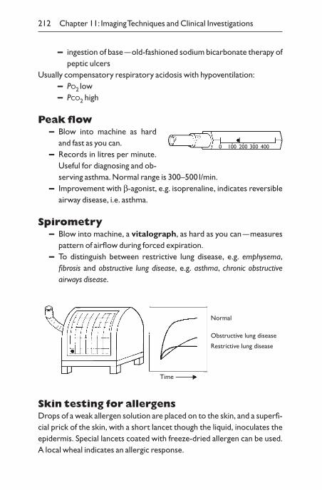
– ingestion of base —old-fashioned sodium bicarbonate therapy ofpeptic ulcers
Usually compensatory respiratory acidosis with hypoventilation:– PO2 low– PCO2 high
Peak flow– Blow into machine as hard
and fast as you can.– Records in litres per minute.
Useful for diagnosing and ob-serving asthma. Normal range is 300–500 l/min.
– Improvement with b-agonist, e.g. isoprenaline, indicates reversibleairway disease, i.e. asthma.
Spirometry– Blow into machine, a vitalograph, as hard as you can —measures
pattern of airflow during forced expiration.– To distinguish between restrictive lung disease, e.g. emphysema,
fibrosis and obstructive lung disease, e.g. asthma, chronic obstructive airways disease.
212 Chapter 11: Imaging Techniques and Clinical Investigations
0 100 200 300 400
Normal
Obstructive lung disease
Restrictive lung disease
Time
Skin testing for allergensDrops of a weak allergen solution are placed on to the skin, and a superfi-cial prick of the skin, with a short lancet though the liquid, inoculates theepidermis. Special lancets coated with freeze-dried allergen can be used.A local wheal indicates an allergic response.
Carbon monoxide transfer factorThe rate of uptake of carbon monoxide from inspired gas determines the lung diffusion capacity. It is reduced in alveolar diseases, e.g. pulmonaryfibrosis.
Ventilation/perfusion scan
Ventilation (V) scan– Inhalation of an isotope allows picture of parenchyma of the lungs to
be taken by a gamma camera.
Perfusion (P) scan– Injection of isotope into the blood stream demonstrates the blood
flow in the lungs.Mismatch of the scans is used to diagnose pulmonary em-bolism, i.e. air reaches all parts of the lung, while the blooddoes not (Fig. 11.16). Matching defects occur with other lungpathologies, e.g. emphysema.
N.B.A perfusion scan showing an area of ischaemia with a nor-mal chest X-ray is generally sufficient to diagnose a pulmonaryembolus. A V/Q scan is needed if there is other lung pathology suspectedor on X-ray (e.g. chronic bronchitis/emphysema), but in practice the results are difficult to interpret.
BronchoscopyFlexible bronchoscopy —under mild sedation, e.g. intravenous diazepam with local anaesthetic spray to pharynx and larynx. Vision by fibreoptics.
– Obstructions can be visualized.– Biopsies can be taken for neoplasms.– Aspiration samples, sometimes after lavage with saline, can be taken
for organisms and malignant cells.Bronchogram —rarely done —a contrast medium is injected into
the bronchial tree to show peripheral dilated bronchi (bronchiectasis).
Respiratory Investigations 213
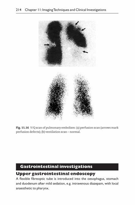
Gastrointestinal investigations
Upper gastrointestinal endoscopyA flexible fibreoptic tube is introduced into the oesophagus, stomach and duodenum after mild sedation, e.g. intravenous diazepam, with localanaesthetic to pharynx.
214 Chapter 11: Imaging Techniques and Clinical Investigations
Fig. 11.16 V/Q scan of pulmonary embolism: (a) perfusion scan (arrows mark
perfusion defects); (b) ventilation scan —normal.
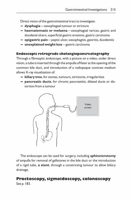
Direct vision of the gastrointestinal tract to investigate:– dysphagia —oesophageal tumour or stricture– haematemasis or melaena —oesophageal varices, gastric and
duodenal ulcers, superficial gastric erosions, gastric carcinoma– epigastric pain —peptic ulcer, oesophagitis, gastritis, duodenitis– unexplained weight loss —gastric carcinoma
Endoscopic retrograde cholangiopancreatographyThrough a fibreoptic endoscope, with a picture on a video, under directvision, a tube is inserted through the ampulla of Vater at the opening of thecommon bile duct, and introduction of a radiopaque contrast medium allows X-ray visualization of:
– biliary tree, for stones, tumours, strictures, irregularities– pancreatic ducts, for chronic pancreatitis, dilated ducts or dis-
tortion from a tumour
Gastrointestinal Investigations 215
Videoscreen
The endoscope can be used for surgery, including sphincterotomyof ampulla for removal of gallstones in the bile duct or the introduction of a rigid tube, a stent, through a constricting tumour to allow biliarydrainage.
Proctoscopy, sigmoidoscopy, colonoscopySee p. 183.
Barium swallow, meal, enemaBarium is drunk (swallow for oesophagus, meal for stomach/duodenum)or introduced rectally (enema) or via a catheter into the duodenum(small-bowel enema).
X-rays are taken with barium coating the mucosa.Air may beintroduced to distend organs and to give double-contrastfilms.
° It outlines physical abnormalities:– strictures, e.g. fibrosis, carcinomata– filling defects, e.g. polyps, carcinomata– craters, e.g. ulcers, diverticula– mucosal irregularities
– mucosal folds radiating from peptic ulcer– clefts in Crohn’s disease of ileum and colon– featureless mucosa of early ulcerative colitis– islands of mucosa in severe ulcerative colitis
An irregularity on a single film needs to be seen on otherviews before an abnormality is confirmed, as peristalsis or gutcontents can mimic defects.
Oral cholecystogramThis procedure is now rarely done as ultrasound is superior.
An initial plain film is taken to show radiopaque gallstones. A ra-diopaque contrast medium is taken by mouth, excreted by the liver andconcentrated in the gallbladder.
– Cholesterol gallstones give filling defects in the gallbladder.– Non-visualization of the gallbladder may occur in some normal sub-
jects, from a stone in the cystic duct or subsequent fibrosis.
Hydrogen breath tests– Lactulose breath test for bacterial overgrowth. Oral
lactulose is given, and excess gut flora in the small bowel or blindloop causes prompt metabolism to provide exhaled hydrogen.
– Lactose breath test for lactase deficiency.– Oral lactose with subnormal exhaled hydrogen.
216 Chapter 11: Imaging Techniques and Clinical Investigations
Renal investigations
Urine testingTesting the urine is part of the routine physical examination. It is most simply done using one of the combination dipsticks.
° Dip the stick in the urine and compare the colours with the key at thetimes specified. Of particular interest are:– pH– protein content (N.B. does not detect Bence Jones protein)– ketones– glucose– bilirubin– urobilinogen– blood/haemoglobin
Urine microscopyUrine should be sent to the laboratory (sterile) for ‘M, C and S’:
– M (microscopy) —for the presence of red cells, white cells, castsand pathogens.
– C (culture) —using appropriate media to detect bacteria and otherpathogens.
– S (sensitivity) —to determine the sensitivity of bacteria to antibiotics.
Creatinine clearancePrecise measurements of the glomerular filtration rate are made isotopically, e.g. chromium EDTA clearance. The creatinine clearance iseasier to organize, although less accurate.
– Collect a blood sample for plasma creatinine.– Collect a 24-hour urine sample for creatinine.
Normal value: 80–120ml/min.
Urine creatininemmol
Plasma creatininemol
Urine volumeml
Duration collectionClearance mI
( )
( )
¥( ) ¥
( )
= ( )
m
103
min
min .
Formula: :U VP T
¥¥
Renal Investigations 217
Intravenous urogramAn initial plain film to show renal or ureteric stones. Contrast medium isinjected intravenously, concentrated in the kidney and excreted.
– Nephrogram phase —kidneys are outlined– observe position, size, shape, filling defects, e.g. tumour
– Excretion phase —renal pelvis– renal papillae may be lost from chronic pyelonephritis, papil-
lary necrosis– calyces blunted from hydronephrosis– pelviureteric obstruction —large pelvis, normal ureters
– Ureters —observe position —displaced by other pathology?– size —dilated from obstruction or recent infection– irregularities —may be contractions and need to be checked in
sequential films
Neurological investigations
ElectroencephalogramApproximately 22 electrodes are applied to the scalp in standard posi-tions and cerebral electrical activity is amplified and recorded.There aremarked normal variations and differences between awake and sleep.
Main uses– Epilepsy
– primary, generalized epilepsy —generalized spike and slow-wave discharges
– partial epilepsy —focal spikes– Disorders of consciousness or coma
– encephalopathy– encephalitis– dementia
The main value of this technique is in showing episodes of abnormalwaves compatible with epilepsy. Large normal variation makes interpreta-tion difficult.
218 Chapter 11: Imaging Techniques and Clinical Investigations
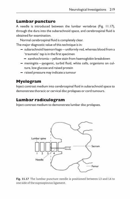
Lumbar punctureA needle is introduced between the lumbar vertebrae (Fig. 11.17),through the dura into the subarachnoid space, and cerebrospinal fluid isobtained for examination.
Normal cerebrospinal fluid is completely clear.The major diagnostic value of this technique is in:
– subarachnoid haemorrhage —uniformly red, whereas blood from a‘traumatic’ tap is in the first specimen– xanthochromia —yellow stain from haemoglobin breakdown
– meningitis —pyogenic, turbid fluid, white cells, organisms on cul-ture, low glucose and raised protein
– raised pressure may indicate a tumour
MyelogramInject contrast medium into cerebrospinal fluid in subarachnoid space todemonstrate thoracic or cervical disc prolapses or cord tumours.
Lumbar radiculogramInject contrast medium to demonstrate lumbar disc prolapses.
Neurological Investigations 219
Fig. 11.17 The lumbar puncture needle is positioned between L3 and L4 to
one side of the supraspinous ligament.
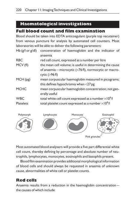
Haematological investigations
Full blood count and film examinationBlood should be taken into EDTA anticoagulant (purple top vacutainer)from venous puncture for analysis by automated cell counters. Mostlaboratories will be able to deliver the following parameters:Hb (g/l or g/dl) concentration of haemoglobin and the indicator of
anaemiaRBC red cell count, expressed as a number per litreMCV (fl) the mean cell volume; is useful in determining the cause
of anaemia —microcytic (<76fl), normocytic or macro-cytic (>96fl)
MCH (pg) mean corpuscular haemoglobin measured in picograms;this defines hypochromia when <27pg
MCHC mean corpuscular haemoglobin concentration; not gen-erally useful
WBC total white cell count expressed as a number ¥109/lPlatelets total platelet count expressed as a number ¥109/l
220 Chapter 11: Imaging Techniques and Clinical Investigations
Polymorph Lymphocyte Monocyte Eosinophil
Pink granules
Most automated blood analysers will provide a five part differential whitecell count, thereby defining by percentage and absolute number of neu-trophils, lymphocytes, monocytes, eosinophils and basophils present.
Blood film examination provides additional morphological informationof blood cells and should always be requested in anaemia of unknowncause, abnormalities of white cell or platelet counts.
Red cellsAnaemia results from a reduction in the haemoglobin concentration —the causes of which include:
– bleeding– haemolysis (premature destruction of red cells) —high reticulocyte
count– bone marrow disease (failure of production)– haematinic deficiency (B12, folate, iron)– renal failure (reduction of erythropoietin)– chronic inflammation and malignancyThe MCV is an indicator of the cause of anaemia:– Microcytic —Fe deficiency, thalassaemia trait.– Macrocytic —B12 or folate deficiency, hypothyroidism, liver dis-
ease, alcohol abuse and bone marrow disease.– Normochromic —chronic disease, renal failure and malignancy.Inspection of the blood film can provide useful information
regarding the aetiology of anaemia. Red cell morphology is important inidentifying causes of haemolysis, e.g. spherocytes, fragmented cells, sicklecells.
Haemoglobin electrophoresis. Electrophoresis of a red cell lysatewill identify haemoglobin variants such as haemoglobin S-HbS.The detec-tion and measurement of HbA2 is very important for the detection of car-riers of thalassaemia. HbA2 >3.5% is suggestive of b-thalassaemia traitcarrier status.
Red cell enzymes. Deficiency of red cell enzymes such as G6PD andPK can lead to a severe haemolytic anaemia. Such enzymes can be assayedin the laboratory.
White cellsAn abnormal white cell count needs attention. Blood film examinationmay identify the presence of abnormal cells such as blasts, or, may simplyshow an elevation or reduction of normal components.The presence ofabnormal white cell morphology may be an indication for a bone marrowbiopsy.
– Neutrophilia —elevated neutrophil count; usually indicative ofbacterial infection.
– Neutropenia —a low neutrophil count can lead to serious infec-tion (gram-negative sepsis) often related to chemotherapy but tem-porarily may follow simple viral infection.
Haematological Investigations 221
– Lymphocytosis —reactive in viral infections such as glandularfever; clonal in lymphoid leukaemias and lymphoma.
– Lymphopenia —common in patients taking steroids, human im-munodeficiency virus (HIV), systemic lupus erythematosus (SLE)and other autoimmune disease.
– Eosinophilia —common in atopy and allergic states. Occurs in as-sociation with drugs, parasitic infection and lymphoma.
CoagulationBlood should be taken into citrate (light blue-topped vacutainer tube).Citrate reversibly binds Ca2+ and prevents the sample from clotting. In thelaboratory the blood is centrifuged and the plasma removed for testing.A source of tissue factor/phospholipid (thromboplastin) is added andCa2+ added.The time to clot in seconds is measured.
° Prothrombin time (PT) (normally 10–14 seconds) is a measure ofthe extrinsic (tissue factor/VII dependent) system. It is very sensitiveto vitamin K-dependent factors (II,VII, IX and X).– The PT is the most sensitive liver function test —prolonged in
liver disease.– The PT is the most sensitive clotting test with which to monitor
warfarin therapy —warfarin inhibits vitamin K-dependent clot-ting factors (II, VII, IX and X). The PT of the patient/PT of poolednormal plasma gives a ratio —the prothrombin ratio. If the PT ratiois multiplied by a correction for the ‘sensitivity’ of the thromboplas-tin used (international sensitivity ratio, ISI) the INR or interna-tional normalized ratio is derived.
Target INR Clinical condition
2.0–3.0 Treatment of deep venous thrombosis (DVT) or pulmonaryembolism (PE), anticoagulation in
3.0–4.5 recurrent DVT or PE, anticoaguation for prosthetic valvesand grafts
° Activated partial thromboplastin time (APTT) —this measures the so-called intrinsic system.This pathway is slower and requires both phos-pholipid and a surface activator (e.g. kaolin —as in the kaolin cephalinthromboplastin time, KCTT). Patients’ plasma from citrated blood is
222 Chapter 11: Imaging Techniques and Clinical Investigations
added to a source of phospholipid, kaolin and Ca2+.The time to clot ismeasured and is usually in the order of 30–40 seconds). The test is used for:– Monitoring heparin when the APTT is usually kept at about 2.5 ¥
normal. N.B. low molecular weight heparin usually does not re-quire monitoring with the exception of renal failure when a factorXa assay is performed.
– This test is prolonged in the presence of the antiphospholipid antibody.
– The test is prolonged in haemophilia and von Willebrand’s disease.
° Other coagulation tests include the thrombin time (TT)which is sensitive to heparin therapy and the fibrinogen level whichis a direct measurement of the fibrinogen concentration of the blood.Disseminated intravascular coagulation usually causes a prolongationof all the above coagulation tests and a reduction in the level of fibrinogen.
° D-dimers —activation of the fibrinolytic system follows the forma-tion of a clot. Plasmin becomes activated and cleaves the polymerizedfibrin into smaller molecules (some of which are called D-dimers). D-dimers can be detected using either a latex agglutination or an ELISA-based test.The detection of D-dimers infers the presence of clot and isnow used in the diagnosis of DVT and PE. Absence of D-dimers impliesabsence of significant thrombosis.
° Thrombophilia tests —a number of components of the blood helpprevent the formation of spontaneous blood clots.These factors workby interupting the coagulation cascade. Deficiencies can make patientssuseptible to thrombosis. Most of these factor deficiencies are inher-ited —taking a family history is very important. Main risk factorsfor thrombosis are:– protein C deficiency– protein S deficiency– antithrombin III deficiency– presence of a lupus anticoagulant (antiphospholipid antibody)– oestrogen therapy —the pill– surgery– malignancy
Haematological Investigations 223
Cross-matching blood for transfusionBefore blood can be safely administered to a patient, the patient’s serummust be screened for red cell antibodies that may cause a transfusion rec-tion should the corresponding antigen be present on the donor red cells.A sample of blood (varies in different laboratories —clot or EDTA) mustbe sent to the transfusion laboratory before blood can be issued. Carefullabelling/identification of all samples is essential. Check with the transfu-sion laboratory as to what samples need to be taken and the system of labelling.The laboratory process involves.
– ABO and RhD blood grouping– serum/plasma from patient is reacted with donor red cells– once the ABO and RhD blood group has been determined and the
absence of red cell antibodies has been confirmed, blood can be issued
Emergency blood for transfusionThere are rare occasions when there is insufficient time to allow forcross-matching. In this situation group O Rh-neg emergency stock can begiven. (Must liase directly with transfusion laboratory.)
With the advent of highly sensitive red cell antibody screening tech-niques, routine cross-matching has been superseded by the electronicissue of ABO Rhesus compatible donor red cells for patients having a re-cent negative antibody screen. (This is not standard practice in all transfu-sion laboratories; refer to local transfusion policy.)
Special requirementsCertain patients have special transfusion requirements —some of theseare listed here:
– irradiated blood product —patients will carry a card– cytomegalovirus (CMV) —negative blood products may be
required in:– patients undergoing organ transplantation– neonates
N.B. all blood issued in the UK is leucocyte depleted.
224 Chapter 11: Imaging Techniques and Clinical Investigations
Bone marrow biopsyThis procedure is usually performed from the iliac crest (most often pos-terior) and is performed in two parts.
– The aspirate is marrow that is sucked out of the marrow cavity andspread on a glass slide, stained and examined under a microscope todetermine cellular morphology. Staining of the marrow with Perl’sPrussian blue stain will give the best indication as to the patient’siron status.
– The trephine involves taking a bone marrow core which is fixed informalin, decalcified and then sectioned in the normal histologicalmanner. The trephine will identify bone marrow infiltration withsecondary carcinoma, fibrosis, haematological malignancies andbest defines the cellularity of the marrow.
The procedure is either carried out with simple infiltration of the periosteum using local anaesthetic or under light sedation.
Bone marrow examination may give the following information:– cellularity —i.e. whether the marrow is empty (aplastic anaemia),
packed (leukaemia) or normal– cytology —whether the cells within the marrow are maturing cor-
rectly and whether there are abnormal forms present– iron status —the ‘gold standard’ against which other measurements
of iron stores are tested
Biochemical tests
° Urea and electrolytes —measurement of sodium, potassium, ureaand creatinine. Urea is useful in assessment of dehydration. It is depen-dant on protein loads —elevated by high protein meals or gastroin-testinal bleeds, reduced by liver dysfunction. Creatinine is the mostreliable test of glomerular function.
° Anion gap —difference in the sum of principal cations (sodium andpotassium) and anions (chloride and bicarbonate) = 14–18mmol/l(represents unmeasured negative charge on plasma proteins). Usefulin investigation of acid–base alterations.
° Liver functions tests —better described as liver profile as thetests do not really reflect liver function.
Biochemical Tests 225
– Albumin —mainly responsible for maintaining colloid osmoticpressure and a useful marker of liver synthetic function. May bedramatically reduced in nephrotic syndrome and protein-losingenteropathy.
– AST and ALT (aspartate transaminase and alanine amino transferase) —these enzymes are released in liver damage, butalso present in red cells, muscle and cardiac cells. May be veryhigh in hepatitis.
– Bilirubin —breakdown product of haemoglobin and thereforeelevated in haemolysis. Also elevated in liver disease.
– ALP (alkaline phosphatase) —an enzyme found in osteoblastsand the hepatobiliary system. Elevated in bone disease and biliaryobstruction.
– GGT (gamma glutamyl-transferase) —increased in alcoholabuse.
– Amylase enzyme produced by the pancreas for digestion ofcomplex carbohydrates. Elevated in pancreatitis.
° Cardiac/muscle markers– AST —intrahepatic enzyme also found in skeletal and cardiac
muscle. Elevated early in myocardial infarction (MI) but not specific.
– LDH —lactate dehydrogenase is found in many tissues. Risesmore slowly in myocardial infarction (MI) and can be useful in retrospective diagnosis of MI.
– CK-MB —Creatine kinase isoenzyme found in cardiac muscle.More specific than AST and LDH but not infallible.
– Troponins (T and I) —most specific and sensitive markers ofmyocardial damage, rising early after myocardial injury.A rise introponin is an indicator or risk in unstable angina and may indi-cate benefit from more aggressive treatment.
° Calcium/bone metabolismMost abundant mineral in the body, though 99% is bound within bone.Plasma levels need adjusting for the albumin concentration before in-terpretation.
Adjusted calcium = (40 - (albumin concentration (g/l)) ¥ 0.02mmol/l
Homeostasis of calcium is affected by parathyroid hormone (PTH) (≠)and vitamin D action.
226 Chapter 11: Imaging Techniques and Clinical Investigations
– Phosphate —most commonly elevated in renal insufficiency.Veryhigh levels are found in tumour lysis. Plasma levels affected by PTHand vitamin D action.
– PTH —released from parathyroid glands in response to a reduc-tion in calcium and results in increased renal tubular absorption ofcalcium and increased phosphate excretion. Also releases calciumand phosphate from bone and leads to renal activation of vitamin D.
– Vitamin D —activated by hydroxylation in liver and kidney. Stimu-lates increased absorption of calcium and phosphate from the gut.Increases osteoblast bone resorption.
° Lipid profile (take samples fasting)– Cholesterol —important membrane structural component. Pre-
cursor of all steroid and bile acid synthesis. Elevated levels correlat-ed with increased risk of cardiovacular disease, especially if LDL iselevated.
– LDL (low density lipoprotein) —principle carrier of choles-terol, attaching to LDL cell surface receptors to allow internaliza-tion. Independent cardiovascular risk factor.
– HDL (high density lipoprotein) —functions to reverse choles-terol transport, carrying cholesterol back to the liver for metabo-lism, therefore, cardioprotective.
– Triglycerides —present in dietary fat and synthesized by liver toprovide store of energy. Independent cardiovascular risk factor. El-evated in liver disease and hypothyroidism.
Endocrinology
Anterior pituitary hormones
° TSH (thryoid stimulating hormone) —stimulates production of thyroixine (T4) and tri-iodothyronine (T3) from the thyroid gland. Diagnosis of hypo- or hyperthyroidism depends on TSH measurement.
° ACTH (adrenocorticotrophic hormone) —increases cortisolproduction from the adrenal glands in response to stress, daily varia-tion, infection, etc.– Cortisol excess is known as Cushing’s syndrome or Cushing’s dis-
ease (pituitary-driven ACTH).
Biochemical Tests 227
– Cortisol deficiency from adrenal failure is known as Addison’s disease.
° GH (growth hormone) —stimulates growth in prepubertal chil-dren and has many diffuse effects on adult metabolism. GH is activatedby insulin-like growth factor 1 (IGF-1) in the liver. An excess of growthhormone in adulthood produces acromegaly.
° Prolactin —milk gland stimulating hormone elevated in pregnancyand lactation.Very high levels in some pituitary adenomata.
° FSH (follicle stimulating hormone) —gonadotrophin which nur-tures the development of the follicle in the first half of the menstraulcycle. In males FSH stimulates spermatogenesis. High levels are foundin post-menopausal women.
° LH (luteinizing hormone) —elevated in the second half of themenstrual cycle producing development of the corpus luteum. Inmales LH stimulates testosterone synthesis.
Posterior pituitary hormone
° ADH (antidireutic hormone) —decreases water loss. Absence ofADH causes diabetes insipidus. Conditions that cause inappropriateADH secretion (various tumours) lead to a fall in plasma sodium. Mea-surement of urine and plasma osmolarity.
Dynamic endocrine tests
° Short Synacthen test —injection of synthetic ACTH at time 0 afterblood sampling. Further blood samples are taken at 30 and 60 minutes.A physiological response results in an elevation of cortisol peak to>570nmol/l. A reduced response is seen in Addison’s disease (adrenalfailure).
° Oral glucose tolerance test (OGTT) —used to diagnose diabetesmellitus by demonstrating abnormal glucose handling.
° Insulin tolerance test —used to demonstrate the normal reactivityof cortisol and growth hormone (both antagonize the action of insulin)to hypoglycaemia induced by insulin. Abnormal response seen in adre-nal failure or growth hormone deficiency.
° Testing for Cushing’s syndrome– Outpatient screening test
228 Chapter 11: Imaging Techniques and Clinical Investigations
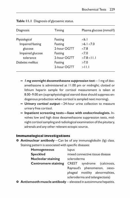
– 1mg overnight dexamethasone suppression test —1mg of dex-amethasone is administered at 11.00 pm or midnight; clotted orlithium heparin sample for cortisol measurement is taken at8.00–9.00 am (supraphysiological steroid dose should suppress en-dogenous production when cortisol is sampled next morning).
– Urinary cortisol output —24-hour urine collection to measureurinary free cortisol.
– Inpatient screening tests —liase with endocrinologists. In-volves low and high dose dexamethasone suppression tests, mid-night cortisol sampling and radiological examination of the pituitary,adrenals and any other relevant ectopic source.
Immunological investigations
° Antinuclear antibody —Can be of any immunoglobulin (Ig) class.Staining pattern is associated with specific diseases:
Homogeneous lupusSpeckled mixed connective tissue diseaseNucleolar staining sclerodermaCentromere staining CREST syndrome (calcinosis,
Raynaud’s phenomenon, oeso-phageal motility abnormalities,scleroderma and telangiectasia)
° Antismooth muscle antibody —elevated in autoimmune hepatitis.
Biochemical Tests 229
Table 11.1 Diagnosis of glycaemic status.
Diagnosis Timing Plasma glucose (mmol/l)
Physiological Fasting <6.1Impaired fasting Fasting >6.1 <7.0
glucose 2-hour OGTT <7.8Impaired glucose Fasting <7.0
tolerance 2-hour OGTT >7.8 <11.1Diabetes mellitus Fasting >7.0
2-hour OGTT >11.1
° Gastric parietal cell —seen in patients with pernicious anaemia(90%).
° Intrinsic factor antibodies —70% of pernicious anaemia patients;more specific.
° Mitochondrial antibody —96% of patients with primary biliary cirrhosis.
° Anti-endomysial and antigliadin antibodies —coeliac disease.
° Thyroid antibody —antithyroglobulin elevated in autoimmune thy-roiditis (90%); antimicrosomal antibody elevations may be seen inGrave’s disease.
° Rheumatoid factor —antibody against human IgG but can be of anyIg class. Positive in 70% of rheumatoid arthritis, particularly extra-articular involvement.Very high levels in cryoglobulinaemia.
° ANCA (antineutrophil cytoplasmic antibodies)– Cytoplasmic ANCA (cANCA) —90% of Wegener’s granulo-
matosis; 40% of patients with microscopic polyangiitis.– Perinuclear ANCA (pANCA) —60% of microscopic polyangi-
itis patients; may also be positive in connective tissue disorders andvasculitic diseases.
Taking blood samples
VenepunctureWear plastic gloves when taking blood.Venous blood is normally takenfrom a vein in the antecubital fossa —it is not necessary to clean the areawith a swab, unless dirt is apparent. DO NOT TAKE BLOOD FROMAN ARM WITH A DRIP.
° Place a tourniquet above the elbow or inflate a cuff to approximately50–80mmHg.
° Ask patient to make a clenched fist repeatedly.
° Inspect for superficial vein. If not seen, palpate with fingertip repeat-edly across antecubital fossa for rebound from a tense vein. Map outcourse of vein.
° Make sure the vessel is not pulsating (i.e. is the brachial artery).
° Decide where you wish to insert needle —a site over the middle of thevein, along line of vein.
230 Chapter 11: Imaging Techniques and Clinical Investigations
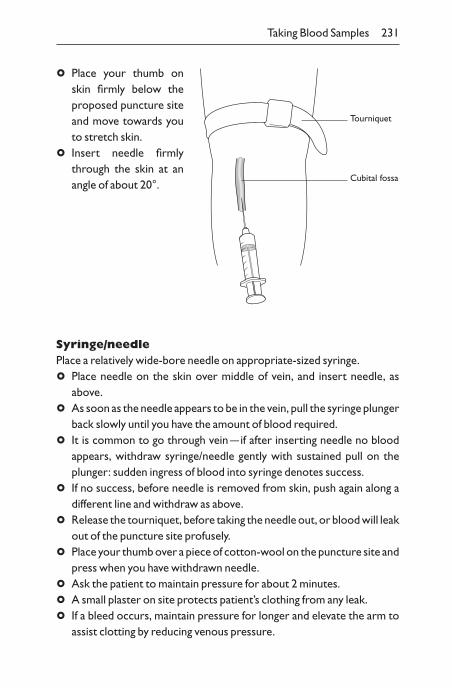
° Place your thumb onskin firmly below theproposed puncture siteand move towards youto stretch skin.
° Insert needle firmlythrough the skin at anangle of about 20°.
Taking Blood Samples 231
Tourniquet
Cubital fossa
Syringe/needlePlace a relatively wide-bore needle on appropriate-sized syringe.
° Place needle on the skin over middle of vein, and insert needle, asabove.
° As soon as the needle appears to be in the vein, pull the syringe plungerback slowly until you have the amount of blood required.
° It is common to go through vein —if after inserting needle no blood appears, withdraw syringe/needle gently with sustained pull on theplunger: sudden ingress of blood into syringe denotes success.
° If no success, before needle is removed from skin, push again along adifferent line and withdraw as above.
° Release the tourniquet, before taking the needle out, or blood will leakout of the puncture site profusely.
° Place your thumb over a piece of cotton-wool on the puncture site andpress when you have withdrawn needle.
° Ask the patient to maintain pressure for about 2 minutes.
° A small plaster on site protects patient’s clothing from any leak.
° If a bleed occurs, maintain pressure for longer and elevate the arm toassist clotting by reducing venous pressure.
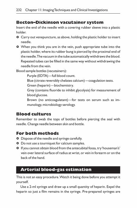
Becton–Dickinson vacutainer systemInsert the end of the needle with a covering rubber sleeve into a plasticholder.
° Carry out venepuncture, as above, holding the plastic holder to insertneedle.
° When you think you are in the vein, push appropriate tube into theplastic holder, where its rubber bung is pierced by the proximal end ofthe needle.The vacuum in the tube automatically withdraws the blood.Repeated tubes can be filled in the same way without withdrawing theneedle from the vein.
Blood sample bottles (vacutainers):Purple (EDTA) —full blood count.Blue (citrate reversibly chelates calcium) —coagulation tests.Green (heparin) —biochemistry.Grey (contains fluoride to inhibit glycolysis) for measurement ofblood glucose.Brown (no anticoagulatant) —for tests on serum such as im-munology, microbiology serology.
Blood culturesRemember to swab the tops of bottles before piercing the seal with needle. Change needle between skin and bottle.
For both methods
° Dispose of the needle and syringe carefully.
° Do not use a tourniquet for calcium samples.
° If you cannot obtain blood from the antecubital fossa, try ‘houseman’s’vein over lateral surface of radius at wrist, or vein in forearm or on theback of the hand.
Arterial blood-gas estimation
This is not an easy procedure.Watch it being done before you attempt ityourself.
Use a 2-ml syringe and draw up a small quantity of heparin. Expel theheparin so just a film remains in the syringe. Pre-prepared syringes are
232 Chapter 11: Imaging Techniques and Clinical Investigations
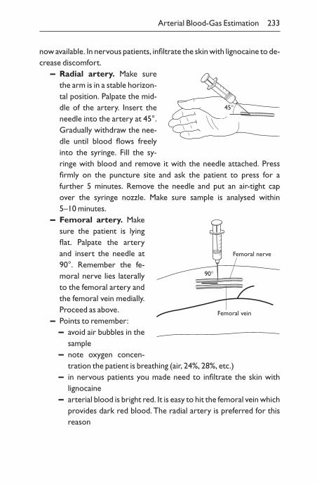
now available. In nervous patients, infiltrate the skin with lignocaine to de-crease discomfort.
– Radial artery. Make surethe arm is in a stable horizon-tal position. Palpate the mid-dle of the artery. Insert theneedle into the artery at 45°.Gradually withdraw the nee-dle until blood flows freelyinto the syringe. Fill the sy-ringe with blood and remove it with the needle attached. Press firmly on the puncture site and ask the patient to press for a further 5 minutes. Remove the needle and put an air-tight cap over the syringe nozzle. Make sure sample is analysed within 5–10 minutes.
– Femoral artery. Makesure the patient is lyingflat. Palpate the arteryand insert the needle at90°. Remember the fe-moral nerve lies laterallyto the femoral artery andthe femoral vein medially.Proceed as above.
– Points to remember:– avoid air bubbles in the
sample– note oxygen concen-
tration the patient is breathing (air, 24%, 28%, etc.)– in nervous patients you made need to infiltrate the skin with
lignocaine– arterial blood is bright red. It is easy to hit the femoral vein which
provides dark red blood. The radial artery is preferred for thisreason
Arterial Blood-Gas Estimation 233
45∞
90∞
Femoral vein
Femoral nerve
Hyper- and hypoglycaemia
Blood glucose
Colorimetric methodsSeveral proprietary glucose-oxidase colour strips are available. Examplesinclude strips for use with meters, or visual use. Make sure the strips havebeen kept in a sealed container.
° Prick finger with a lancet, and squeeze fingertip to produce drop ofblood.
° Put one large drop of blood on to the pad on the strip or meter’s de-tector —a smear will not do.
° Follow instructions for visual reading, maybe to time precisely for 1minute (use second hand of watch or digital watch).– Some methods require you to wipe off blood using cotton wool or
tissue paper; wait 1 more minute before reading strip.
° Use a colorimeter for a more precise reading, particularly in the hypo-glycaemic range.
Amperometric methodsSome machines give a direct current output from a glucose-oxidase strip.
– After applying blood to the strip, the result is shown after about 20seconds.
234 Chapter 11: Imaging Techniques and Clinical Investigations
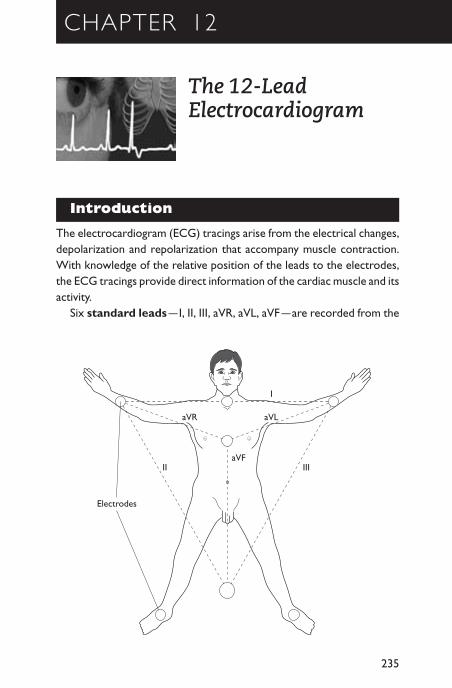
CHAPTER 12
The 12-LeadElectrocardiogram
Introduction
The electrocardiogram (ECG) tracings arise from the electrical changes,depolarization and repolarization that accompany muscle contraction.With knowledge of the relative position of the leads to the electrodes,the ECG tracings provide direct information of the cardiac muscle and itsactivity.
Six standard leads —I, II, III, aVR, aVL, aVF —are recorded from the
235
aVR aVL
aVFIII
I
II
Electrodes
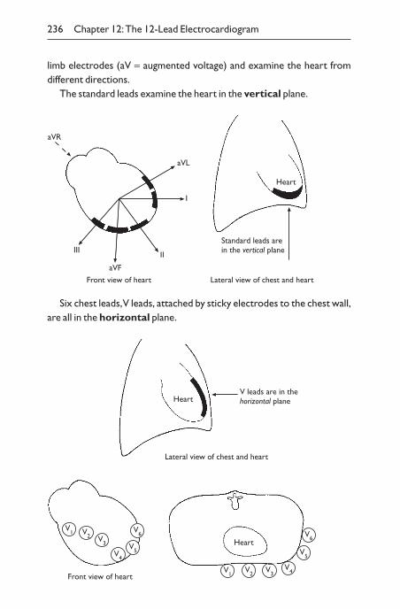
Six chest leads,V leads, attached by sticky electrodes to the chest wall,are all in the horizontal plane.
236 Chapter 12:The 12-Lead Electrocardiogram
limb electrodes (aV = augmented voltage) and examine the heart from different directions.
The standard leads examine the heart in the vertical plane.
Heart
Standard leads arein the vertical plane
Front view of heart Lateral view of chest and heart
aVL
aVF
I
IIIII
aVR
V leads are in thehorizontal planeHeart
Lateral view of chest and heart
Front view of heart
V1 V2 V3
V4
V5
V6
V1V2 V3
V4
V5
V6Heart
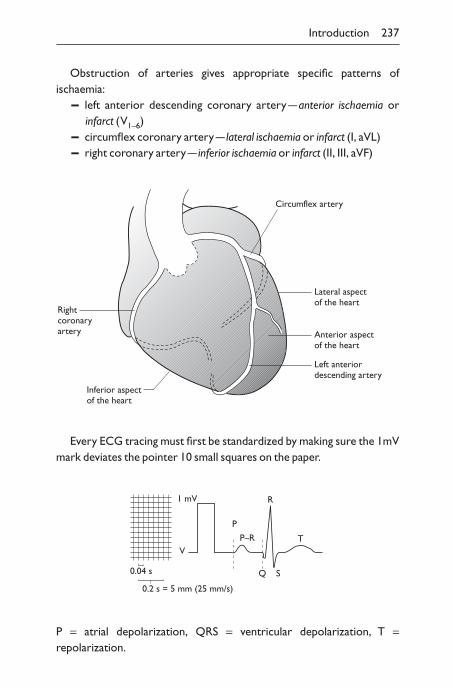
Obstruction of arteries gives appropriate specific patterns of ischaemia:
– left anterior descending coronary artery —anterior ischaemia or infarct (V1–6)
– circumflex coronary artery — lateral ischaemia or infarct (I, aVL)– right coronary artery — inferior ischaemia or infarct (II, III, aVF)
Introduction 237
Rightcoronaryartery
Inferior aspectof the heart
Left anteriordescending artery
Anterior aspectof the heart
Lateral aspectof the heart
Circumflex artery
Every ECG tracing must first be standardized by making sure the 1mVmark deviates the pointer 10 small squares on the paper.
1 mV
V
0.04 s
0.2 s = 5 mm (25 mm/s)
R
P
P–R
Q S
T
P = atrial depolarization, QRS = ventricular depolarization, T =repolarization.
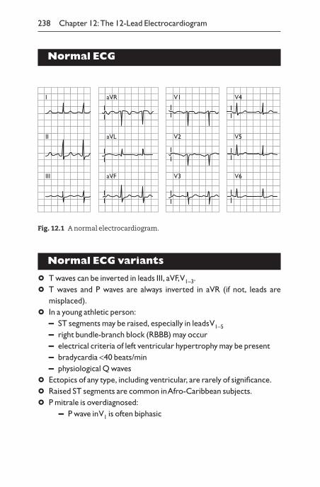
Normal ECG
238 Chapter 12:The 12-Lead Electrocardiogram
I
II
III
aVR
aVL
aVF
V1
V2
V3
V4
V5
V6
Normal ECG variants
° T waves can be inverted in leads III, aVF,V1–3.
° T waves and P waves are always inverted in aVR (if not, leads are misplaced).
° In a young athletic person:– ST segments may be raised, especially in leads V1–5
– right bundle-branch block (RBBB) may occur– electrical criteria of left ventricular hypertrophy may be present– bradycardia <40 beats/min– physiological Q waves
° Ectopics of any type, including ventricular, are rarely of significance.
° Raised ST segments are common in Afro-Caribbean subjects.
° P mitrale is overdiagnosed:– P wave in V1 is often biphasic
Fig. 12.1 A normal electrocardiogram.
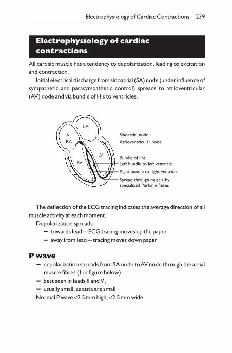
Electrophysiology of cardiaccontractions
All cardiac muscle has a tendency to depolarization, leading to excitationand contraction.
Initial electrical discharge from sinoatrial (SA) node (under influence ofsympathetic and parasympathetic control) spreads to atrioventricular(AV) node and via bundle of His to ventricles.
Electrophysiology of Cardiac Contractions 239
Sinoatrial nodeAtrioventricular node
Bundle of HisLeft bundle to left ventricle
Right bundle to right ventricle
Spread through muscle byspecialized Purkinje fibres
LA
RA
RV
LV
The deflection of the ECG tracing indicates the average direction of allmuscle activity at each moment.
Depolarization spreads:– towards lead —ECG tracing moves up the paper– away from lead —tracing moves down paper
P wave– depolarization spreads from SA node to AV node through the atrial
muscle fibres (1 in figure below)– best seen in leads II and V1
– usually small, as atria are smallNormal P wave <2.5mm high, <2.5mm wide
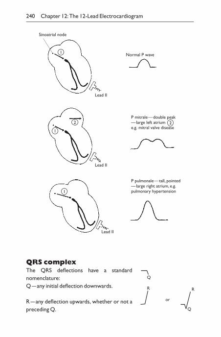
QRS complexThe QRS deflections have a standard nomenclature:Q —any initial deflection downwards.
R —any deflection upwards, whether or not apreceding Q.
240 Chapter 12:The 12-Lead Electrocardiogram
Sinoatrial node
Normal P wave
P mitrale—double peak—large left atriume.g. mitral valve disease
P pulmonale—tall, pointed—large right atrium, e.g.pulmonary hypertension
Lead II
Lead II
Lead II
1
1
2
1
2
Q
R
or
Q
R
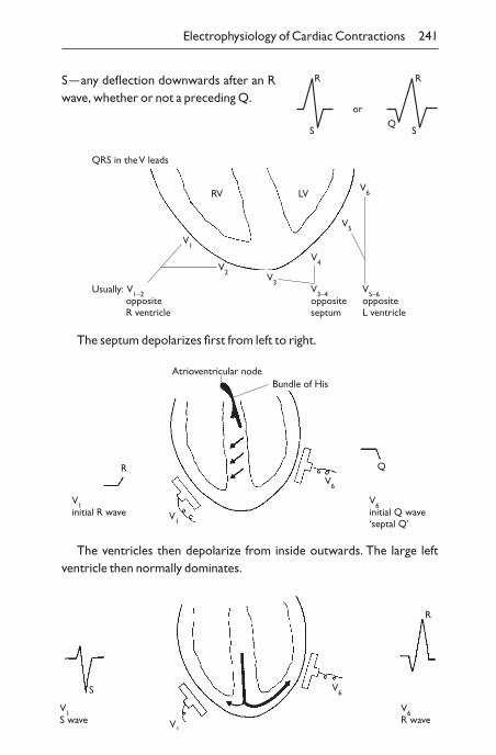
Electrophysiology of Cardiac Contractions 241
RV LV
V1
V2V3
V4
V5
V6
V5–6oppositeL ventricle
V3–4oppositeseptum
Usually: V1–2 opposite R ventricle
QRS in the V leads
The septum depolarizes first from left to right.
V1
V6
Atrioventricular node
V1initial R wave
R
Bundle of His
Q
V6initial Q wave‘septal Q’
The ventricles then depolarize from inside outwards. The large leftventricle then normally dominates.
S
V1
V1S wave
V6
V6R wave
R
S —any deflection downwards after an Rwave, whether or not a preceding Q.
orQ
R
S
R
S
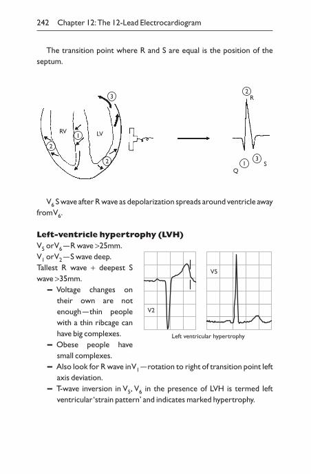
The transition point where R and S are equal is the position of the septum.
242 Chapter 12:The 12-Lead Electrocardiogram
12
2
1
3
3
2R
LVRV
QS
V6 S wave after R wave as depolarization spreads around ventricle awayfrom V6.
Left-ventricle hypertrophy (LVH)V5 or V6 —R wave >25mm.V1 or V2 —S wave deep.Tallest R wave + deepest Swave >35mm.
– Voltage changes ontheir own are notenough —thin peoplewith a thin ribcage canhave big complexes.
– Obese people havesmall complexes.
– Also look for R wave in V1 —rotation to right of transition point leftaxis deviation.
– T-wave inversion in V5, V6 in the presence of LVH is termed left ventricular ‘strain pattern’ and indicates marked hypertrophy.
V2
V5
Left ventricular hypertrophy
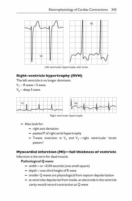
Right-ventricle hypertrophy (RVH)The left ventricle is no longer dominant.V1 —R wave > S wave.V6 —deep S wave.
Electrophysiology of Cardiac Contractions 243
V2
V5
Left ventricular hypertrophy with strain
Right ventricular hypertrophy
V1 V6
– Also look for:– right axis deviation– peaked P of right atrial hypertrophy– T-wave inversion in V2 and V3 —right ventricular ‘strain
pattern’
Myocardial infarction (MI)—full thickness of ventricleInfarction is the term for dead muscle.
Pathological Q wave:– width = or >0.04 seconds (one small square)– depth > one-third height of R wave– smaller Q waves are physiological from septum depolarization– as ventricles depolarize from inside, an electrode in the ventricle
cavity would record contraction as Q wave
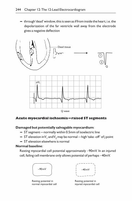
– through ‘dead’ window, this is seen as if from inside the heart, i.e. thedepolarization of the far ventricle wall away from the electrodegives a negative deflection
244 Chapter 12:The 12-Lead Electrocardiogram
2
1
2
1
Q
Dead tissue
Q wave
aVL
Acute myocardial ischaemia —raised ST segments
Damaged but potentially salvagable myocardium:– ST segment —normally within 0.5mm of isoelectric line– ST elevation in V1 and V2 may be normal —high ‘take -off ’ of j point– ST elevation elsewhere is normal
Normal baseline:Resting myocardial cell potential approximately -90mV. In an injuredcell, failing cell membrane only allows potential of perhaps -40mV.
–90 mV –40 mV
Resting potential innormal myocardial cell
Resting potential ininjured myocardial cell
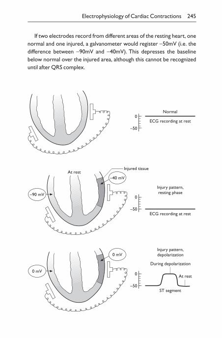
If two electrodes record from different areas of the resting heart, onenormal and one injured, a galvanometer would register -50mV (i.e. thedifference between -90mV and -40mV). This depresses the baselinebelow normal over the injured area, although this cannot be recognizeduntil after QRS complex.
Electrophysiology of Cardiac Contractions 245
0
–50
Normal
ECG recording at rest
0
–50
Injury pattern,resting phase
ECG recording at rest
Injured tissueAt rest
–90 mV
–40 mV
0 mV
0 mV
0
–50
Injury pattern,depolarization
ST segment
During depolarization
At rest
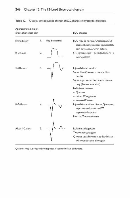
246 Chapter 12:The 12-Lead Electrocardiogram
Table 12.1 Classical time sequence of onset of ECG changes in myocardial infarction.
Approximate time of
onset after chest pain ECG changes
Immediately ECG may be normal. Occasionally ST
segment changes occur immediately
pain develops, or even before
0–2 hours 2. ST segments rise—occluded artery Æinjury pattem
3–8 hours 3. Injured tissue remains
Some dies (Q waves = myocardium
death)
Some improves to become ischaemic
only (T-wave inversion)
Full infarct pattern:
– Q waves
– raised ST segments
– inverted T waves
8–24 hours 4. Injured tissue either dies Æ Q wave or
improves and abnormal ST
segments disappear
Inverted T waves remain
After 1–2 days 5. Ischaemia disappears
T waves upright again
Q waves usually remain, as dead tissue
will not not come alive again
Q waves may subsequently disappear if scarred tissue contracts.
1. May be normal

Raised ST segment:– acute ischaemic injury of ventricle– pericarditis– normal athletes– normal West Indians
Anterior infarction (Figs 12.2 and 12.3)– changes in leads V1–6
– occlusion of left anterior descending coronary artery
Inferior infarction (Fig. 12.4)– changes in leads II, III, aVF
– occlusion of right coronary artery
Electrophysiology of Cardiac Contractions 247
V1
V2
V3
V4
V5
V6
Fig. 12.2 Acute anterior infarct: ST ≠ V2–6 at 3–8hours.
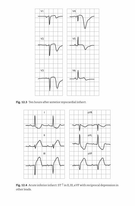
V1
V3
V2
V4
V6
V5
Fig. 12.3 Ten hours after anterior myocardial infarct.
I
II
III
aVR
aVL
aVF
Fig. 12.4 Acute inferior infarct: ST ≠ in II, III,a VF with reciprocal depression in
other leads.
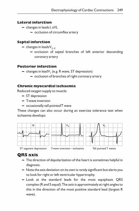
Lateral infarction– changes in leads I, aVL
– occlusion of circumflex artery
Septal infarction– changes in leads V2–3
– occlusion of septal branches of left anterior descending coronary artery
Posterior infarction– changes in lead V1 (e.g. R wave, ST depression)
– occlusion of branches of right coronary artery
Chronic myocardial ischaemiaReduced oxygen supply to muscle:
– ST depression– T-wave inversion– occasionally tall pointed T wave
These changes can also occur during an exercise tolerance test when ischaemia develops:
Electrophysiology of Cardiac Contractions 249
ST segment depression
III
T-wave inversion—ischaemic
V6
Tall pointed T waves
V2
QRS axis– The direction of depolarization of the heart is sometimes helpful in
diagnosis.– Note the axis deviation on its own is rarely significant but alerts you
to look for right or left ventricular hypertrophy.– Look at the standard leads for the most equiphasic QRS
complex (R and S equal).The axis is approximately at right angles tothis in the direction of the most positive standard lead (largest Rwave).
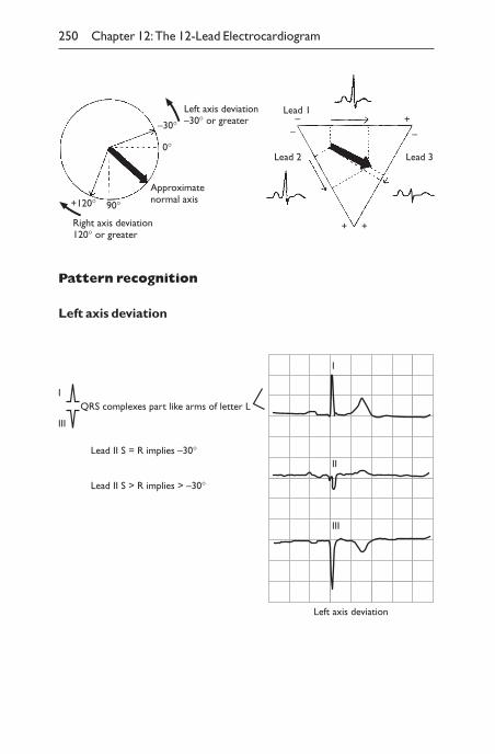
Pattern recognition
Left axis deviation
250 Chapter 12:The 12-Lead Electrocardiogram
–30°
0°
90°+120°
Right axis deviation120° or greater
Approximatenormal axis
Left axis deviation–30° or greater
Lead 1
––
Lead 2
+ +
+–
Lead 3
Left axis deviation
I
II
III
QRS complexes part like arms of letter L
Lead II S = R implies –30°
Lead II S > R implies > –30°
I
III
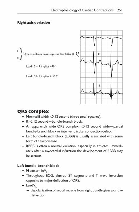
Right axis deviation
Electrophysiology of Cardiac Contractions 251
I
II
III
QRS complexes point together like letter R
Lead I S = R implies +90°
Lead I S > R implies > +90°
I
III
QRS complex– Normal if width <0.12 second (three small squares).– If >0.12 second —bundle-branch block.– An apparently wide QRS complex, <0.12 second wide —partial
bundle-branch block or interventricular conduction defect.– Left bundle-branch block (LBBB) is usually associated with some
form of heart disease.– RBBB is often a normal variation, especially in athletes. Immedi-
ately after a myocardial infarction the development of RBBB may be serious.
Left bundle-branch block– M pattern in V6.– Throughout ECG, slurred ST segment and T wave inversion
opposite to major deflection of QRS.– Lead V6
– depolarization of septal muscle from right bundle gives positivedeflection
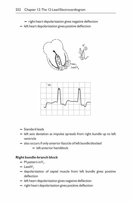
– right heart depolarization gives negative deflection– left heart depolarization gives positive deflection
252 Chapter 12:The 12-Lead Electrocardiogram
1
32
2
13
Lead V6
R1
R
V6
– Standard leads– left axis deviation as impulse spreads from right bundle up to left
ventricle– also occurs if only anterior fascicle of left bundle blocked
– left anterior hemiblock
Right bundle-branch block– M pattern in V1.– Lead V1
– depolarization of septal muscle from left bundle gives positive deflection
– left heart depolarization gives negative deflection– right heart depolarization gives positive deflection
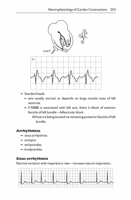
– Standard leads– axis usually normal, as depends on large muscle mass of left
ventricle– if RBBB is associated with left axis, there is block of anterior
fascicle of left bundle —bifascicular blockAll heart is being excited via remaining posterior fascicle of left
bundle.
Arrhythmias– sinus arrhythmia– ectopics– tachycardias– bradycardias
Sinus arrhythmiaNormal variation with respiratory rate —increase rate on inspiration.
Electrophysiology of Cardiac Contractions 253
1
3
2 2
1
3
Lead V1
R1R
V1
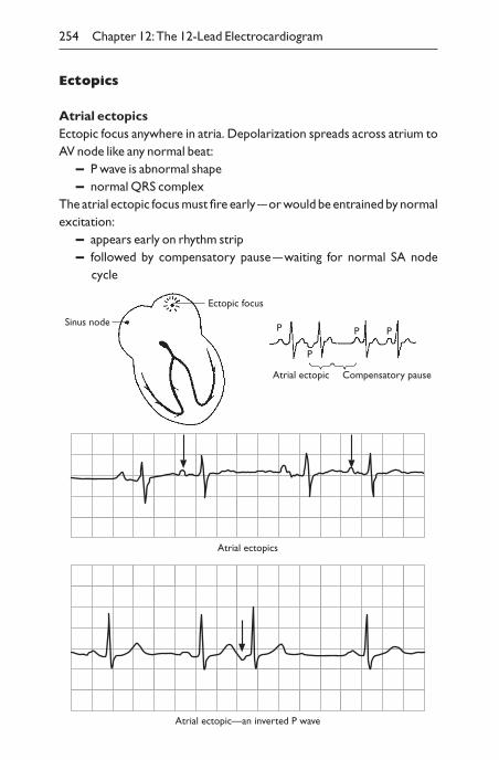
Ectopics
Atrial ectopicsEctopic focus anywhere in atria. Depolarization spreads across atrium toAV node like any normal beat:
– P wave is abnormal shape– normal QRS complex
The atrial ectopic focus must fire early —or would be entrained by normalexcitation:
– appears early on rhythm strip– followed by compensatory pause —waiting for normal SA node
cycle
254 Chapter 12:The 12-Lead Electrocardiogram
P
P
P P
Compensatory pauseAtrial ectopic
Ectopic focus
Sinus node
Atrial ectopic—an inverted P wave
Atrial ectopics
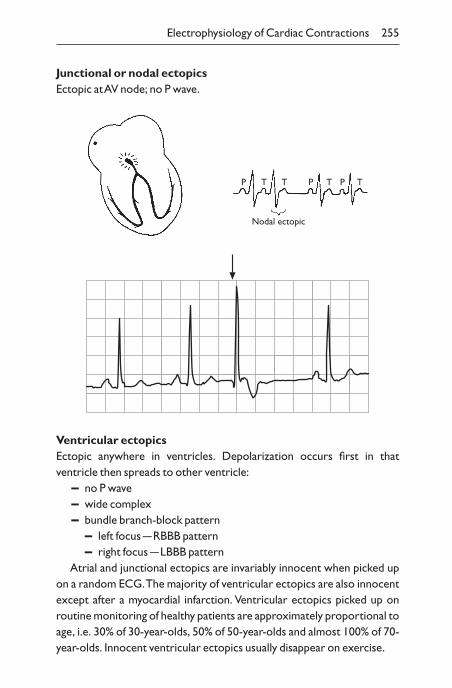
Junctional or nodal ectopicsEctopic at AV node; no P wave.
Electrophysiology of Cardiac Contractions 255
P
Nodal ectopic
T T P T TP
Ventricular ectopicsEctopic anywhere in ventricles. Depolarization occurs first in that ventricle then spreads to other ventricle:
– no P wave– wide complex– bundle branch-block pattern
– left focus —RBBB pattern– right focus —LBBB pattern
Atrial and junctional ectopics are invariably innocent when picked upon a random ECG.The majority of ventricular ectopics are also innocentexcept after a myocardial infarction. Ventricular ectopics picked up onroutine monitoring of healthy patients are approximately proportional toage, i.e. 30% of 30-year-olds, 50% of 50-year-olds and almost 100% of 70-year-olds. Innocent ventricular ectopics usually disappear on exercise.
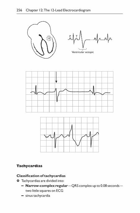
Tachycardias
Classification of tachycardias
° Tachycardias are divided into:– Narrow-complex regular —QRS complex up to 0.08 seconds —
two little squares on ECG– sinus tachycardia
256 Chapter 12:The 12-Lead Electrocardiogram
P
Ventricular ectopic
T
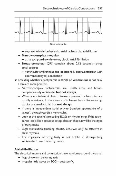
– supraventricular tachycardia, atrial tachycardia, atrial flutter– Narrow-complex irregular
– atrial tachycardia with varying block, atrial fibrillation– Broad-complex —QRS complex about 0.12 seconds —three
small squares– ventricular arrhythmias and occasionally supraventricular with
aberrant (delayed) conduction
° Deciding whether a tachycardia is atrial or ventricular is not easy.Here are some pointers.– Narrow-complex tachycardias are usually atrial and broad-
complex usually ventricular, but not always.– When acute ischaemic heart disease is present, tachycardias are
usually ventricular. In the absence of ischaemic heart disease tachy-cardias are usually atrial, but not always.
– If there is independent atrial activity (random appearance of p values), the tachycardia is ventricular.
– Look at the patient’s preceding ECGs or rhythm strip. If the tachy-cardia looks like a previous ectopic beat in shape, it will be that typeof tachycardia.
– Vagal stimulation (rubbing carotid, etc.) will only be effective in atrial rhythms.
– The regularity or irregularity is not helpful in distinguishing ventricular from atrial arrhythmias.
Atrial fibrillationThe electrical impulse and contraction travel randomly around the atria:
– ‘bag-of-worms’ quivering atria– irregular little waves on ECG —best seen V1
Electrophysiology of Cardiac Contractions 257
Sinus tachycardia
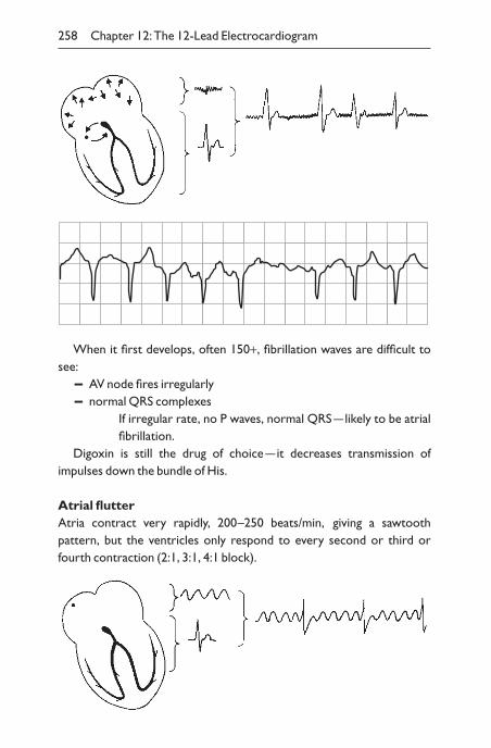
When it first develops, often 150+, fibrillation waves are difficult tosee:
– AV node fires irregularly– normal QRS complexes
If irregular rate, no P waves, normal QRS —likely to be atrialfibrillation.
Digoxin is still the drug of choice —it decreases transmission of impulses down the bundle of His.
Atrial flutterAtria contract very rapidly, 200–250 beats/min, giving a sawtooth pattern, but the ventricles only respond to every second or third orfourth contraction (2:1, 3:1, 4:1 block).
258 Chapter 12:The 12-Lead Electrocardiogram
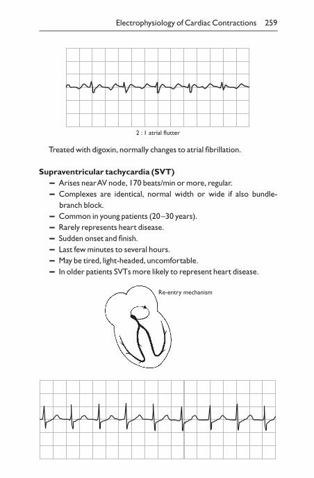
Treated with digoxin, normally changes to atrial fibrillation.
Supraventricular tachycardia (SVT)– Arises near AV node, 170 beats/min or more, regular.– Complexes are identical, normal width or wide if also bundle-
branch block.– Common in young patients (20–30 years).– Rarely represents heart disease.– Sudden onset and finish.– Last few minutes to several hours.– May be tired, light-headed, uncomfortable.– In older patients SVTs more likely to represent heart disease.
Electrophysiology of Cardiac Contractions 259
2 : 1 atrial flutter
Re-entry mechanism
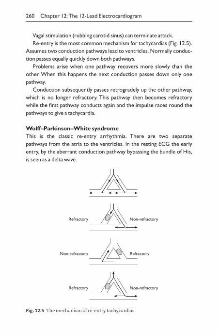
Vagal stimulation (rubbing carotid sinus) can terminate attack.Re-entry is the most common mechanism for tachycardias (Fig. 12.5).
Assumes two conduction pathways lead to ventricles. Normally conduc-tion passes equally quickly down both pathways.
Problems arise when one pathway recovers more slowly than theother. When this happens the next conduction passes down only onepathway.
Conduction subsequently passes retrogradely up the other pathway,which is no longer refractory. This pathway then becomes refractorywhile the first pathway conducts again and the impulse races round thepathways to give a tachycardia.
Wolff–Parkinson–White syndromeThis is the classic re-entry arrhythmia. There are two separate pathways from the atria to the ventricles. In the resting ECG the earlyentry, by the aberrant conduction pathway bypassing the bundle of His,is seen as a delta wave.
260 Chapter 12:The 12-Lead Electrocardiogram
Non-refractoryRefractory
RefractoryNon-refractory
Non-refractoryRefractory
Fig. 12.5 The mechanism of re-entry tachycardias.
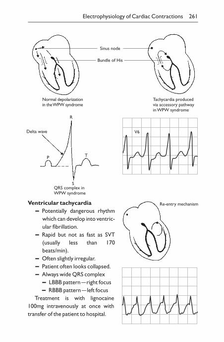
Electrophysiology of Cardiac Contractions 261
Sinus node
Bundle of His
Normal depolarizationin the WPW syndrome
Tachycardia producedvia accessory pathwayin WPW syndrome
Delta wave
R
P
SQRS complex inWPW syndrome
T
V6
Ventricular tachycardia– Potentially dangerous rhythm
which can develop into ventric-ular fibrillation.
– Rapid but not as fast as SVT(usually less than 170beats/min).
– Often slightly irregular.– Patient often looks collapsed.– Always wide QRS complex
– LBBB pattern —right focus– RBBB pattern —left focus
Treatment is with lignocaine100mg intravenously at once withtransfer of the patient to hospital.
Re-entry mechanism
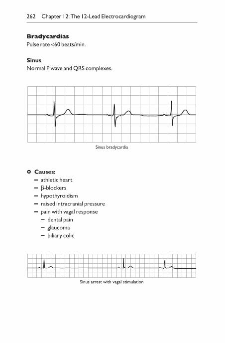
BradycardiasPulse rate <60 beats/min.
SinusNormal P wave and QRS complexes.
262 Chapter 12:The 12-Lead Electrocardiogram
Sinus bradycardia
° Causes:– athletic heart– b-blockers– hypothyroidism– raised intracranial pressure– pain with vagal response
– dental pain– glaucoma– biliary colic
Sinus arrest with vagal stimulation
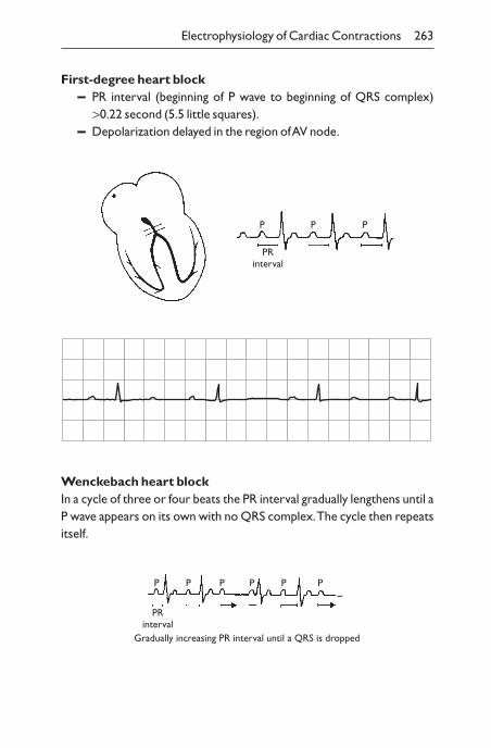
First-degree heart block– PR interval (beginning of P wave to beginning of QRS complex)
>0.22 second (5.5 little squares).– Depolarization delayed in the region of AV node.
Electrophysiology of Cardiac Contractions 263
PRinterval
P P P
Gradually increasing PR interval until a QRS is dropped
PRinterval
P P P P P P
Wenckebach heart blockIn a cycle of three or four beats the PR interval gradually lengthens until aP wave appears on its own with no QRS complex.The cycle then repeatsitself.
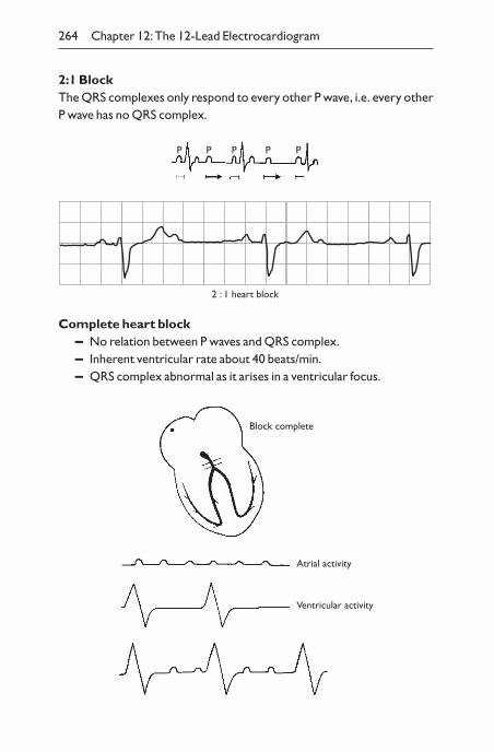
2:1 BlockThe QRS complexes only respond to every other P wave, i.e. every otherP wave has no QRS complex.
264 Chapter 12:The 12-Lead Electrocardiogram
2 : 1 heart block
Complete heart block– No relation between P waves and QRS complex.– Inherent ventricular rate about 40 beats/min.– QRS complex abnormal as it arises in a ventricular focus.
P P P P P
Block complete
Atrial activity
Ventricular activity
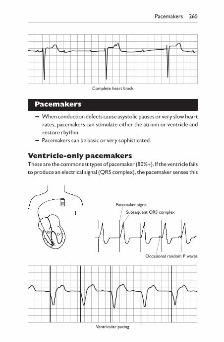
Pacemakers
– When conduction defects cause asystolic pauses or very slow heartrates, pacemakers can stimulate either the atrium or ventricle andrestore rhythm.
– Pacemakers can be basic or very sophisticated.
Ventricle-only pacemakersThese are the commonest types of pacemaker (80%+). If the ventricle failsto produce an electrical signal (QRS complex), the pacemaker senses this
Pacemakers 265
Complete heart block
Pacemaker signal
Subsequent QRS complex
Occasional random P waves
RV LV
Ventricular pacing
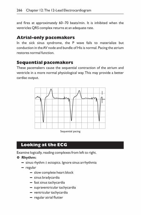
and fires at approximately 60–70 beats/min. It is inhibited when the ventricles QRS complex returns at an adequate rate.
Atrial-only pacemakersIn the sick sinus syndrome, the P wave fails to materialize but conduction in the AV node and bundle of His is normal. Pacing the atriumrestores normal function.
Sequential pacemakersThese pacemakers cause the sequential contraction of the atrium andventricle in a more normal physiological way. This may provide a bettercardiac output.
266 Chapter 12:The 12-Lead Electrocardiogram
Sequential pacing
Looking at the ECG
Examine logically, reading complexes from left to right.
° Rhythm:– sinus rhythm ± ectopics. Ignore sinus arrhythmia– regular
– slow complete heart block– sinus bradycardia– fast sinus tachycardia– supraventricular tachycardia– ventricular tachycardia– regular atrial flutter
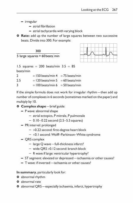
– irregular– atrial fibrillation– atrial tachycardia with varying block
° Rate: add up the number of large squares between two successivebeats. Divide into 300. For example:
1.5 squares = 200 beats/min 3.5 = 85beats/min2 = 150 beats/min 4 = 75 beats/min2.5 = 120 beats/min 5 = 60 beats/min3 = 100 beats/min 6 = 50 beats/min
If the simple formula does not work for irregular rhythm —then add upnumber of complexes in 6 seconds (sometimes marked on the paper) andmultiply by 10.
° Complex shape —brief guide:– P wave: abnormal shape
– atrial ectopics, P mitrale, P pulmonale– 0.10–0.22 second (2.5–5.5 squares)
– PR interval: prolonged– >0.22 second: first-degree heart block– <0.1 second: Wolff–Parkinson–White syndrome
– QRS complex– large Q wave —full-thickness infarct?– wide QRS >0.12 second: branch block– R wave if large: ventricular hypertrophy?
– ST segment: elevated or depressed —ischaemia or other causes?– T wave: if inverted —ischaemia or other causes?
In summary, particularly look for:
° abnormal rhythm
° abnormal rate
° abnormal QRS —especially ischaemia, infarct, hypertrophy
3005 60 =large squares beats min
Looking at the ECG 267
CHAPTER 13
Interpretation ofInvestigations
Sensitivity, specificity and efficiency
These terms have specificmeanings which indicate theclinical usefulness of investiga-tions. Sensitivity and specificityassess the frequency of resultsin relation to the correct answers.
° Sensitivity —how oftenthe correct positive answer is obtained in those who have the disease:
268
truepositive
falsepositive
falsenegative
truenegative
+ –Correct diagnosis
Testresult
+
–
It also expresses the likelihood that a negative test result correctly indi-cates disease is not present: 95% sensitivity means five false-negatives in100 patients with the disease.
° Specificity —how often the correct negative answer is obtained inthose who do not have the disease:
=+
True PositiveTrue positive false negative
i e-
- - , . .
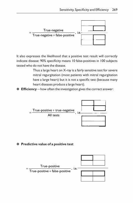
It also expresses the likelihood that a positive test result will correctly indicate disease: 90% specificity means 10 false-positives in 100 subjectstested who do not have the disease.
Thus a large heart on X-ray is a fairly sensitive test for severemitral regurgitation (most patients with mitral regurgitationhave a large heart) but it is not a specific test (because manyheart diseases produce a large heart).
° Efficiency —how often the investigation gives the correct answer:
Sensitivity, Specificity and Efficiency 269
=+
True negativeTrue negative false positive
i e-
- - , . .
=+True positive true negative
All testsi e
- -
, . .
° Predictive value of a positive test
=+
True positiveTrue positive false positive
i e-
- - , . .
Interpretation
The reliance put on the result of an investigation depends on the a priorichance of the result being abnormal. Thus a high plasma calcium in awoman with breast cancer would be taken to indicate either bone metas-tases or the non-metatastic hypercalcaemia (due to tumour production ofa parathormone-like peptide), whereas a similar value in an apparentlynormal medical student would be regarded as being a false-positive untilrechecked.Where the prior probability of an event is known, Bayes theo-rem can be used to calcuate the current probability.The prevalence of anabnormality in the population therefore assists interpretation of an indi-vidual patient’s results.
Prevalence and incidence
Please note the difference between prevalence and incidence.
° Prevalence —the number of cases of a disease in a designated popu-lation, e.g. 10% of males aged 40–60 years.
° Incidence —the number of new cases during a specific period, e.g. 10per 100000 population per annum.
270 Chapter 13: Interpretation of Investigations
CHAPTER 14
Laboratory Results —Normal Values
Introduction
Normal ranges are the most frequently used reference interval. Forsome situations, specific diagnostic reference intervals are appro-priate, e.g. twice normal value of plasma creatine kinase for diagnosingDuchenne muscular dystrophy.Action limits can be set which aid decision-taking, e.g. a cholesterolvalue in the upper normal range (>6.5 mmol/l) may require therapy.Patient-specific reference intervals are sometimes required fortherapeutic purposes, e.g. specific glucose control criteria for different diabetic patients.
Methods and their normal ranges vary from laboratory to laboratoryand according to the sex and age distribution of the reference healthypopulation.The following results are a general guide for adults’ values andmay not be apposite for your laboratory.
Haematology
Male FemaleHaemoglobin 13.5–18.0 g/dl 11.5–16.0 g/dlPacked cell volume (PCV) 40–54% 37–47%Red cell count 4.5–6.5 ¥ 1012/l 3.9–5.6 ¥ 1012/lMean cell volume (MCV) 76–100 flMean cell haemoglobin 27–32 pgMean cell haemoglobinconcentration 32–36 g/dlReticulocyte count 0.8–2%White cell count 4.0–11.0 ¥ 109/l
271
Platelets 150–450 ¥ 109/lProthrombin time 10–14 sActivated partialthromboplastin time 30–40 sINR therapeutic rangefor treatment of DVT 2.0–3.0Erythrocyte sedimentationrate (ESR)Westergren at 1 hour Male Female
0–10 mm 0–15 mm(higher values of ESR may occur innormal elderly patients)
Cerebrospinal fluid
Cells 0–5 white cells0 red cells
Glucose 2.8–4.2 mmol/lPressure 70–180 mmH2OProtein 0.15–0.45 g/l
Clinical chemistry (in SI units)
Serum or plasmaACE (angiotensin-converting enzyme) 20–54 U/lAcid phosphatase (total) 1–5 iu/lAcid phosphatase (prostatic) 0–1 iu/lACTH (adrenocorticotrophic hormone) <80 mg/lAlbumin 35–50 g/lAldosterone, recumbent (doublesafter 30 min in upright posture) 100–500 pmol/lAlkaline phosphatase (adult) 80–250 iu/lAlpha-1antitrypsin 107–209 mg/dlAmylase 25–180Somogyi units/dlAnion gap 7–16 mmol/lAspartate aminotransferase (AST) 15–42 iu/l
272 Chapter 14: Laboratory Results —Normal Values
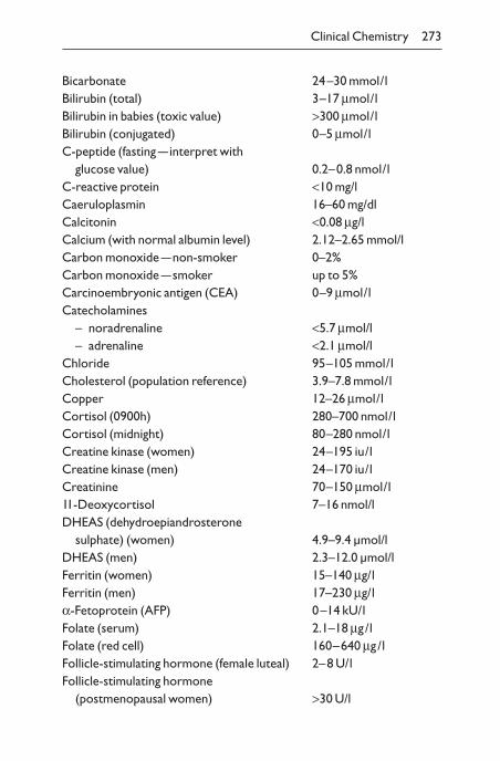
Bicarbonate 24–30 mmol/lBilirubin (total) 3–17 mmol/lBilirubin in babies (toxic value) >300 mmol/lBilirubin (conjugated) 0–5 mmol/lC-peptide (fasting —interpret with
glucose value) 0.2–0.8 nmol/lC-reactive protein <10 mg/lCaeruloplasmin 16–60 mg/dlCalcitonin <0.08 mg/lCalcium (with normal albumin level) 2.12–2.65 mmol/lCarbon monoxide —non-smoker 0–2%Carbon monoxide —smoker up to 5%Carcinoembryonic antigen (CEA) 0–9 mmol/lCatecholamines
– noradrenaline <5.7 mmol/l– adrenaline <2.1 mmol/l
Chloride 95–105 mmol/lCholesterol (population reference) 3.9–7.8 mmol/lCopper 12–26 mmol/lCortisol (0900h) 280–700 nmol/lCortisol (midnight) 80–280 nmol/lCreatine kinase (women) 24–195 iu/lCreatine kinase (men) 24–170 iu/lCreatinine 70–150 mmol/l11-Deoxycortisol 7–16 nmol/lDHEAS (dehydroepiandrosterone
sulphate) (women) 4.9–9.4 µmol/lDHEAS (men) 2.3–12.0 µmol/lFerritin (women) 15–140 mg/lFerritin (men) 17–230 mg/la-Fetoprotein (AFP) 0–14 kU/lFolate (serum) 2.1–18 mg /lFolate (red cell) 160–640 mg /lFollicle-stimulating hormone (female luteal) 2–8 U/lFollicle-stimulating hormone
(postmenopausal women) >30 U/l
Clinical Chemistry 273
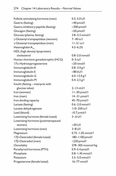
Follicle-stimulating hormone (men) 0.5–5.0 U/lGastrin (fasting) <40 pmol/lGastro-inhibitory peptide (fasting) <300 pmol/lGlucagon (fasting) <50 pmol/lGlucose (plasma, fasting) 3.8–5.5 mmol/lg-Glutamyl transpeptidase (women) 7–40 iu/lg-Glutamyl transpeptidase (men) 11–51 iu/lHaemoglobin A1c 4.5–6.2%HDL (high density lipoprotein)
cholesterol 0.8–2.0 mmol/lHuman chorionic gonadotrophin (HCG) 0–5 iu/l17a-Hydroxyprogesterone <20 nmol/lImmunoglobulin A 0.8–3.0 g/lImmunoglobulin E <80 kU/lImmunoglobulin G 6.0–13.0 g/lImmunoglobulin M 0.4–2.5 g/lInsulin (fasting —interpret with
glucose value) 2–13 mU/lIron (women) 11–30 mmol/lIron (men) 14–31 mmol/lIron-binding capacity 45–70 mmol/lLactate (fasting) 0.6–2.0 mmol/lLactate dehydrogenase 110–250 iu/lLead (blood) <0.7 mmol/lLuteinizing hormone (female luteal) 3–6 U/lLuteinizing hormone (postmenopausal
women) >30 U/lLuteinizing hormone (men) 3–8 U/lMagnesium 0.75–1.05 mmol/l17b-Oestradiol (female luteal) 180–1100 pmol/l17b-Oestradiol (men) <220 pmol/lOsmolality 278–305 mosmol/kgParathyroid hormone (PTH) 0.9–5.4 pmol/lPhosphate 0.8–1.45 mmol/lPotassium 3.5–5.0 mmol/lProgesterone (female luteal) 16–77 nmol/l
274 Chapter 14: Laboratory Results —Normal Values
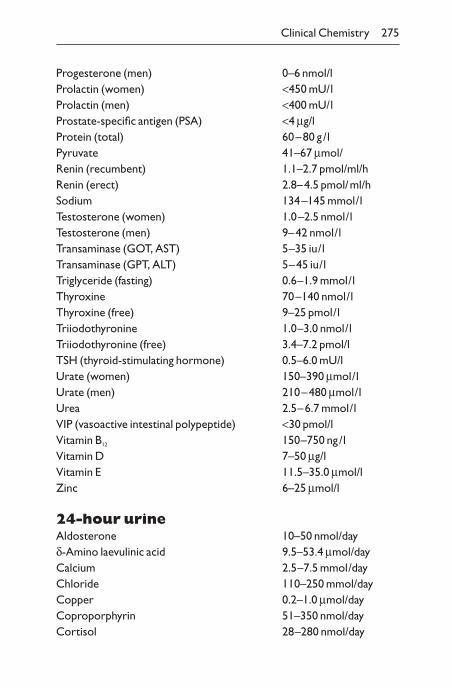
Progesterone (men) 0–6 nmol/lProlactin (women) <450 mU/lProlactin (men) <400 mU/lProstate-specific antigen (PSA) <4 mg/lProtein (total) 60–80 g/lPyruvate 41–67 mmol/Renin (recumbent) 1.1–2.7 pmol/ml/hRenin (erect) 2.8–4.5 pmol/ml/hSodium 134–145 mmol/lTestosterone (women) 1.0–2.5 nmol/lTestosterone (men) 9–42 nmol/lTransaminase (GOT, AST) 5–35 iu/lTransaminase (GPT, ALT) 5–45 iu/lTriglyceride (fasting) 0.6–1.9 mmol/lThyroxine 70–140 nmol/lThyroxine (free) 9–25 pmol/lTriiodothyronine 1.0–3.0 nmol/lTriiodothyronine (free) 3.4–7.2 pmol/lTSH (thyroid-stimulating hormone) 0.5–6.0 mU/lUrate (women) 150–390 mmol/lUrate (men) 210–480 mmol/lUrea 2.5–6.7 mmol/lVIP (vasoactive intestinal polypeptide) <30 pmol/lVitamin B12 150–750 ng/lVitamin D 7–50 mg/lVitamin E 11.5–35.0 mmol/lZinc 6–25 mmol/l
24-hour urineAldosterone 10–50 nmol/dayd-Amino laevulinic acid 9.5–53.4 mmol/dayCalcium 2.5–7.5 mmol/dayChloride 110–250 mmol/dayCopper 0.2–1.0 mmol/dayCoproporphyrin 51–350 nmol/dayCortisol 28–280 nmol/day
Clinical Chemistry 275
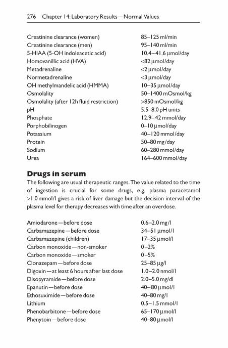
Creatinine clearance (women) 85–125 ml/minCreatinine clearance (men) 95–140 ml/min5-HIAA (5-OH indoleacetic acid) 10.4–41.6 mmol/dayHomovanillic acid (HVA) <82 mmol/dayMetadrenaline <2 mmol/dayNormetadrenaline <3 mmol/dayOH methylmandelic acid (HMMA) 10–35 mmol/dayOsmolality 50–1400 mOsmol/kgOsmolality (after 12h fluid restriction) >850 mOsmol/kgpH 5.5–8.0 pH unitsPhosphate 12.9–42 mmol/dayPorphobilinogen 0–10 mmol/dayPotassium 40–120 mmol/dayProtein 50–80 mg/daySodium 60–280 mmol/dayUrea 164–600 mmol/day
Drugs in serumThe following are usual therapeutic ranges.The value related to the timeof ingestion is crucial for some drugs, e.g. plasma paracetamol>1.0 mmol/l gives a risk of liver damage but the decision interval of theplasma level for therapy decreases with time after an overdose.
Amiodarone —before dose 0.6–2.0 mg/lCarbamazepine —before dose 34–51 mmol/lCarbamazepine (children) 17–35 mmol/lCarbon monoxide —non-smoker 0–2%Carbon monoxide —smoker 0–5%Clonazepam —before dose 25–85 mg/lDigoxin —at least 6 hours after last dose 1.0–2.0 nmol/lDisopyramide —before dose 2.0–5.0 mg/dlEpanutin —before dose 40–80 mmol/lEthosuximide —before dose 40–80 mg/lLithium 0.5–1.5 mmol/lPhenobarbitone —before dose 65–170 mmol/lPhenytoin —before dose 40–80 mmol/l
276 Chapter 14: Laboratory Results —Normal Values
Salicylate 0.4–2.5 mmol/lTheophylline —before dose 55–110 mmol/lValproate —before dose 0.3–0.7 mmol/l
Toxic levelsBarbiturate —potentially fatal
– short-acting 35 mmol/l– medium-acting 105 mmol/l– long-acting 215 mmol/l
Ethanol (physiological <0.2nmol/l)– legal limit for driving <17.4 nmol/l
Paracetamol —risk of liver damage– at 4 hours >1.32 mmol/l– at 15 hours <0.2 mmol/l
Salicylate >2.5 mmol/l
MiscellaneousFaecal fat <18 mmol/daySweat chloride 6–40 mmol/lExtractable nuclear antigen-binding association
Anti-Ro SLE, cutaneous lupusAnti-La SLE, Sjögren’s diseaseAnti-Sm SLE (specific)Anti-RNP SLE, mixed connective
tissue diseaseAnti-Scl-70 Progressive systemic
sclerosisAnti-Jo 1 Polymyositis
Clinical Chemistry 277
CHAPTER 15
Common EmergencyTreatments
Introduction
You will see patients being treated. The following notes provide a guide to the therapies that are employed. In each case a diagnosis needs to be made. The treatments apply to many situations. The specific causes may require additional therapy.These therapies were appropriate in May2002, but with time other therapies may become more appropriate.
Cardiovascular
Myocardial infarction (classic crushing, centralchest pain with radiation to arms, pallor, sweating, distressed ±electrocardiogram (ECG) changes)
– Give 100% oxygen.– Chew an aspirin —300mg.– Give diamorphine intravenously (i.v.) 2.5–5.0mg or morphine i.v.
5.0–10.0mg (± anti-emetic if necessary).– Attach ECG monitor.– If ST ≥2mm new elevation in two or more contiguous chest leads or
= 1mm in standard leads or left bundle-branch block (LBBB):– institute thrombolysis with streptokinase, e.g. i.v. 1500000U
over 1 hour or tissue plasminogen activator if no contraindi-cation, e.g. bleeding, active peptic ulceration, recent opera-tion, recent cerebral bleed or transient ischaemic attack,aortic aneurysm
– hydrocortisone i.v. 100mg if allergic reaction to streptokinase– If normal blood pressure (BP), well-perfused (warm hands), no
heart failure give i.v. b-blocker, e.g. atenolol 5mg.
278
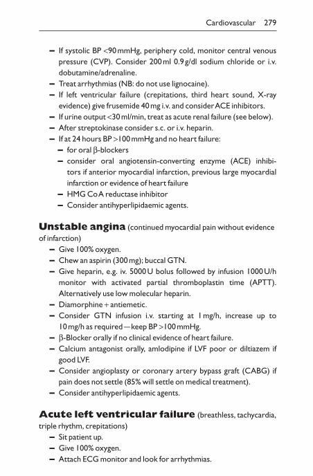
– If systolic BP <90mmHg, periphery cold, monitor central venouspressure (CVP). Consider 200ml 0.9g/dl sodium chloride or i.v.dobutamine/adrenaline.
– Treat arrhythmias (NB: do not use lignocaine).– If left ventricular failure (crepitations, third heart sound, X-ray
evidence) give frusemide 40mg i.v. and consider ACE inhibitors.– If urine output <30ml/min, treat as acute renal failure (see below).– After streptokinase consider s.c. or i.v. heparin.– If at 24 hours BP >100mmHg and no heart failure:
– for oral b-blockers– consider oral angiotensin-converting enzyme (ACE) inhibi-
tors if anterior myocardial infarction, previous large myocardialinfarction or evidence of heart failure
– HMG Co A reductase inhibitor– Consider antihyperlipidaemic agents.
Unstable angina (continued myocardial pain without evidenceof infarction)
– Give 100% oxygen.– Chew an aspirin (300mg); buccal GTN.– Give heparin, e.g. iv. 5000U bolus followed by infusion 1000U/h
monitor with activated partial thromboplastin time (APTT).Alternatively use low molecular heparin.
– Diamorphine + antiemetic.– Consider GTN infusion i.v. starting at 1mg/h, increase up to
10mg/h as required —keep BP >100mmHg.– b-Blocker orally if no clinical evidence of heart failure.– Calcium antagonist orally, amlodipine if LVF poor or diltiazem if
good LVF.– Consider angioplasty or coronary artery bypass graft (CABG) if
pain does not settle (85% will settle on medical treatment).– Consider antihyperlipidaemic agents.
Acute left ventricular failure (breathless, tachycardia,triple rhythm, crepitations)
– Sit patient up.– Give 100% oxygen.– Attach ECG monitor and look for arrhythmias.
Cardiovascular 279
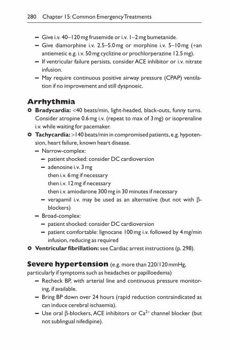
– Give i.v. 40–120mg frusemide or i.v. 1–2mg bumetanide.– Give diamorphine i.v. 2.5–5.0mg or morphine i.v. 5–10mg (+an
antiemetic e.g. i.v. 50mg cyclizine or prochlorperazine 12.5mg).– If ventricular failure persists, consider ACE inhibitor or i.v. nitrate
infusion.– May require continuous positive airway pressure (CPAP) ventila-
tion if no improvement and still dyspnoeic.
Arrhythmia
° Bradycardia: <40 beats/min, light-headed, black-outs, funny turns.Consider atropine 0.6mg i.v. (repeat to max of 3mg) or isoprenalinei.v. while waiting for pacemaker.
° Tachycardia: >140 beats/min in compromised patients, e.g. hypoten-sion, heart failure, known heart disease.– Narrow-complex:
– patient shocked: consider DC cardioversion– adenosine i.v. 3mg
then i.v. 6mg if necessarythen i.v. 12mg if necessarythen i.v. amiodarone 300mg in 30 minutes if necessary
– verapamil i.v. may be used as an alternative (but not with b- blockers)
– Broad-complex:– patient shocked: consider DC cardioversion– patient comfortable: lignocane 100mg i.v. followed by 4mg/min
infusion, reducing as required
° Ventricular fibrillation: see Cardiac arrest instructions (p. 298).
Severe hypertension (e.g. more than 220/120mmHg,particularly if symptoms such as headaches or papilloedema)
– Recheck BP, with arterial line and continuous pressure monitor-ing, if available.
– Bring BP down over 24 hours (rapid reduction contraindicated ascan induce cerebral ischaemia).
– Use oral b-blockers, ACE inhibitors or Ca2+ channel blocker (butnot sublingual nifedipine).
280 Chapter 15:Common Emergency Treatments
– Or i.v. nitroprusside with arterial monitoring.– Treat any complications, e.g. left ventricular failure, encephalopathy.
Respiratory
Acute bronchospasm (breathless, wheeze, distress)– Give 100% oxygen unless known chronic airways disease (see
below).– Ventolin by continuous nebulizer (not inhalers) 5ml in 2ml
water.– Hydrocortisone 100mg i.v. or oral prednisolone 30–50mg.– Aminophylline 5mg/kg i.v. by slow injection (10–15 minutes) but
not if patient has been taken theophyllines already.– Do blood gases:
PO2 (in pKa) PCO2 (in pKa)– Mild <10 <4– Moderate 8–10 <4– Severe <8 4–6: watch carefully– Desperate <7 >6: consider ventilation
– Monitor fatigue —consider ventilation if patient becomes exhausted.
Acute exacerbation in chronic obstructiveairways disease (usually breathless, cough, sputum and coarsecrepitations)
– Give 24% oxygen and increase if PCO2 not raised.– Blood gases:
– PO2Ø and PCO2 Ø ‘pink puffer’: increase oxygen content– PO2 Ø and PCO2 ≠ ‘blue bloater’: increase oxygen carefully, re-
peating blood gases, as removal of hypoxic drive may decreaserespiratory volume and rate. Then reduce PO2 and considerdoxapram. Ventilation may be indicated when there is a goodprognosis
– Physiotherapy to cough up sputum.– Culture sputum, chest X-ray, consider antibiotics.
Respiratory 281
Gastrointestinal
Acute gastrointestinal haemorrhage (suddencollapse, haematemasis or red/black sticky stools; BP <100mmHg,pulse >100 beats/min)
– Assess whether cirrhosis/portal hypertension, peptic ulcer.– i.v. access (2 large venflons) and take blood for haematology,
biochemistry and crossmatching.– If BP <90mmHg, 500ml 0.9g/dl sodium chloride or colloid in 30
minutes.– If no BP, consider group 0 rhesus-negative blood.– If no central pulse, for cardiorespiratory resuscitation.– Monitor CVP.– Give blood as required to raise BP and CVP.– Urinary catheter if severe blood loss.– Alert surgical team and arrange urgent endoscopy.
Acute hepatic failure (jaundice, foetor, liver flap,confusion)
– If systolic BP <90mmHg, 500ml i.v. 5% dextrose or colloid in 30minutes.
– Monitor CVP.– Monitor blood glucose —if <4mmol/l, infuse dextrose 10% and
recheck.– Look for drugs, including paracetamol overdose.– Look for infection —blood, chest, urine, ascites.
– Look for occult bleeding, including increasing plasma urea:– consider fresh frozen plasma to correct clotting– Start oral lactulose, consider neomycin.– Prevent stress ulcers with H2-blocker or proton pump blocker.– Vitamins B and K i.v.– Restrict salt and water intake.– Monitor drugs, electrolyte, liver function tests, clotting, pH.
282 Chapter 15:Common Emergency Treatments
Neurological
Epileptic attack (tonic/clonic movements, usuallyunconscious)
– Oxygen.– Diazepam i.v. 5–10mg over 2 minutes, then i.v. 2mg/min for 20
minutes or until fit ceases.Watch for respiratory depression.– Bedside test for glucose —?hypoglycaemia.– Phenytoin i.v. 50mg/min.– If fit continues:
– chlormethiazole or phenytoin– general anaesthesia and ventilate
Unconsciousness with no overt cause– Clear airway and give 100% oxygen.– Prone recovery position unless airway protected by endotracheal
tube.– Examine for head injury, neurological deficit, neck stiffness.– Enquire whether diabetic or access to insulin, any tablets or
whether a suicide risk.– Prevent fitting (see above).– If respiratory rate <10 breaths/min give i.v. naloxone.– Check blood glucose.– If BP <90mmHg systolic, give 500ml 0.9g/dl sodium chloride or
colloid i.v.– Check blood gases.– Take blood and urine for drug tests.– Document level of consciousness on Glasgow Coma Scale.
Meningitis (headache, neck stiffness, vomiting, photophobia,febrile)
– N.B. if purpuric rash immediately start i.v. antibiotic —ceftriaxone 2g —after taking blood cultures.
– Check for signs of raised intracranial pressure, e.g. papilloedema.– Lumbar puncture if no signs of raised intracranial pressure:
– note pressure
Neurological 283
– cerebrospinal fluid (CSF) for culture —bacterial, PCR for viruses,biochemistry and microscopy
– Cloudy CSF (white cells) —prompt i.v. antibiotics after blood cultures.
– Blood-stained —assess whether bloody tap, i.e. blood at first then clearing, or subarachnoid haemorrhage (consistent bloodwith xanthochromia of CSF after centrifuging down red cells).
Other systems
Acute renal failure (rapid increase in plasma creatinine,urine output <30ml/h)
– Consider prerenal cause (patient ‘dehydrated’ —dry tongue,low skin turgor, empty veins, low CVP, low blood pressure) —give fluid challenge and continue until JVP is 2–3cm above themanubriosternal junction.
– Consider postrenal cause (e.g. enlarged prostate, bilateral ure-teric stones, renal/pelviureteric obstruction). If large prostate andlarge bladder, consider passing catheter.
– If no obvious cause of renal failure, ultrasound abdomen —?dilatedureters or dilated renal pelves or small kidneys, indicating chronicrenal failure.
– Check plasma potassium, sodium, creatinine, urea (if potassium >6mmol/l and ECG changes, give i.v. glucose/insulin, i.v. calcium gluconate and rectal cation exchange resin).
– Check urine sodium and osmolality– in prerenal failure, urine osmolality >400mosmol/kg and
sodium <30mmol/l– in renal failure, <400mosmol/kg and >30mmol/l, respectively
– Microscope urine sediment for red cells, white cells, casts and bacteria.
– Check arterial pH.– If incipient renal tubular necrosis, for i.v. frusemide 80–500mg.– When fluid-replete, restrict fluid to 500ml per day + previous day’s
losses.
284 Chapter 15:Common Emergency Treatments
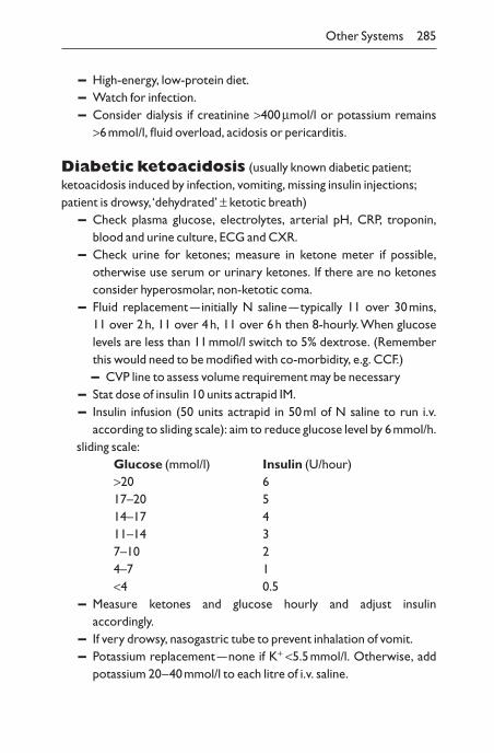
– High-energy, low-protein diet.– Watch for infection.– Consider dialysis if creatinine >400mmol/l or potassium remains
>6mmol/l, fluid overload, acidosis or pericarditis.
Diabetic ketoacidosis (usually known diabetic patient;ketoacidosis induced by infection, vomiting, missing insulin injections;patient is drowsy,‘dehydrated’ ± ketotic breath)
– Check plasma glucose, electrolytes, arterial pH, CRP, troponin,blood and urine culture, ECG and CXR.
– Check urine for ketones; measure in ketone meter if possible,otherwise use serum or urinary ketones. If there are no ketonesconsider hyperosmolar, non-ketotic coma.
– Fluid replacement —initially N saline —typically 11 over 30mins,11 over 2h, 11 over 4h, 11 over 6h then 8-hourly. When glucoselevels are less than 11mmol/l switch to 5% dextrose. (Rememberthis would need to be modified with co-morbidity, e.g. CCF.)– CVP line to assess volume requirement may be necessary
– Stat dose of insulin 10 units actrapid IM.– Insulin infusion (50 units actrapid in 50ml of N saline to run i.v.
according to sliding scale): aim to reduce glucose level by 6mmol/h.sliding scale:
Glucose (mmol/l) Insulin (U/hour)>20 617–20 514–17 411–14 37–10 24–7 1<4 0.5
– Measure ketones and glucose hourly and adjust insulin accordingly.
– If very drowsy, nasogastric tube to prevent inhalation of vomit.– Potassium replacement —none if K+ <5.5mmol/l. Otherwise, add
potassium 20–40mmol/l to each litre of i.v. saline.
Other Systems 285
Hypoglycaemia (symptoms include drowsy/unconscious,perspiring, tachycardia, bounding pulse, usually in insulin-treated diabeticdue to missing snack or increased exercise. N.B. many diabetic patientsare asymptomatic with hypoglycaemia)
– Check plasma glucose. (Do not await result from laboratory —treatstraight away.)
– Keep airway clear.– If no i.v. access give 1mg glucagon i.m., acts in 5–10 minutes (but
not if hypoglycaemia due to insulinoma).– If emergency, e.g. fitting, 50ml 50g/dl i.v. glucose followed by 50ml
of 0.9g/dl saline to wash sclerosant, hypertonic glucose out of vein.
Septicaemia (febrile >39°C, rigors)– Give 100% oxygen.– Look for source of septicaemia.– If systolic BP <90mmHg, 500ml i.v. sodium chloride or colloid i.v.
in 30 minutes.– Monitor CVP.– Antibiotics i.v. after blood cultures, culture of urine, throat or
pustules.– If BP Ø, pH Ø or consciousness level Ø —transfer to intensive care
unit.
Poisoning or overdose– Give 100% oxygen, except in paraquat poisoning.– Check paracetamol and aspirin levels in all patients.– Give naloxone if respiratory rate <10 breaths/min. Measure blood
gases and consider ventilation.– Correct hypotension: if BP <90mmHg, 500ml i.v. sodium chloride
in 30 minutes.– Consider gastric lavage —intubate first if unconscious.– Paracetamol overdose —acetylcysteine according to blood levels of
paracetamol.– Aspirin overdose:
– gastric lavage up to 12 hours– watch pH
286 Chapter 15:Common Emergency Treatments
– consider forced alkaline diuresis– Amphetamine poisoning:
– beware sudden airway oedema– have available intubation equipment, adrenaline, chlorpheni-
ramine, hydrocortisone– Consider activated charcoal per oral/gastric tube.– If potentially an unusual poison, phone poison centre for advice.
Anaphylactic response– Give 100% oxygen.– Chlorpheniramine 10mg i.v. in 1 minute.– Hydrocortisone 100mg i.v.– If severe, adrenaline 0.5–1mg i.v. slowly over 1–2 minutes.– If BP <90mmHg, 500ml i.v. sodium chloride or colloid.
Death
Whilst diagnosis of death per se does not require emergency therapy,the required procedures are an important aspect of medicine.
If there is sudden loss of consciousness, consider cardiopulmonary resuscitation —see Cardiac arrest instructions (p. 298).
– Pale, pulseless, apnoeic —listen at mouth, observe chest.– No heart sounds —listen with diaphragm.– Fixed pupils.– Head and eyes move together when head moved, i.e. no oculo-
cephalic reflex movement or ‘doll’s eye’ movement.– No corneal response.– No response to any stimulus.If patient cold, <35°C, or major drug overdose, e.g. barbiturate,
patients can appear dead. If in doubt look at retina with ophthalmoscopeto see if ‘trucking’ of non-flowing segments of blood in veins.
Brain death criteria– If the patient is on a ventilator because of apnoea, test:
– at least 6 hours after onset of coma– at least 24 hours after cardiac arrest/circulation restoration
Death 287
– by two independent consultants if feasible– Whether patient has condition that could lead to irremediable
brain damage.– There are no reflex responses or epileptic jerks.– No hypothermia —temperature >35°C.– No drug intoxication —off therapy for 48 hours
– particularly depressants, neuromuscular-blocking (relaxant)drugs
– No hypoglycaemia, acidosis, gross electrolyte imbalance.– All brainstem reflexes absent, confirmed by two physicians:
– no pupil response to light– no corneal reflexes– no vestibular-ocular reflexes:
– visualize tympanic membranes– 20ml cold water in each ear– no eye movements
– no cranial motor responses:– no gag reflex– no cough reflex to bronchial stimulaton
– no respiratory effort when ventilator is stopped:– PCO2 rise to 6.7kPa
– Repeat tests at least 2 hours later, usually after 24 hours.– Time of second test is legally the time of death.
N.B. Spinal reflexes and electroencephalogram are irrelevant. Warn family that reflex leg movements can exist after cessation of brainstemfunction and are not of relevance.
288 Chapter 15:Common Emergency Treatments
Appendices
Appendix 1: Jaeger reading chart
Jaeger types assess visual acuity for close tasks. It provides the easiestquick method of assessment. The patient should use his spectacles nor-mally required for reading. Ask the patient to read the smallest type he can read, if read with few mistakes, ask him to read the next size down.Record the size of type that can be read with each eye separately.
289
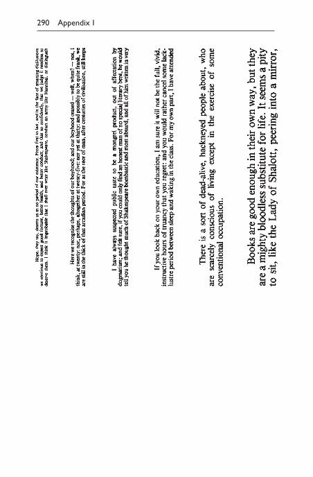
290 Appendix 1
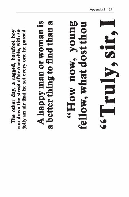
Appendix 1 291
Appendix 2:Visual acuity 3m chart
The 3-m Snellen chart should be held at 3m from the patient, with goodlighting, with each of the patient’s eyes covered in turn. Use the patient’susual spectacles for this distance. If the patient cannot read 6/6 (e.g. 6/12is best vision in one eye), repeat without spectacles and with a ‘pinhole’that largely nullifies refractive errors. Note for each eye the best acuityobtained and the method used, e.g. L 6/9 R 6/6 with spectacles.
292 Appendix 2

Appendix 2 293
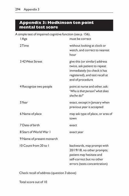
Appendix 3: Hodkinson ten pointmental test score
A simple test of impaired cognitive function (see p. 156).1Age must be correct
2Time without looking at clock orwatch, and correct to nearesthour
3 42 West Street give this (or similar) addresstwice, ask patient to repeat immediately (to check it hasregistered), and test recall atend of procedure
4 Recognize two people point at nurse and other, ask:‘Who is that person? what doesshe/he do?’
5Year exact, except in January when previous year is accepted
6 Name of place may ask type of place, or area oftown
7 Date of birth exact
8 Start of World War 1 exact year
9 Name of present monarch
10 Count from 20 to 1 backwards, may prompt with20/19/18, no other prompts;patient may hesitate and self-correct but no other errors (tests concentration)
Check recall of address (question 3 above)
Total score out of 10
294 Appendix 3

Communication problems (e.g. deafness, dysphasia) or abnormal mood(e.g. depression) may affect the mental test score, and should be noted.(After Qureshi, K. & Hodkinson, H. Evaluation of a ten-question mentaltest in the institutional elderly. Age Ageing 1974;3:152.)
Appendix 3 295
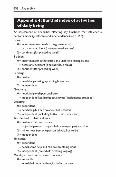
Appendix 4: Barthel index of activities of daily living
An assessment of disabilities affecting key functions that influence a person’s mobility, self-care and independence (see p. 157).Bowels:
0 = incontinent (or needs to be given enema)1 = occasional accident (once per week or less)2 = continent (for preceding week)
Bladder:0 = incontinent or catheterized and unable to manage alone1 = occasional accident (once per day or less)2 = continent (for preceding week)
Feeding:0 = unable1 = needs help cutting, spreading butter, etc.2 = independent
Grooming:0 = needs help with personal care1 = independent face/hair/teeth/shaving (implements provided)
Dressing:0 = dependent1 = needs help but can do about half unaided2 = independent (including buttons, zips, laces, etc.)
Transfer bed to chair and back:0 = unable, no sitting balance1 = major help (one strong/skilled or two people), can sit up2 = minor help from one person (physical or verbal)3 = independent
Toilet use:0 = dependent1 = needs some help, but can do something alone2 = independent (on and off, dressing, wiping)
Mobility around house or ward, indoors:0 = immobile1 = wheelchair independent, including corners
296 Appendix 4
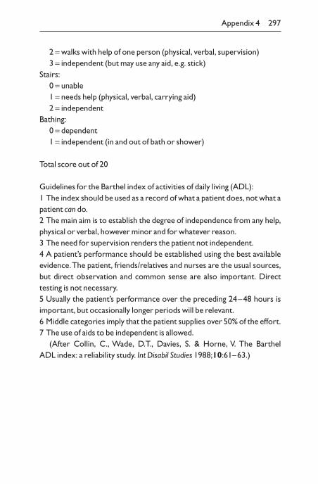
2 = walks with help of one person (physical, verbal, supervision)3 = independent (but may use any aid, e.g. stick)
Stairs:0 = unable1 = needs help (physical, verbal, carrying aid)2 = independent
Bathing:0 = dependent1 = independent (in and out of bath or shower)
Total score out of 20
Guidelines for the Barthel index of activities of daily living (ADL):1 The index should be used as a record of what a patient does, not what apatient can do.2 The main aim is to establish the degree of independence from any help,physical or verbal, however minor and for whatever reason.3 The need for supervision renders the patient not independent.4 A patient’s performance should be established using the best availableevidence.The patient, friends/relatives and nurses are the usual sources,but direct observation and common sense are also important. Directtesting is not necessary.5 Usually the patient’s performance over the preceding 24–48 hours isimportant, but occasionally longer periods will be relevant.6 Middle categories imply that the patient supplies over 50% of the effort.7 The use of aids to be independent is allowed.
(After Collin, C., Wade, D.T., Davies, S. & Horne, V. The Barthel ADL index: a reliability study. Int Disabil Studies 1988;10:61–63.)
Appendix 4 297
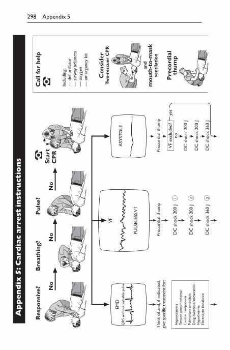
298 Appendix 5
Res
pons
ive?
EMD
QR
S w
ithou
t pa
lpab
le p
ulse
Thi
nk o
f and
, if i
ndic
ated
,gi
ve s
peci
fic tr
eatm
ent f
or:
Hyp
ovol
aem
iaTe
nsio
n pn
eum
otho
rax
Car
diac
tam
pona
dePu
lmon
ary
embo
lism
Dru
g ov
erdo
se/in
toxi
catio
nH
ypot
herm
iaEl
ectr
olyt
e im
bala
nce
Prec
ordi
al t
hum
p
DC
sho
ck 2
00 J
1
DC
sho
ck 2
00 J
2
DC
sho
ck 3
60 J
3
VF
PULS
ELES
S V
T
ASY
STO
LE
Prec
ordi
al t
hum
p
DC
sho
ck 2
00 J
DC
sho
ck 2
00 J
DC
sho
ck 3
60 J
yes
VF
excl
uded
?no
Cal
l fo
r he
lp
Incl
udin
g:—
def
ibri
llato
r—
air
way
adj
unct
s—
oxy
gen
— e
mer
genc
y ki
t
Co
nsid
erTw
o-r
escu
er C
PR
and
mou
th-t
o-m
ask
vent
ilati
on
Pre
cord
ial
thum
p
Bre
athi
ng?
Pul
se?
No
No
No
Sta
rtC
PR
Ap
pen
dix
5:C
ard
iac
arr
est
inst
ruct
ion
s
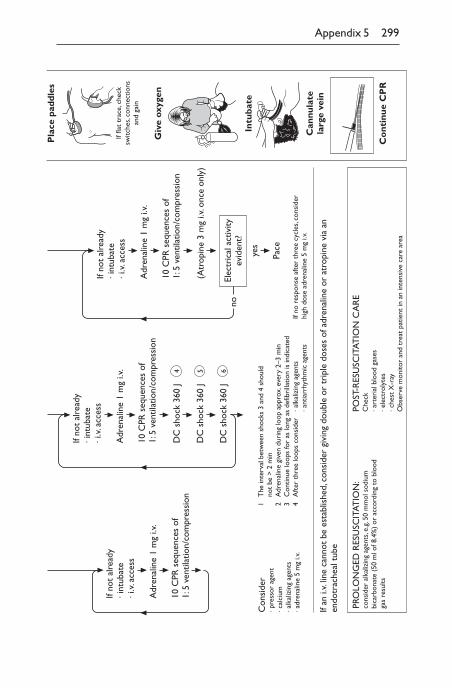
Appendix 5 299
no
Adr
enal
ine
1 m
g i.v
.
If no
t al
read
y· i
ntub
ate
· i.v
. acc
ess
10 C
PR s
eque
nces
of
1: 5
ven
tilat
ion/
com
pres
sion
10 C
PR s
eque
nces
of
1: 5
ven
tilat
ion/
com
pres
sion
If no
t al
read
y· i
ntub
ate
· i.v
. acc
ess
Adr
enal
ine
1 m
g i.v
.
DC
sho
ck 3
60 J
DC
sho
ck 3
60 J
DC
sho
ck 3
60 J
4 5 6
10 C
PR s
eque
nces
of
1: 5
ven
tilat
ion/
com
pres
sion
If no
t al
read
y· i
ntub
ate
· i.v
. acc
ess
Adr
enal
ine
1 m
g i.v
.
(Atr
opin
e 3
mg
i.v. o
nce
only
)
Elec
tric
al a
ctiv
ityev
iden
t?
yes
Pace
Con
side
r· p
ress
or a
gent
· cal
cium
· alk
aliz
ing
agen
ts· a
dren
alin
e 5
mg
i.v.
1 2 3 4
The
inte
rval
bet
wee
n sh
ocks
3 a
nd 4
sho
uld
not
be >
2 m
inA
dren
alin
e gi
ven
duri
ng lo
op a
ppro
x. e
very
2–3
min
Con
tinue
loop
s fo
r as
long
as
defib
rilla
tion
is in
dica
ted
Aft
er t
hree
loop
s co
nsid
er· a
lkal
izin
g ag
ents
· ant
iarr
hyth
mic
age
nts
If no
res
pons
e af
ter
thre
e cy
cles
, con
side
rhi
gh d
ose
adre
nalin
e 5
mg
i.v.
If an
i.v.
line
cann
ot b
e es
tabl
ishe
d, c
onsi
der
givi
ng d
oubl
e or
tri
ple
dose
s of
adr
enal
ine
or a
trop
ine
via
anen
dotr
ache
al t
ube
PRO
LON
GED
RES
USC
ITAT
ION
:co
nsid
er a
lkal
izin
g ag
ents
, e.g
. 50
mm
ol s
odiu
mbi
carb
onat
e (5
0 m
l of 8
.4%
) or
acc
ordi
ng t
o bl
ood
gas
resu
lts
POST
-RES
USC
ITAT
ION
CA
RE
Che
ck· a
rter
ial b
lood
gas
es· e
lect
roly
tes
· che
st X
-ray
Obs
erve
mon
itor
and
trea
t pa
tient
in a
n in
tens
ive
care
are
a
Pla
ce p
addl
es
If fla
t tr
ace,
che
cksw
itche
s, co
nnec
tions
and
gain
Giv
e ox
ygen
Intu
bate
Can
nula
tela
rge
vein
Co
ntin
ue C
PR
ABC of clinical medicine 2–3abdomen/abdomen examination 87–100,
162auscultation 94–5common illnesses 97–9general inspection 87–8genitals 96herniae 95–6inspection 88–90lymph nodes 39masses 93notes 165pain 13palpation 90–1
of organs 91–3per rectum 96–7percussion 93–4radiography 189–91shifting dullness 94system-oriented 99–100ultrasound 182, 284vaginal 97
abdominal reflexes, superficial 140–1abducens nerve 123–7abnormal findings 166ABO blood groups 224acalculia 115acanthosis nigricans Plate 5accessory nerve 131–2accommodation reflex 122–3acne 31acoustic testing 130acromegaly 29, 43, 228, Plate 1action limits of laboratory investigations
271activated partial thromboplastin time
(APTT) 222–3, 279activities of daily living 157–8
Barthel index 296–7activity 16Addison’s disease 43, 228adrenaline 279, 287adrenals
failure 228nuclear medicine 196
adrenocorticotrophic hormone (ACTH)227–8
aegophony 83affect 157agnosia 115airway oedema 287alanine amino transferase (ALT) 226albumin 226alcohol abuse 87
dementia 116GGT levels 226intoxication 109toxic levels 277
alcohol consumption 22alkaline phosphatase (ALP) 226allergens, skin testing 212alveolitis
allergic 85breath sounds 81
amaurosis fugax 15, 17amblyopia 124aminophylline 281amlodipine 279amoebal infection 14amphetamine poisoning 287ampicillin rash Plate 3amylase 226anaemia
haemolytic 221investigations 220–1iron deficiency 27, 87, Plate 2
Index
300
Note: page numbers in italics refer to figures, those in bold refer to tables. Plates are referredto by Plate number and are located between pages 150 and 151
Index 301
macrocytic 211microcytic 211retina 37signs 87
anal fissures 96anaphylactic response 287aneurysm
aortic 74, 93, 193digital subtraction imaging 193dissecting 53left ventricular 57subarachnoid haemorrhage 192ultrasound examination 183
anginamedical treatment 200unstable 279
angiography 193, 200angioplasty 279angiotensin converting enzyme (ACE)
inhibitors 279, 280angular stomatitis 87anion gap 211, 225ankle 136
clonus 134reflex 138–9swelling 12
ankylosing spondylitis 48anorexia nervosa 108anti-endomysial antibodies 230antibiotics 281, 283, 284, 286anticoagulation 222antidiuretic hormone (ADH) 228antigliadin antibodies 230antineutrophil cytoplasmic antibodies
(ANCA) 230antinuclear antibody 229antiphospholipid antibody 223antismooth muscle antibody 229anxiety 16, 103, 105, 108
blood pressure measurement 209concentration impairment 116
aortachest radiograph 187palpation 93ultrasound examination 182unfolded 93
aortic aneurysm 74, 93aortography 193
aortic incompetence 51, 53, 56, 57, 69murmurs 60, 61, 62, 63, 64, 65
aortic regurgitation 65aortic sclerosis 60, 65aortic stenosis 51, 53, 57, 69, 207
heart sounds 59, 60, 61murmurs 60, 61, 62, 64, 65
aortic valve pressure 206, 207aortography 193apex beat 56–7, 78aphasia 114appearance 101, 102appendicitis 98approach, analytical 2apraxia 115Argyll Robertson pupils 122, 123arms
cerebellar signs 150examination 163nervous system assessment 133–4, 152,
153arterial blood gas measurement 209–12,
232–3bronchospasm 281unconsciousness 283
arterial bruits, abdominal 94–5arterial insufficiency Plate 4
Buerger’s test 72arteriography 192–3arteriosclerosis 51arthralgia 18, 46arthritis 18, 46arthropathy 46articulation 114artrial fibrillation 59ascites 190aspartate transaminase (AST) 226aspirin 278, 279
overdose 286–7assessment
of illness 1of problem 2
asthma 84–5, 210, 212ataxia, sensory 145atheroma 53atopy 85atrial asystole 208atrial ectopic beats 254atrial fibrillation 51, 55, 257–8, 259, 267atrial flutter 258, 259, 266atrial myxoma, left 65atrial septal defect 59, 65, 70atrial tachycardia 208, 267atrioventricular node 239
ectopic beats 255first-degree heart block 263
atropine 280attention 116
302 Index
auditory nerve 129–30auscultation 27, 58–60, 61, 62–3, 64, 65Austin Flint murmur 65, 69
b-agonists 212b-blockers 278, 279, 280Babinski response 140bacterial endocarditis see infectious
endocarditisbacterial overgrowth 216balanitis 96barium swallow/meal/enema 216Barthel index 157, 296–7basal cell carcinoma 31, Plate 5Becton–Dickinson vacutainer system
232behaviour 101, 102beliefs, abnormal 101, 103–4bereavement 110bicarbonate 210, 211biceps reflex 138biliary tree endoscopy 215bilirubin 226black-outs 12blindness, ocular 123blood
biochemical tests 225–30normal values 272–5, 276–7
coagulation 222–3count 220crossmatching 224cultures 232, 284, 285, 286endocrinology tests 227–9faecal occult 97groups 224–5samples 230–2
blood film examination 220, 221blood pressure 52–3, 280
24-hour recording 208–9anaphylactic response 287gastrointestinal haemorrhage 282hepatic failure 282myocardial infarction 278, 279overdose 286septicaemia 286unconsciousness 283
blood products 224blood tests for drugs 283blood transfusion 224, 282blood vessels, ultrasound examination 183blue bloater 85, 281bone
deformity 46
metabolism 226–7metastases 189, 194MRI 197nuclear medicine 196
bone marrowbiopsy 221, 225imaging 196
bowelhabit 13radiology 190, 191sounds 94ultrasound examination 182
bowel gas pattern 190brachial vessel cannulation 206bradycardia 51, 238, 262–4, 265, 266
emergency treatment 280bradykinesia 150brain
MRI 197ultrasound examination 183see also cerebellar dysfunction; cerebral
entriesbrain death criteria 287–8brainstem
dysfunction 15function 288
breast cancer 41breasts, examination 40–1breath
smell 33–4, 88sounds 81, 82, 83, 85
breathlessness 11–12Broca’s area lesion 114bronchial carcinoma 28, 76, Plate 2
endoscopy 184bronchiectasis 28, 76, 83, 85bronchitis 213bronchoscopy 184, 213Bruce protocol 199–200bulimia nervosa 108bullae 31bundle branch block 267
see also left bundle-branch block; rightbundle-branch block
bundle of His 239, 258, 260
C-reative protein (CRP) 285café-au-lait patches 31calcification, radiology 191calcium antagonists 279, 280calcium/bone metabolism 226–7calcium gluconate 284calculation 116
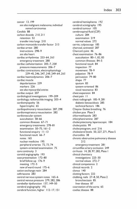
Index 303
cancer 13, 199see also malignant melanoma; individual
named carcinomasCandida 88carbon dioxide 210, 211
retention 52transfer into lungs 210
carbon monoxide transfer factor 213cardiac arrest 280
instructions 298–9see also heart
cardiac arrhythmias 253–64, 265emergency treatment 280
cardiac catheterization 205–7, 208pressure measurements 206–7
cardiac contractions, electrophysiology239–45, 246, 247, 248, 249–64, 265
cardiac haemodynamics 206–7cardiac muscle
depolarization 239markers 226see also myocardial entries
cardiac output 207cardiological investigations 199–209cardiology, radionuclide imaging 203–4cardiomyopathy 56
hypertrophic 65cardiopulmonary resuscitation 287, 298cardiorespiratory resuscitation 282cardiovascular system
auscultation 58–66common illnesses 67–71emergency treatments 278–81examination 50–75, 161–2functional enquiry 11–13functional result 66–7notes 165nuclear medicine 195peripheral arteries 72, 73, 74system-oriented examination 75
care continuity 3carotid angiography 193case presentation 172–80
brief follow-up 176–9meeting 172–5new of ward round 175–6
cation exchange resin 284ceftriaxone 283central nervous system notes 165–6central venous pressure 279, 282, 286cerebellar dysfunction 137, 149–50cerebral angiography 192cerebral function, higher 112–17, 151
cerebral hemispheres 192cerebral scintigraphy 195cerebral tumour 105cerebrospinal fluid (CSF)
culture 284examination 219normal values 272
cervix, colposcopy 185charcoal, activated 287Charcot’s joint 46chest examination 76–86
auscultation 80–1, 82, 83common illnesses 84–5functional result 84inspection 77movement 78–9palpation 78–9percussion 79–80shape 77sputum 83system-oriented 86vocal resonance 83
chest expansion 84chest pain 11chest radiograph 185–9, 281
diabetic ketoacidosis 285technical factors 186
Cheyne–Stokes breathing 76chicken pox Plate 3chlormethiazole 283chlorpheniramine 287cholecystectomy, laparoscopic 184cholecystitis 99cholecystogram, oral 216cholesterol levels 50, 227, 271, Plate 5choroiditis 38chronic obstructive pulmonary disease
212emergency treatment 281
circumflex artery occlusion 249cirrhosis 14, 28, 97, 282, Plate 1clinical chemistry
investigations 225–30normal values 272–7
clinical competence 2clinical records 2clonus 140clotting factors 222clubbing, nails 27–8, 50, Plate 2
bronchiectasis 85liver disease 87
coarctation of the aorta 65coeliac disease 88
304 Index
cognitive function 101, 105assessment 115–17, 156
cognitive impairment 156cold sores 32, Plate 6colloid 282, 283, 286, 287colon
carcinoma 184, 196polyps 184
colonoscopy 184colour-flow Doppler 205colposcopy 185coma 113, 218, 283
diabetic 211communication 5
difficulties 158, 295history taking 8within team 179–80
computed tomography 191–2concentration, impaired 116confabulation 116confusional states 105, 114, 154
elderly patients 160Conn’s tumour 196consciousness/conscious level 113
disorders 218septicaemia 286sudden loss 287see also coma
continuous positive airway pressure(CPAP) 280
coordination 137, 138core biopsy 185corneal arcus Plate 5corneal reflex 128coronary arteries
disease 192ischaemia patterns 237occlusion 247, 248, 249
coronary arteriography 192coronary artery bypass graft 279cortisol 227, 228
urinary output 229cough 12, 131Courvoisier’s law 99cranial nerves
examination 117–32, 151, 163–4palsies 17–18, Plate 6
creatine kinase (CK-MB) 226, 271creatinine 284, 285
clearance 217, 225cremasteric reflex 141crepitations 81Crohn’s disease 88, 216
Cushing’s syndrome 29, 43, 88, 227,228–9, Plate 1
cyanosiscentral 33, 50, 85, 88peripheral 27, 33, 50
cyclizine 280cystic fibrosis 28cystoscopy 184cytomegalovirus (CMV)-negative blood
products 224
D-dimers 223data interpretation 5DC cardioversion 280deafness 295death 287–8deep vein thrombosis 193, 222, 223dehydration 98, 225, 284, 285delirium 103, 109, 116delusions 104, 109dementia 103, 105, 107, 110, 116
alcohol-related 116assessment 154cerebral scintigraphy 195electroencephalogram 218
depression 16, 103, 105, 107–8, 109,116
disability 157mental test score 295pharmaceutical treatment 157
dermatitis 31dermatomes 143dexamethasone suppression test 229dextrose 282, 285diabetes 14, 34, 43–4
autonomic neuropathy 125diagnosis 168glucose control criteria 271hypoglycaemia 286peripheral vascular disease 72sliding scale 285unconsciousness 283
diabetic coma 211diabetic foot Plate 4diabetic ketoacidosis 285diabetic retinopathy 36, 37, 38, 44,
Plate 6diagnosis 2, 166–71
differential 2, 167information required 3provisional 1, 2
diagnostic investigations 167diagnostic labels 179
Index 305
dialysis 285diamorphine 278, 279, 280diaphragm 80
depressed 85diarrhoea 13diazepam 283digit span recall 116digital subtraction 193, 194digoxin 258, 259diltiazem 279diplopia 17–18, 124–5disability 154
analysis 158–9assessment 154–60causes 159–60functional 19index 296–7instruments for assessment 157–8objective setting 158–9
diseases 167anxiety 20experience of 3see also illness
disorientation 116disseminated intravascular coagulation
(DIC) 223dizziness 15dobutamine 279doctors, case presentation to 172–9Doppler ultrasound cardiography 204–5Down’s syndrome 71doxapram 281drive 157drug abuse 107drug reactions
masquerading as mental illness 105pupils 122rash 30, 31, Plate 3
drug tests, blood/urine 283drugs, recreational 22
overdose 287drugs, therapeutic 22
overdose 286–7serum level normal ranges 276–7toxic levels 277
Duchenne muscular dystrophy 271ductus arteriosus, patent 70Dupuytren’s contracture 28, 87, Plate 4dysarthria 114, 131dysdiadochokinesia 149dysgraphia 115dyslexia 115dyspareunia 14
dysphagia 13, 215dysphasia 114, 295
expressive 114mixed 115receptive 114
dysphonia 114dyspnoea 11, 50dyspraxia 155dysuria 14
ear disease 15echocardiography 200, 202, 203
Doppler 202M-mode 202, 203two-dimensional 200, 202
ectopic beats 238, 254–5, 256atrial 267
eczema 31, Plate 3editorials 4ejection fraction 204elbow 134, 135elderly patients
causes of disability 159–60communication difficulties 158disability assessment 159Romberg’s test 146
electrocardiography (ECG) 235–6724-hour tape recording 208, 209complex shape 267diabetic ketoacidosis 285examining 266–7exercise 199–200, 201left ventricular failure 279myocardial infarction 278normal/normal variants 238rate 267rhythm 266–7standard leads 235–6standardization 237
electroencephalogram 218electrolyte plasma levels 285emergency treatments
anaphylactic response 287bronchospasm 281cardiac arrest 298–9cardiac arrhythmia 280chronic obstructive pulmonary disease
281death 287–8diabetic ketoacidosis 285epilepsy 283gastrointestinal haemorrhage 282hepatic failure 282

306 Index
hypertension 280–1hypoglycaemia 286left ventricular failure 279–80meningitis 283–4myocardial infarction 278–9overdose 286–7poisoning 286–7renal failure 284–5septicaemia 286unconsciousness 283unstable angina 279
emotional state 154emphysema 80, 212, 213empyema 28, 76, 188encephalitis 218encephalopathy 218, 281endoscopic retrograde
cholangiopancreatography (ERCP)215
endoscopy 183–5gastrointestinal haemorrhage 282upper gastrointestinal 214–15
eosinophilia 222epididymis 96epigastric pain 215epilepsy
cerebral scintigraphy 195electroencephalogram 218emergency treatment 283grand mal 15–16see also fits
erythema multiforme 31–2erythema nodosum 31, 32, Plate 3evidence-based medicine 5examination, basic 161–4
notes 164–6examination, general 26–50, 161–2
appearance of patient 26breasts 40–1eye 34–8hands 27–8locomotor system 44–8lumps 40lymph nodes 38–9mouth 33–4skin 29–33system-oriented 48–9thyroid 41–3
exercise testing 155, 199–200, 201exercise tolerance test 249expiration time 84extensor response 140extrapyramidal dysfunction 150extrasystole 51
eyeblindness 123examination 34–8, 49functional enquiry 16–18movement Plate 6
eyelids 34, Plate 1ptosis 125
facial muscles 129facies 48, Plate 1
cardiovascular illnesses 71faecoliths 191faints 15–16Fallot’s tetralogy 33, 65falls, elderly patients 160family history 20–1
thrombosis 223fasciculation 133Felty’s syndrome 99femoral artery 96
blood gas sampling 233bruits 94–5
femoral vessel cannulation 206fibrinogen 223fibrosing alveolitis 28, 76Fick principle 207fine-needle aspiration 185fingers 135
movement 133fits 15–16, 286fluid replacement 285fluid restriction 284folate deficiency 87follicle-stimulating hormone (FSH) 228foot, diabetic 44, Plate 4foot drop 146fresh frozen plasma 282friends, history taking 10frusemide 279, 280, 284functional enquiry 10–19
cardiovascular system 11–12eye 16–18gastrointestinal system 13–14genitourinary system 14locomotor system 18–19mental state 16nervous system 15–16respiratory system 11–12thyroid disease 19
fundus examination 35–8, 120–2fungal infections of skin 32
gag reflex 130–1gait 15, 45, 144–7, 164
Index 307
assessment 137ataxic 145, 149cerebellar 146cerebellar signs 150hysterical 146shuffling 146spastic 145
gallbladder, palpable 99gallstones 14, 99
oral cholecystogram 216radiology 191sphincterotomy 215ultrasound examination 182
gamma glutamyl-transferase (GGT) 226gastric emptying studies 196gastric lavage 286gastric neoplasms 93gastric parietal cell 230gastrointestinal haemorrhage 196, 282gastrointestinal system
emergency treatments 282functional enquiry 13–14intestinal obstruction 98investigations 214–16stomach radiography 190see also bowel; colon
gastroscopy 183general knowledge assessment 117genitals, examination 96genitourinary system, functional enquiry
14Glasgow Coma Scale 113, 283glaucoma 36glomerular filtration rate 217, 225glossopharyngeal nerve 130–1glucagon 286glucose
blood levels 234, 282, 283intravenous 284plasma levels 285
glue intoxication 109glycaemic status 229glyceryl trinitrate (GTN) 279gout 18, 28, 48, Plate 2Graham Steell murmur 65, 70growth hormone (GH) 228gynaecomastia 87
H2-blockers 282habitus, general 44–5haematemesis 215, 282, Plate 6haematological investigations 220–5
normal values 271–2haematuria 14
haemoglobin electrophoresis 221haemophilia 223haemoptysis 12haemorrhoids 13, 96Hallpike manoeuvre 126–7hallucinations 104, 109handedness, left/right 114handicap 154
analysis 158–9hands
examination 27–8, Plate 2joints 28palms 28wasting Plate 6
head injury 283headache 15, 280
meningitis 283hearing 15heart
chest radiograph 187ultrasound examination 183see also cardiac entries; cardiovascular
system; pericardial entriesheart block
2:1 264complete 56, 59, 208, 264, 265, 266first-degree 263, 267Wenckebach 51, 263
heart diseasecongenital 28, 33, 188, Plate 2cyanotic 50supraventricular tachycardia 259
heart failure 59breath sounds 81emergency treatment 279
heart sounds 58–60, 61, 62–3, 64, 65added 59–60, 61, 62–3, 64, 65canter rhythm 59–60ejection click 60normal 59opening snap 60splitting of second 60see also murmurs
heart valves, prosthetic 70, 222hemiplegia 131, 132
hysterical 132Henoch–Schönlein syndrome Plate 3heparin
intravenous 279low molecular weight 223, 279
hepatic encephalopathy 98, 155hepatic failure
emergency treatment 282see also liver
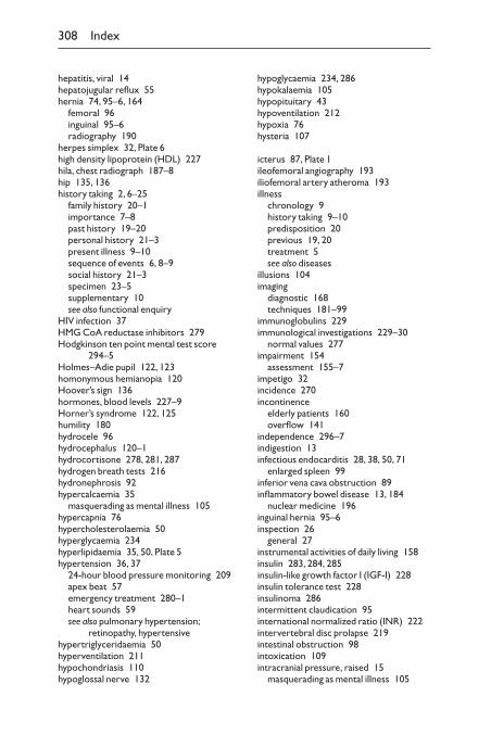
308 Index
hepatitis, viral 14hepatojugular reflux 55hernia 74, 95–6, 164
femoral 96inguinal 95–6radiography 190
herpes simplex 32, Plate 6high density lipoprotein (HDL) 227hila, chest radiograph 187–8hip 135, 136history taking 2, 6–25
family history 20–1importance 7–8past history 19–20personal history 21–3present illness 9–10sequence of events 6, 8–9social history 21–3specimen 23–5supplementary 10see also functional enquiry
HIV infection 37HMG CoA reductase inhibitors 279Hodgkinson ten point mental test score
294–5Holmes–Adie pupil 122, 123homonymous hemianopia 120Hoover’s sign 136hormones, blood levels 227–9Horner’s syndrome 122, 125humility 180hydrocele 96hydrocephalus 120–1hydrocortisone 278, 281, 287hydrogen breath tests 216hydronephrosis 92hypercalcaemia 35
masquerading as mental illness 105hypercapnia 76hypercholesterolaemia 50hyperglycaemia 234hyperlipidaemia 35, 50, Plate 5hypertension 36, 37
24-hour blood pressure monitoring 209apex beat 57emergency treatment 280–1heart sounds 59see also pulmonary hypertension;
retinopathy, hypertensivehypertriglyceridaemia 50hyperventilation 211hypochondriasis 110hypoglossal nerve 132
hypoglycaemia 234, 286hypokalaemia 105hypopituitary 43hypoventilation 212hypoxia 76hysteria 107
icterus 87, Plate 1ileofemoral angiography 193iliofemoral artery atheroma 193illness
chronology 9history taking 9–10predisposition 20previous 19, 20treatment 5see also diseases
illusions 104imaging
diagnostic 168techniques 181–99
immunoglobulins 229immunological investigations 229–30
normal values 277impairment 154
assessment 155–7impetigo 32incidence 270incontinence
elderly patients 160overflow 141
independence 296–7indigestion 13infectious endocarditis 28, 38, 50, 71
enlarged spleen 99inferior vena cava obstruction 89inflammatory bowel disease 13, 184
nuclear medicine 196inguinal hernia 95–6inspection 26
general 27instrumental activities of daily living 158insulin 283, 284, 285insulin-like growth factor I (IGF-I) 228insulin tolerance test 228insulinoma 286intermittent claudication 95international normalized ratio (INR) 222intervertebral disc prolapse 219intestinal obstruction 98intoxication 109intracranial pressure, raised 15
masquerading as mental illness 105
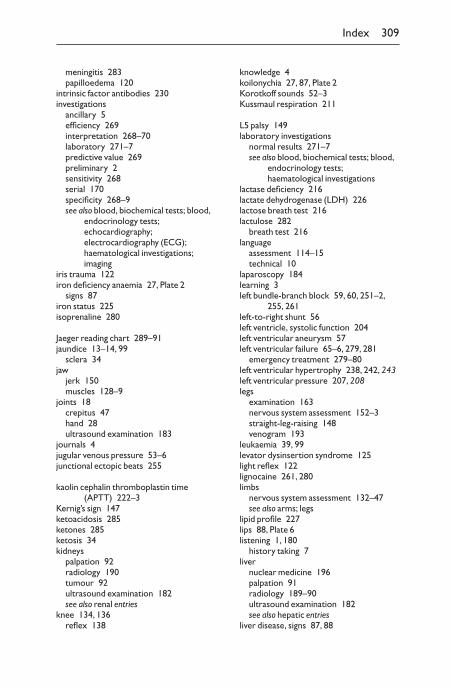
Index 309
meningitis 283papilloedema 120
intrinsic factor antibodies 230investigations
ancillary 5efficiency 269interpretation 268–70laboratory 271–7predictive value 269preliminary 2sensitivity 268serial 170specificity 268–9see also blood, biochemical tests; blood,
endocrinology tests;echocardiography;electrocardiography (ECG);haematological investigations;imaging
iris trauma 122iron deficiency anaemia 27, Plate 2
signs 87iron status 225isoprenaline 280
Jaeger reading chart 289–91jaundice 13–14, 99
sclera 34jaw
jerk 150muscles 128–9
joints 18crepitus 47hand 28ultrasound examination 183
journals 4jugular venous pressure 53–6junctional ectopic beats 255
kaolin cephalin thromboplastin time(APTT) 222–3
Kernig’s sign 147ketoacidosis 285ketones 285ketosis 34kidneys
palpation 92radiology 190tumour 92ultrasound examination 182see also renal entries
knee 134, 136reflex 138
knowledge 4koilonychia 27, 87, Plate 2Korotkoff sounds 52–3Kussmaul respiration 211
L5 palsy 149laboratory investigations
normal results 271–7see also blood, biochemical tests; blood,
endocrinology tests;haematological investigations
lactase deficiency 216lactate dehydrogenase (LDH) 226lactose breath test 216lactulose 282
breath test 216language
assessment 114–15technical 10
laparoscopy 184learning 3left bundle-branch block 59, 60, 251–2,
255, 261left-to-right shunt 56left ventricle, systolic function 204left ventricular aneurysm 57left ventricular failure 65–6, 279, 281
emergency treatment 279–80left ventricular hypertrophy 238, 242, 243left ventricular pressure 207, 208legs
examination 163nervous system assessment 152–3straight-leg-raising 148venogram 193
leukaemia 39, 99levator dysinsertion syndrome 125light reflex 122lignocaine 261, 280limbs
nervous system assessment 132–47see also arms; legs
lipid profile 227lips 88, Plate 6listening 1, 180
history taking 7liver
nuclear medicine 196palpation 91radiology 189–90ultrasound examination 182see also hepatic entries
liver disease, signs 87, 88
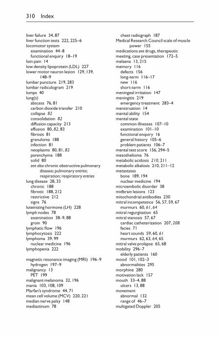
310 Index
liver failure 34, 87liver function tests 222, 225–6locomotor system
examination 44–8functional enquiry 18–19
loin pain 14low density lipoprotein (LDL) 227lower motor neuron lesion 129, 139,
148–9lumbar puncture 219, 283lumbar radiculogram 219lumps 40lung(s)
abscess 76, 81carbon dioxide transfer 210collapse 82consolidation 82diffusion capacity 213effusion 80, 82, 83fibrosis 81granuloma 188infection 81neoplasms 80, 81, 82parenchyma 188solid 80see also chronic obstructive pulmonary
disease; pulmonary entries;respiration; respiratory entries
lung disease 28, 33chronic 188fibrotic 188, 212restrictive 212signs 76
luteinizing hormone (LH) 228lymph nodes 78
examination 38–9, 88groin 90
lymphatic flow 196lymphocytosis 222lymphoma 39, 99
nuclear medicine 196lymphopenia 222
magnetic resonance imaging (MRI) 196–9hydrogen 197–9
malignancy 13PET 199
malignant melanoma 32, 196mania 103, 108, 109Marfan’s syndrome 44, 71mean cell volume (MCV) 220, 221median nerve palsy 148mediastinum 78
chest radiograph 187Medical Research Council scale of muscle
power 155medications see drugs, therapeuticmeeting, case presentation 172–5melaena 13, 215memory 116
defects 156long-term 116–17new 116short-term 116
meningeal irritation 147meningitis 219
emergency treatment 283–4menstruation 14mental ability 154mental state
common illnesses 107–10examination 101–10functional enquiry 16general history 105–6problem patients 106–7
mental test score 156, 294–5mesothelioma 76metabolic acidosis 210, 211metabolic alkalosis 210, 211–12metastases
bone 189, 194nuclear medicine 194
microembolic disorder 38midbrain lesions 123mitochondrial antibodies 230mitral incompetence 56, 57, 59, 67
murmurs 60, 61, 64mitral regurgitation 65mitral stenosis 57, 67
cardiac catheterization 207, 208facies 71heart sounds 59, 60, 61murmurs 62, 63, 64, 65
mitral valve prolapse 65, 68mobility 296–7
elderly patients 160mood 101, 102–3
abnormalities 295morphine 280motivation lack 157mouth 33–4, 88
ulcers 13, 88movement
abnormal 132range of 46–7
multigated Doppler 205
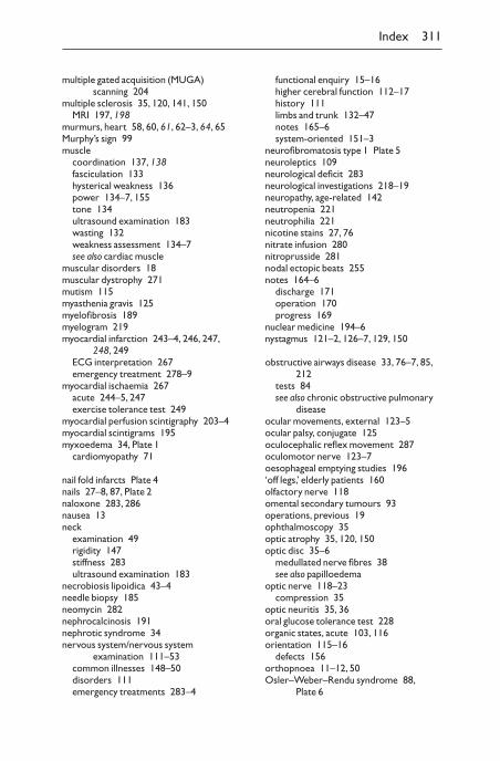
Index 311
multiple gated acquisition (MUGA)scanning 204
multiple sclerosis 35, 120, 141, 150MRI 197, 198
murmurs, heart 58, 60, 61, 62–3, 64, 65Murphy’s sign 99muscle
coordination 137, 138fasciculation 133hysterical weakness 136power 134–7, 155tone 134ultrasound examination 183wasting 132weakness assessment 134–7see also cardiac muscle
muscular disorders 18muscular dystrophy 271mutism 115myasthenia gravis 125myelofibrosis 189myelogram 219myocardial infarction 243–4, 246, 247,
248, 249ECG interpretation 267emergency treatment 278–9
myocardial ischaemia 267acute 244–5, 247exercise tolerance test 249
myocardial perfusion scintigraphy 203–4myocardial scintigrams 195myxoedema 34, Plate 1
cardiomyopathy 71
nail fold infarcts Plate 4nails 27–8, 87, Plate 2naloxone 283, 286nausea 13neck
examination 49rigidity 147stiffness 283ultrasound examination 183
necrobiosis lipoidica 43–4needle biopsy 185neomycin 282nephrocalcinosis 191nephrotic syndrome 34nervous system/nervous system
examination 111–53common illnesses 148–50disorders 111emergency treatments 283–4
functional enquiry 15–16higher cerebral function 112–17history 111limbs and trunk 132–47notes 165–6system-oriented 151–3
neurofibromatosis type 1 Plate 5neuroleptics 109neurological deficit 283neurological investigations 218–19neuropathy, age-related 142neutropenia 221neutrophilia 221nicotine stains 27, 76nitrate infusion 280nitroprusside 281nodal ectopic beats 255notes 164–6
discharge 171operation 170progress 169
nuclear medicine 194–6nystagmus 121–2, 126–7, 129, 150
obstructive airways disease 33, 76–7, 85,212
tests 84see also chronic obstructive pulmonary
diseaseocular movements, external 123–5ocular palsy, conjugate 125oculocephalic reflex movement 287oculomotor nerve 123–7oesophageal emptying studies 196‘off legs,’ elderly patients 160olfactory nerve 118omental secondary tumours 93operations, previous 19ophthalmoscopy 35optic atrophy 35, 120, 150optic disc 35–6
medullated nerve fibres 38see also papilloedema
optic nerve 118–23compression 35
optic neuritis 35, 36oral glucose tolerance test 228organic states, acute 103, 116orientation 115–16
defects 156orthopnoea 11–12, 50Osler–Weber–Rendu syndrome 88,
Plate 6
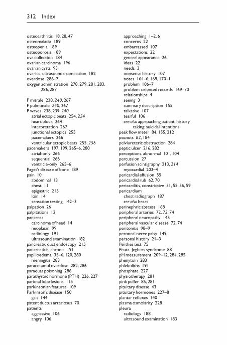
312 Index
osteoarthritis 18, 28, 47osteomalacia 189osteopenia 189osteoporosis 189ova collection 184ovarian carcinoma 196ovarian cysts 93ovaries, ultrasound examination 182overdose 286–7oxygen administration 278, 279, 281, 283,
286, 287
P mitrale 238, 240, 267P pulmonale 240, 267P waves 238, 239, 240
atrial ectopic beats 254, 254heart block 264interpretation 267junctional ectopics 255pacemakers 266ventricular ectopic beats 255, 256
pacemakers 197, 199, 265–6, 280atrial-only 266sequential 266ventricle-only 265–6
Paget’s disease of bone 189pain 10
abdominal 13chest 11epigastric 215loin 14sensation testing 142–3
palpation 26palpitations 12pancreas
carcinoma of head 14neoplasm 99radiology 191ultrasound examination 182
pancreatic duct endoscopy 215pancreatitis, chronic 191papilloedema 35–6, 120, 280
meningitis 283paracetamol overdose 282, 286paraquat poisoning 286parathyroid hormone (PTH) 226, 227parietal lobe lesions 115parkinsonian features 109Parkinson’s disease 150
gait 144patent ductus arteriosus 70patients
aggressive 106angry 106
approaching 1–2, 6concerns 22embarrassed 107expectations 22general appearance 26ideas 22needs 3nonsense history 107notes 164–6, 169, 170–1problem 106–7problem-oriented records 169–70relationships 4seeing 3summary description 155talkative 107tearful 106see also approaching patient; history
taking; suicidal intentionspeak flow meter 84, 155, 212peanuts 82, 184pelviureteric obstruction 284peptic ulcer 216, 282perceptions, abnormal 101, 104percussion 27perfusion scintigraphy 213, 214
myocardial 203–4pericardial effusion 55pericardial rub 62, 70pericarditis, constrictive 51, 55, 56, 59pericardium
chest radiograph 187see also heart
perinephric abscess 168peripheral arteries 72, 73, 74peripheral neuropathy 145peripheral vascular disease 72, 74peritonitis 98–9peroneal nerve palsy 149personal history 21–3Perthes test 75Peutz–Jeghers syndrome 88pH measurement 209–12, 284, 285phenytoin 283phleboliths 191phosphate 227physiotherapy 281pink puffer 85, 281pituitary disease 43pituitary hormones 227–8plantar reflexes 140plasma osmolarity 228pleura
radiology 188ultrasound examination 183

Index 313
pleural rub 81pleurisy 81pneumonia 81, 82pneumoperitoneum 190pneumothorax 80, 82, 188poisoning 286–7poisons centre 287polyarthritis 46polycystic disease 92porphyria 105portal hypertension 89, 97, 99, 282position sense 142positron emission tomography (PET) 199postural instability 150posture 132potassium
depletion 211plasma levels 284replacement 285
PR interval 263, 267precordial thump 298precordium 56–8pregnancy 93prevalence 270problems list 166–71prochlorperazine 280proctoscopy 183progressive supranuclear palsy 127prolactin 228proprioception 142prostate 97
enlarged 284prostatism 14prosthetic heart valves 70, 222prothrombin time 222proton pump blocker 282psoas lines 191psoriasis 28, 32, 48, Plate 3psychosis 104, 108, 109ptosis 125pulmonary angiography 193
see also lungpulmonary artery atresia 188pulmonary embolism 188, 222, 223
computed tomography 192pulmonary angiography 193V/Q scan 214
pulmonary fibrosis 213pulmonary hypertension 55
Graham Steell murmur 65pulmonary system, nuclear medicine
195pulmonary valve
incompetence 62
regurgitation 65stenosis 55, 59, 65
pulmonary vessels 188pulses
peripheral 72, 73radial 51–2
pulsus alternans 52pulsus bigeminus 51pulsus paradoxus 51, 85pupils 122–3pyloric stenosis 13, 98pyramidal lesions 133pyrexia of unknown origin 168pyrophosphate scanning 204
Q waves 238pathological 243–4
QRS axis 249–50, 251QRS complex 240–2, 245, 249, 251–64,
265atrial ectopic beats 254heart block 263, 264interpretation 267left axis deviation 250pacemakers 265–6right axis deviation 251tachycardias 256–7, 258ventricular ectopic beats 255ventricular tachycardia 261Wolff–Parkinson–White syndrome 261
questionnaires 155, 157–8questions 8
functional enquiry 10–11previous illnesses 20
R wave 240–2, 243QRS complex 249
radial artery blood gas sampling 233radial nerve palsy 149radial pulse palpation 51–2, 73radio-opaque contrast 207radiology 185–93, 194radionuclide imaging, cardiology 203–4radionuclide ventriculography 204râles 81, 85rash
drug reactions 30, 31, Plate 3purpuric 283
Raynaud’s syndrome 27, Plate 2reading chart
Jaeger 289–91Snellen 292–3
reasoning 117reassurance 180
314 Index
recordsproblem-oriented 169–70see also notes
rectal examination 96–7red cells 220–1
antibody screening techniques 224enzymes 221
reference intervals 271reflex testing 138–40
jerks 139trunk 140–1
rehabilitation 157relationships 4relatives, history taking 10, 107renal angiography 193
see also kidneysrenal arteries, bruits 94–5renal arteriovenous fistula 193renal artery stenosis 95, 193renal failure
acute 279, 284–5chronic 284
renal investigations 217–18renal obstruction 284renal scintigraphy 195renal stones 191renal tubular necrosis 284renography 195respiration
investigations 209–13, 214pattern 76
respiratory acidosis 210, 212respiratory alkalosis 210, 211respiratory failure 76
arterial blood gases 209–10chronic 210see also chronic obstructive pulmonary
disease; obstructive airways diseaserespiratory rate 76, 283
overdose 286respiratory system
emergency treatments 281examination 162functional enquiry 11–13notes 165see also lung(s)
reticuloendothelial system 196retina examination 36–8retinal vein occlusion 38retinitis pigmentosa 38retinopathy
diabetic 36, 37, 38, 44, Plate 6hypertensive 36, 37, Plate 6
Rhesus blood group 224rheumatic fever 71rheumatoid arthritis 18, 28, 47, Plate 2rheumatoid factor 230rhonchi 81right bundle-branch block 59, 238, 252–3,
255, 261right ventricular atresia 188right ventricular failure 66right ventricular hypertrophy 57, 243rigidity 150Rinne’s test 130rodent ulcer 31, Plate 5Romberg’s test 146Roth’s spots 38
S wave 241–2, 243QRS complex 249
S1 palsy 149saccades 127saline, normal 285saphenous veins 74sarcoid 168scabies 32schizophrenia 103, 104, 108–9
cerebral scintigraphy 195sciatic nerve 148sciatica 148scleroderma Plate 4scotoma 120scrotum 96
ultrasound examination 183Seldinger technique 206self-care 296–7self-reliance 2Sengstaken tube 282sensation
examination 141–7loss 147
sensory inattention 120, 143sensory perception loss 150septic arthritis 18septicaemia 286serum levels, normal ranges 276–7sexual intercourse 14short Synacthen test 228shoulder 135sick sinus syndrome 266sigmoidoscopy 184silence 180sinoatrial node 239sinus arrest 262sinus arrhythmia 51, 253
Index 315
sinus bradycardia 262, 266sinus rhythm 266sinus tachycardia 266sixth nerve lesion Plate 6skeleton
nuclear medicine 194radiology 189
skin Plate 3allergen testing 212diseases 31–3inspection 29lesions 29–31see also rash
skull palpation 117sleep 16smiling 180smoking 13, 22social history 21–3sodium 284sodium chloride 279, 282, 283, 286, 287soft-tissue tumours 197somatization 110speech 101, 103
assessment 114–15scanning 150staccato 150
sphincter disturbance 15sphincterotomy 215spider naevi 87, Plate 1spinal cord
lesions 197tumours 219
spinecurvature 45, 77examination 162inspection 117see also ankylosing spondylitis
spirometry 212spleen
enlarged 99nuclear medicine 196palpation 92radiology 190ultrasound examination 182
sputum 12, 83, 85culture 281plug 82, 184
squamous cell carcinoma 32ST segment 238
changes 208depression 200, 249elevation 244, 245, 247interpretation 267
left bundle-branch block 251myocardial infarction 278
staff relationships 4Staphylococcus aureus 168statistical analyses 5status asthmaticus 51steatorrhoea 13stents 215stereognosis 143sterilization 184stomach radiography 190strabismus 125
non-paralytic 124streptokinase 278, 279stress testing 199–200subarachnoid haemorrhage 219, 284subarachnoid space 219subtraction angiography, background 193,
194suicidal intentions 103, 106, 283superior vena cava obstruction 71, 89supinator reflex 138supraventricular tachycardia (SVT)
259–60, 266swallowing 13, 131symptoms 7, 8
checklist 10–19description 9–10
syncope 12syndrome of inappropriate antidiuretic
hormone (SIADH) 228syphilis 122, 125
tabes dorsalis 147systemic lupus erythematosus (SLE) 99
T waves 238, 242interpretation 267inversion 249left bundle-branch block 251
T1 palsy 148, Plate 6tabes dorsalis 147tachycardia 51, 256–61, 266
atrial 208, 267classification 256–7emergency treatment 280re-entry 260supraventricular 259–60, 266ventricular 261, 266
tamponade 51, 55tardive dyskinesia 109teeth 33, 88telangiectasia 87, Plate 1, Plate 6
gastrointestinal 88
316 Index
tendon reflexes 138–40jerks 139
testes 96testicular torsion 195textbooks 4thalassaemia 221thallium scanning 203–4therapy, information required 3thermal sensation 143thermodilution technique 207thinking 101, 103third nerve palsy 122, 123, 125thoracocentesis 188thrombin time 223thrombophilia tests 223thrombosis 223
see also deep vein thrombosis;pulmonary embolism
thyroidantibody 230cystic 42hormones 227nuclear medicine studies 196
thyroid disease 28, 41–3, 227, Plate 1eye 34, Plate 1functional enquiry 19masquerading as mental illness 105muscle 125
thyroid-stimulating hormone (TSH) 227thyrotoxic nodules 196thyrotoxicosis 42–3, Plate 1
apex beat 57atrial fibrillation 71pulse volume 52
tissue plasminogen activator (t-PA) 278tongue 33, 87, 88
hypoglossal nerve 132touch, light 143trachea 78transverse myelitis 150trapezius muscle 131travel abroad 13, 14treadmill exercise test 155tremor 132, 150
intention 137Parkinson’s disease 144
Trendelenburg test 74–5tricuspid incompetence 56, 69tricuspid regurgitation 65tricuspid stenosis 55, 65trigeminal nerve 127–9triglycerides 227trochlear nerve 123–7
troponins 226, 285trunk reflexes 140–1tuberculosis 188Turner’s syndrome 71two-point discrimination 143
ulcerative colitis 88, 184, 216ulnar nerve
lesion Plate 6palsy 148–9
ultrasound cardiography, Doppler 204–5ultrasound examination 181–3, 284
Doppler 204–5unconsciousness
emergency treatment 283see also coma
understanding of condition 101, 105upper motor neuron
lesion 129, 139, 149signs 150
urea measurement 225, 284ureters 218
stones 284urinary bladder
cystoscopy 184enlarged 93palpation 141
urine 14culture 285, 286ketones 285microscopy 217, 284osmolarity 228, 284output in myocardial infarction 279retention 141, 150
urine testing 217normal values 275–6
urogenital system, nuclear medicine 195urogram, intravenous 218urticaria 32–3uterus, ultrasound examination 182
vacutainer system 232vaginal bleeding 14vaginal discharge 14vaginal examination 97vagus nerve 131
stimulation 260varicose ulcers 74–5, Plate 4varicose veins 164vasculitis 30, 37, Plate 3vein filling 90venepuncture 230–1venography 192–3
Index 317
venous blood sampling 193ventilation, mechanical 280, 281, 283, 287ventilation/perfusion (V/Q) scan 213, 214ventilatory failure 209ventolin 281ventricles 239
fibrosed 59see also left ventricular entries; right
ventricular entriesventricular ectopic beats 255, 256ventricular fibrillation 261, 280ventricular function 204ventricular hypertrophy 267ventricular septal defect 65, 70ventricular tachycardia 208verapamil 280vertigo 15vibration sense 142Virchow’s node 88vision 15, 17–18
blindness 123visual acuity 118–19
chart 292–3visual fields 119–20
defects 122vitalograph 212vitamin B 282vitamin B12 deficiency 87vitamin D 226, 227vitamin K 282
vitiligo 33vocal fremitus 83vocal resonance 83vomiting 13von Willebrand’s disease 223
walkingheel-to-toe 144see also gait
ward round, case presentation 175–6warfarin therapy 222Weber’s test 130weight loss, unexplained 215Wenckebach heart block 263Wernicke’s area lesion 114wheezing 81, 84whispering pectoriloquy 83white-band keratopathy 35white cells 221–2white coat effect 209Wolff–Parkinson–White syndrome 260,
261, 267wrist 134, 135wrist drop 132
xanthelasma 34, 50xanthelasma–cholesterol deposits
Plate 5xanthochromia 219, 284xanthoma 50