clinical presentation and management of african horse
TRANSCRIPT

Clinical presentation and management of African Horse
Sickness in two dogs.
Journal: Veterinary Record Case Reports
Manuscript ID vetreccr-2018-000664.R1
Manuscript Type: Companion or pet animals
Species: Dogs
Date Submitted by the Author: 30-Jul-2018
Complete List of Authors: Whitehead, Zandri; TAH Specialist Medicine Clinic le Roux, Christelle; University of Pretoria, Companion Animal Clinical Studies O'Dell, Nicolize; University of Pretoria, Paraclinical Sciences Hanekom, Josef; University of Pretoria, Companion Animal Clinical Studies
Keywords: African horse sickness, Dogs, Treatment, Radiography, Cytology
Topics: African horse sickness, Vector-borne diseases, Orbiviruses
Abstract:
A ten-month-old, female, spayed Beagle (case 1) and an unrelated two-year-old, female, intact Labrador Retriever (case 2), both living in Pretoria, South Africa, presented individually on separate occasions with acute onset dyspnoea and severe hypoxia. Thoracic radiographs demonstrated severe, diffuse interstitial to alveolar lung patterns with mild pleural and mediastinal effusion. Mixed airway inflammation was seen on trans-tracheal aspirate cytology in case 1. Both cases received supportive therapy but only one dog survived (case 2).
African Horse Sickness (AHS) was diagnosed at necropsy based on histopathology and immunohistochemistry in case 1 and ante-mortally in case 2, using reverse-transcriptase polymerase chain reaction (RT-PCR) on whole blood. To the authors’ knowledge, this is the first report to detail the haematological-, biochemical-, thoracic radiological-, arterial blood gas- and trans-tracheal aspirate cytology findings of AHS in dogs. This report also describes the treatment of a dog surviving clinical AHS infection.

Revised August 2017 Page 1 of 9
TITLE OF CASE
Clinical presentation and management of African Horse Sickness in two dogs.
SUMMARY
A ten-month-old, female, spayed Beagle (case 1) and an unrelated two-year-old, female,
intact Labrador Retriever (case 2), both living in Pretoria, South Africa, presented
individually on separate occasions with acute onset dyspnoea and severe hypoxia. Thoracic
radiographs demonstrated severe, diffuse interstitial to alveolar lung patterns with mild
pleural and mediastinal effusion. Mixed airway inflammation was seen on trans-tracheal
aspirate cytology in case 1.
Both cases received supportive therapy but only one dog survived (case 2).
African Horse Sickness (AHS) was diagnosed at necropsy based on histopathology and
immunohistochemistry in case 1 and ante-mortally in case 2, using reverse-transcriptase
polymerase chain reaction (RT-PCR) on whole blood.
To the authors’ knowledge, this is the first report to detail the haematological-, biochemical-,
thoracic radiological-, arterial blood gas- and trans-tracheal aspirate cytology findings of AHS
in dogs. This report also describes the treatment of a dog surviving clinical AHS infection.
BACKGROUND
What is AHS
African Horse Sickness (AHS) is a World Organisation for Animal Health (OIE)- listed, non-
contagious disease of equids, caused by an arthropod-borne orbivirus of the family
Reoviridae.(1-3) The African horse sickness virus (AHSV) is endemic in Sub-Saharan Africa with intermittent outbreaks reported in the Near- and Middle East, northern Africa and
Europe.(2, 4) In naïve horses, the disease is usually peracute to acute with a greater than
90% mortality rate.
Dogs are the only known non-equid species that contract a highly fatal form of AHS.(2)

Revised August 2017 Page 2 of 9
African horse sickness virus has been shown to affect dogs after experimental intravenous
inoculation with infected horse blood.(5) Serological surveys have reported evidence of
widespread natural AHS infection among various African carnivore species with
seroconversion in dogs as high as 34-43% reported in endemic areas. (4, 6-9) Infection in
these carnivores is proposed to result from ingestion of meat and organs from AHS-infected
prey species.(4) Sporadic outbreaks of AHS in domestic dogs have occurred in association
with ingestion of virus-contaminated horse meat.(10-12) In 2013, the first case of AHS in a
dog without apparent ingestion of horse meat was reported(6) and from 2006-2017, 33
spontaneous cases of canine AHS have been diagnosed post-mortally at the Section of
Pathology, Department of Paraclinical Sciences, Faculty of Veterinary Science, University of
Pretoria.(13)
The previously reported signs of AHS in dogs have ranged from asymptomatic to transient
pyrexia(12), “classical horse sickness symptoms”, including hyperpnoea, moist rales on
auscultation, white foam around the nostrils, coughing and death.(10, 12) Descriptions
beyond basic clinical signs and pathological findings are scant and many reports were
published more than 50 years ago. The complete clinical, laboratory and radiological findings
of AHS in dogs has not been reported to date.
The authors report the clinical presentation and treatment of two cases of canine AHS, as well as the necropsy findings of one dog and emphasise the consideration of this disease as
a differential diagnosis in dogs with acute respiratory signs in areas where equine AHS
occurs.
CASE PRESENTATION
Two unrelated dogs presented individually to the Onderstepoort Veterinary Academic
Hospital (OVAH), Pretoria, South Africa on separate occasions with acute respiratory
distress.
History and Clinical Assessment
Case 1
A ten-month-old spayed female Beagle presented with a one-day history of lethargy,
vomiting and acute collapse in May of 2016. The dog was part of the Onderstepoort Teaching
Animal Unit (OTAU) breeding colony and was housed on the campus of the University of
Pretoria, Faculty of Veterinary Science, in a roofed enclosure with grass pen access, in close
proximity to horse paddocks. The faculty is located in an enzootic AHS area. (8)
All horses that reside on the campus are vaccinated annually with a live attenuated African
Horse Sickness vaccine 1 (containing AHS serotype 1, 3 and 4) in October and three weeks later with the OBP African Horse Sickness vaccine 2 (containing AHS serotypes 2, 6, 7, and
8) according to manufacturer’s instructions (African Horse Sickness Vaccine for horses,
mules and donkeys, Onderstepoort Biological Products). Foals are vaccinated at weaning and
again six months later.
Nose-to-nose contact between dogs and horses, and sharing of water points is plausible
when the dogs are taken on regular walks, but the dogs do not enter the OVAH, Equine
Department buildings, where clinically ill or injured horses are stabled. No other dogs in the
colony were reported to show clinical abnormalities. Vaccinations of case 1 were fully up to
date and a veterinary-formulated, commercial diet was fed solely.
On physical examination, the dog was collapsed with depressed mentation and a body
condition score (BCS) of 4/9. Tachypnoea (60 breaths per minute) with severe inspiratory-
expiratory dyspnoea, costo-abdominal breathing and bilaterally referred lung sounds and
crackles were evident. The remainder of the clinical examination was unremarkable.
Case 2
A client-owned, two-year-old intact female Labrador Retriever presented with a three-day
history of partial anorexia and one day of non-productive retching and lethargy in February
of 2017. Vaccination history was unclear. The dog was a sole-pet from a low-density
suburban, residential area, approximately 15km from the Veterinary Faculty. The dog had no
contact with horses and no horses were stabled in the vicinity. The dog had no known access
Revised August 2017 Page 3 of 9
to horse meat and was fed a commercial diet.
On physical examination, the dog was depressed with BCS 5/9. Marked inspiratory-
expiratory dyspnoea with referred lung sounds and crackles were evident. Mild hypothermia
(37°C), tachycardia (168 beats per minute) and tachypnoea (100 breaths per minute) was
seen. The remainder of the clinical examination was unremarkable.
INVESTIGATIONS
Case 1
Complete blood count revealed haemoconcentration (haematocrit 0.65 L/L, ref: 0.37 –
0.55), lymphopenia (0.6 x 10⁹/L, ref: 1 – 4.8), eosinopenia (0.00 x 10⁹/L, ref: 0.1 – 1.25)
and thrombocytopenia (118 x 109/L, ref: 200 – 500). Biochemistry revealed
hypoproteinaemia (41.9 g/L, ref: 56 – 73), hypoalbuminaemia (27.2 g/L, ref: 28 – 41),
hypoglobulinaemia (14.7 g/L, ref: 20 – 41), increased alkaline phosphatase (496 U/L, ref: 20
– 165), low creatinine (33 umol/L, ref:59 – 109), hyponatraemia (133 mmol/L, ref: 142 –
151) and hypochloraemia (102 mmol/L, ref: 107 – 117).
Distemper antigen ELISA lateral flow test (Antigen rapid CDV Ag Test Kit, BioNote
Inc.) tested negative. This test was chosen for its speed and availability. The dog arrived at
the clinic prior to a weekend and serological testing was not available until the following Monday. The rapid death of this dog precluded further ante-mortem testing for distemper.
Unsedated thoracic radiographs (Fig. 1 A & B) revealed a severe perihilar to hilar alveolar
lung pattern, extending into the dorso-caudal lungs, with fissure lines and reverse fissure
lines indicating mild pleural and mediastinal effusion, respectively. The alveolar pattern had
a distinct peribronchial “track-like” appearance.
Arterial blood gas analysis revealed severe hypoxaemia (PaO2 53.4 mmHg, ref 81 – 103),
acute lung injury (PaO2/FiO2 273) and an increased arterial oxygen gradient (A-a) of 49.2.
The A-a gradient did not improve after oxygen administration.
A modified trans-tracheal wash (TTW) was performed under local anaesthesia (lidocaine
injection 2%, Bayer Animal Health). Cytological evaluation of the obtained sample revealed a
moderately mucoid background with a high number of nucleated cells. The nucleated cells
consisted of 30% non-degenerate, mature neutrophils, 5% small lymphocytes and 62,5 %
large macrophages that were markedly vacuolated and phagocytosing cellular debris. A high
number of ciliated and non-ciliated epithelial cells and plaques of basophilic basal epithelial
cells (indicating trauma) was visible (Fig. 2 A & B). No microorganisms were seen.
On presentation of this case in 2016, AHS in dogs was not widely known and clinical findings
such as radiographic findings had not been described, thus AHS was not considered as a
differential diagnosis, especially with case 1 being from the controlled colony of dogs that are
not fed horse meat. Prior AHS diagnoses in dogs at the pathology section were all made in
dogs referred from other veterinary clinics, or brought in by owners for necropsy. The post-
mortem diagnosis was made after TTW fluid and EDTA blood samples were already
discarded, hence no RT-PCR was performed in case 1. Retrospective RT-PCR was attempted
on wax-imbedded histological samples after RNA extraction, but this proved fruitless.
Case 2
Haematology revealed mature neutrophilia (27.2 x 10⁹/L, ref: 6-15), monocytosis (2.72 x
109/L, ref: 0.15 – 1.35), eosinopenia (0.00 x 10⁹/L, ref: 0.1 – 1.25) and basophilia (0.27 x
109/L, ref: 0.1 x 10⁹/L). The biochemistry and urinalysis findings were within normal limits.
Unsedated thoracic radiographs (Fig. 3 A & B) were taken within one hour of presentation
and a moderate diffuse interstitial to alveolar pattern affecting all the lungs lobes was
present, with an asymmetrical more severely affected right lung appearance. Mild fissure
and reverse fissure lines were present. Radiographs were repeated 38 hours later (Fig. 3 C &
D) and demonstrated a marked improvement of the lung pattern, and improvement of the
fissure and reverse fissure lines.
Peripheral oxygen saturation (Sp02) (Nonin Model 9847V, Nonin Medical Inc.) on
presentation was 85% and remained below 90% despite oxygen supplementation.
Revised August 2017 Page 4 of 9
Based on an increased awareness of AHS in dogs and a recent diagnosis of AHS in Case 1
with similar presentation, AHSV specific duplex real-time reverse transcriptase quantitative
PCR (RT-PCR) was performed ante-mortally in case 2, which tested positive for AHS. The
test methodology was performed as described by Guthrie et al.(14) This RT-PCR test is OIE
accredited (15), and was run at the Veterinary Genetics Laboratory (Equine Research
Centre), Faculty of Veterinary Science, University of Pretoria – Onderstepoort.
DIFFERENTIAL DIAGNOSIS
Based on the radiographic and arterial blood gas findings, differential diagnoses included
non-cardiogenic pulmonary oedema, most likely as a result of acute respiratory distress
syndrome (ARDS). Pneumonia was considered unlikely given the haematology and the
radiological findings. Pulmonary haemorrhage with haemothorax and mediastinal
haemorrhage was also unlikely as there was no history of trauma or toxin ingestion in either
dog, as well as a lack of cytological evidence based on the TTW cytology in dog 1.
TREATMENT AND OUTCOME
Case 1
The dog received 0.3mg/kg promethazine hydrochloride (Phenergan, Sanofi-aventis) IM,
0.125 mg/kg dexamethasone (Kortico injection, Bayer Pty Ltd) IV once, and 20mg/kg
ampicillin (Ampicillin-Fresenius, Fresenius Kabi SA Pty Ltd) IV q8h. The dog was maintained
in an oxygen tank with oxygen supplementation at 3L/min and a constant rate infusion
(22mL/h) of lactated Ringer’s (Ringer-Lactate solution, Fresenius Kabi SA Pty Ltd.) was
administered.
Case 2
Oxygen was supplemented at 3L/min via intranasal cannula and 0.2mg/kg butorphanol (Dolorex®, Zoetis) IV was given once. The dog then received 0.1 mg/kg dexamethasone
(Kortico injection®, Bayer Pty Ltd) IV once, 2mg/kg furosemide (Salix®, MSD Intervet) IV
repeated after one hour and continued q8h for three days and thereafter 2mg/kg furosemide
(Mylan Furosemide®, Mylan) was given PO q8h for one day and q12h PO for another three
days. Additionally Amikacin sulphate at 15mg/kg (Amikacin Fresenius, Fresenius Kabi) IV
q24h for five days and 8.75 mg/kg amoxicillin-clavulanic acid (Synulox RTU, Zoetis) SC once
followed by 20mg/kg amoxicillin-clavulanic acid (Clavet, Cipla) PO q12h for three days. A
constant rate infusion of lactated Ringer’s (Ringer-Lactate solution, Fresenius Kabi SA Pty
Ltd) was given at 72mL/h.
OUTCOME AND FOLLOW-UP
Case 1
Despite supportive management, the dog’s vital signs declined and cardiopulmonary arrest
and death ensued 17 hours after initial presentation.
Gross post mortal examination of case 1 revealed generalised congestion, severe pulmonary
congestion and oedema associated with diffuse interstitial pneumonia and severe
hydrothorax. Histologically, the lungs were characterised by diffuse congestion, protein-rich
oedema and small amounts of fibrin filling the alveolar spaces associated with numerous
alveolar macrophages. The alveolar walls were distended by mononuclear cells and activated
endothelial cells. Small scattered areas of haemorrhage were present throughout the lung parenchyma (Fig. 4 A). African Horse Sickness-specific NS4 immunohistochemical
labelling(16) following the standard immunohistochemical protocol(17) revealed numerous
positive-labelling intra-alveolar macrophages, confirming an unexpected diagnosis of AHS
(Fig. 4 B). The details of this test with both positive and negative controls are described at
length by Zwart et al. (16)
No serum was stored in Case 1 to perform retrospective serology. Due to the low awareness
of the disease, AHS was not considered as a differential for the symptoms at the time the
dog presented.
Case 2
Gradual improvement and resolution of hypoxia was seen and the dog was discharged after
72 hours of hospitalisation. At the time of manuscript submission, the dog was reported to

Revised August 2017 Page 5 of 9
be doing well and no respiratory abnormalities were observed by the owners. A definitive
diagnosis of AHS by a positive, AHSV specific duplex real-time reverse transcriptase
quantitative PCR was confirmed only after discharging the animal from the hospital.
Case 2 was brought in for a re-evaluation eight months post-recovery, was clinically healthy,
and tested positive for AHS titres using VP7-based AHSV iELISA run at the Onderstepoort
Veterinary Institute according to the protocol described in Maree et al.(18) This test is not
validated for canines, but there are no currently available serological tests that are.
DISCUSSION
This case report is, as far as the authors are aware, the first report of the complete
haematological, biochemical, thoracic radiographic, arterial blood gas and trans-tracheal
wash cytology findings in dogs with AHS.
The clinical signs seen in these two cases are in agreement with previously reported signs of
AHS in dogs, including progressive inappetence, depressed habitus, pyrexia, hyperpnoea,
tachypnoea, coughing, moist rales on auscultation and white foam around the nostrils.(6, 10,
12, 13) Additional signs of diarrhoea and seizures have been reported by Haig, but some
dogs in that report were concurrently affected by Distemper viral infection.(10) More
recently, excessive salivation in addition to the abovementioned signs has been described.
(13) Not all dogs that contract AHS develop severe clinical signs and some dogs only display
mild pyrexia.(12)
The clinical signs were comparable to what is seen in horses affected by the pulmonary form
of AHS, with pyrexia, severe respiratory distress and frothy white to blood-tinged fluid at the
nostrils.(19) Horses also develop a more subacute or cardiac form of the disease that is
associated with pyrexia, subcutaneous oedema, conjunctival congestion, petechiation and
signs of colic.(19) Whether these clinical forms are also seen in dogs require verification. The
abovementioned signs occur as a result of increased vascular permeability caused by direct
and indirect injury to endothelial cells, particularly in organs such as the lungs.(1) It can only
be speculated that a similar underlying pathophysiological mechanism occurs in dogs.
The haematological and biochemical findings of the two affected dogs were non-specific. The
haemoconcentration in case 1 can be ascribed to dehydration. The lymphopenia, eosinopenia in case 1, and the stress leukogram in case 2, are non-specific findings and are expected in
ill dogs. The panhypoproteinaemia and low creatinine in case 1 can be attributed to the dog’s
young age and thin body condition. Prolonged prothrombin time, activated partial
thromboplastin time and increased fibrin degradation products have been described in horses
with AHS(20), but coagulation testing was not performed in the reported cases.
Although an alveolar pattern is not a specific finding on radiographs, the severity and diffuse
distribution may aid the diagnosis, and follow-up radiographs would be useful to monitor
progression of the disease. Mild pleural and mediastinal effusion are compatible with
vasculitis, but in this case, no other body cavity effusions could be confirmed on radiographs,
which may be expected given the mechanism of disease.
The alveolar arterial oxygen gradient (A-a) of 49.2 was indicative of a severe pulmonary ventilation perfusion mismatch or diffusion impairment. Failure of A-a gradient improvement
after oxygen administration was indicative of either alveolar collapse, flooding of alveoli with
fluid (pulmonary oedema/acute respiratory distress syndrome ARDS) or alveolar
consolidation.(21)
Airway cytological findings have not been reported previously in dogs with AHS. The TTW
cytology results in case 1 were consistent with severe inflammation but the presence of
macrophages was suggestive of a more sub-acute to chronic condition, although the clinical
signs were acute. A mixed inflammatory response is frequently seen in non-infectious
pulmonary disease such as inhalation pneumonia, lung lobe torsion or necrosis secondary to
a neoplastic lesion.(22) A bronco-alveolar lavage might have enabled collection of a more
representative sample, but the dog was not deemed stable enough to perform the
procedure.
An ante-mortem diagnosis of AHS can be confirmed by semi-quantitative RT-PCR on EDTA
Revised August 2017 Page 6 of 9
whole blood.(14) At necropsy, fresh lung tissue can be submitted for RT-PCR and in 10%
formalin for routine histopathology and IMP testing.(13, 16, 23) It was attempted to perform
IMP retrospectively on the cytological preparations of the TTW sample of case 1 but no
positive labelling was observed.
In previous reports, mortality in dogs has ranged from 20-95%.(8, 12, 13, 24) Not all dogs
that were fed infective meat developed clinical signs but once symptoms of respiratory
embarrassment had set in, the prognosis was poor and mortality high.(12) Case 2 in the
current report survived despite severe respiratory disease and hypoxia.
No specific therapy for equine AHS infection is available and treatment is supportive and
symptomatic.(19) Based on the tropism and multiplication of the virus in myocardial tissue,
regardless of the clinical form of AHS(25), current treatment recommendations by the OVAH
equine clinic include the addition of furosemide, angiotensin converting enzyme inhibitor and
pimobendan in AHS-affected horses.(26) There are no recommendations regarding
treatment in dogs with AHS. The addition of diuretic therapy could have influenced the
recovery in case 2 but no conclusions can be made in this regard.
The route of transmission of AHS to dogs that have not ingested horse meat, such as the
two dogs involved here, is unknown. Previous studies found that the major vector of AHS,
Culicoides imicola, is probably not attracted, or only very slightly attracted to dogs, thus
dogs are unlikely to act as vectors for AHS virus from infected dogs to horses.(27)
Mosquitoes may play a role in transmission of AHS amongst equine populations and do feed
on dogs in the absence of other preferable hosts.(27) The apparent increase in the incidence
of AHS in dogs, in an endemic AHS area warrants further investigation into the prevalence of
the disease in the canine population, the route of transmission to dogs and the current role
of canines in the maintenance cycle of AHS.
The apparent increase in AHS occurrence in dogs in South Africa raises the question whether
this represents an emerging canine disease. Additionally, this may have implications on
international transport and trade of canines in the future.
Control methods in horses in endemic AHS areas are limited to vaccination(28) with the live
attenuated AHS vaccination (African Horse Sickness Vaccine for horses, mules and donkeys,
Onderstepoort Biological Products) and midge avoidance strategies including stabling during
high midge activity, vector proofing stables and insect repellents.(29) On a national and
international level, the movement of horses from AHS area is strictly controlled with testing
of horses prior to movement.(3)
Due to the lack of knowledge of the current method of transmission, clear control or
prevention strategies are premature. Currently, further research is underway to determine
the sero-prevelance of AHS in dogs in the Pretoria area as well as potential epidemiological
factors that would help shape further studies into prevention strategies. Avoiding the feeding
of horse meat to dogs, keeping dogs indoors during times of high midge activity and
avoiding direct contact with sick horses (and recently vaccinated horse) or their water bowls
would be the presumable preventative measures that owners could adopt.
African horse sickness should be considered as a differential diagnosis in dogs with acute
respiratory signs, hypoxia and thoracic radiographic evidence of alveolar lung pattern and
pleural/mediastinal effusion in areas where equine AHS occurs.
LEARNING POINTS/TAKE HOME MESSAGES
• Dogs with AHS virus infection may present with acute respiratory distress,
tachypnoea, referred lung sound and crackles.
• Hypoxia and severe pulmonary ventilation perfusion mismatch was seen on arterial
blood gas analysis.
• Thoracic radiographic findings include a marked diffuse hilar to perihilar alveolar lung
pattern with mild pleural and mediastinal effusion.
• The prognosis of dogs that present in acute respiratory distress is poor but treatment with diuretic therapy, antibiotics and oxygen supplementation might be beneficial.
• Increased incidence of AHS in dogs can be anticipated due to the increased
Revised August 2017 Page 7 of 9
awareness of this condition and relative ease of diagnosis.
REFERENCES
References
1. Burrage TG, Laegreid WW. African horsesickness: Pathogenesis and immunity.
Comparative Immunology, Microbiology and Infectious Diseases. 1994;17(3):275-85.
2. MacLachlan NJ, Guthrie AJ. Re-emergence of bluetongue, African horse sickness, and
other Orbivirus diseases. Veterinary Research. 2010;41(6):41:35.
3. OIE-Listed diseases, infections and infestations in force in 2018.
http://www.oie.int/animal-health-in-the-world/oie-listed-diseases-2018/: World Organization of
Animal Heath; 2018.
4. Alexander KA, Kat PW, House J, House C, O'Brien SJ, Laurenson MK, et al. African
horse sickness and African carnivores. Veterinary Microbiology. 1995;47(1/2):133-40.
5. Theiler A. The susceptibility of the dog to african horse-sickness. Journal of Comparative
Pathology and Therapeutics. 1910;23:315-25.
6. Sittert SJv, Drew TM, Kotze JL, Strydom T, Weyer CT, Guthrie AJ. Occurrence of
African horse sickness in a domestic dog without apparent ingestion of horse meat. Journal of the
South African Veterinary Association. 2013;84(1):Art. #948.
7. Baba SS, Olaleye OD, Ayanbadejo OA. Haemagglutination-inhibiting antibodies against
African horse sickness virus in domestic animals in Nigeria. Veterinary research. 1993;24(6):483-
7.
8. McIntosh BM. Horsesickness antibodies in the sera of dogs in enzootic areas. Journal of
the South African Veterinary Association. 1955;26(4):269-72.
9. Awad FI, Amin MM, Salama SA, Aly MM. The incidence of African horse sickness
antibodies in animals of various species in Egypt. Bulletin of animal health and production in
Africa Bulletin des sante et production animales en Afrique. 1981;29(3):285-7.
10. Haig DA, McLNtosh BM, Cumming RB, Hempstead JFD. An outbreak of horsesickness,
complicated by distemper, in a pack of foxhounds. Journal of the South African Veterinary
Medical Association. 1956;27:245-9.
11. Piercy SE. Some observations on African horse-sickness including an account of an
outbreak amongst dogs. East African Agricultural Journal. 1951;17:62-4.
12. Rensburg IBJv, Clerk Jd, Groenewald HB, Botha WS. An outbreak of African
horsesickness in dogs. Journal of the South African Veterinary Association. 1981;52(4):323-5.
13. O'Dell N, Arnot L, Janisch CE, Steyl J. Clinical presentation and pathology of suspected
vector transmitted African horse sickness in South African domestic dogs from 2006 to 2017. The
Veterinary record. 2018.
14. Guthrie AJ, MacLachlan NJ, Joone C, Lourens CW, Weyer CT, Quan M, et al. Diagnostic
accuracy of a duplex real-time reverse transcription quantitative PCR assay for detection of
African horse sickness virus. Journal of Virological Methods. 2013;189(1):30-5.
15. Manual of Diagnostic Tests and Vaccines for Terrestrial Animals 2018.
http://www.oie.int/fileadmin/Home/eng/Health_standards/tahm/2.05.01_AHS.pdf: World
Organisation For Animal Health; 2018.
16. Zwart L, Potgieter CA, Clift SJ, van Staden V. Characterising Non-Structural Protein NS4
of African Horse Sickness Virus. PloS one. 2015;10(4):e0124281.
17. Ramos-Vara J, Miller M. When tissue antigens and antibodies get along: revisiting the
technical aspects of immunohistochemistry—the red, brown, and blue technique. Veterinary
pathology. 2014;51(1):42-87.
18. Maree S, Paweska JT. Preparation of recombinant African horse sickness virus VP7
antigen via a simple method and validation of a VP7-based indirect ELISA for the detection of
group-specific IgG antibodies in horse sera. Journal of Virological Methods. 2005;125(1):55-65.
19. Reed SM, Bayly WM, Sellon DC. Equine internal medicine: Elsevier Health Sciences;
2009.
20. Wilson D. Clinical veterinary advisor: The horse: Elsevier Health Sciences; 2010.
21. Bach JF. Hypoxemia: a quick reference. Vet Clin North Am Small Anim Pract.

Revised August 2017 Page 8 of 9
2008;38(3):423-6, vii.
22. Raskin RE, Meyer DJ. Canine and Feline cytology: A color atlas and interpretation Guide.
Third Edition ed. St. Louis, Missouri: Elsevier; 2016.
23. Ramos-Vara JA, Webster JD, DuSold D, Miller MA. Immunohistochemical evaluation of
the effects of paraffin section storage on biomarker stability. Veterinary pathology.
2014;51(1):102-9.
24. Theiler A. Transmission of horse sickness into dogs. Rep Govern Vet Bacteriol.
1906:160-2.
25. Gomez-Villamandos JC, Sanchez C, Carrasco L, Laviada MM, Bautista MJ, Martinez-
Torrecuadrada J, et al. Pathogenesis of African horse sickness: ultrastructural study of the
capillaries in experimental infection. J Comp Pathol. 1999;121(2):101-16.
26. Hewertson M, Shaik T, editors. Myocardial dysfunction in horses with AHS – should we
be reconsidering our treatment strategy? South African Equine Veterinary Association Congress;
2018; Western Cape Province, South Africa
27. Braverman Y, Chizov-Ginzburg A. Role of dogs (Canis domesticus) as hosts for African
horse sickness virus. Veterinary Microbiology. 1996;51(1):19-25.
28. Sanchez-Vizcaino JM. Control and eradication of African horse sickness with vaccine.
Developments in biologicals. 2004;119:255-8.
29. Robin M, Page P, Archer D, Baylis M. African horse sickness: The potential for an
outbreak in disease-free regions and current disease control and elimination techniques. Equine
Veterinary Journal. 2016;48(5):659-69.
FIGURE/VIDEO CAPTIONS
FIG 1: Right-lateral recumbent (A) and dorsoventral (B) thoracic radiographs of Dog 1. Note
the severe perihilar to hilar alveolar lung pattern with extension into the dorso-caudal lungs.
The alveolar pattern has a distinct peribronchial “track-like” appearance There are mild
fissure lines and reverse fissure lines present.
FIG 2: Transtracheal aspirate sediment cytology of Dog 1. The sample demostrated a mucoid
background and high cellularity with non-degenerate, hypersegmented neutrophils and
markedly vacuolated macrophages (A). A high number of ciliated and non-ciliated respiratory
epithelial cells were seen (B).
FIG 3: Right-lateral recumbent (A and C) and dorsoventral (B and D) thoracic radiographs of
Dog 2. Radiographs A and B were taken at presentation with the follow up images (C and D)
acquired 38 hours later. A moderate mainly perihilar interstitial to alveolar pattern was
present. The right lungs were more severely affected. Mild fissure and reverse fissure lines
were present. Note the marked improvement of the lung pattern on the follow up images.
There is mild focal persistence of the alveolar pattern in the ventral left cranial lung, and
subtle improvement of the fissure and reverse fissure lines.
FIG 4: Photomicrograph of the lungs of Dog 1 demonstrating diffuse congestions and
protein-rich oedema. Many of the alveolar spaces were filled with fibrin and numerous
alveolar macrophages. The alveolar walls were thickened by mononuclear cells and activated
endothelial cells (A). AHS NS4 immunoperoxidase labelling of the lung of Dog 1 with
numerous intra-alveolar macrophages and endothelial cells displaying positive solid nuclear
labelling (brown). For detailed interpretation please see Zwart et al. (16) (B).

FIG 1: Right-lateral recumbent (a) and dorsoventral (b) thoracic radiographs of Dog 1. Note the severe perihilar to hilar alveolar lung pattern with extension into the dorso-caudal lungs. The alveolar pattern has a distinct peribronchial “track-like” appearance There are mild fissure lines and reverse fissure lines present.
78x63mm (220 x 220 DPI)
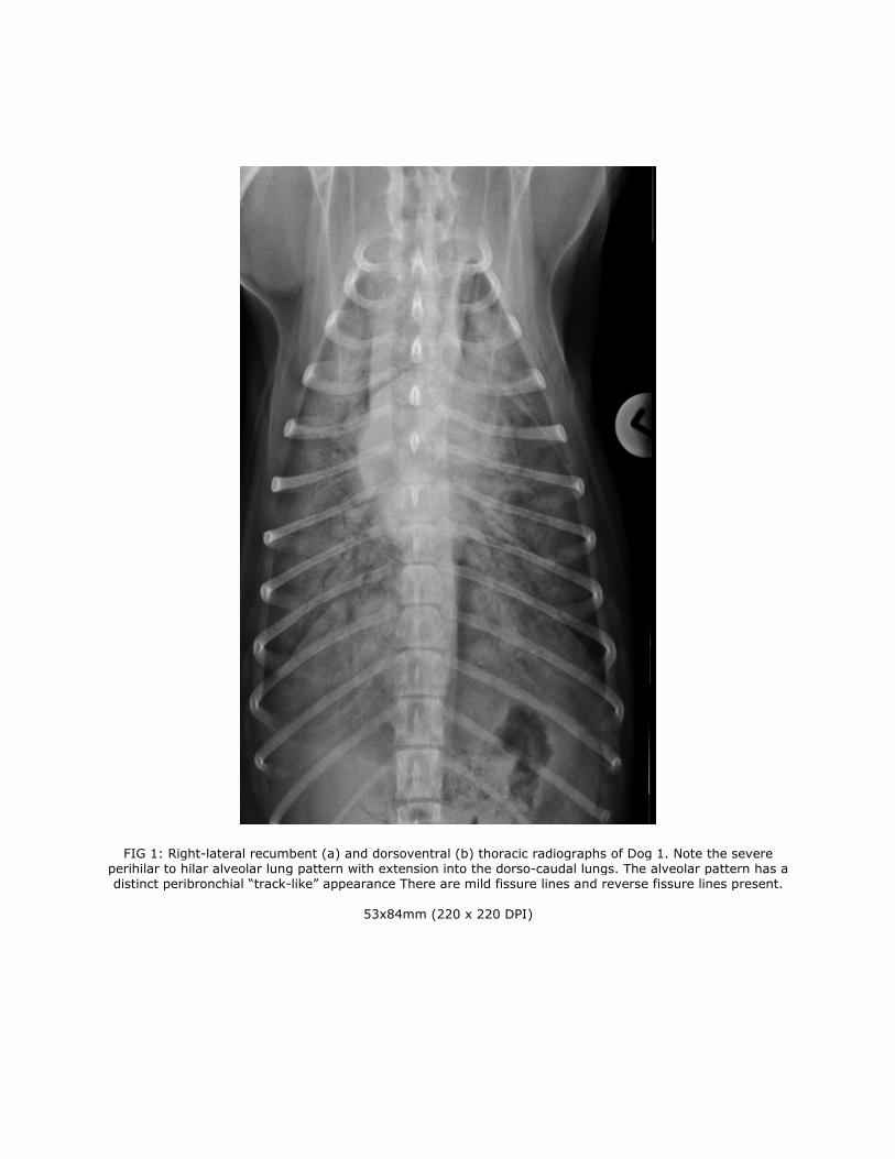
FIG 1: Right-lateral recumbent (a) and dorsoventral (b) thoracic radiographs of Dog 1. Note the severe perihilar to hilar alveolar lung pattern with extension into the dorso-caudal lungs. The alveolar pattern has a distinct peribronchial “track-like” appearance There are mild fissure lines and reverse fissure lines present.
53x84mm (220 x 220 DPI)
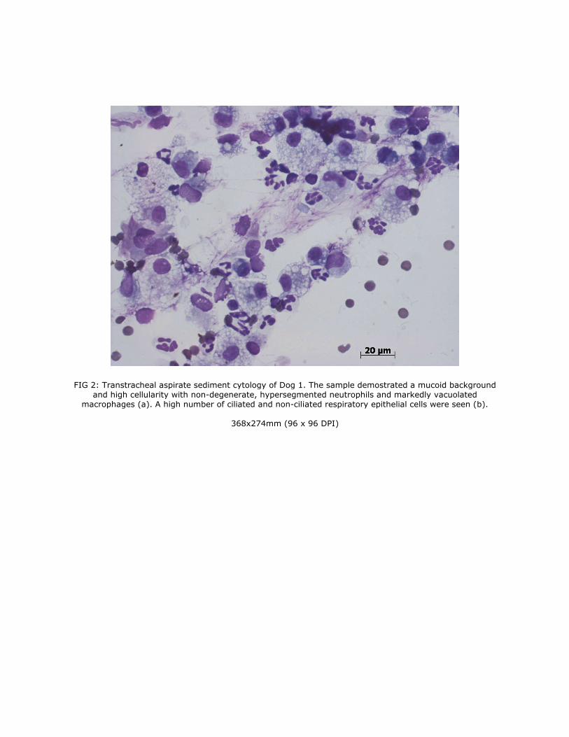
FIG 2: Transtracheal aspirate sediment cytology of Dog 1. The sample demostrated a mucoid background and high cellularity with non-degenerate, hypersegmented neutrophils and markedly vacuolated
macrophages (a). A high number of ciliated and non-ciliated respiratory epithelial cells were seen (b).
368x274mm (96 x 96 DPI)
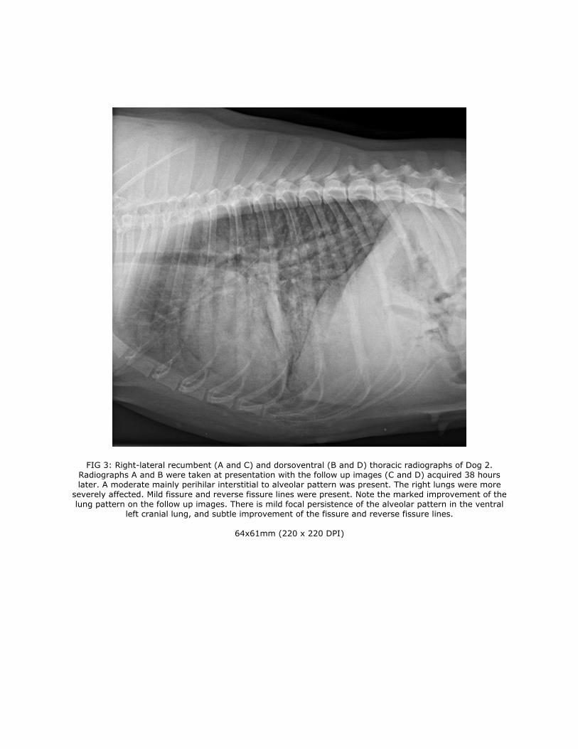
FIG 3: Right-lateral recumbent (A and C) and dorsoventral (B and D) thoracic radiographs of Dog 2. Radiographs A and B were taken at presentation with the follow up images (C and D) acquired 38 hours later. A moderate mainly perihilar interstitial to alveolar pattern was present. The right lungs were more
severely affected. Mild fissure and reverse fissure lines were present. Note the marked improvement of the lung pattern on the follow up images. There is mild focal persistence of the alveolar pattern in the ventral
left cranial lung, and subtle improvement of the fissure and reverse fissure lines.
64x61mm (220 x 220 DPI)

FIG 3: Right-lateral recumbent (A and C) and dorsoventral (B and D) thoracic radiographs of Dog 2. Radiographs A and B were taken at presentation with the follow up images (C and D) acquired 38 hours later. A moderate mainly perihilar interstitial to alveolar pattern was present. The right lungs were more
severely affected. Mild fissure and reverse fissure lines were present. Note the marked improvement of the lung pattern on the follow up images. There is mild focal persistence of the alveolar pattern in the ventral
left cranial lung, and subtle improvement of the fissure and reverse fissure lines.
60x67mm (220 x 220 DPI)
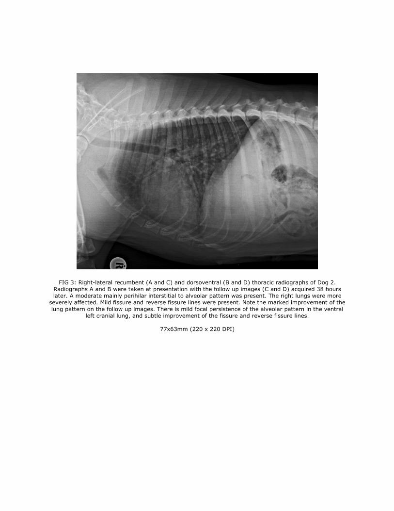
FIG 3: Right-lateral recumbent (A and C) and dorsoventral (B and D) thoracic radiographs of Dog 2. Radiographs A and B were taken at presentation with the follow up images (C and D) acquired 38 hours later. A moderate mainly perihilar interstitial to alveolar pattern was present. The right lungs were more
severely affected. Mild fissure and reverse fissure lines were present. Note the marked improvement of the lung pattern on the follow up images. There is mild focal persistence of the alveolar pattern in the ventral
left cranial lung, and subtle improvement of the fissure and reverse fissure lines.
77x63mm (220 x 220 DPI)

FIG 3: Right-lateral recumbent (A and C) and dorsoventral (B and D) thoracic radiographs of Dog 2. Radiographs A and B were taken at presentation with the follow up images (C and D) acquired 38 hours later. A moderate mainly perihilar interstitial to alveolar pattern was present. The right lungs were more
severely affected. Mild fissure and reverse fissure lines were present. Note the marked improvement of the lung pattern on the follow up images. There is mild focal persistence of the alveolar pattern in the ventral
left cranial lung, and subtle improvement of the fissure and reverse fissure lines.
62x82mm (220 x 220 DPI)

FIG 4: Photomicrograph of the lungs of Dog 1 demonstrating diffuse congestions and protein-rich oedema. Many of the alveolar spaces were filled with fibrin and numerous alveolar macrophages. The alveolar walls were thickened by mononuclear cells and activated endothelial cells (A). AHS NS4 immunoperoxidase
labelling of the lung of Dog 1 with numerous intra-alveolar macrophages and endothelial cells displaying positive solid nuclear labelling (brown). For detailed interpretation please see Zwart et al. (16) (B).
64x48mm (220 x 220 DPI)

FIG 4: Photomicrograph of the lungs of Dog 1 demonstrating diffuse congestions and protein-rich oedema. Many of the alveolar spaces were filled with fibrin and numerous alveolar macrophages. The alveolar walls were thickened by mononuclear cells and activated endothelial cells (A). AHS NS4 immunoperoxidase labelling of the lung of Dog 1 with numerous intra-alveolar macrophages and endothelial cells displaying positive solid nuclear labelling (brown). For detailed interpretation please see Zwart et al. (16) (B).
64x48mm (220 x 220 DPI)