clinical practice guideline the diagnosis and … · clinical practice guideline the diagnosis and...
TRANSCRIPT

DRAFT
CLINICAL PRACTICE GUIDELINE THE DIAGNOSIS AND MANAGEMENT OF ECTOPIC PREGNANCY
1
CLINICAL PRACTICE GUIDELINE
THE DIAGNOSIS AND MANAGEMENT OF ECTOPIC PREGNANCY
Institute of Obstetricians and Gynaecologists, Royal College of Physicians of Ireland
and Directorate of Clinical Strategy and Programmes,
Health Service Executive
Version: 1.0 Publication date: November 2014
Guideline No: 33 Revision date: November 2017
DRAFT
CLINICAL PRACTICE GUIDELINE THE DIAGNOSIS AND MANAGEMENT OF ECTOPIC PREGNANCY
2
Contents
1. Key recommendations 3
2. Purpose and Scope 4
3. Background and Introduction 4
4. Methodology 5
5. Clinical guideline 6
5.1 Clinical assessment 6
5.2 Ultrasound scanning 6
5.3 Human Chorionic Gonadotrophin 7
5.4 Expectant Management 7
5.5 Medical Management 8
5.6 Surgical Management 9
5.7 Non-tubal ectopic pregnancies 10
5.7.1 Interstitial pregnancy 10
5.7.2 Cornual pregnancy 11
5.7.3 Cervical pregnancy 11
5.7.4 Caesarean scar pregnancy 11
5.7.5 Ovarian pregnancy 12
5.7.6 Abdominal pregnancy 12
5.8 Anti-D Rhesus prophylaxis 13
5.9 Counselling 13
6. References 14
7. Implementation Strategy 17
8. Key Metrics 17
9. Qualifying Statement 17
10. Appendices 18
DRAFT
CLINICAL PRACTICE GUIDELINE THE DIAGNOSIS AND MANAGEMENT OF ECTOPIC PREGNANCY
3
Key Recommendations
1. Women of reproductive age presenting acutely with gastrointestinal symptoms, particularly diarrhoea, as well as those presenting with abdominal pain and/or
vaginal bleeding should have a urinary hCG test. 2. A transvaginal ultrasound service should be provided to all acute hospitals or
maternity units for the initial investigation of women with a suspected ectopic pregnancy (EP).
3. If appropriate ultrasound services are not available, referral to an Early Pregnancy
Assessment Unit (EPAU) is recommended provided that the woman’s condition is
stable.
4. Ideally, treatment should be based on a positive identification of the EP. 5. Expectant management is an option in selected women with probable EP who have
a small mass with low and falling hCG provided they have minimal symptoms and are compliant with follow-up.
6. Methotrexate (MTX) therapy may be considered for women with an adnexal mass 35 mm whose initial hCG is less than 1500 IU/L, provided they have minimal
symptoms and compliance with follow-up is anticipated.
7. A laparoscopic approach is preferred for the surgical management of EP. Either salpingectomy or salpingotomy may be used. Both have a similar outcome regarding future pregnancy success.
8. Salpingectomy is appropriate for women who have a healthy contralateral tube and
it may be necessary in cases of uncontrolled bleeding or extensive tubal damage. 9. Salpingotomy has a greater risk of persistent trophoblastic activity requiring follow
up with serial hCG monitoring. MTX therapy is appropriate for treatment of persistent trophoblastic activity.
10. A woman diagnosed with ectopic pregnancy, and her partner, need to have the
diagnosis and treatment options communicated sensitively and clearly.
DRAFT
CLINICAL PRACTICE GUIDELINE THE DIAGNOSIS AND MANAGEMENT OF ECTOPIC PREGNANCY
4
2. Purpose and Scope
The purpose of this guideline is to improve the management of women who may have an ectopic pregnancy. It is for use by all healthcare professionals involved in women’s
care and particularly obstetricians, nurses/midwives, sonographers, radiologists and general practitioners who provide care to women in early pregnancy. The guideline
should be read in conjunction with Clinical Practice Guideline No. 1 Ultrasound Diagnosis of Early Pregnancy Miscarriage and Clinical Practice Guideline No. 10 Management of Early Pregnancy Miscarriage. The guideline aids clinical judgment and
does not replace it. In individual cases a healthcare professional may, after careful consideration, decide not to follow the guideline if it is deemed to be in the best
interest of the woman.
3. Background and Introduction
Worldwide, ectopic pregnancy (EP) remains the leading cause of maternal death in the first trimester (Autry, 2013). The most recent figure for the rate of EP in Ireland is 14.8 per 1,000 maternities (HIPE, 2012). In Ireland, as in most of the developed
world, there has been a reduction in mortality from EP reflecting a success story of modern management. A life-threatening surgical emergency in a woman with a
positive pregnancy test and haemodynamic shock has been converted to a non-urgent medical condition in many cases. In the US from 1970 until 1992 the mortality rate decreased by 90% despite a simultaneous 6-fold increase in incidence of EP. Death
rates from ectopic pregnancy in the UK have almost halved from an estimated 31.2 (95% CI 16.8–57.9) per 100,000 estimated ectopic pregnancies for 2003-05 to 16.9
(95% CI 7.6–37.6) for 2006-08 (The Eighth Report of the Confidential Enquiries into Maternal Deaths in the United Kingdom, 2011). The major improvement in mortality came as a result of earlier and more accurate diagnosis, made possible by the
development of high-resolution ultrasonography and radioimmunoassay for human chorionic gonadotropin (hCG) and also the widespread availability of laparoscopy
(Lipscomb, 2010).
Serial hCG measurement is widely used in the diagnosis of early pregnancy complications. In the US this is combined with an aggressive strategy of intervention by uterine curettage to distinguish a non-viable intrauterine pregnancy (IUP) from an
EP. Transabdominal scanning by non-specialists is still widely used in emergency room assessment in the US. A different approach has been taken in Europe where Early
Pregnancy Assessment Units (EPAU)’s have been introduced to provide specialist assessment. The preferred option for first line investigation of a woman who is symptomatic in the first trimester is transvaginal ultrasound scanning (TVS). In cases
of suspected EP serial hCG measurement may be combined with an extended period of observation and follow-up scanning after an interval of a week or more (NICE,
2012). Every Irish maternity unit now has an EPAU with an evolving standardisation of practice along the European model.
Despite improvements in prompt diagnosis of this potentially fatal condition there are avoidable factors in over half of the associated deaths. EP remains responsible for 6%
of maternal deaths which mainly occur after an acute initial presentation. Women who present with signs of hypovolemia demand rapid diagnosis and management. Yet in about half of those with EP presenting to emergency departments the diagnosis is
missed at first assessment (CMACE, 2011).
DRAFT
CLINICAL PRACTICE GUIDELINE THE DIAGNOSIS AND MANAGEMENT OF ECTOPIC PREGNANCY
5
The development of algorithms for diagnosis and medical management using MTX has allowed one third of EP’s to be managed without surgery (Hoover et al., 2010). This
results in fewer complications of treatment and lower costs. Surgery, when performed, is more likely to be laparoscopic. By 2007 a woman in the US was twice as
likely to have laparoscopic surgery as laparotomy.
4. Methodology
Medline, EMBASE and Cochrane Database of Systematic Reviews were searched using
terms relating to ectopic pregnancy, diagnosis and management. Searches were limited to humans and restricted to English language articles published between January 2000 and June 2013. Relevant meta-analyses, systematic reviews,
interventional and observational studies were included.
The guideline was developed by Professor Michael Gannon. The guideline was peer-
reviewed by Dr Anne Bergin (Midwifery), Dr Brian Cleary (Pharmacy), Dr Sam Coulter-Smith (Rotunda), Dr Nadine Farah (Coombe), Ms Síle Gill (Midwifery), Ms Caroline Keegan (Midwifery), Ms Valerie Kinsella (Midwifery), Ms Máiread McGuire (Pharmacy),
Dr Mary Moran (Midwifery), Dr Aoife Mullally (Obstetrics), Ms Janet Murphy (Midwifery), Ms Mary O’Reilly (Midwifery) Professor Michael Turner (HSE Director,
Clinical Care Programme in Obstetrics and Gynaecology) and Dr Julia Unterscheider (Obstetrics).
The sample Patient Information Leaflet was reviewed by Ectopic Pregnancy Ireland and Miscarriage Association Ireland.
DRAFT
CLINICAL PRACTICE GUIDELINE THE DIAGNOSIS AND MANAGEMENT OF ECTOPIC PREGNANCY
6
5. Clinical guideline
5.1 Clinical assessment
About 5% of women with EP present in haemorrhagic shock. Pallor, tachycardia and
hypotension should alert the examiner to major abdominal bleeding, regardless of the intensity of abdominal pain. Shoulder pain occurs from irritation of the diaphragm. Vomiting and diarrhoea may be the presenting symptoms of abdominal bleeding.
Women of reproductive age presenting acutely with gastrointestinal symptoms
particularly diarrhoea and dizziness, as well as those presenting with abdominal pain and/or vaginal bleeding, should have a urinary hCG test. This readily available test which is highly sensitive and specific for the beta subunit of hCG narrows the
differential diagnosis to pregnancy-related problems.
The common presentation of EP - vaginal bleeding and lower abdominal pain in a woman with delayed menses – also occurs in early pregnancy miscarriage. Women who have an EP typically complain of brown vaginal discharge soon after a missed
period, sometimes progressing to heavier bleeding similar to a miscarriage. This is often followed by pain associated with tubal distension and intraperitoneal bleeding at
the fimbrial end of the tube (Crochet et al., 2013). Clinical suspicion is the key to identifying women who need prompt and careful
evaluation. The risk of EP increases 2-fold for infertility, 3-fold for tubal pathology and 4-fold for documented salpingitis. One third of pregnancies in women who have been
sterilised and one half in women with a LNG-IUS are ectopic. The risk of recurrence is approximately 10% for women with one previous EP and at least 25% for women having two or more previous EP’s. Women with a history of EP should be given early
access to an EPAU in a future pregnancy (NICE, 2012).
5.2 Ultrasound scanning
Speculum and bimanual examination are uncomfortable and have a limited diagnostic value. However the widespread availability of TVS allows this modality to be the initial
investigation of choice for women with suspected EP. It is desirable that TVS is carried out by an experienced sonographer with good equipment. If appropriate scanning facilities are not immediately available and provided that the woman’s condition is
stable referral to an early pregnancy unit within 48-72 hours is recommended. Women with an interim diagnosis of pregnancy of unknown location (PUL) with a history of a
previous ectopic pregnancy, and women at high risk of ectopic pregnancy (e.g. assisted conception, IUD in situ) should be seen by an obstetrician and a management plan formulated. Women and their partners should be informed that scanning cannot
be guaranteed to be 100% accurate in early pregnancy (NICE, 2012).
The presence of an intrauterine gestational sac containing a yolk sac (from 5.5 weeks) or a fetus (from 6 weeks) constitutes an IUP (Barnhart, 2009). Fetal cardiac activity
may be apparent from 6.5 weeks. When no IUP is seen on TVS careful examination of the adnexae and cul-de-sac should be carried out. Approximately 60% of EP’s are seen as a nonhomogeneous mass adjacent to the ovary, 20% appear as a
hyperechoic ring (bagel sign) and 13% have an obvious gestational sac with a fetal pole. Failure to detect either an intra or extrauterine pregnancy by scan in a woman
DRAFT
CLINICAL PRACTICE GUIDELINE THE DIAGNOSIS AND MANAGEMENT OF ECTOPIC PREGNANCY
7
with a positive pregnancy test is termed a PUL which is an interim diagnosis requiring further investigation (Barnhart et al., 2011).
5.3 Human Chorionic Gonadotropin
Diagnostic accuracy can be improved by measuring the rate of change of serum hCG, which is predictable during the early weeks of a normal IUP. First detectable in
maternal serum as early as 8-10 days after ovulation the level of serum hCG rises to between 50 and 100 IU/L by the time of the expected menstrual period. In general,
levels double every 1.4 – 2.1 days in early pregnancy and peak at 50,000 - 100,000 IU/L between 8 and 10 weeks gestation. An interval of 48 hours is used to monitor progress in early pregnancy from paired serum hCG levels. If the expected doubling is
observed this is a reliable indicator of viability provided an intrauterine sac is seen. A lesser rise is also compatible with normal pregnancy and a minimum increase of 53%
in 48 hours was seen in 99% of viable IUP’s. Below this level a pregnancy is likely to be nonviable (Barnhart, 2009).
The accumulated experience in ultrasound and hCG assessment has led to a clearer picture of the natural history of EP. Women who present early are more likely to have
a rising hCG. In about 20% of EPs the rise in hCG is the same as for a potentially viable IUP (Barnhart, 2009). More than 70% of women with an EP will have a rise in hCG that is slower than the minimal rise for a viable pregnancy or a decline that is
slower than the minimal rate of fall in a complete miscarriage. A fall in hCG reflects a natural failure of the pregnancy and in 8% of women with EP the rate of fall is the
same as that found after a complete miscarriage, at least 15% over 48 hours. With higher initial levels expect a quicker fall, at least 20% for an initial level of 500 IU/L and 30% for a level of 2000 IU/L or more. The differential for a slowly falling hCG is
an EP, a non-viable missed miscarriage or an incomplete miscarriage.
The absolute value of hCG is also useful in diagnosis: above 1,000 – 1,500 IU/L (the discriminatory zone) a viable IUP should be visible on TVS. Although this may be helpful, experience has shown that pregnancies identified by the discriminatory zone
are not always ectopic. The majority of non-visualised pregnancies are failed IUP’s or incomplete miscarriages with small amounts of retained products or the aftermath of
miscarriage when initial levels were high. The discriminatory zone should be interpreted with caution and always in conjunction with the clinical features. When the situation is not acute and the diagnosis is unclear it is usually preferable to continue
observation until a positive diagnosis is made (Jurkovic and Wilkinson, 2011).
5.4 Expectant Management
Extended observation of women who have an uncertain prognosis in early pregnancy has shown that many EPs resolve spontaneously. Expectant management is an option
in selected women with probable EP provided they have minimal symptoms and are compliant with follow-up (Jurkovic and Wilkinson, 2011). In the presence of a nonhomogenous adnexal mass it has been shown that expectant management may
have a success rate of over 80% provided that the initial hCG is less than 1,000 IU/L and falling by at least 13% over 48 hours.
DRAFT
CLINICAL PRACTICE GUIDELINE THE DIAGNOSIS AND MANAGEMENT OF ECTOPIC PREGNANCY
8
Continuing outpatient observation is appropriate if the woman is clinically stable (van Mello et al., 2013). She should be given written information explaining her condition and the possible complications of EP. She should understand the importance of
compliance with follow-up and have easy 24 hour access to emergency hospital gynaecological care. Women managed expectantly should be followed at least weekly
with serial hCG measurements and TVS to ensure a rapidly decreasing hCG level (ideally less than 50% of its initial level within seven days) and a reduction in the size of an adnexal mass by seven days. As tubal rupture has been reported with low and
declining hCG concentrations follow-up should be continued until hCG is at a non-pregnant level (RCOG, 2010).
Considering the potentially serious risks of tubal rupture and haemorrhage and the established safety and effectiveness of medical and surgical treatment of EP, it seems
prudent that expectant management should be reserved for asymptomatic patients with very low and falling hCG levels. Any plateau or rise in the hCG measurements
should prompt medical or surgical treatment (Jurkovic and Wilkinson, 2011). Expectant management may have to be abandoned in patients who become symptomatic whether or not hCG levels are falling.
5.5 Medical Management
Systemic MTX (MTX) is a safe and effective treatment for EP. MTX avoids anaesthesia and is simpler, less invasive and less costly than surgery. MTX is a folic acid
antagonist that inhibits DNA and RNA synthesis by inactivating dihydrofolate reductase. Rapidly proliferating tissues such as trophoblast cells are particularly
vulnerable to its actions. MTX has a half-life of 8-15 hours. Women with EP who have minimal symptoms and a low level of hCG should be
considered for medical treatment (Mol et al., 2008). MTX therapy can be considered for those whose initial hCG is less than 1,500 IU/L with an adnexal mass not greater
than 35 mm (NICE, 2012). Although medical therapy can be successful at higher levels of hCG there is a price to be paid in longer follow-up and a higher rate of surgical re-intervention. A candidate for medical management with MTX should be
haemodynamically stable with no evidence of acute intra-abdominal bleeding and not in severe or persistent pain. She should have no contraindication to MTX therapy and
should have a reliable commitment to comply with follow-up surveillance (ASRM, 2008).
Contraindications include: pre-existing blood dyscrasias, such as bone marrow hypoplasia, leukopenia,
thrombocytopenia, or significant anaemia serious, acute or chronic infections such as tuberculosis, HIV or other
immunodeficiency syndromes ulcers of the oral cavity and known active gastrointestinal ulcer disease breast-feeding
concurrent vaccination with live vaccines (http://www.hpra.ie/homepage/site-tools/search?query=MTX)
MTX is directly toxic to hepatocytes and is cleared from the body by renal excretion; therefore it should not be used in women with hepatic or renal disease. For women
with suspected hepatic or renal disease screening should be performed by liver function tests and serum creatinine (Clark et al., 2012).

DRAFT
CLINICAL PRACTICE GUIDELINE THE DIAGNOSIS AND MANAGEMENT OF ECTOPIC PREGNANCY
9
The single dose MTX protocol was developed to reduce the incidence of side effects, eliminate the need for folic acid rescue and increase convenience of administration. The single dose MTX has proved successful in treating EP and is the regime favored in
the UK (Kirk et al., 2007). The regime involves a single administration of MTX by intramuscular injection with monitoring of hCG levels on day 4 and day 7. The dose of
MTX can be calculate in either of two ways. The first method is based on a calculation of 1mg/kg body weight. The second method is based on body surface area. Please see Appendix 2 for calculations. All calculations should be double-checked by two
people prior to administration. If the calculated dose is above 100mg, please recheck dose carefully. When MTX is not available on the wards out of hours, and the
pharmacy is closed, provisions should be made to ensure that there is no delay in obtaining MTX at the appropriate dose for the patient. Prepacks can be made up that contain 2 x vials of MTX, patient information leaflet and prescribing and administration
information.
If there is less than the expected 15% decrease in hCG, the dose of MTX can be repeated. Measure hCG at weekly intervals until non-pregnant levels are reached. At least 15% of medically treated women require a second dose of MTX. Surgery is
indicated for failure of satisfactory medical reduction in hCG level.
Almost 75% of women experience abdominal pain following treatment (RCOG, 2010).
This may reflect tubal bleeding or tension from a haematoma. Increasing or severe pain should prompt re-evaluation, preferably as an inpatient, to determine if
observation and medical treatment can be continued safely. Surgery should be performed for suspected tubal rupture, which may occur in approximately 7% of women.
The use of MTX in a presumed EP which is subsequently identified as a viable intrauterine pregnancy may cause exposure of the fetus during a critical stage of
embryogenesis. Great care must be taken to confirm the diagnosis of EP prior to administration of MTX (Nurmohamed et al., 2011). Although MTX may persist in body tissues for extended periods of time after treatment has stopped, there are no
reported cases of MTX embryopathy in a baby exposed to MTX from maternal treatment of a prior ectopic pregnancy. However, it is important to advise women
who receive MTX that they should wait at least three months before attempting to become pregnant again (Hackmon et al., 2011).
5.6 Surgical Management
Surgery is preferable if the hCG level is >1500 IU/L or if there is a visible EP sac with fetal cardiac activity or if there is a mass of greater than 35 mm (NICE 2012). In appropriate cases surgery provides rapid confirmation of the diagnosis with shorter
resolution time of the EP thus avoiding prolonged monitoring. Surgery also allows an accurate assessment of the pelvis which is helpful for counselling. It is preferable to
demonstrate an EP sac or adnexal mass on TVS prior to surgery. A laparoscopic approach to the surgical management of EP is preferred. A woman with
haemodynamic instability due to intraperitoneal haemorrhage should be treated by the most expedient surgical method to gain rapid haemostasis. Without evidence for
an open or laparoscopic approach for a woman in shock, laparotomy has traditionally been favoured (RCOG, 2010). More recent evidence suggests that laparoscopic
DRAFT
CLINICAL PRACTICE GUIDELINE THE DIAGNOSIS AND MANAGEMENT OF ECTOPIC PREGNANCY
10
treatment is safe and effective for suitably trained and experienced staff (Odenjinmi et al., 2011).
Salpingectomy is recommended for recurrent EP in the same Fallopian tube, extensive damage to the involved tube, uncontrolled bleeding or for women who have
completed childbearing. Salpingotomy is preferred in those women who do not have a healthy contralateral tube.
Two recent randomised trials have found no significant difference in fertility between women who had salpingectomy and those who had salpingotomy. The 2-year rates of
IUP were 64% and 70% respectively in one reported study (Fernandez et al., 2013) and and the 3-year rates were 56% and 61% after salpingectomy and salpingotomy respectively in the second study (Mol et al., 2014). There were no significant
differences in recurrent EP’s which were 12% vs 8% (Fernandez et al., 2013) and 5% vs 8% (Mol et al., 2014) after salpingectomy and salpingotomy respectively.
Persistent trophoblast developed in 7% of the salpingotomy group (Mol et al., 2014), 15 times more than after salpingectomy. Prophylactic use of single shot MTX after salpingotomy significantly lowers the persistent trophoblast rate. However, ten women
need to be treated with MTX to prevent one woman with persistent trophoblast. Monitoring serum hCG concentrations seems a better option (Hajenius et al., 2009). A
fall of <50% on postoperative day 1 is three times more likely to proceed to persistent trophoblast than a fall of >50%. Serum hCG should be followed weekly
until it decreases to a non-pregnant level. Those who have a rise or plateau in hCG indicating persistent trophoblast should be treated with MTX according to the regime outlined above.
The choice of salpingectomy or salpingotomy should take into account both the clinical
situation and the woman’s preference.
5.7 Non-tubal ectopic pregnancies
Ultrasound is an essential tool in the diagnosis of non-tubal EPs. As is the case with tubal EPs expectant or medical management can be employed and is more likely to
succeed with a smaller pregnancy and lower baseline hCG. Close monitoring is needed as it may take several months for hCG to return to normal and there is also a slow
resorption of pregnancy tissue seen on scan.
5.7.1 Interstitial pregnancy
The pregnancy is implanted in the tubal segment which is within the muscular wall of
the uterus (Moawad et al., 2010). Pain is more common than bleeding at clinical presentation. A significant number of women have a history of damaged tubes and surgery including salpingectomy (Oliver et al., 2007).
Many are diagnosed at first trimester scanning by the presence of an eccentric
gestational sac. A thin surrounding myometrial layer helps to distinguish this from an angular intrauterine pregnancy. A further sonographic sign is the presence of an echogenic line running from the endometrial cavity to the gestational sac.
For failed medical treatment or when rupture is suspected surgery is usually
necessary. Laparoscopic linear cornuostomy is carried out in a similar manner to salpingostomy for EP including allowing spontaneous closure of the corneal incision.
DRAFT
CLINICAL PRACTICE GUIDELINE THE DIAGNOSIS AND MANAGEMENT OF ECTOPIC PREGNANCY
11
Cornual resection is another option that may also be carried out by laparotomy. Post-operative monitoring by hCG should be performed.
5.7.2 Cornual pregnancy
This is a pregnancy implanting in the contralateral rudimentary horn of a unicornuate uterus (Mavrelos et al., 2007). Presentation may be delayed and is usually with
abdominal pain. About 50% present after rupture and morbidity is high.
The sensitivity of ultrasound diagnosis is low. The appearance is of a gestation sac separate from an empty unicornuate uterus which is identified by the single interstitial tube. The sac is mobile and surrounded by a thick myometrial layer. A vascular
pedicle may be seen joining the gestational sac and the lateral aspect of the empty unicornuate uterus. Surgical cornual excision is usually preferred either by
laparoscopy or open surgery and avoids the risk of recurrence.
5.7.3 Cervical pregnancy
Implantation is within the cervical canal. Common predisposing factors are curettage,
caesarean section or cervical surgical procedures. Usually the first complaint is of painless vaginal bleeding and speculum examination may reveal an open external cervical os with a fleshy mass protruding (Fylstra, 2012).
Ultrasound shows a gestation sac distal to a closed internal cervical os. Doppler
demonstration of surrounding vasculature helps distinguish a cervical pregnancy from a displaced intrauterine pregnancy. In addition, gentle pressure with the transvaginal probe may elicit the “sliding sign” whereby a miscarrying sac is seen to slide within
the cervical canal unlike the cervical pregnancy which is fixed.
Cervical dilation and curettage may provoke bleeding. Infiltration of the cervix with a haemostatic vasoconstricting agent, followed by the placement of cervical sutures to temporarily occlude the descending branches of the uterine arteries followed by
suction curettage (without dilation) and post-curettage cervical canal balloon tamponade has proven successful in treating first trimester cervical pregnancies.
Another treatment option is uterine artery embolisation which has been used in combination with MTX (Zakaria et al., 2011).
5.7.4 Caesarean scar pregnancy
Caesarean scar pregnancy differs from placenta accreta in that the pregnancy is situated outside the endometrial cavity and completely surrounded by myometrium and scar tissue (Ash et al., 2007). It may present as early as 5–6 weeks with light
painless bleeding or as an incidental scan finding (Rotas et al., 2006). Depending on its location in the uterine wall growth may be toward the cavity resulting in profuse
bleeding or toward the abdominal cavity resulting in severe pain and rupture. There is a substantial risk of catastrophic haemorrhage if the pregnancy continues past the first trimester. The mortality rate of nearly 1 in 500 is more than 10 times that for all
EPs (CMACE, 2011).
DRAFT
CLINICAL PRACTICE GUIDELINE THE DIAGNOSIS AND MANAGEMENT OF ECTOPIC PREGNANCY
12
Diagnosis by TVS relies on an empty uterine cavity and a gestation sac in the anterior uterine isthmus with a thin myometrium between sac and bladder. There is no contact between the sac and the uterine cavity and the cervical canal is empty. Colour flow
Doppler may show distinct circular peritrophoblastic perfusion surrounding the gestation sac.
Expectant management carries a significant risk of uterine rupture and hysterectomy. Medical treatment using MTX is an appropriate first line measure. Blind uterine
curettage, which may not reach the gestation sac, may result in heavy bleeding and should be discouraged. Surgical removal can be carried out by operative hysteroscopy
or laparoscopy, with the choice depending on the location of the pregnancy sac. Open surgical treatment by wedge resection should be considered in women who do not respond to these methods and for women who present after rupture or if facilities and
expertise for operative endoscopy are not available.
5.7.5 Ovarian pregnancy
The clinical features are similar to those for tubal EP, current use of an IUD is more likely in cases of ovarian pregnancy (Odejinmi et al., 2009).
Apart from the few cases with a clear cut yolk sac or fetal pole visible in the ovary ultrasound diagnosis is difficult. The ring surrounding an EP usually shows greater
echogenicity than the surrounding ovarian tissue unlike the ring of a corpus luteum cyst which is less echogenic. If laparoscopy for suspected EP reveals that the tubes
are normal a close inspection of the ovaries should be performed. Typically an ovarian EP has the appearance of a cystic haemorrhagic mass.
Optimum management is resection of the ovarian pregnancy with preservation of healthy ovarian tissue. Follow-up hCG monitoring is recommended. MTX is appropriate
for persistent trophoblast and has also been used for primary treatment but is limited in this regard due to the need for laparoscopic and histologic confirmation of diagnosis.
5.7.6 Abdominal pregnancy
Implantation in the Pouch of Douglas is the most common site for abdominal pregnancy. Diagnosis is difficult and is usually made intraoperatively (Oliver et al.,
2007).
Primary abdominal pregnancy which presents in the first trimester can be removed laparoscopically with minimal morbidity. Secondary abdominal pregnancies following tubal ectopic rupture are usually advanced at presentation and require an open
procedure. If the placenta is firmly attached with no significant bleeding it can be left in situ after trimming the cord and membranes. Postoperative MTX has been used to
assist the involution process.

DRAFT
CLINICAL PRACTICE GUIDELINE THE DIAGNOSIS AND MANAGEMENT OF ECTOPIC PREGNANCY
13
5.8 Anti-D Rhesus prophylaxis
Non-sensitised women who are Rhesus negative with a confirmed or suspected ectopic or suspected ectopic pregnancy should be offered anti-D immunoglobulin 250 iu (50
micrograms) as soon as possible (RCOG, 2010).
5.9 Counselling
Healthcare professionals providing care for women with early pregnancy loss must be mindful of the emotional impact of pregnancy loss. The unavoidable delay that
frequently attends the diagnosis of ectopic pregnancy may cause additional and significant emotional distress. It is important that the purpose of each investigation is clearly explained and the treatment options are unambiguously outlined so that the
woman and her partner may understand the treatment choices available to her. The choice of treatment is jointly agreed between the woman and the attending
obstetrician. Information is communicated in a sympathetic manner that takes cognisance of the couple’s social and cultural needs. At the discretion of the attending
obstetrician, or if requested by the couple, a referral for bereavement counselling may be recommended. Staff providing care to women with early pregnancy loss should receive training in communicating bad news.
DRAFT
CLINICAL PRACTICE GUIDELINE THE DIAGNOSIS AND MANAGEMENT OF ECTOPIC PREGNANCY
14
6. References
1. American College of Obstetricians and Gynecologists (2008). Medical management of ectopic pregnancy. Obstetrics & Gynecology, ACOG Practice Bulletin, 111 (94),
1479–1485.
2. American Society for Reproductive Medicine, ASRM (2008). Medical treatment of ectopic pregnancy. Fertility and Sterility, 90, S206-212.
3. Ash A, Smith A and Maxwell D (2007). Caesarean scar pregnancy. Obstetrics & Gynaecology, ACOG Practice Bulletin, 114, 253-263.
4. Autry AM (2013). Medical treatment of ectopic pregnancy: is there something new?
Obstetrics & Gynecology, 122 (4), 733-734.
5. Barnhart KT (2009). Ectopic pregnancy. New England Journal Medicine, 361, 379-
387.
6. Barnhart KB, van Mello NM, Bourne T, Kirk E, Van Calster B, Bottomley C, et al. (2011). Pregnancy of unknown location: a consensus statement of nomenclature, definitions, and outcome. Fertility & Sterility, 95 (3), 857-866.
7. Clark LE, Bhagavath B, Wheeler CA, Frishman GN and Carson SC (2012). Role of
routine monitoring of liver and renal function during treatment of ectopic pregnancies with single-dose MTX protocol. Fertility & Sterility, 98 (1), 84-88.
8. Centre for Maternal and Child Enquiries, CMACE (2011). Saving Mothers’ Lives: reviewing maternal deaths to make motherhood safer, 2006–08: the eighth report
on confidential enquiries into maternal deaths in the United Kingdom. British Journal Obstetrics & Gynaecology, 118 (S1), 1–203.
9. Fernandez H, Capmas P, Lucot JP, Resch B, Panel P and Bouyer J for the GROG (Research Group in Obstetrics & Gynaecology) (2013). Fertility after ectopic
pregnancy: the DEMETER randomized trial. Human Reproduction, 28 (5), 1247-1253.
10. Fylstra DL (2012). Ectopic pregnancy not within the (distal) fallopian tube: etiology, diagnosis and treatment. American Journal Obstetrics & Gynecology, 206
(4), 289-299. 11. Hajenius PJ, Mol F, Mol BWJ, Bossuyt PMM, Ankum WM and Van der Veen F
(2007). Interventions for tubal ectopic pregnancy. Cochrane Database Syst Rev. Issue 1. CD000324. doi/10.1002/14651858.CD000324.pub2/.
12. Hackmon R, Sakaguchi S and Koren G (2011). Effect of MTX treatment of ectopic
pregnancy on subsequent pregnancy. Canadian Family Physician, 57 (1), 37-39.
13. Hospital Inpatient Enquiry (HIPE) Ireland (2012). Healthcare Pricing Office (HPO)
Portal Data.
14. Hoover KW, Tao G and Kent CK (2010). Trends in the diagnosis and treatment of ectopic pregnancy in the United States. Obstetrics & Gynecology, 115 (3), 495-502.
DRAFT
CLINICAL PRACTICE GUIDELINE THE DIAGNOSIS AND MANAGEMENT OF ECTOPIC PREGNANCY
15
15. Jurkovic D and Wilkinson H (2011). Diagnosis and management of ectopic
pregnancy. British Medical Journal, 342, d3397.
16. Kirk E, Condous G, Van Calster B, Haider Z, Van Huffel S, Timmerman D and
Bourne T (2007). A validation of the most commonly used protocol to predict the success of single-dose MTX in the treatment of early pregnancy. Human Reproduction, 22 (3), 858-863.
17. Lipscomb GH (2010). Ectopic pregnancy still cause for concern. Obstetrics &
Gynecology, 115 (3), 487-488. 18. Mavrelos D, Sawyer E, Helmy S, Holland TK, Ben-Nagi J and Jurkovic D (2007).
Ultrasound diagnosis of ectopic pregnancy in the non-communicating horn of a unicornuate uterus (cornual pregnancy). Ultrasound Obstetrics & Gynecology, 30
(5), 765-770. 19. Moawad NS, Mahajan ST, Moniz MH, Taylor SE and Hurd WW (2010). Current
diagnosis and treatment of interstitial pregnancy. American Journal Obstetrics & Gynecology, 202 (1), 15-29.
20. Mol F, Mol BW, Ankum WM, van der Veen F and Hajenius PJ (2008). Current
evidence on surgery, systemic MTX and expectant management in the treatment of tubal ectopic pregnancy: a systemic review and meta-analysis. Human Reproduction Update, 14 (4), 309-319.
21. Mol F, van Mello NM, Strandell A, Strandell K, Jurkovic D and Ross J for the
European Surgery in Ectopic Pregnancy (ESEP) study group (2014). Salpingotomy versus salpingectomy in women with tubal pregnancy (ESEP study): an open-label, multicentre, randomized controlled trial. Lancet, 383, 1483-1489.
22. National Institute for Clinical Excellence, NICE (2012). Ectopic pregnancy and
miscarriage: diagnosis and initial management in early pregnancy of ectopic pregnancy and miscarriage. Clinical Guideline No. 154. Manchester: NICE.
23. Nurmohamed L, Moretti ME, Schechter T, Einarson A, Johnson D, Lavigne SV, Erebara A, Koren G and Finkelstein Y (2011). Outcome following high-dose MTX in
pregnancies misdiagnosed as ectopic. American Journal Obstetrics & Gynecology, 205 (6), 533.e1-3.
24. Odejinmi F, Rizzuto MI, MacRae R, Olowu, O and Hussain M (2009). Diagnosis and laparoscopic management of 12 consecutive cases of ovarian pregnancy and
review of literature. Journal Minimal Invasive Gynecology, 16 (3), 354-359. 25. Odejinmi F, Sangrithi M and Olowu, O (2011). Operative laparoscopy as the
mainstay method in management of hemodynamically unstable patients with ectopic pregnancy. Journal Minimal Invasive Gynecology, 18 (2), 179-183.
26. Oliver R, Malik M, Coker A and Morris J (2007). Management of extra-tubal and
rare ectopic pregnancies: case series and review of current literature. Archives
Gynecology & Obstetrics, 276 (2), 125-131.

DRAFT
CLINICAL PRACTICE GUIDELINE THE DIAGNOSIS AND MANAGEMENT OF ECTOPIC PREGNANCY
16
27. Rotas MA, Haberman S and Levgur M (2006). Cesarean scar ectopic pregnancies. Obstetrics & Gynecology, 107 (6), 1373-1381.
28. Royal College of Obstetricians and Gynaecologists, RCOG (2010). The management of tubal pregnancy. Clinical Guideline No. 21 (revised). London:
RCOG Press. 29. Seeber BE and Barnhart KT (2006). Suspected ectopic pregnancy. Obstetrics &
Gynecology, 107 (2pt Pt 1), 399-413.
30. van Mello NM, Mol F, Verhoeve HR, van Wely M, Adriaanse AH, Boss EA, Dijkman AB, Bayram N, Emanuel MH, Friederich J, van der Leeuw-Harmsen L, Lips JP, Van Kessel MA, Ankum WM, van der Veen F, Mol BW and Hajenius PJ (2013). MTX or
expectant management in women with an ectopic pregnancy or pregnancy of unknown location and low serum hCG concentrations? A randomized comparison.
Human Reproduction, 28 (1), 60-67. 31. Zakaria MA, Abdallah ME, Shavell VI, Berman JM, Diamond MP and Kmak DC
(2011). Conservative management of cervical ectopic pregnancy: utility of uterine artery embolization. Fertility & Sterility, 95 (2), 872-876. CRN 2131300 page
number: 2. http://www.hpra.ie/homepage/site-tools/search?query=MTX http://www.medicines.org.uk/emc/search (Accessed 17/07/2013).
DRAFT
CLINICAL PRACTICE GUIDELINE THE DIAGNOSIS AND MANAGEMENT OF ECTOPIC PREGNANCY
17
7. Implementation Strategy Distribution of guideline to all members of the Institute of Obstetrics and
Gynaecology, to all maternity units and to all acute general hospitals. Implementation through HSE Obstetrics and Gynaecology Programme local
implementation boards. Distribution to other interested parties and professional bodies.
8. Key Metrics
a) Total number of ectopic pregnancies diagnosed b) % treated expectantly
c) % treated with MTX only d) % treated laparoscopically
e) % treated using laparotomy f) % requiring MTX for persistent throphoblastic activity g) Number of caesarian section scar ectopic pregnancies.
9. Qualifying Statement
These guidelines have been prepared to promote and facilitate standardisation and consistency of practice using a multidisciplinary approach. Clinical material offered in
this guideline does not replace or remove clinical judgment or the professional care and duty necessary for each pregnant woman. Clinical care carried out in accordance
with this guideline should be provided within the context of locally available resources and expertise.
This guideline does not address all elements of standard practice and assumes that individual clinicians are responsible for:
Discussing care with women in an environment that is appropriate and which
enables respectful confidential discussion
Advising women of their choices and ensure informed consent is obtained Meeting all legislative requirements and maintaining standards of professional
conduct Applying standard precautions and additional precautions, as necessary, when
delivering care
Documenting all care in accordance with local and mandatory requirements.

DRAFT
CLINICAL PRACTICE GUIDELINE THE DIAGNOSIS AND MANAGEMENT OF ECTOPIC PREGNANCY
18
10. Appendices
Appendix 1: List of National Guidelines
Appendix 2: MTX Regime
Appendix 3: Sample Patient Information Leaflet

DRAFT
CLINICAL PRACTICE GUIDELINE THE DIAGNOSIS AND MANAGEMENT OF ECTOPIC PREGNANCY
19
Appendix 1: List of associated national guidelines
Ultrasound Diagnosis of Early Pregnancy Miscarriage (Clinical Practice Guideline No. 1,
issued December 2010). http://www.rcpi.ie/content/docs/000001/647_5_media.pdf
Management of Early Pregnancy Miscarriage (Clinical Practice Guideline No. 10, issued
April 2012). http://www.rcpi.ie/content/docs/000001/655_5_media.pdf
DRAFT
CLINICAL PRACTICE GUIDELINE THE DIAGNOSIS AND MANAGEMENT OF ECTOPIC PREGNANCY
20
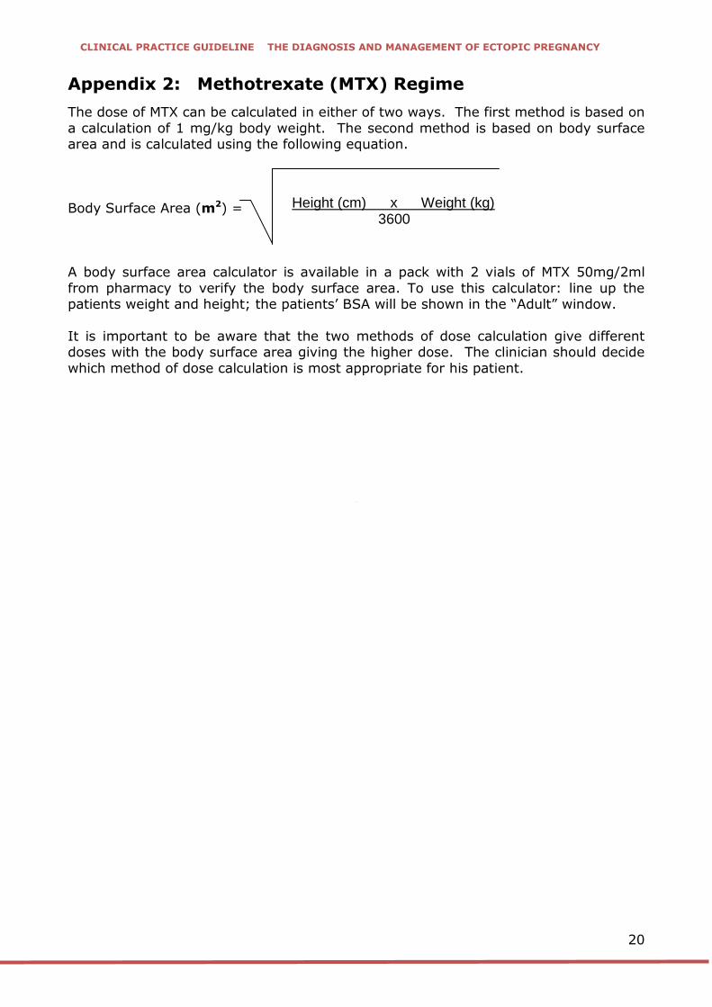
Appendix 2: Methotrexate (MTX) Regime
The dose of MTX can be calculated in either of two ways. The first method is based on
a calculation of 1 mg/kg body weight. The second method is based on body surface area and is calculated using the following equation.
Body Surface Area (m2) =
A body surface area calculator is available in a pack with 2 vials of MTX 50mg/2ml
from pharmacy to verify the body surface area. To use this calculator: line up the patients weight and height; the patients’ BSA will be shown in the “Adult” window.
It is important to be aware that the two methods of dose calculation give different doses with the body surface area giving the higher dose. The clinician should decide
which method of dose calculation is most appropriate for his patient.
Height (cm) x Weight (kg) 3600

DRAFT
CLINICAL PRACTICE GUIDELINE THE DIAGNOSIS AND MANAGEMENT OF ECTOPIC PREGNANCY
21
Appendix 3. Sample Patient Information Leaflet
This information leaflet is intended for women who are suspected of having, or are
confirmed to have, an ectopic pregnancy.
What is an ectopic pregnancy?
The normal process of pregnancy usually begins when the sperm and egg meet in the fallopian tube. It is in the fallopian tube that fertilisation takes place. Within a week
of fertilisation the fertilised egg moves into the womb (uterus) but if this does not happen, the fertilised egg will implant in the fallopian tube or, less commonly, elsewhere such as the ovary or the abdomen. Very rarely it may implant in the cervix
(neck of the womb). Ectopic Pregnancy occurs in approximately 1.5-2% of pregnancies and may be psychologically devastating for a woman and her partner.
What causes an ectopic pregnancy?
The precise cause of ectopic pregnancy may not be known. However, certain women
are at greater risk of experiencing an ectopic pregnancy and these include women who have a history of;
pelvic infection abdominal surgery previous ectopic pregnancy
an Intrauterine Contraceptive Device (IUCD) in situ at the time of conception become pregnant while taking the progesterone only pill
assisted In Vitro Fertilization (IVF) pregnancy.
What are the symptoms of ectopic pregnancy?
The most common symptom is abdominal pain. Not all women are aware that they are pregnant when they experience abdominal pain and may associate the pain with
gastro-intestinal upset. Some women experience pain while passing urine which may lead them to suspect a urinary tract infection. There may also be vaginal bleeding, spotting or a brownish discharge after a missed period. If there is a likelihood that
pregnancy has occurred, a pregnancy test will alert to the possibility of an ectopic pregnancy. However, the first pregnancy test may not always be positive and when
ectopic pregnancy is suspected the test will be repeated next day. Nausea and pain at the tip of the shoulder may also be experienced. Most women attend their family doctor because of some, or many, of the above symptoms. Rarely, a woman may
faint or collapse and should this happen she must be brought immediately to the nearest hospital. It is possible though rarely occurs that a woman who has had a
tubal ligation (sterilisation) may experience an ectopic pregnancy.
How is an ectopic pregnancy diagnosed?
A woman with a suspected ectopic pregnancy will be referred ideally to the Early Pregnancy Assessment Unit (EPAU) in a maternity hospital or to the Emergency
Department out of Hours in an acute hospital with a gynaecological service. The doctor or midwife will take a detailed medical history and perform a pregnancy test
even if a test has already been performed by the woman herself or by her GP. The doctor will examine the abdomen and may do a vaginal examination and/or a speculum examination. As ectopic pregnancy is difficult to diagnose a transvaginal
scan will, in most cases, be performed. A transvaginal scan involves inserting a probe about the width of a tampon into the vagina to allow the doctor or sonographer to
look at the womb, ovaries and fallopian tubes. However, if the pregnancy is ongoing
DRAFT
CLINICAL PRACTICE GUIDELINE THE DIAGNOSIS AND MANAGEMENT OF ECTOPIC PREGNANCY
22
for less than 6 or 7 weeks this examination may not be able to confirm an ectopic pregnancy and may need to be repeated some days later. The earlier a woman is in her pregnancy the more difficult it is to diagnose an ectopic pregnancy. If a woman is
not acutely ill it may be recommended that the repeat scan is delayed for a day or two. To assist in the diagnosis a blood test measuring hCG (the pregnancy hormone)
will be carried out and if necessary repeated at intervals.
What is the management for ectopic pregnancy?
The choice may be based on the woman’s symptoms. It may take a few days to confirm the diagnosis but when this happens the doctor will discuss and recommend
one of three treatment options. If at all possible, that is if pain and other symptoms are not severe, ‘expectant management’ will be recommended. If symptoms are more severe ‘medical management’ will be considered. ‘Surgical management’ will be
required for women experiencing very severe symptoms e.g. internal bleeding, severe pain or if a fetal heartbeat has been identified in a pregnancy outside the womb.
What is expectant management?
Expectant management is essentially a ‘wait and see’ management option as up to
40% of women will not need to have either medical or surgical intervention. This option is suitable for women who have mild or no symptoms and have no vaginal
bleeding. The lower the pregnancy hormone, the more likely it is that the pregnancy will end naturally. If this is the case a woman is advised to go home and return to the hospital for a blood test and a transvaginal scan at regular intervals for two to four
weeks. Before going home, a woman will be provided with verbal and written information about her treatment plan. If pain increases or if bleeding becomes heavier
it will be necessary to return to hospital where the doctor will advise if medical or surgical treatment has become necessary.
What is involved in medical management?
Medical management avoids anaesthesia and is less invasive than surgery and does
not require an overnight stay in hospital. A medication called methotrexate (MTX) is administered by intramuscular injection. In a small percentage of women a second injection may be required some days later. Nearly 75% of women treated with MTX
will experience some abdominal pain. However, if the pain increases or if bleeding occurs it is advisable to return to hospital to determine if surgical treatment will be
necessary.
What is involved in surgical treatment?
Hospital admission is necessary when surgical treatment is required. Ideally admission to a gynaecological ward will be arranged. As with many surgical
procedures a woman will be given an anaesthetic. The operation will be performed through ‘key-hole surgery’ (laparoscopy). Two small incisions are made close to the
umbilicus (belly button) and the ectopic pregnancy is removed. In some, but not all cases, the fallopian tube will be saved. If the pain is very severe and if there is internal bleeding a more invasive surgical procedure called a laparotomy will be
performed. This involves only one but a wider incision in the lower abdomen. Provided the fallopian tube is not damaged it will be preserved during the operation.
Does a woman have any say in the treatment she receives?
Yes. The doctor will discuss the management options with each woman and, where
appropriate, her partner or family. Treatment recommendations will be based on the results of each woman’s investigations and her treatment preference. The diagnosis
DRAFT
CLINICAL PRACTICE GUIDELINE THE DIAGNOSIS AND MANAGEMENT OF ECTOPIC PREGNANCY
23
and all management options should be discussed on an on-going basis with the woman and, if appropriate, those supporting her. The only occasion when a choice is not offered is when a woman presents in an emergency situation where there is
severe pain and internal bleeding. If a woman has any questions or concerns she should not hesitate to ask questions.
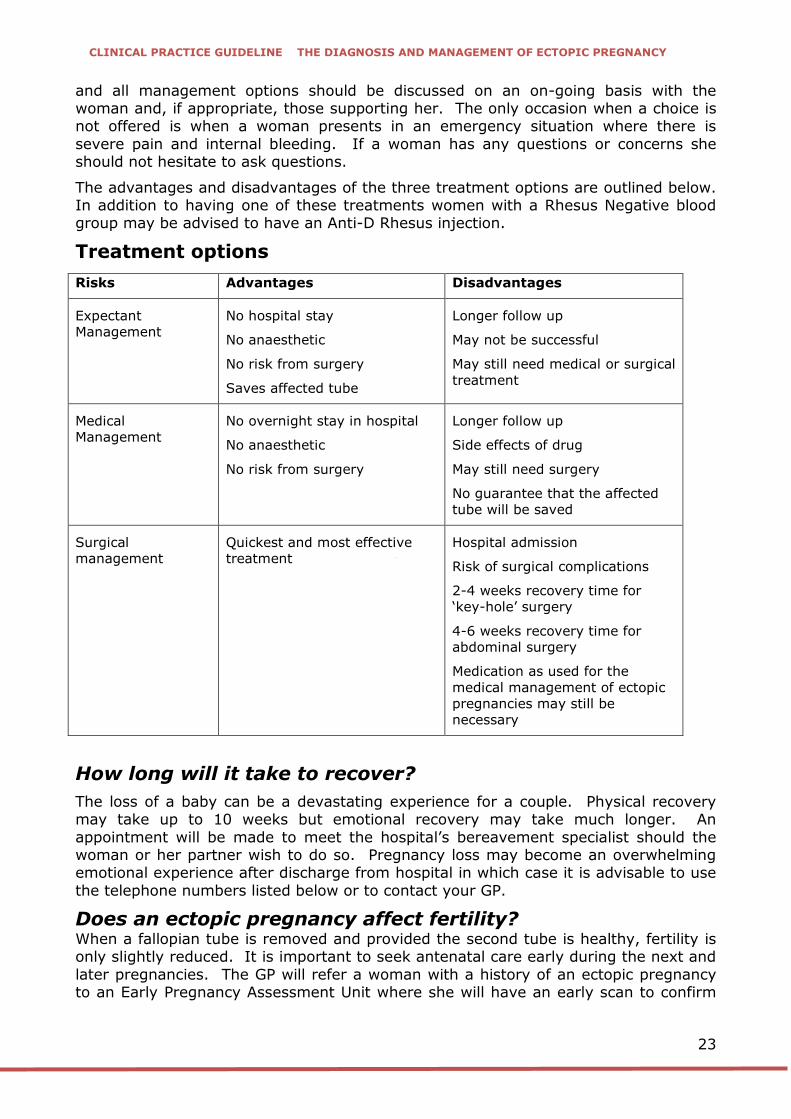
The advantages and disadvantages of the three treatment options are outlined below. In addition to having one of these treatments women with a Rhesus Negative blood group may be advised to have an Anti-D Rhesus injection.
Treatment options
Risks Advantages Disadvantages
Expectant
Management
No hospital stay
No anaesthetic
No risk from surgery
Saves affected tube
Longer follow up
May not be successful
May still need medical or surgical
treatment
Medical
Management
No overnight stay in hospital
No anaesthetic
No risk from surgery
Longer follow up
Side effects of drug
May still need surgery
No guarantee that the affected
tube will be saved
Surgical
management
Quickest and most effective
treatment
Hospital admission
Risk of surgical complications
2-4 weeks recovery time for
‘key-hole’ surgery
4-6 weeks recovery time for
abdominal surgery
Medication as used for the
medical management of ectopic
pregnancies may still be
necessary
How long will it take to recover?
The loss of a baby can be a devastating experience for a couple. Physical recovery may take up to 10 weeks but emotional recovery may take much longer. An
appointment will be made to meet the hospital’s bereavement specialist should the woman or her partner wish to do so. Pregnancy loss may become an overwhelming emotional experience after discharge from hospital in which case it is advisable to use
the telephone numbers listed below or to contact your GP.
Does an ectopic pregnancy affect fertility?
When a fallopian tube is removed and provided the second tube is healthy, fertility is only slightly reduced. It is important to seek antenatal care early during the next and
later pregnancies. The GP will refer a woman with a history of an ectopic pregnancy to an Early Pregnancy Assessment Unit where she will have an early scan to confirm
DRAFT
CLINICAL PRACTICE GUIDELINE THE DIAGNOSIS AND MANAGEMENT OF ECTOPIC PREGNANCY
24
the pregnancy is in her womb. This is important because women who have had an ectopic pregnancy are at a slightly higher risk of having a second ectopic pregnancy.
How soon can I try for another baby?
The length of time to physically and emotionally recover from an ectopic pregnancy
will vary between individuals. Some couples will feel ready to conceive sooner than others. It is recommended that women treated with MTX should avoid pregnancy for 3 months. In all other cases pregnancy is best delayed until after the next period or
until a woman feels ready to try again. Future pregnancies will in all likelihood be more stressful and it is advisable that a woman and her partner feel emotionally
ready. In the meantime a woman’s GP will advise on the method of contraception that is best for her. Ectopic Pregnancy Ireland (www.ectopicireland.ie) provides information and support to those who have been affected by an ectopic pregnancy.
All volunteers have first-hand experience of ectopic pregnancy and can be contacted by telephone: 089 436 5742 or by email [email protected]. The Miscarriage
Association of Ireland (www.miscarriage.ie) also provides a support service for couples who have experienced an early pregnancy loss. The association can be contacted by telephone: 01 873 5702 or by email [email protected].
Is there anything one should do in advance of becoming pregnant?
It is important to enjoy a well-balanced diet, take regular exercise and take folic acid
supplements daily prior to and during pregnancy. Folic acid enriched foods will not provide an adequate amount of folic acid for women preparing for or during early
pregnancy. Cigarettes and alcohol are best avoided prior to and during pregnancy. Information on folic acid and healthy eating can be found at http://www.hse.ie/eng/search?q=pregnancy%20diet.
Useful telephone numbers
Hospital Telephone Number: ________________
Gynaecological Ward: ________________
Early Pregnancy Assessment Unit: ________________
Ectopic Pregnancy Ireland (EPI): www.ectopicireland.ie
EPI Telephone Number: 089 436 5742
EPI email: [email protected]
Miscarriage Association of Ireland (MAI): http://www.miscarriage.ie/
MAI Telephone Number: 01 873 5702
MAI email: [email protected].