clinical ,hematological and diagnostic studies of ... · clinical ,hematological and diagnostic...
TRANSCRIPT
ISI Impact Factor:3.461 . ,20164Bas.J.Vet.Res.Vol.15,No.
37
CLINICAL ,HEMATOLOGICAL AND DIAGNOSTIC STUDIES OF MYCOPLASMA WENYONII INFECTION IN CATTLE OF
BASRAH GOVERNORATE BASRAH ,IRAQ
Ali Jarad , Kamal M.Alsaad
Department of internal and preventive medicine, College of Veterinary Medicine, University of Basrah , Basrah, Iraq
(Received 15 May 2016,Accepted 31 May,2016)
Keywords: Cattle, Lethargy, TRBCs.
ABSTRACT The present work were conducted on (225) local cattle breeds of both sexes , and of
different ages in Basarh governorate (Basrah –Iraq). Two hundred local cattle breeds
were naturally infected with. Mycoplasma wenyonii and (25) clinically normal cattle
breeds served as controls. According to age, diseased animals were divided into four
age groups (50) animals for each. Animals were found clinically infected
with Mycoplasma wenyonii which diagnosed based on Giemsa stain blood smears and
confirmed with PCR test technique. Diseased cattle show sings of partial or complete
loss of appetite ,anemia with pale and / or icteric mucous membranes , decrease milk
production , rapid and difficult respiration, enlargement of superficial lymph nodes ,
rough coat , lethargy , weight loss and edema of lower hind limbs, In addition , body
temperature ,respiratory rate ,heart rate and capillary refilling time were increased
statistically , compared with controls ,Furthermore statistically significant decrease
(P<0.05) were encountered in ruminal contractions .
Results were also indicated that TRBCs, Hb, and PCV values of diseased cattle were
significantly decrease than controls thereby macrocytic hypochromic type of anemia
was indicated. Results were also shown a significant increase in TLC as a result of
significant increase lymphocytes, Mycoplasma wenyonii appear coccoid or rod shape,
structures, However it might found individually or in chains on the erythrocyte cell
wall, Moreover diagnosis were confirmed by PCR,Since out of (96) blood samples
(80) (83.3%) were found positive. Results were also revealed that animals of 2-3
years old were highly infected compared with other age groups. Changes of blood
clotting factor indices were also noticed in diseased cattle compared with controls and
the results showed significant decrease (P<0.05) in the mean values of total

ISI Impact Factor:3.461 . ,20164Bas.J.Vet.Res.Vol.15,No.
38
thrombocytes count and Fibrinogen time ,whereas significant increase (P<0.05) were
detected in thrombocytes volume, thrombocytes distribution width, prothrombin time
, clotting time, and activated partial thromboplastin time. It have been concluded that
Mycoplasma wenyonii infected local cattle breeds of Basrah Governorates and lead to
substantial effect including decrease milk yield and anemia's ,which might terminated
with economic losses ,Therefore all cattle reared in this area must be screened.
INTRODUCTION
Mycoplasma wenyonii (Formerly call Eperythrozoon wenyonii) are eperythrocytic
rickettsial parasites of the family Bartonellaceae.(1) It also call Hemotropic
Mycoplasmas or Hemoplasmas (2).
The organisms are procaryotic forms occurring out side on the surface of the red
blood cells , Positive smears, stained with Giemsa-stain show a pleomorphic coccoid,
rod, and ring-shaped structures might found individually or in chains on the red cell
and have gram-negative staining because of the lack of a cell wall ,However none of
the hemoplasmas have been cultured outside their hosts (3). Infections are common
worldwide in cattle and other animals .(1,4).
The species Mycoplasma wenyonii cause infectious anemia in several
mammalians, Their effects were vary from mild effect to death , they were
reclassified as genus Mycoplasma depending on 16S rRNA sequences and
morphologic similarities, and have been identified in different countries in the world
,their disease characterized by transient fever , anemia, lethargy , decreased milk
yield, enlargement of superficial lymph nodes , anorexia, weight loss, edema of
different body parts specially hind limbs ,and rough coat However, the infection
might remains subclinical and / or chronic, although acute form were also suspected
and diagnosed (5,6).
The organisms are found singular or in different chains on the surface of
erythrocytes (7), Even though it not invade the erythrocyte ,However Groebel et al (8)
were find out that the Mycoplasma suis can invade erythrocytes.
It have been documented that Mycoplasma wenyonii can infect different animals
and shown to be transmitted by various blood sucking ecto-parasites, including ticks,
flies, lice, fleas, and also mosquitoes, Moreover it have been shown that the main
route of transmission of these organisms were uncharacterized, Nevertheless
ISI Impact Factor:3.461 . ,20164Bas.J.Vet.Res.Vol.15,No.
39
mechanical and transplacental routes have been recorded(7),Furthermore Aedes
aegypt are thought to play an important role in the transmission(9).In addition
Messick, (10) was mention other route of transmission as direct contact, and vertical
route and transmission via contaminated food but these routes of transmission need
further investigation.
The disease caused by Mycoplasma wenyonii have been registered in Mosul ,Iraq
previously (11,12) Whereas they were recorded for the first time in cattle of Basrah
governorate ,Therefore the present study were aimed to investigated and study the
clinical , hematological and diagnostic methods of Mycoplasma wenyonii infection in
this area .
MATERIALS AND METHODS
The present work were conducted on (225) local cattle breeds of both sexes , and
of different ages in Basarh governorate (Basrah –Iraq). Two hundred local cattle
breeds were naturally infected with. Mycoplasma wenyonii and (25) clinically normal
cattle breeds served as controls. According to age, diseased animals were divided into
four age groups (50) animals for each (New born calves1-5 days old ,calves under
one year old, cows of 2-3 years old and Old cows more than 5 years ). Complete
clinical examinations had been carried out in all infected and control cows, Moreover
their fecal samples are screened for parasitic loud using the usual scientific methods.
Ten milliliter of blood were drained from each animal by jugular vein-puncture,
from these (2.5) milliliter of blood mixed with EDTA used to determine Total
erythrocyte count (TRBc), Hemoglobin concentration (Hb), packed cell volume
(PCV), mean corpuscular volume (MCV), mean corpuscular hemoglobin
concentration (MCHC), Thrombocytes count , mean thrombocytes volume ,
thrombocytes distribution width ,and total leukocytes count, (Hematology analyzer,
Genex, USA),Furthermore differential leukocytes count were done according to
Weiss and Wardrop (13) . Another (2.5) milliliter of blood mixed with Trisodium
citrate (used plasma) were used to determine Fibrinogen time ,prothrombine time and
activated partial thromboplastine time ( Biolabo / France ). Clotting time was also
estimated according to Bush (14).
Blood smears had been stained with Wright and Giemsa stain for detection of
mycoplasma wenyonii on erythrocyte surface under light microscope at

ISI Impact Factor:3.461 . ,20164Bas.J.Vet.Res.Vol.15,No.
40
1000X,positive sample with mycoplasma wenyonii was recorded if one infected
erythrocyte was found in 200 observed RBCs.
For the PCR assay, the blood was stored in EDTA tube until assayed.DNA was
extracted from 200µl of blood using a commercial kit(Bioingentech Genomic DNA
Purification Kit, BioInGentch,Chile). According to manufacture protocol of
( Mycoplasma wenyonii Detection Kit, BioInGentch,Chile) that used for the specific
amplification of a region(180-basepair) of 16S RNA gen from Mycoplasma wenyonii,
The kit consists of the following components that are enough for amplification of
genomic DNA,such as, Mycoplasma wenyonii Pre-mixture, PCR internal control,
Mycoplasma wenyonii PCR Positive control, DNase/Ranse free water, PCR negative
control, mineral oil solution and brig™molecular weight marker. Preparation of
Mycoplasma wenyonii PCR mixture was done according to protocol of the same
detection kit.
The amplification condition was include, one cycle Initial Denaturation at 94ċ for
2 minutes,30 cycle for denaturation at 94ċ , annealing at 57ċ and extension at 72ċ for
30 second, final extension one cycle at 72ċ for 5 minutes using an automatic cycler.
Amplification products were electrophoresed on 1.5% agarose gels. Gels were
stained with ethidium bromide and examined with ultraviolet illumination. Bands
seen at the expected location (180- bp).
The significance of variations between infected cows and healthy animals were
statistically analyzed using (SPSS) student t-test, (15).
RESULTS
Results were showed that out of 200 suspected cases of Mycoplasma wynonii
infection (140) were detected positive by microscopical examination ( Giemsa stain
smears ) with an infection rate of 70%.,Clinically infected animals showed different
clinical manifestations which include partial or complete loss of appetite , anemia
which manifested with pale and / or icteric mucous membranes which were detected
on conjunctival ,nictitating and vaginal membranes, however icteration were more
clear on sclera ,Furthermore decrease milk production were mentioned by the cow
owners in milk producing cattle , rapid and difficult respiration, enlargement of
superficial lymph nodes specially prescapuler lymph nodes ,in addition other animals
were suffering from rough coat , lethargy and weight loss, However ,edema of lower
hind limbs were detected in some infected animals .(Table 1).

ISI Impact Factor:3.461 . ,20164Bas.J.Vet.Res.Vol.15,No.
41
Table 1: Clinical signs of infected cattle with Mycoplasma wenyonii Clinical signs Infected cattle
n=140 %
Partial or complete loss of appetite 130 92.8 Anemia with pale and / or icteric mucous membranes 128 91.4 Decrease milk production 34 24.2 Rapid and difficult respiration 128 91.4 Enlargement of superficial lymph nodes 102 72.8 Rough coat 98 70 Lethargy 82 58.5 weight loss 82 58.5 Edema of lower hind limbs 34 24.2
Statistically significant increase (p<0.05) were encountered in body temperature,
respiratory and heart rates, capillary refilling time, However ruminal contractions
was decreased significantly (Table 2).
Table 2: Body temperature, respiratory and heart rate ,capillary refilling time
and ruminal contractions of diseased cattle and controls. Diseased cattle
n=140 Controls
n=25 Parameters
40.5 ±2.8 ** 38.36 ± 0.29 Body temperature C °
81.5 ±9.2 ** 22.72 ±6.37 Respiratory rate/ mint
121.3 ±18.6 ** 57.6 ±3.5 Heart rate/ mint
5.63 ± 0.82 ** 1.33 ± 0.57 Capillary refilling time / mint
1.64± 1.43 ** 3.53± 0.72 Ruminal contractions / 5 mints
Values are mean ± standard error of mean. ** (P<0.05).
Mycoplasma wenyonii appears coccoid or rod shape, structures as well , However it
might found individually or in chains on the erythrocyte cell wall . Figure,1and 2.

ISI Impact Factor:3.461 . ,20164Bas.J.Vet.Res.Vol.15,No.
42
Figure 1:Mycoplasma wenyonii.on the erythrocyte cell wall Giemsa stain ×1000
Figure: 2: Infected erythrocytes with High parasiteamia.
Wright stain ×1000
ISI Impact Factor:3.461 . ,20164Bas.J.Vet.Res.Vol.15,No.
43
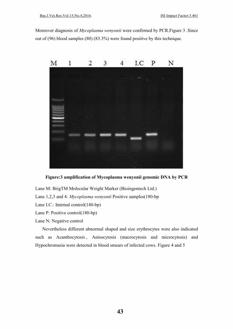
Moreover diagnosis of Mycoplasma wenyonii were confirmed by PCR.Figure 3 .Since
out of (96) blood samples (80) (83.3%) were found positive by this technique.
Figure:3 amplification of Mycoplasma wenyonii genomic DNA by PCR
Lane M: BrigTM Molecular Weight Marker (Bioingentech Ltd.)
Lane 1,2,3 and 4: Mycoplasma wenyonii Positive samples(180-bp
Lane I.C.: Internal control(140-bp)
Lane P: Positive control(180-bp)
Lane N: Negative control
Nevertheless different abnormal shaped and size erythrocytes were also indicated
such as Acanthocytosis , Anisocytosis (macrocytosis and microcytosis) and
Hypochromasia were detected in blood smears of infected cows. Figure 4 and 5

ISI Impact Factor:3.461 . ,20164Bas.J.Vet.Res.Vol.15,No.
44
Figure 4:Acanthocytes in blood smear of infected cow .Giemsa stain ×1000
Figure: 5 Infected erythrocytes of cattle with Hypochromasia Giemsa stain ×1000
ISI Impact Factor:3.461 . ,20164Bas.J.Vet.Res.Vol.15,No.
45
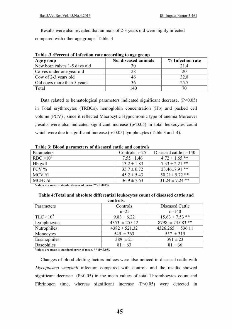
Results were also revealed that animals of 2-3 years old were highly infected
compared with other age groups. Table .3
Table .3 :Percent of Infection rate according to age group Age group No. diseased animals % Infection rate New born calves 1-5 days old 30 21.4 Calves under one year old 28 20 Cow of 2-3 years old 46 32.8 Old cows more than 5 years 36 25.7 Total 140 70
Data related to hematological parameters indicated significant decrease, (P<0.05)
in Total erythrocytes (TRBCs), hemoglobin concentration (Hb) and packed cell
volume (PCV) , since it reflected Macrocytic Hypochromic type of anemia Moreover
,results were also indicated significant increase (p<0.05) in total leukocytes count
which were due to significant increase (p<0.05) lymphocytes (Table 3 and 4).
Table 3: Blood parameters of diseased cattle and controls
Values are mean ± standard error of mean. ** (P<0.05).
Table 4:Total and absolute differential leukocytes count of diseased cattle and
controls. Parameters Controls
n=25 Diseased Cattle
n=140 TLC ×103 9.83 ± 6.22 15.63 ± 7.53 ** Lymphocytes 4353 ± 255.12 8798 ± 735.83 ** Nutrophiles 4382 ± 521.32 4326.265 ± 536.11 Monocytes 549 ± 363 557 ± 315 Eosinophiles 389 ± 21 391 ± 23 Basophiles 81 ± 63 81 ± 66 Values are mean ± standard error of mean. ** (P<0.05).
Changes of blood clotting factors indices were also noticed in diseased cattle with
Mycoplasma wenyonii infection compared with controls and the results showed
significant decrease (P<0.05) in the mean values of total Thrombocytes count and
Fibrinogen time, whereas significant increase (P<0.05) were detected in
Diseased cattle n=140 Controls n=25 Parameters 4.72 ± 1.65 ** 7.55± 1.46 RBC ×106
7.33 ± 2.21 ** 13.2 ± 1.83 Hb g/dl 23.46±7.91 ** 35.7 ± 6.72 PCV % 50.21± 5.72 ** 45.2 ± 5.43 MCV /fl 31.24 ± 7.24 ** 36.9 ± 7.63 MCHC/dl

ISI Impact Factor:3.461 . ,20164Bas.J.Vet.Res.Vol.15,No.
46
Thrombocytes volume, Thrombocytes distribution width, clotting time, prothrombin
time and activated partial thromboplastin time. Table (5).
Table 5:Indices of clotting factors in diseased cattle and controls.
Parameters Controls
n=25 Diseased Cattle
n=140 Total Thrombocytes count × 103 569.533 ± 76.744 332.233 ± 62.53** Thrombocytes volume /fl 11.535 ± 6.141 16.289 ± 1.922 ** Thrombocytes distribution width / % 14.653 ± 1.864 24.292 ± 5.788 ** Fibrinogen time / sec 20.54± 2.27 13.26 ± 5.21** Clotting time / mint 3.563 ± 1.723 5.234 ± 2.755 ** Prothrombin time /sec 14.276 ± 2.551 31.423 ± 3.622 ** Activated partial thromboplastin time /sec 52.534±6.443 69.455± 13.663 **
Values are mean ± standard error of mean ,** (P<0.05).
DISCUSSION There were no scientific document clarify the registration of Mycoplasma
wenyonii in Basrah governorate and other south parts of Iraq in cattle , Nevertheless
bovine clinical infection hade been documented and seen in Mosul ,North of Iraq , (11
and 12) whom mentioned that , It seems these concurrent infections are in animals
imported from the neighboring countries such as Turkey, Saudi Arabia and may be
Iran .
Hemoplasmas , occurs in most domesticated animals such as cattle , Buffaloes
,sheep, Swine, llamas, dogs and cats and has greater clinical occurrence in those
animals ,However latent Hemoplasmas might also affected mules, deer, elk and goats
since the organisms mostly appear to be species specific.(16).Those organisms were
incriminated and involved to rickettsial (Mycoplasma) parasite of the mammalian
erythrocytic cell membrane worldwide causing a febrile and haemolytic clinical
disease in a different livestock, especially food animals (17).
In the current study infected animals show different clinical manifestations which
were agreed with those mentioned by (1,2 and 3), since difficult and rapid respiration
which have been detected in diseased animals were due to Anemic hypoxia, as
decrease erythrocytes count and hemoglobin concentration were affected the oxygen
transmitted to body tissues, thereby failure of tissues to receive an adequate supply of
oxygen will occur, and panting with Dyspnea of diseased animals were detected
clinically, (16). The presences of pale mucus membranes will exhibited the
development of anemia and reduction of blood indices concentration was due to
destruction and removal of parasisetized erythrocytes by the reticulo-endothelial
ISI Impact Factor:3.461 . ,20164Bas.J.Vet.Res.Vol.15,No.
47
system ,Whereas icteric mucus membranes which were also seen reflected the
progressive anemia and bilirubinemia, developed in advance diseased animals (18).
Lethargy which had been shown by diseased cattle might occur due too decrease
muscle mass confirmed by decrease values of serum creatinine, presumably
associated with the poor body condition(19).
It have been proved that following experimental infection there is a variable
prepatent period, usually last for 1-3 weeks, which is followed by a period of intense
parasitemia, Ring form, coccoid and rod-shaped organisms are evident in stained
blood smears, Moreover , The organism is epicellular, infecting the surface and
periphery of erythrocytes, Nevertheless it were also could found free in the plasma in
blood examinations.(7). On the other hand there is a profound hypoglycemia during
the parasitemic phase which is believed to be due to direct consumption of glucose by
the parasite.(20). The period of intense parasitemia lasts for a period of 5-10 days
following which visible organisms in the blood become much less frequent and
anemia develops(21). Parasitized erythrocytes are removed from the circulation by the
spleen , It is believed that the parasite alters the erythrocyte membrane, exposing new
antigenic determinants and stimulating the development of antierythrocyte antibodies,
Moreover, the severity and duration of the anemia varies between individuals but
commonly lasts from 1-2 months , In addition during recovery stage there may be
further cycles of parasitemia and anemia which might become less severe However,
death which will follow occur due to anemic hypoxia (22).
Some diseased animals were also show edema of lower limbs and this were also
mention by (11). As well the development of edema always need a change in one or
more of a forces in a direction that might supported an increase in net filtration,
However This can be produced by an increase in capillary hydrostatic pressure,
capillary permeability, or interstitial usual venous pressure, or by a reduction in the
plasma oncotic pressure, Furthermore, edema it also can be induced by obstruction of
lymphatic tissue parts , since the fluid that is normally filtered is not returned to the
systemic circulation.(23).Moreover Diskin et al (24) were also added that increased
venous pressures due to central or regional venous obstruction or to an expansion in
plasma volume are transmitted to the capillary bed, thereby increasing hydrostatic
pressure and predisposing to edema , However hypoprotenemia which may expected
to occur were also play good role .
ISI Impact Factor:3.461 . ,20164Bas.J.Vet.Res.Vol.15,No.
48
Enlargement of superficial lymph node were mention in the current study as one of
the clinical manifestations encountered by diseased animals , it have been reported
that some plasma and cells in the interstitial space, along with certain cellular
material, antigens, and foreign particles enter lymphatic vessels, becoming lymphatic
fluid However, Lymph nodes filter the lymphatic fluid on its way to the central
venous circulation, removing cells and other material, Moreover The filtering process
also presents antigens to the lymphocytes contained within the nodes, Furthermore,
the immune response from these lymphocytes involves cellular proliferation, which
can cause the nodes to enlarge (lymphadenopathy),(25), In addition Smith (19) ,
added that pathogenic microorganisms carried in the lymphatic fluid can directly
infect the nodes, causing lymphadenitis .
Increase body temperature ,respiratory and heart rate were also mentioned by
Adresi and saki (1) which reflected the acute phase of the disease, However decrease
ruminal contractions reflected the atony of ruminal smooth muscles which mostly
reflected by lack of ruminal fibers followed by anorexia (16).
Results were also indicated increase capillary refilling time in diseased cattle
compared with controls .It have been documented that the capillary refilling time is a
quick test done used to monitor some disease problems such as dehydration ,shock
,peripheral vascular disease and hypothermia, thereby prolong time of the test might
reflected the less amount of blood flow reaches to tissues which were indicated in
infected animals of current study (21).
Mycoplasma wenyonii appears coccoid or rod shape, structures however it might
found individually or in chains on the red blood cells, same results were also mention
by others (7) ,However ,Sudan et al (17) added that microscopic examination of
Giemsa stained blood smear evidenced characteristic light pinkish to blue stained
cocci (0.5–1.0 μm diameter) and/or short rod (1–3 μm longer) shaped rickettsial
pathogens nesting in the depressions on the periphery of erythrocyte cell membrane as
well as extra cellular free rickettsial bodies in the plasma, some studies have also
favored the use of PCR as an aid in diagnosing bovine hemoplasmosis(6) , PCR
amplification can be perform directly from whole blood for detecting blood
organisms, since this test were detected the organism even in very small amounts, as
the method allows direct detection of pathogens in a blood sample (26). In this study,
(96) cows blood samples were used to molecular analysis to confirm the presence
of Mycoplasma wenyonii, Therefore infected animals gave a strong bands on this

ISI Impact Factor:3.461 . ,20164Bas.J.Vet.Res.Vol.15,No.
49
technique, Moreover, this finding is agreed with the results that obtained by
others(27).
High infection rate were indicated in age group of 2-3 years old ,since this result
were consistent with (3 and 16).
Anemia which were indicated in the present work occur because of significant
decrease in values of TRBc, Hb and PCV ,Moreover Macrocytic hypochromic type
were indicated ,same results were also documented by (18), Moreover Radostitis et
al(16) were also mention that the hemolysis caused by hemoplasma infections is
typically extra vascular and results in regenerative anemia with erythrocyte
agglutination may be present, In addition the increase in MCV shows the appearance
of immature red blood cells and is the index of regenerative anemia (17).Increase in
total leukocytes counts and lymphocytosis might indicated increase in immune system
capability (cellular immune excess) which were agreed with (18 and 28).
Little document had been mention the relation of hemoplasma infection and the
effect on clotting factors indices, Nevertheless in infected animals thrombocytopaenia
might occurs regularly in acute stages of the disease although the reduction of
platelets count does not always result in marked hemorrhages, even though the cause
of decrease platelets count is not completely clear , However megakaryocytes lysis
and reduced production of thrombocytes by megakaryocytes, with increased
consumption of platelets in the periphery, and defects of its functions might all been
suggested as factors predispose to decline its levels (19).Moreover hemorrhagic
diathesis were only indicated when platelets count are too low (29).
In the present study clotting factors indices were indicated clear disturbances in
clotting system of diseased animal with imbalanced regulation may lead to hyper
coagulation and / or hypo coagulation which might indicated the initiation of
disseminated intravascular coagulation (30).

ISI Impact Factor:3.461 . ,20164Bas.J.Vet.Res.Vol.15,No.
50
دمیة وتشخیصیة لخمج االبقار بالمایكوبالزما الدمویة في محافظة ،دراسة سریریة العراق ، البصرة
كمال الدین مھلھل السعد ، علي جراد
جامعة البصرة ،كلیة الطب البیطري ،فرع الطب الباطني والوقائي
العراق ،البصرة
الخالصة
اذ ش�ملت الدراس�ة فح�ص، ةالمحلی بقارالمایكوبالزما الدمویة في االدراسة وتشخیص مرض تم في ھذا البحث
ة وم�ن ك�ال الجنس�ین وبحس�ب الفئ�ات العمری�ة قس�مت الحیوان�ات إل�ى المحلی� بق�ارم�ن اال مئت�ان وخم�س وعش�رون
، عجول بعم�ر اق�ل م�ن س�نة ،یوم 5-1عجول حدیثة الوالدة بعمر (لكل مجموعة ) خمسون حیواناً (اربعة مجامیع
حیوان�ات . الع�راق –البص�رة ، ف�ي محافظ�ة البص�رة ) سنة وابقار بعمر اكثر من خمسة س�نوات 3-2ر بعمر ابقا
والت��ي شخص��ت باالعتم��اد عل��ى فح��ص Mycoplasma wenyonii نوعالدراس��ة كان��ت خمج��ھ س��ریریا ب��ال
م�ا ت�م فح�ص ك لس�لالمتس البلم�رة المسحات الدمویة المصبوغة بصبغة كمزا وتم تأكید التشخیص باستخدام فح�ص
اظھرت الحیوانات المریض�ة عالم�ات س�ریریة .كمجوعة سیطرة تعد اً سریری ةسویخمس وعشرون بقرة محلیة
تزای�د ،انخفاض انت�اج الحلی�ب ،شحوب االغشیة المخاطیة وبخاصة المبطنة للعین او المھبل ،تمثلت بفقدان الشھیة
وذم�ة االط�راف الخلفی�ة م�ع فق�دان ،الخم�ود،خش�ونة الجل�د ،الس�طحیة تضخم العقد اللمفیة،ترداد التنفس وصعوبتة
ومعدالت ترداد التنفس وضربات القلب وزمن رج�وع ،فضال عن ذلك فقد ارتفعت درجات حرارة الجسم . الوزن
اكم�، م�ع حیوان�ات مجموع�ة الس�یطرة الدم في االوعیة الدمویة وبش�كل معن�وي ف�ي الحیوان�ات الخمج�ة بالمقارن�ة
ف�ي المس�حات الدموی�ة Mycoplasma wenyonii ن�وعل�وحظ ال .مع�دالت تقلص�ات الك�رش ل�وحظ تن�اقص ف�ي
كم�ا ، بشكل منفرد أو بھیئة سالسل مفردة اً متجمعوعلى جدار كریات الدم الحمر المكور والعصوي متطفال ھبشكل
لنت�ائج ان وق�د س�جلت ا، ة للفح�صب�موج ت المفحوصة كانتمن الحاال%) 80.3(اكد فحص البلمرة المتسلسل إن
ازدادت وبش�كل معن�وي .سنة بالمقارنة مع الفئات العمری�ة االخ�رى 3-2اعلى نسبة خمج سجلت في االبقار بعمر
تركی��ز خض��اب ال��دم وحج��م خالی��ا ال��دم المرصوص��ة ف��ي الحیوان��ات ، مع��دالت الع��دد الكل��ي لكری��ات ال��دم الحم��ر
ر الدم من النوع ذي الكریات كبی�رة الحج�م س�ویة اذا سجل فق، المریضة بالمقارنة مع حیوانات مجموعة السیطرة
كما اتضح من نتائج الدراسة حدوث زیادة معنویة في العدد الكلي لخالیا الدم البیض بسبب تزاید الخالیا، الصباغ
كما سجلت نت�ائج الدراس�ة ح�دوث االخ�تالف ف�ي عوام�ل تخث�ر ال�دم إذ ل�وحظ تن�اقص معن�وي ف�ي . اللمفیة معنویاً
منشيء اللیفین في ح�ین س�جل تزای�د ف�ي مع�دالت حج�م الص�فیحات وقت،الكلي للصفیحات الدمویة ددمعدالت الع
بالمقارن��ة م��ع زم��ن س��ابق الخث��رین وزم��ن ح��رك الخث��رین الجزئ��ي، زم��ن تخث��ر ال��دم ،الدموی��ة ومع��دل انتش��ارھا
Mycoplasma ان االبق��ار المحلی��ة تص��اب ب��النوعاس��تنتج م��ن ھ��ذه الدراس��ة . مجموع��ة حیوان��ات الس��یطرة
wenyonii مما یؤدي الى تأثیرات جانبیة كبیرة قد تنتھي بموت الحیوان المصاب.
REFERENCES
ISI Impact Factor:3.461 . ,20164Bas.J.Vet.Res.Vol.15,No.
51
1-Adresi, Y and Saki ,CE.(2009). Clinical Eperythrozoon wenyoni (Adler and
Ellenbogen,1934) and Haemobartonella bovis (Donatin and Lestoquard,1934)
Infection in A Cattle. F.Ü.Sağ.Bil.Vet.Derg. 23 (2): 117 – 118.
2- Kahn, CM.(2010). The Merck Veterinary manual. 10th ed. Merck &Co.
3-Messick, JB. (2004). Hemotrophic mycoplasmas (hemoplasmas): a review and new
insights into pathogenic potential. Vet.Clin. Pathol. 33: 2–13.
4-Smith, JA., Thrall, MA., Smith, JL., Salman, MD., Ching, SV and Collins, JK.
(1990). Eperythrozoon wenyonii infection in dairy cattle. J. Am. Vet. Med.
Assoc. 196: 1244–1250.
5-Fujihara, Y., Sasaoka, F., Suzuki, J., Watanabe, Y., Fujihara, M., Ooshita, K., Ano,
H. and Harasawa, R. (2011). Prevalence of hemoplasma infection among
cattle in the western part of Japan. J. Vet. Med. Sci. 73: 1653–1655.
6-McAuliffe, L., Lawes, J., Bell, S., Barlow, A., Ayling, R. and Nicholas, R. (2006).
The detection of Mycoplasma (formerly Eperythrozoon)wenyonii by 16S
rRNA PCR and denaturing gradient gel electrophoresis. Vet. Microbiol. 117:
292–296.
7-Urquhart, Gm., Armour, J., Duncan, JL., Dunn, A.M., Jennings, Fw.
(2003)Veterinary parasitology, 2nd ed. Black well science Ltd., Oxford. Pp:
252.
8-Groebel, K., Hoelzle, K., Wittenbrink, MM., Ziegler,U and Hoelzle, LE. (2009).
Mycoplasma suis invades porcine erythrocytes. Infec.Immunol. 77: 576-584.
9- Hendrix, CM.(1998). Diagnostic Veterinary Parasitology 2nd Edition. Mosby Inc,
United State of America . 266-267.
10-Messick, JB. (2003). New perspectives about Hemotrophic mycoplasma
(formerly,Haemobartonella and Eperythrozoon species)infections in dogs and
cats. Vet Clin North Am Small Anim Pract. 33: 1453-1465
11-Al-Badrani, BA. and Rhaymah, MSH. (2012). A clinical and diagnostic study of
Mycoplasma wenyonii and Haemobartonella bovis infections in cattle of
Mosul City, Iraq. Res. Opin. Anim. Vet. Sci. 2(1), 27-30.
12-Hasan, MH.(2012). Diagnosis of some blood parasites in cattle and sheep in
Mosul, Iraq. Iraqi J. Vet. Sci. 26. Supp II :57-61.
13-Weiss, DJ and Wardrop ,KJ. (2010). Schalm's Veterinary Hematology, 6th Ed,
Ames, Wiley-182 Blackwell.
ISI Impact Factor:3.461 . ,20164Bas.J.Vet.Res.Vol.15,No.
52
14- Bush, BM. (1975).Veterinary laboratory manual. 1st ed., the Gresham
press,London. pp: 113-167.
15-Leech, NL., Barrett, KC and Morgan, GA. (2007).SPSS for intermediate statistics:
use and interpretation .1st Ed. Lawrence Erlbaum Asso.USA. 20-51.
16- Radostitis, OM., Gay,CC., Blood,DC and Hinchliff, KW.(2007). Veterinary
Medicine. A text book of the diseases of cattle, sheep, goats and horses.10th
ed, WB Saunders Co.pp:1483-1498.
17-Sudan,V.,Sharma,RL., Gupta, SR., Borah, MK. and Mishra, R.(2012). An
occurrence of clinical eperythrozoonosis in a German Shepherd dog and its
therapeutic management. J Parasit Dis.36(2):181–183.
18-Tagawa, M., Matsumoto, K., Yokoyama, N., and Inokuma, H. (2010). Comparison
of the effect of two Hemoplasma species on hematological parameters in
cattle. J. Vet. Med.Sci.72: 113-115.
19-Smith, BP.(2004). Large animal internal medicine, 4th ed., New York, Mosby.
20 -Love, JN and McEwen, EG.(1972). Hypoglycemia associated with
Haemobartonella-like infection in splenectomized calves. Am J Vet Res.
33:2087–2089.
21-Suzanne, G., Genova, RN., Streeter, KE., Velguth, TA., Snider, KM
and Katharine, MS.(2011). Severe anemia associated with Mycoplasma
wenyonii infection in a mature cow.Can Vet J. 52(9): 1018–1021.
22-Hoelzle, K., Winkler, M., Kramer, MM., Wittenbrink, MM.,
Dieckmann, SM. and Hoelzle, LE. (2011). Detection of Candidatus
Mycoplasma haemobos in cattle with anaemia. Vet. J. 187:408–410.
23- Shaun, C., Edwin, MDJ., Atwood, MD.(2002). Peripheral Edema. Amer. J.
Med.113.580-586.
24-Diskin, CJ., Stokes, TJ., Dansby, LM., Carter,TB., Radcliff,L., Thomas ,SG
.(1999).Towards an understanding of oedema. BMJ. 318:1610–1613.
25- Salgado, BS., Battaglia, CT., Stuchi, RS., Cadioli, FA., Rozza, DB., Machado,
GF.(2011). What is your diagnosis? Lymphadenopathy in a cow with sever
anemia . Vet Clin Pathol.40/1 ,103–104c.
26-Basima. A and Baraa. A.(2016). First Documented Study of Mycoplasma
Wenyonii of Cattle in Iraq. IJSR.5(2):515-520.
27-Wang, J., Zhu ,Y.,Qin, J.,Zhang,F ., Zhao,Y.(2009).Detection of Eperythrozoon
wenyoni by PCR assay.Front.Agri.china.3(1):100-103.

ISI Impact Factor:3.461 . ,20164Bas.J.Vet.Res.Vol.15,No.
53
28- Reaganf,WJ., Carrym, . A., Thrall,S ., Colgan, J., Hutchisona,NDM., Weiser, G.
(1990). The Clinicopathologic, Light, and Scanning Electron Microscopic
Features of Eperythrozoonosis in Four Naturally Infected Llamas. Vet Pathol.
27:426431.
29-Pantanowitz, L.( 2003) Mechanism of thrombocytopenia in tick born diseases. In J
Inf Dis.2:1-7.
30- Bick, RL.( 2003). Disseminated intravascular coagulation: Current concepts of
etiology, pathophysiology, diagnosis and treatment. Hematol Oncol Clin
North Am.17:149.