clinical experience of infantile posthemorrhagic ... · pdf fileorg/licenses/by-nc/3.0/) ......
TRANSCRIPT
106 Copyright © 2015 Korean Neurotraumatology Society
Introduction
Infantile posthemorrhagic hydrocephalus (IPHH) is de-fined as ventricular dilatation after germinal matrix (GM) hemorrhage in a preterm infant. The GM is a gelatinous, highly cellular, and vascularized structure located near the head of the caudate nucleus, just beneath the ependymal lin-ing. This structure develops to its maximal size by 23 weeks
of gestation and involutes by 36 weeks.1,5,8,10,17) Preterm in-fants have a high risk of GM hemorrhage because of expo-sure of the fragile and immature vessels in the GM to stress-ful conditions, such as blood pressure fluctuations, increased cerebral perfusion pressure, and coagulopathy.1,7,20) Dilata-tion of the ventricle occurs by alterations of cerebrospinal flu-id (CSF) circulation resulting from GM hemorrhage into the ventricle.
Although the incidence of GM hemorrhage in preterm infants has decreased because of advances in neonatal care, the rate of GM hemorrhage is still as high as 30%.12,13) Fur-thermore, the incidence of GM hemorrhage increases with decreasing birth weight. Temporary neurosurgical proce-dures for hydrocephalus include lumbar puncture, ventric-ular tap, external ventricular drainage (EVD), ventricular access device, and placement of a ventricular subgaleal shunt. Placement of a ventriculo-peritoneal (VP) shunt, which is a permanent procedure, is required in cases where CSF circu-
Clinical Experience of Infantile Posthemorrhagic Hydrocephalus Treated with Ventriculo-Peritoneal Shunt
Hae Min Kim, MD and Ki Hong Kim, MDDepartment of Neurosurgery, Daegu Catholic University College of Medicine, Daegu, Korea
Objective: Infantile posthemorrhagic hydrocephalus (IPHH) is the most common cause of infantile acquired hydrocephalus. We present and discuss our experience of treatment of six IPHH patients treated by a ventriculo-peritoneal (VP) shunt.Methods: Six preterm infants treated by a VP shunt due to germinal matrix hemorrhage and hydrocephalus were included in our study. External ventricular drainage (EVD) was performed in patients with symptomatic ventricular dilatation, and a VP shunt was placed in the case of no improvement of the ventricular index despite several rounds of EVD. Radiographic findings and surgical outcomes were analyzed retrospectively.Results: Four patients were male and two were female. Mean gestational age was 25 weeks and mean weight at birth was 868.3 g. One patient had a Papile grade II (16.7%) hemorrhage, three had a grade III (50%) hemorrhage, and two had a grade IV (33.3%) hemorrhage. EVD complications (one case of ventriculitis and one case of a ventricular abscess) occurred in two patients. VP shunt complications occurred in two patients (33.3%). Three cases had an isolated 4th ventricle; two of these cases had a VP shunt placed whereas the other case had a VP shunt placed in addition to aqueductoplasty using a neuroendoscope. At the last follow-up, three of the six patients had severe neurodevelopmental delay, two had mild neuro-developmental delay, and one had normal development status.Conclusion: In our study, although it is difficult to present the significant result for management of IPHH, we think that var-ied efforts are required to treat IPHH patients. (Korean J Neurotrauma 2015;11(2):106-111)
KEY WORDS: Infant ㆍCerebrospinal fluid shunt ㆍHemorrhage ㆍHydrocephalus.
Received: July 22, 2015 / Revised: September 11, 2015Accepted: October 12, 2015Address for correspondence: Ki Hong Kim, MDDepartment of Neurosurgery, Daegu Catholic University College of Medicine, 33 Duryugongwon-ro 17-gil, Nam-gu, Daegu 42472, KoreaTel: +82-53-650-4258, Fax: +82-53-650-4932E-mail: [email protected] cc This is an Open Access article distributed under the terms of Cre-ative Attributions Non-Commercial License (http://creativecommons.org/licenses/by-nc/3.0/) which permits unrestricted noncommercial use, distribution, and reproduction in any medium, provided the original work is properly cited.
CLINICAL ARTICLEKorean J Neurotrauma 2015;11(2):106-111
pISSN 2234-8999 / eISSN 2288-2243
http://dx.doi.org/10.13004/kjnt.2015.11.2.106
Hae Min Kim and Ki Hong Kim
http://www.kjnt.org 107
lation fails to be restored. However, because there are many complications associated with invasive procedures, such as infection, ventricular isolation by infection, CSF leakage, shunt malfunction, over-drainage, and skin breakdown over the shunt device, meticulous procedures are required for these patients.23) Here, we describe our experience of the treatment and discuss the results of six IPHH patients treat-ed by a VP shunt.
Materials and Methods
Between April 2010 and December 2014, six preterm in-fants had a VP shunt placed due to GM hemorrhage and hydrocephalus at our institution. Medical records and ra-diographic findings were analyzed retrospectively. All pa-tients in the neonatal intensive care unit were scanned by cranial ultrasonography (USG) at the first 2 days of life and thereafter every week. GM hemorrhage and ventricle dila-tation on USG were classified according to the Papile clas-sification.15) Ventricle dilatation was estimated by the ven-tricular index (VI), which is the distance between the falx and lateral wall of the anterior horn in the coronal plane at the level of the third ventricle.2) Computed tomography (CT) was performed to evaluate the posterior fossa and to con-firm the trajectory and the length of the intracranial cathe-ter before VP shunt placement. EVD was performed in pa-
tients with symptomatic ventricular dilatation. The placement of EVD was maintained for 1 week and about 20 cc of CSF was drained per day. After serial rounds of USG, a VP shunt was placed in cases where there was no improvement in VI despite several rounds of EVD, no evidence of CSF in-fection, and patient weight over 1,500 g. In the operating room, we inserted an intracranial shunt catheter along the axis of the lateral ventricle via the occipito-parietal point. Other VP shunt procedures were performed in a routine fash-ion with a programmable valve. In one case with multiloc-ulated hydrocephalus confirmed by preoperative imaging findings, we used a neuroendoscope for fenestration of the pseudomembrane, septum pellucidum, and Sylvius aque-duct. Patient symptoms, such as neurodevelopmental delay, seizure, and cerebral palsy, were evaluated as neurological outcomes.
Results
Clinical data (gender, gestational age, weight at birth, VI, Papile grade, weight at VP shunt placement, associated prob-lems) and surgical outcomes (time from birth to EVD, num-ber of times EVD, time from EVD to VP shunt placement, complications of EVD, neuroendoscopic procedures, com-plications of shunt, final neurological deficits) are summa-rized in Tables 1 and 2. VP Shunt was placed in six patients
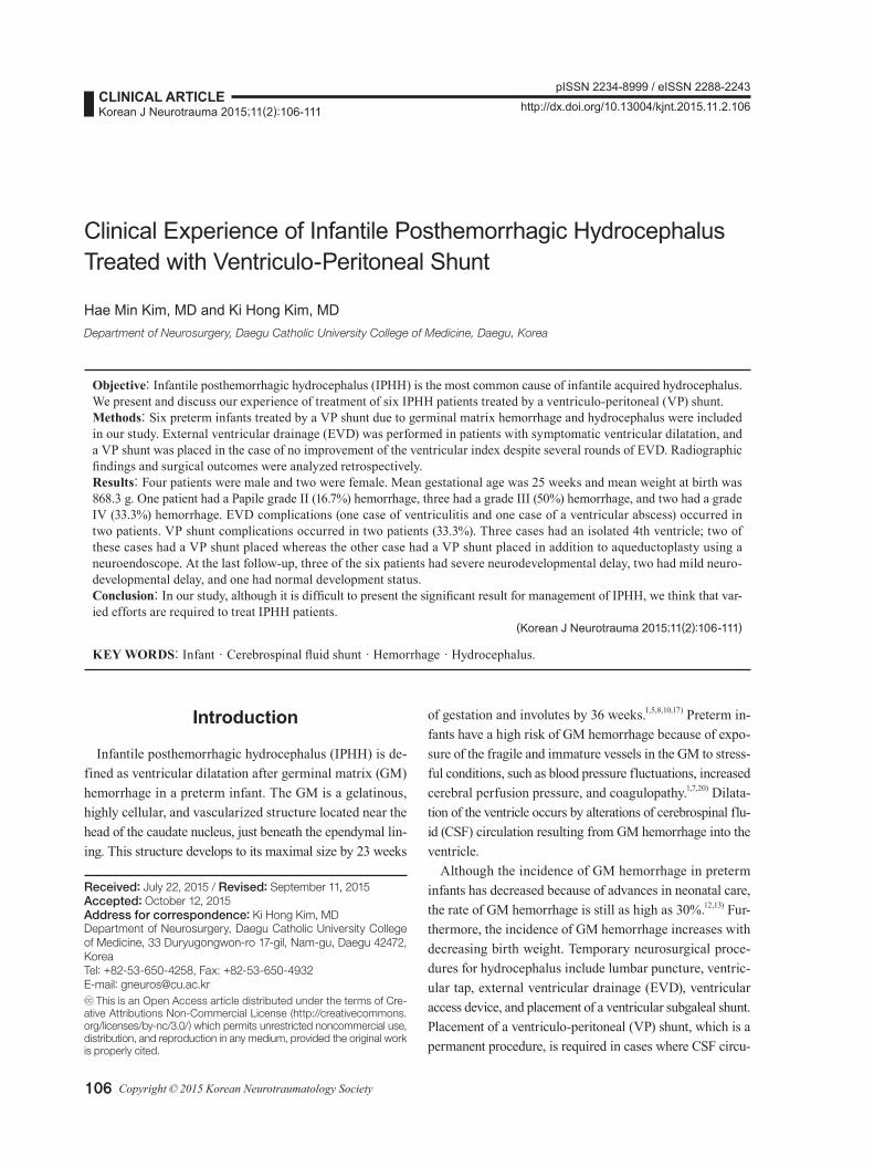
TABLE 1. Clinical characteristics of infantile posthemorrhagic hydrocephalus in 6 patients
Case No. Gender Gestational
age (week)Weight at birth (g) VI (mm) Papile
gradeWeight at
VP shunt (g) Associated problems
1 F 26 990 23 4 1,700 Isolated 4th ventricle2 M 25 830 21 3 2,500 -
3 M 27 1,040 14 4 1,830 Multiloculated lateral ventricle4 M 27 1,140 19 2 2,050 Isolated 4th ventricle
5 M 23 760 16 3 2,120 Multiloculated lateral ventricle, isolated 4th ventricle
6 F 22 450 21 3 1,730 -
VI: ventricular index, VP: ventriculo-peritoneal
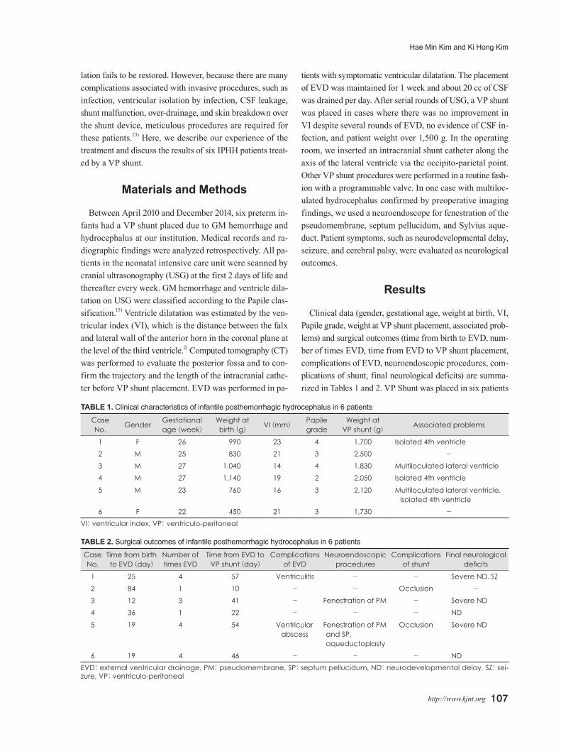
TABLE 2. Surgical outcomes of infantile posthemorrhagic hydrocephalus in 6 patients
Case No.
Time from birth to EVD (day)
Number of times EVD
Time from EVD to VP shunt (day)
Complications of EVD
Neuroendoscopic procedures
Complications of shunt
Final neurological deficits
1 25 4 57 Ventriculitis - - Severe ND, SZ2 84 1 10 - - Occlusion -
3 12 3 41 - Fenestration of PM - Severe ND4 36 1 22 - - - ND
5 19 4 54 Ventricular abscess
Fenestration of PM and SP, aqueductoplasty
Occlusion Severe ND
6 19 4 46 - - - NDEVD: external ventricular drainage, PM: pseudomembrane, SP: septum pellucidum, ND: neurodevelopmental delay, SZ: sei-zure, VP: ventriculo-peritoneal
108 Korean J Neurotrauma 2015;11(2):106-111
Clinical Experience of Infantile Posthemorrhagic Hydrocephalus
after the diagnosis of IPHH. Four patients were male (66.7%) and two were female (33.3%). Mean gestational age was 25 weeks (range, 22 to 27 weeks) and mean weight at birth was 868.3 g (range, 450 to 1,140 g). Mean VI was 19 mm (range, 14 to 23 mm), and the VI of all patients was over the 97th centile. One patient (16.7%) had a Papile grade II hemor-rhage, three patients had a grade III hemorrhage (50%), and two patients had a grade IV hemorrhage (33.3%). Mean weight at VP shunt surgery was 1,988.3 g (range, 1,700 to 2,500 g). Three patients had an isolated 4th ventricle.
The mean number of EVD sessions was 2.8 (range, 1 to 4) and the mean time from EVD to VP shunt placement was 38.3 days (range, 10 to 57 days). EVD complications oc-curred in two patients: one case of ventriculitis and one case of a ventricular abscess. Neuroendoscopic procedures were done in two patients: one patient underwent fenestra-tion of the pseudomembrane while the other underwent fenestration of the pseudomembrane and septum pellu-cidum as well as aqueductoplasty due to multiloculated hy-drocephalus. VP shunt complications (occlusion of the intra-cranial catheter) occurred in two patients (33.3%). Three of six patients had severe neurodevelopmental delay, two had mild neurodevelopmental delay, and one showed a normal development status at the end of the study. There was no mortality and no shunt infection.
Illustrative cases
Case 1 (patient No. 2)The patient was born at 25 weeks gestational age by nor-
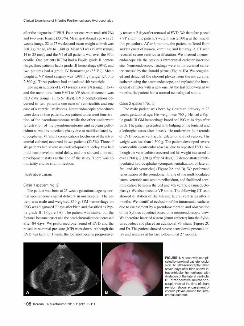
mal spontaneous vaginal delivery in our hospital. The pa-tient was male and weighted 830 g. GM hemorrhage on USG was diagnosed 7 days after birth and classified as Pap-ile grade III (Figure 1A). The patient was stable, but the fontanel became tenser and the head circumference increased after 84 days. We performed one round of EVD and the raised intracranial pressure (ICP) went down. Although the EVD was kept for 1 week, the fontanel became progressive-
ly tenser at 2 days after removal of EVD. We therefore placed a VP shunt; the patient’s weight was 2,500 g at the time of this procedure. After 6 months, the patient suffered from sudden onset of nausea, vomiting, and lethargy. A CT scan revealed severe ventricular dilatation. We inserted a neuro-endoscope via the previous intracranial catheter insertion site. Neuroendoscopic findings were an intracranial cathe-ter encased by the choroid plexus (Figure 1B). We coagulat-ed and detached the choroid plexus from the intracranial catheter using the neuroendoscope, and replaced the intra-cranial catheter with a new one. At the last follow-up at 49 months, the patient had a normal neurological status.
Case 2 (patient No. 5)The male patient was born by Cesarean delivery at 23
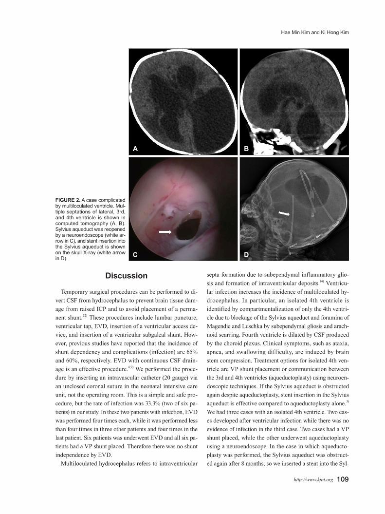
weeks gestational age. His weight was 760 g. He had a Pap-ile grade III GM hemorrhage based on USG at 14 days after birth. The patient presented with bulging of the fontanel and a lethargic status after 1 week. He underwent four rounds of EVD because ventricular dilatation did not resolve. His weight was less than 1,500 g. The patient developed severe ventriculitis (ventricular abscess) due to repeated EVD. Al-though the ventriculitis recovered and his weight increased to over 1,500 g (2,120 g) after 54 days, CT demonstrated multi-loculated hydrocephalus (compartmentalization of lateral, 3rd, and 4th ventricles) (Figure 2A and B). We performed fenestration of the pseudomembrane of the multiloculated lateral ventricle and septum pellucidum, and facilitated com-munication between the 3rd and 4th ventricle (aqueducto-plasty). We also placed a VP shunt. The following CT scan showed dilatation of the 4th and lateral ventricles after 8 months. We identified occlusion of the intracranial catheter due to encasement by a pseudomembrane and obstruction of the Sylvius aqueduct based on a neuroendoscopic view. We therefore inserted a stent (shunt catheter) into the Sylvi-us aqueduct and placed an additional VP shunt (Figure 2C and D). The patient showed severe neurodevelopmental de-lay and seizures at his last follow-up at 27 months.
FIGURE 1. A case with compli-cated by proximal catheter occlu-sion. A: Ultrasonography taken seven days after birth shows in-traventricular hemorrhage with dilatation of the lateral ventricle. B: Intraoperative neuroendo-scopic view at the time of shunt revision shows encasement of choroid plexus around the intra-cranial catheter.A B
Hae Min Kim and Ki Hong Kim
http://www.kjnt.org 109
Discussion
Temporary surgical procedures can be performed to di-vert CSF from hydrocephalus to prevent brain tissue dam-age from raised ICP and to avoid placement of a perma-nent shunt.22) These procedures include lumbar puncture, ventricular tap, EVD, insertion of a ventricular access de-vice, and insertion of a ventricular subgaleal shunt. How-ever, previous studies have reported that the incidence of shunt dependency and complications (infection) are 65% and 60%, respectively. EVD with continuous CSF drain-age is an effective procedure.4,9) We performed the proce-dure by inserting an intravascular catheter (20 gauge) via an unclosed coronal suture in the neonatal intensive care unit, not the operating room. This is a simple and safe pro-cedure, but the rate of infection was 33.3% (two of six pa-tients) in our study. In these two patients with infection, EVD was performed four times each, while it was performed less than four times in three other patients and four times in the last patient. Six patients was underwent EVD and all six pa-tients had a VP shunt placed. Therefore there was no shunt independence by EVD.
Multiloculated hydrocephalus refers to intraventricular
septa formation due to subependymal inflammatory glio-sis and formation of intraventricular deposits.14) Ventricu-lar infection increases the incidence of multiloculated hy-drocephalus. In particular, an isolated 4th ventricle is identified by compartmentalization of only the 4th ventri-cle due to blockage of the Sylvius aqueduct and foramina of Magendie and Luschka by subependymal gliosis and arach-noid scarring. Fourth ventricle is dilated by CSF produced by the choroid plexus. Clinical symptoms, such as ataxia, apnea, and swallowing difficulty, are induced by brain stem compression. Treatment options for isolated 4th ven-tricle are VP shunt placement or communication between the 3rd and 4th ventricles (aqueductoplasty) using neuroen-doscopic techniques. If the Sylvius aqueduct is obstructed again despite aqueductoplasty, stent insertion in the Sylvius aqueduct is effective compared to aqueductoplasty alone.3) We had three cases with an isolated 4th ventricle. Two cas-es developed after ventricular infection while there was no evidence of infection in the third case. Two cases had a VP shunt placed, while the other underwent aqueductoplasty using a neuroendoscope. In the case in which aqueducto-plasty was performed, the Sylvius aqueduct was obstruct-ed again after 8 months, so we inserted a stent into the Syl-
C D
A B
FIGURE 2. A case complicated by multiloculated ventricle. Mul-tiple septations of lateral, 3rd, and 4th ventricle is shown in computed tomography (A, B). Sylvius aqueduct was reopened by a neuroendoscope (white ar-row in C), and stent insertion into the Sylvius aqueduct is shown on the skull X-ray (white arrow in D).
110 Korean J Neurotrauma 2015;11(2):106-111
Clinical Experience of Infantile Posthemorrhagic Hydrocephalus
vius aqueduct using a neuroendoscope. No complications associated with stent insertion were observed at the last follow-up.
Generally, neurodevelopmental outcome of IPHH is de-termined by the grade of GM hemorrhage, coexistent paren-chymal injury, and progression of ventriculomegaly.18,19) Patients can suffer mental retardation, seizure, and cerebral palsy. The timing of GM hemorrhage is an important fac-tor that affects neurodevelopmental outcome. Especially, patients who develop GM hemorrhage within 6 hours of birth have a poor neurodevelopmental outcome.19) Progres-sion of ventricular dilatation and signs of raised ICP are also important factors to consider. A patient who develops ventricular dilatation without symptoms related raised ICP has a more favorable prognosis than symptomatic patients.6) Three of our patients had severe neurodevelopmental de-lays and seizures. The mean Papile grade of these three pa-tients was 3.6, while the mean Papile grade of the other three patients was 2.6. Although we could not evaluate the significance of this result because of the small number of patients, Papile grade could affect neurodevelopmental out-come. However, because two of three patients with severe neurodevelopmental delay also had ventricular infection, we could not verify that Papile grade is an important factor affecting neurodevelopmental outcome. A large number of patients and multivariate analysis will be necessary to test this hypothesis.
A VP shunt is the traditional CSF diversion treatment for hydrocephalus. However, it has many complications, such as infection, CSF leakage, disconnection, shunt malfunc-tion, over-drainage, and skin breakdown over the shunt de-vice. Because preterm infants have immature immune sys-tems, infection associated with the shunt device as a foreign body develops frequently. Furthermore, skin breakdown due to a thin scalp and scalp thinning associated with in-creasing head size may occur.11) Endoscopic third ventric-ulostomy (ETV) is usually performed for noncommuni-cating hydrocephalus of children older than 2 years. ETV had not been considered as a treatment modality for IPHH. However, studies about effectiveness of ETV have been re-ported recently as a treatment option for hydrocephalus of IPHH.16) And there were reports addressing medical treat-ments and alternative temporary surgical procedures (ven-tricular access device and ventriculosubgaleal shunt inser-tion) to reduce a rate of complication.13,21) We placed a VP shunt in all IPHH patients. No shunt-associated complica-tions except for occlusion in two patients occurred in our study.
One patient (patient No. 2) had an excellent neurodevelop-
mental outcome as evidenced by normal cognitive function, no developmental delay, no seizures, and no cerebral palsy. Despite a GM hemorrhage and ventricular dilatation of Pap-ile grade III, symptoms and signs associated with raised ICP were not observed for 84 days. Gestational age, weight at birth, VI, and Papile grade were not different from those of the other patients, so these cannot be considered to be fac-tors associated with a good outcome. Delayed symptom development may be an important factor that affects neu-rodevelopmental outcome. Because late developing symp-toms are associated with fewer EVD procedures, there is less chance of EVD complications, and the rate of infection and VP shunt complications are likely to be lower. However, more cases need to be examined to confirm this hypothesis.
Conclusion
In our study, although it is difficult to present the signifi-cant result for management of IPHH because of the small number of patients, we think that varied efforts associated with neonatal intensive care, meticulous procedures, and less invasive operations are required to treat IPHH patients who are in poor condition and generally have a high rate of com-plications. A large number of patients and long-term follow-up are required to determine factors that affect neurode-velopmental outcome.
■ The authors have no financial conflicts of interest.
REFERENCES1) Ballabh P. Intraventricular hemorrhage in premature infants:
mechanism of disease. Pediatr Res 67:1-8, 20102) Brouwer MJ, de Vries LS, Pistorius L, Rademaker KJ, Groenendaal
F, Benders MJ. Ultrasound measurements of the lateral ventricles in neonates: why, how and when? A systematic review. Acta Pae-diatr 99:1298-1306, 2010
3) Cinalli G, Spennato P, Savarese L, Ruggiero C, Aliberti F, Cuomo L, et al. Endoscopic aqueductoplasty and placement of a stent in the cerebral aqueduct in the management of isolated fourth ventri-cle in children. J Neurosurg 104(1 Suppl):21-27, 2006
4) Cornips E, Van Calenbergh F, Plets C, Devlieger H, Casaer P. Use of external drainage for posthemorrhagic hydrocephalus in very low birth weight premature infants. Childs Nerv Syst 13:369-374, 1997
5) Duncan CC, Ment LR. Intraventricular hemorrhage and prema-turity. Neurosurg Clin N Am 4:727-734, 1993
6) Fletcher JM, Landry SH, Bohan TP, Davidson KC, Brookshire BL, Lachar D, et al. Effects of intraventricular hemorrhage and hydrocephalus on the long-term neurobehavioral development of preterm very-low-birthweight infants. Dev Med Child Neurol 39: 596-606, 1997
7) Folkerth RD. Germinal matrix haemorrhage: destroying the brain’s building blocks. Brain 134(Pt 5):1261-1263, 2011
8) Hambleton G, Wigglesworth JS. Origin of intraventricular haem-orrhage in the preterm infant. Arch Dis Child 51:651-659, 1976
Hae Min Kim and Ki Hong Kim
http://www.kjnt.org 111
9) Januschek E, Machado LS, Steinthal B, Ulrich PT. Posthemor-rhagic hydrocephalus in very low birth weight infants--a new gen-tle surgical technique for external ventricular drainage. Childs Nerv Syst 27:991-994, 2011
10) Kedzia A. Characteristics of periventricular matrix vascularization in image computer transformation system. Folia Neuropathol 33: 267-270, 1995
11) Kim SY, Cho JH, Kim KH. Endoscopic coagulation of choroid plexus in hydranencephaly. J Korean Neurosurg Soc 55:375-378, 2014
12) Lemons JA, Bauer CR, Oh W, Korones SB, Papile LA, Stoll BJ, et al. Very low birth weight outcomes of the National Institute of Child health and human development neonatal research network, January 1995 through December 1996. NICHD Neonatal Re-search Network. Pediatrics 107:E1, 2001
13) Limbrick DD Jr, Mathur A, Johnston JM, Munro R, Sagar J, Inder T, et al. Neurosurgical treatment of progressive posthemorrhagic ventricular dilation in preterm infants: a 10-year single-institution study. J Neurosurg Pediatr 6:224-230, 2010
14) Oi S, Abbott R. Loculated ventricles and isolated compartments in hydrocephalus: their pathophysiology and the efficacy of neuroen-doscopic surgery. Neurosurg Clin N Am 15:77-87, 2004
15) Papile LA, Burstein J, Burstein R, Koffler H. Incidence and evolu-tion of subependymal and intraventricular hemorrhage: a study of infants with birth weights less than 1,500 gm. J Pediatr 92:529-534, 1978
16) Peretta P, Ragazzi P, Carlino CF, Gaglini P, Cinalli G. The role of
Ommaya reservoir and endoscopic third ventriculostomy in the management of post-hemorrhagic hydrocephalus of prematurity. Childs Nerv Syst 23:765-771, 2007
17) Takashima S, Tanaka K. Microangiography and vascular perme-ability of the subependymal matrix in the premature infant. Can J Neurol Sci 5:45-50, 1978
18) Vohr B, Allan WC, Scott DT, Katz KH, Schneider KC, Makuch RW, et al. Early-onset intraventricular hemorrhage in preterm neonates: incidence of neurodevelopmental handicap. Semin Peri-natol 23:212-217, 1999
19) Vohr BR, Allan WC, Westerveld M, Schneider KC, Katz KH, Makuch RW, et al. School-age outcomes of very low birth weight infants in the indomethacin intraventricular hemorrhage preven-tion trial. Pediatrics 111(4 Pt 1):e340-e346, 2003
20) Volpe JJ. Intraventricular hemorrhage in the premature infant--cur-rent concepts. Part I. Ann Neurol 25:3-11, 1989
21) Whitelaw A, Jary S, Kmita G, Wroblewska J, Musialik-Swietlins-ka E, Mandera M, et al. Randomized trial of drainage, irrigation and fibrinolytic therapy for premature infants with posthemor-rhagic ventricular dilatation: developmental outcome at 2 years. Pediatrics 125:e852-e858, 2010
22) Whitelaw A, Thoresen M, Pople I. Posthaemorrhagic ventricular dilatation. Arch Dis Child Fetal Neonatal Ed 86:F72-F74, 2002
23) Wu Y, Green NL, Wrensch MR, Zhao S, Gupta N. Ventriculoperi-toneal shunt complications in California: 1990 to 2000. Neurosur-gery 61:557-562; discussion 562-563, 2007