clinical application of endodontic...
TRANSCRIPT

MAXIL!LOFACIAL PROSTHETICS l DENTAL IMPLANTS SECTION EDITORS
I. KENNETH ADISMAN RONALD I’. DESJARDINS
Clinical application of endodontic implants
Sandra Madison, D.D.S., M.S.,* and Arne M. Bjorndal, D.D.S., MS.** University of North Carolina. School of Dentistry, Chapel Hill, N.C., and University of Iowa, College of Dentistry, Iowa City, Iowa
P reservation of the natural dentition and restqration of the oral cavity to a normal functional state is a primary goal in dentisiry. In addition to advances in cariology and periodontics, the use of intrabone implants has provided a method for maintaining a functional oral environment. There are two types of dental implants; prosthetic implants that replace missing teeth and end- odontic impla:nts that use remaining teeth for anchor- age.
The endoda’ntic implant described by Orlay’ uses the root canal space in an existing tooth as a pathway for the implant to extend into the apical bone. A major advan- tage of the endodontic implant, in comparison with the prosthetic implant, is that it provides a closed environ- ment that does not communicate with the oral cavity. Complete separation from the oral environment reduces the complications of periodontal breakdown often responsible fo:r implant failures.
Frank2 reported the use of endodontic implants for three patients to improve crown-root ratio, thereby decreasing clinical mobility. Others have reported suc- cessful use of endodontic implants.3-6
Fig. 1. Preoperative radiograph. Mandibular right cen- tral incisor with silver cone perforating laterally.
This article describes uses of endodontic implants to maintain com:promised teeth. The technique for prepa- ration and placement of endodontic implants is similar to that reported by Frank.2 The root canals were chemome- chanically prepared to a minimum of a size 50 file. After canal preparation the bone preparations were done with 40 mm reamers, sizes 45 to 120 set at the desired length for the implant termination. The osseous preparations were made with increasing file sizes corresponding to
that of the implant to be used. The implants were sectioned at the coronal termini and the portions of the implant to remain in the root were coated with an endodontic sealer. The implants were seated and the access openings were closed.
TREATMENT APPLICATIONS Perforation of root and mobility
A 24-year-old man was treated at the endodontic clinic 1 year after trauma and root canal therapy on the
mandibular left central incisor. The preoperative radio- graph revealed a silver cone in the corona1 half of the root canal perforating the lateral surface of the root (Fig. 1). Apical root resorption was also observed. The tooth exhibited class II mobility and was tender to percussion. Treatment involved removal of the silver cone, chemo- mechanical preparation of the root canal, and prepara- tion and placement of an intraosseous endodontic implant through the canal into the apical bone. Apical curettage was performed during the surgical procedure and the lateral root perforation was repaired (Fig. 2). No fixation was used following implant placement and tooth mobility decreased immediateiy. At the 2-year evaluation, no symptoms were reported and there were no radiographic signs of pathosis associated with the implant (Fig. 3). The apical resorptive defect was still present; however, the lamina dura remained intact. Continued evaluation was recommended.
Pulpal necrosis and chronic apical periodontitis
*Assistant Professor, Department of Endodontics, University of North Carolina, Schwl of Dentistry; formerly Assistant Professor, Depart- ment of Endodontics, University of Iowa, College of Dentistry.
**Professor Emeritus, University of Iowa, College of Dentistry.
A 69-year-old man was treated for extraction of mandibular anterior teeth and construction of a complete mandibular denture to oppose a maxillary denture. After examination, a treatment plan was devised that permit-
THE JOURNAL OF PROSTHETIC DENTISTRY 603
MADISON AND BJORNDAL
Fig. 2. Radiograph immediately after endodontic implant placement and perforation repair.
Fig. 3. Two-year follow-up radiograph of tooth shown in Fig. 1. No periapical pathosis noted.
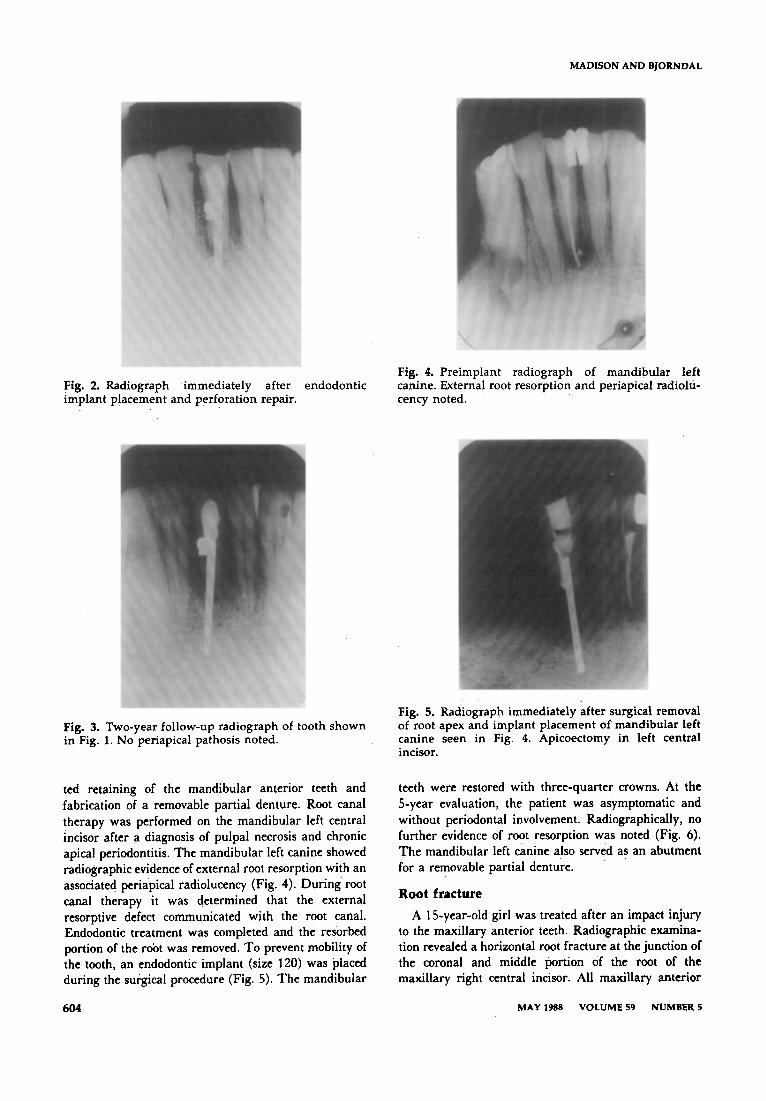
ted retaining of the mandibular anterior teeth and fabrication of a removable partial denture. Root canal therapy was performed on the mandibular left central incisor after a diagnosis of pulpal necrosis and chronic apical periodontitis. The mandibular left canine showed radiographic evidence of external root resorption with an associated periapical radiolucency (Fig. 4). During root canal therapy it was determined that the external resorptive defect communicated with the root canal. Endodontic treatment was completed and the resorbed portion of the root was removed. To prevent mobility of the tooth, an endodontic implant (size 120) was placed during the surgical procedure (Fig. 5). The mandibular
604
Fig. 4. Preimplant radiograph of mandibular left canine. External root resorption and periapical radiolu- cency noted.
Fig. 5. Radiograph immediately after surgical removal of root apex and implant placement of mandibular left canine seen in Fig. 4. Apicoectomy in left central incisor.
teeth were restored with three-quarter crowns. At the S-year evaluation, the patient was asymptomatic and without periodontal involvement. Radiographically, no further evidence of root resorption was noted (Fig. 6). The mandibular left canine also served as an abutment for a removable partial denture.
Root fracture
A 1 S-year-old girl was treated after an impact injury to the maxillary anterior teeth. Radiographic examina- tion revealed a horizontal root fracture at the junction of the coronal and middle portion of the root of the maxillary right central incisor. All maxillary anterior
MAY 1988 VOLUME 59 NUMBER 5
CLINICAL APPLICATION OF ENDODONTIC IMPLANTS
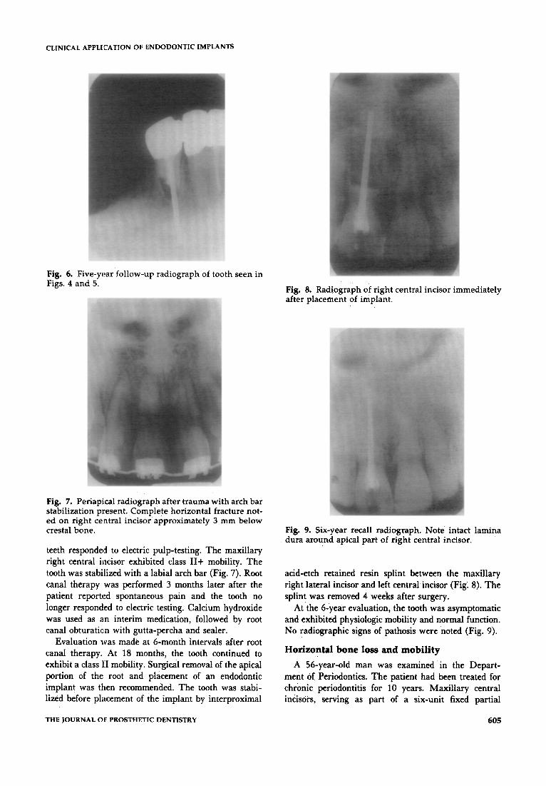
Fig. 6. Five-year follow-up radiograph of tooth seen in Figs. 4 and 5.
Fig. 7. Periapical radiograph after trauma with arch bar stabilization present. Complete horizontal fracture not- ed on right central incisor approximately 3 mm below crestal bone.
teeth responded to electric pulp-testing. The maxillary right central incisor exhibited class II+ mobility. The tooth was stabilized with a labial arch bar (Fig. 7). Root canal therapy was performed 3 months later after the patient reported spontaneous pain and the tooth no longer responded to electric testing. Calcium hydroxide was used as an interim medication, followed by root canal obturation with gutta-percha and sealer.
Evaluation was made at 6-month intervals after root canal therapy. At 18 months, the tooth continued to exhibit a class 111 mobility. Surgical removal of the apical portion of the root and placement of an endodontic implant was then recommended. The tooth was stabi- lized before placement of the implant by interproximal
Fig. 8. Radiograph of right central incisor immediately after placement of implant.
Fig. 9. Six-year recall radiograph. Note intact lamina dura around apical part of right central incisor.
acid-etch retained resin splint between the maxillary right lateral incisor and left central incisor (Fig: 8). The splint was removed 4 weeks after surgery.
At the 6-year evaluation, the tooth was asymptomatic and exhibited physiologic mobility and normal function. No radiographic signs of pathosis were noted (Fig. 9).
Horizontal bone loss and mobility
A 56-year-old man was examined in the Depart- ment of Periodontics. The patient had been treated for chronic periodontitis for 10 years. Maxillary central incisors, serving as part of a six-unit fixed partial
THE JOURNAL OF PROSTHETIC DENTISTRY 605
MADISON AND BJORNDAL
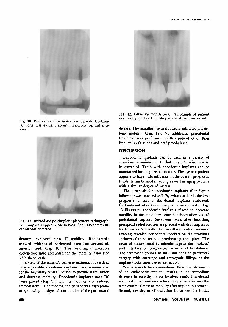
Fig. 10. Pretreatment periapical radiograph. Horizon- tal bone loss evident around maxillary central inci- sors.
Fig. 11. Immediate postimplant placement radiograph. Both implants appear close to nasal floor. No communi- cation was detected.
denture, exhibited class II mobility. Radiographs showed evidence of horizontal bone loss around all anterior teeth (Fig. 10). The resulting unfavorable crown-root ratio accounted for the mobility associated with these teeth.
In view of the patient’s desire to maintain his teeth as long as possible, endodontic implants were recommended for the maxillary central incisors to provide stabilization and decrease mobility. Endodontic implants (size 70) were placed (Fig. 11) and the mobility was reduced immediately. At 55 months, the patient was asymptom- atic, showing no signs of continuation of the periodontal
Fig. 12 Fifty-five month recall radiograph of patient seen in Figs. 10 and 11. No periapical pathosis noted.
disease. The maxillary central incisors exhibited physio- logic mobility (Fig. 12). No additional periodontal treatment was performed on this patient other than frequent evaluations and oral prophylaxis.
DISCUSSION
Endodontic implants can be used in a variety of situations to maintain teeth that may otherwise have to be extracted. Teeth with endodontic implants can be maintained for long periods of time. The age of a patient appears to have little influence on the overall prognosis. Implants can be used in young as well as aging patients with a similar degree of success.
The prognosis for endodontic implants after S-year follow-up was reported as 91 %,I which to date is the best prognosis for any of the dental implants evaluated. Certainly not all endodontic implants are successful. Fig. 13 illustrates endodontic implants placed to decrease mobility in.the maxillary central incisors after loss of periodontal support. Seventeen years after insertion, periapical radiolucencies are present with draining sinus tracts associated with the maxillary central incisors. Probing revealed periodontal pockets on the proximal surfaces of these teeth approximating the apices. The cause of failure could be microleakage at the implant/ root interface or progressive periodontal breakdown. The treatment options at this time include periapical surgery with curettage and retrograde fillings at the implant/tooth interface or extraction.
We have made two observations. First, the placement of an endodontic implant results in an immediate decrease in mobility of the involved tooth. Interdental stabilization is unnecessary for some patients because the teeth exhibit almost no mobility after implant placement. Second, the degree of occlusion influences the initial
606 MAY 1988 VOLUME 59 NUMBER 5

CLINICAL APPLI’CATION OF ENDODONTIC IMPLANTS
Fig. 13. Endodontic implants immediately after placement in maxillary central incisors. A, Note presence of radiolucencies around both apices. B, Three years after implant placement, periapical areas appear normal. C, After 17 years, radiolucencies are again present around both root apices.
outcome. Teeth in a maximum occlusal interrelationship exhibit more mobility after placement of endodontic implants than those with minimal or no occlusion.
Patient selection is the most important consideration for implantation recommendations. Endodontic implants are not appropriate for all teeth that are mobile, nor will their placement resolve advancing periodontal disease.8 For some patients, however, endodontic implants may enable maintenance of the natural dentition.
The criteria used in patient selection for endodontic implants include:
1. Current periodontal status of the tooth. Active periodontal disease with continued loss of bone support may lead to fa:ilure of the implant, as will communica- tion of the implant with the oral cavity as a result of soft tissue breakdown.
2. Degree of mobility. Teeth with mobility beyond physiologic limits that compromise function should be considered as candidates for implants. Splinting is anoth- er treatment option that may give the desired results without compromising the periodontium.
3. Anatomic considerations. Anatomic structures in close proximity to the apex of the root may prevent adequate extension of the implant. In addition, inclina- tion of the ro,ot in the surrounding bone should be evaluated before implant placement.
4. Patient’s acceptance of the procedure. Endodon- tic implantation is presently regarded as experimental by the American Dental Association Council on Dental Materials, Instruments, and Equipment’ and should be explained to the patient before treatment.
THE JOURNAL OF PROSTHETIC DENTISTRY
The dental profession often evaluates dental therapies in terms of longevity, with the desire for treatments to last indefinitely. Physicians consider a 3- to 5-year cure rate for certain malignancieslO or a 7 to 10 year longevity for hip prostheses” as successful treatment. Treatment of compromised oral conditions that provides a functional, natural dentition for an additional number of years should be considered appropriate and successful treat- ment.
CONCLUSION
Patient selection and use of endodontic endosseous implants in periodontally compromised teeth or teeth exhibiting extreme mobility due to loss of root structure or loss of bone support have been presented. Not all situations in which endodontic implants might be used are discussed; nevertheless, a variety of different instances in which endodontic implants may be applica- ble have been described. After treating many patients with endodontic implants, we conclude that the primary factor affecting the prognosis of treatment is patient selection.
REFERENCES
1. Orlay HG. Endodontic splinting treatment in periodontal dis- ease. Br Dent J 1960;108:118-21.
2. Frank AL. Improvement of the crown-root ratio by endodontic endosseous implants. J Am Dent Assoc 1967;74:451-62.
3. Frank AL. Resorption, perforations, and fractures. Dent Clin North Am 1974;18:465-87.
4. Silverband H, Rabkin M, Cranin AN. The uses of endodontic implant stabilizers in post-traumatic and periodontal disease. Oral h-g 1978;45:920-9.
607
MADISON AND BJORNDAL
5. Scopp IW, Dictrow RL, Lichtenstein BS. Endodontic endosseous implants: a conservative method of stabilization in geriatric patients. J Periodontol 1969;40:664-6.
6. Shaykin JB. Endodontic implant. J Am Dent Assoc 1964; 68:704-7.
7. Cranin AN, Rabkin MF, Garfinkel L. A statistical evaluation of 952 endosteal implants in humans. J Am Dent Assoc 1977; 94:315-20.
8. Frank AL. Endodontic endosseous implants and treatment of the wide open apex. Dent Clin North Am 1967;Nov:675-700.
9. American Dental Association: Report of the Council on Dental Materials, Instruments, and Equipment. J Am Dent Assoc 1980;100:247.
10. Seidman H, Mushinski MH, Gelb SK, Silverberg 15. Probabili- ties of eventually developing or dying of cancer-United States, 1985. CA-A Cancer J for Clin 1985;35:36-56.
11. Dobbs HS. Survivorship of total hip replacements. J Bone Joint Surg 1980;62-B: 168-73.
Reprint requests to: DR. SANDRA MADISON UNIVERWY OF NORTH CAROLINA SCHOOL OF DENTISTRY CHAPEL HILL, NC 27514
A simplified prosthesis for the treatment of burns to the oral cavity
Joe P. Ampil, D.M.D., M.Sc.,* Lisa Newell, D.D.S.,** and Paul Taylor, D.D.S., M.S.*** University of Texas Health Science Center at Dallas, and Baylor College of Dentistry, Dallas, Tex.
liosthetic treatment for electrical burns to the oral cavity was described by Ryan.’ Although these burns are most often caused by electricity, chemical burns are also seen. The burns often result in scar tissue formation at the oral commissure, which restricts oral opening.
A simple technique for fabrication of a splint to limit
*Director, Maxillofacial Section, Associate Professor of Clinical Oral Surgery, University of Texas Health Science Center at Dallas.
**Graduate student in pedodontics, Baylor College of Dentistry. ***Director Emeritus of Dentistry, Children’s Medical Center; for-
merly Chairman, Department of Pedodontics, Baylor College of Dentistry.
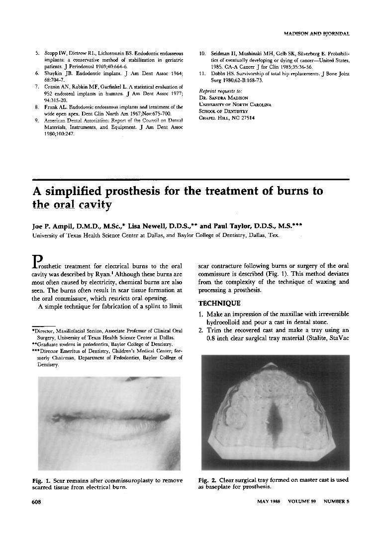
Fig. 1. Scar remains after commissuroplasty to remove scarred tissue from electrical burn.
scar contracture following burns or surgery of the oral commissure is described (Fig. 1). This method deviates from the complexity of the technique of waxing and processing a prosthesis.
TECHNIQUE
1. Make an impression of the maxillae with irreversible hydrocolloid and pour a cast in dental stone.
2. Trim the recovered cast and make a tray using an 0.8 inch clear surgical tray material (Stalite, StaVac
Fig. 2. Clear surgical tray formed on master cast is used as baseplate for prosthesis.
MAY 1988 VOLUME 59 NUMBER 5