clinical and translational pathology innovations high ... · proficiency testing challenges....
TRANSCRIPT

ALSO IN THIS ISSUE:
High Blood Pressure PreventionClinical and Translational
Pathology InnovationsA Publication of the Robert J. Tomsich Pathology & Laboratory Medicine Institute | Summer 2015
Feature StoryQuantification of drugs/metabolites in urine by liquid chromatography-tandem mass spectrometry for pain management services
Molecular Diagnostics in BCR-ABL1 NegativeMyeloproliferative Neoplasms | p 6
STAT6 Rabbit Monoclonal Antibody for the Diagnosis of Solitary Fibrous Tumor | p 10
Alumni Connect | p 15
News | p 16

2
Pathology Innovations | Clinical | Translational SPRING | 2015
Pain management drugs are among the most prescribed medications in the United States and are often abused.1, 2 It is estimated that between 9 to 41% of patients receiving opioids for chronic pain abuse the prescription.2-5 Chronic pain patients also are estimated to use illicit drugs at a rate of 14 to 34%.2 A serious public health problem is caused by the diversion of prescribed pain management drugs.6
Monitoring the drugs and metabolites of the prescribed medication and potentially abused drugs plays an important role in ensuring patient compliance. Urine is the preferred specimen due to the ease of sample collection and the acquired background knowledge for this sample type. Urine drug testing has often been used to verify adherence to prescribed pain management regimens and to detect illicit drug use.7 Both routine and random drug testing can significantly improve patient compliance.8-10 The test results have important implications regarding the patient’s eligibility for additional pain management medications. Therefore, accuracy in determining both negative and positive results is extremely important.
Though immunoassays are easy to run with fast turnaround time, they may lack the needed sensitivity and/or specificity.11,12 While gas chromatography-mass
spectrometry (GC-MS) methods are considered the gold standard, sample preparation is labor intensive. There is increasing use of liquid chromatography-tandem mass spectrometry (LC-MS/MS), which generally requires much simpler sample preparation with high specificity and sensitivity.13
Some drugs/metabolites, such as morphine and codeine, are present in urine as both free and glucuronide conjugated forms. Percentage of the conjugates may vary significantly depending on both sample collection time and individual metabolism rates.14 Conjugated analytes may be hydrolyzed prior to LC-MS/MS analysis in order to improve sensitivity and consistency. While chemical hydrolysis is fast and efficient, it may result in undesired reactions and loss of important information. For example, acid hydrolysis converts heroin and its metabolite, 6-acetylmorphine, to morphine.15 In contrast, enzymatic hydrolysis is more specific, but requires longer incubation time to achieve high efficiency (> 90%).16
While direct injection of diluted urine samples for LC-MS/MS analysis has been reported, the matrix effect may compromise sensitivity and accuracy.17 Both off-line and online solid phase extraction is widely used for sample purification prior to LC-MS/MS analysis18-21 to reduce the matrix effects. Mueller et al. reported an online turbulent flow extraction method using two TurboFlow columns with different stationary phases to extract hundreds of drugs/metabolites.22 Most of the analytes important for pain management are measured in the positive electrospray ionization (ESI) mode,18-20, 23 while tetrahydrocannabinol carboxylic acid (THCA), the major metabolite of marijuana, is known to ionize more efficiently in negative ESI mode.24
At Cleveland Clinic, we have developed and validated a novel LC-MS/MS method for simultaneously measuring 20 drugs and metabolites in urine to monitor the use of 16 prescription or illicit drugs (morphine, codeine,
Quantification of drugs/metabolites in urine by liquid chromatography-tandem mass spectrometry for pain management servicesBy Dustin Bunch, Katherine Lembright and Sihe Wang, PhD*

3
Pathology Innovations | Clinical | Translational SPRING | 2015
dihydrocodeine, oxycodone, oxymorphone, hydrocodone, hydromorphone, methadone, fentanyl, tramadol, buprenorphine, amphetamine, methamphetamine, cocaine, heroin and marijuana).25, 26 The chromatogram is shown in Figure 1. Sample preparation includes enzymatic hydrolysis followed by online turbulent flow extraction. Recovery for all analytes ranges between 93.2% and 111.4%. The lower limit of quantification (LLOQ) is between 5 and 25 ng/mL for all analytes. Accuracy varies between 85.8% and 119.4%. Intra-assay and total CVs at three different levels are 0.2% – 12.7%.
Significant inter-laboratory variation has been reported when measuring conjugated urine drugs, especially for codeine.27 The high variation was likely due to different hydrolysis methods used in each laboratory.15 Enzymatic hydrolysis efficiency is greatly affected by the type of glucuronidase and the reaction condition used. In addition, hydrolysis rates of different drugs vary significantly under the same reaction condition.28 Therefore, a robust and reproducible hydrolysis method is important for consistent measurement. Compared to other glucuronidases, the glucuronidase from Patella vulgate showed a superior efficiency to liberate morphine from its glucuronide conjugates.28 We found that reproducible and near complete (> 94%) hydrolysis for all analytes can be achieved using this enzyme with an overnight incubation (≥ 16 h) at 60°C.
Simultaneously measuring a large panel of pain management drugs in one LC-MS/MS run has obvious benefits. However, significant variances in their chemical properties can make concurrent measurement technically challenging. As a result, separate sample preparation and separate MS methods were required to quantify different classes of drugs in the past. We use two TurboFlow columns with compensatory stationary phases to simultaneously extract 20 analytes. These two TurboFlow columns in tandem allow a clinically meaningful LLOQ to be achieved for all analytes. Another advantage of online extraction is reduced labor while achieving a high reproducibility.
In summary, we have established a novel quantitative urine drug analysis to simultaneously monitor the use of 16 drugs. This method has been successfully used to support pain management clinics in the last six years with ~85,000 samples analyzed. This method also has successfully passed multiple CAP proficiency testing challenges.
References:
1. Compton P, Athanasos P. Chronic pain, substance abuse and addiction. Nurs Clin North Am 2003;38:525-37.
2. Manchikanti L, Cash KA, Damron KS, Manchukonda R, Pampati V, McManus CD. Controlled substance abuse and illicit drug use in chronic pain patients: An evaluation of multiple variables. Pain Physician 2006;9:215-25.
3. Chabal C, Erjavec MK, Jacobson L, Mariano A, Chaney E. Prescription opiate abuse in chronic pain patients: Clinical criteria, incidence, and predictors. Clin J Pain 1997;13:150-5.
4. Manchikanti L, Pampati V, Damron KS, Fellows B, Barnhill RC, Beyer CD. Prevalence of opioid abuse in interventional pain medicine practice settings: A randomized clinical evaluation. Pain Physician 2001;4:358-65.
5. Katz NP, Sherburne S, Beach M, Rose RJ, Vielguth J, Bradley J, Fanciullo GJ. Behavioral monitoring and urine toxicology testing in patients receiving long-term opioid therapy. Anesth Analg 2003;97:1097-102, table of contents.
6. Volkow ND, McLellan TA. Curtailing diversion and abuse of opioid analgesics without jeopardizing pain treatment. JAMA 2011;305:1346-7.
7. Reisfield GM, Salazar E, Bertholf RL. Rational use and interpretation of urine drug testing in chronic opioid therapy. Ann Clin Lab Sci 2007;37:301-14.
8. Manchikanti L, Manchukonda R, Pampati V, Damron KS, Brandon DE, Cash KA, McManus CD. Does random urine drug testing reduce illicit drug use in chronic pain patients receiving opioids? Pain Physician 2006;9:123-9.
9. Katz N, Fanciullo GJ. Role of urine toxicology testing in the management of chronic opioid therapy. Clin J Pain 2002;18:S76-82.
10. Christo PJ, Manchikanti L, Ruan X, Bottros M, Hansen H, Solanki DR, et al. Urine drug testing in chronic pain. Pain Physician 2011;14:123-43.
11. Moriya F, Chan KM, Hashimoto Y. Concentrations of morphine and codeine in urine of heroin abusers. Leg Med (Tokyo) 1999;1:140-4.

4
Pathology Innovations | Clinical | Translational SPRING | 2015
12. Fraser AD, Worth D. Urinary excretion profiles of 11-nor-9-carboxy-delta9-tetrahydrocannabinol: A delta9-thccooh to creatinine ratio study. J Anal Toxicol 1999;23:531-4.
13. Porter WH. Clinical toxicology. In: Burtis CA, Ashwood EA, Bruns DE, eds., Vol., 2006.
14. Chen ZR, Somogyi AA, Reynolds G, Bochner F. Disposition and metabolism of codeine after single and chronic doses in one poor and seven extensive metabolisers. Br J Clin Pharmacol 1991;31:381-90.
15. Hackett LP, Dusci LJ, Ilett KF, Chiswell GM. Optimizing the hydrolysis of codeine and morphine glucuronides in urine. Ther Drug Monit 2002;24:652-7.
16. Jennison TA, Wozniak E, Nelson G, Urry FM. The quantitative conversion of morphine 3-beta-d glucuronide to morphine using beta-glucuronidase obtained from patella vulgata as compared to acid hydrolysis. J Anal Toxicol 1993;17:208-10.
17. Eichhorst JC, Etter ML, Rousseaux N, Lehotay DC. Drugs of abuse testing by tandem mass spectrometry: A rapid, simple method to replace immunoassays. Clin Biochem 2009;42:1531-42.
18. de Castro A, Concheiro M, Shakleya DM, Huestis MA. Development and validation of a liquid chromatography mass spectrometry assay for the simultaneous quantification of methadone, cocaine, opiates and metabolites in human umbilical cord. J Chromatogr B Analyt Technol Biomed Life Sci 2009;877:3065-71.
19. Rook EJ, Hillebrand MJ, Rosing H, van Ree JM, Beijnen JH. The quantitative analysis of heroin, methadone and their metabolites and the simultaneous detection of cocaine, acetylcodeine and their metabolites in human plasma by high-performance liquid chromatography coupled with tandem mass spectrometry. J Chromatogr B Analyt Technol Biomed Life Sci 2005;824:213-21.
20. Badawi N, Simonsen KW, Steentoft A, Bernhoft IM, Linnet K. Simultaneous screening and quantification of 29 drugs of abuse in oral fluid by solid-phase extraction and ultraperformance lc-ms/ms. Clin Chem 2009;55:2004-18.
21. Huwyler J, Rufer S, Kusters E, Drewe J. Rapid and highly automated determination of morphine and morphine glucuronides in plasma by on-line solid-phase extraction and column liquid chromatography. J Chromatogr B Biomed Appl 1995;674:57-63.
22. Mueller DM, Duretz B, Espourteille FA, Rentsch KM. Development of a fully automated toxicological lc-ms(n) screening system in urine using online extraction with turbulent flow chromatography. Anal Bioanal Chem 2010.
23. Feng J, Wang L, Dai I, Harmon T, Bernert JT. Simultaneous determination of multiple drugs of abuse and relevant metabolites in urine by lc-ms-ms. J Anal Toxicol 2007;31:359-68.
24. Teixeira H, Verstraete A, Proenca P, Corte-Real F, Monsanto P, Vieira DN. Validated method for the simultaneous determination of delta9-thc and delta9-thc-cooh in oral fluid, urine and whole blood using solid-phase extraction and liquid chromatography-mass spectrometry with electrospray ionization. Forensic Sci Int 2007;170:148-55.
25. Yuan C, Heideloff C, Kozak M, Wang S. Simultaneous quantification of 19 drugs/metabolites in urine important for pain management by liquid chromatography-tandem mass spectrometry. Clin Chem Lab Med 2012;50:95-103.
26. Yuan C, Lembright K, Heideloff C, Wang S. Quantification of buprenorphine, norbuprenorphine and 6-monoacetylmorphine in urine by liquid chromatography-tandem mass spectrometry. J Chromat Separation Techniq 2013;4.
27. Corcione S, Pichini S, Badia R, Segura J, de la Torre R. Quantitative aspects of drugs of abuse in urine samples: Intercollaborative studies conducted in the european union. Ther Drug Monit 1999;21:653-60.
28. Combie J, Blake JW, Nugent TE, Tobin T. Morphine glucuronide hydrolysis: Superiority of beta-glucuronidase from patella vulgata. Clin Chem 1982;28:83-6.
5
Pathology Innovations | Clinical | Translational SPRING | 2015
About the Authors
Dustin R. Bunch
Dustin R. Bunch received his bachelor’s degree in biochemistry from Case Western Reserve University and is currently a Senior Research Technologist in the Department of Clinical Pathology and a graduate student in the Clinical and Bioanalytical Chemistry program at Cleveland State University. His major duties involve managing the research team and performing development and validation of liquid chromatography tandem mass spectrometry (LC-MS/MS) methods. He has authored or co-authored more than 50 peer-reviewed abstracts and publications and has been a member of the American Association for Clinical Chemistry (AACC) since 2008. Upon completion of his PhD, Dustin plans to join a Commission on Accreditation in Clinical Chemistry (ComACC) approved post-doctoral fellowship program in clinical chemistry. He currently serves as the Internet Coordinator for the Northeast Ohio Section of AACC and the Ohio Collaborative Laboratory Conference. Mr. Bunch can be reached at [email protected] or 216.444.7003.
Katherine Lembright
Katherine Lembright is an ASCP-certified Medical Technologist. She received her bachelor’s degree in zoology/medical technology from Ohio University and completed a medical technology internship at Cleveland Clinic. Ms. Lembright has worked as a Medical Technologist for the past 30 years in the Cleveland Clinic Special Chemistry Laboratory, where she analyzes multiple analytes using liquid chromatography tandem mass spectrometry (LC-MS/MS), Fourier Transform Infrared Spectrometry (FT-IR) and inductively coupled plasma mass spectrometry (ICP-MS) methods. She has been involved with the development and validation of liquid chromatography tandem mass spectrometry (LC-MS/MS) methods. Ms. Lembright can be reached at [email protected] or 216.444.2525.
Sihe Wang, PhD, DABCC, FACB
Sihe Wang, Phd, DABCC, FACB, is Section Head and Medical Director of the Clinical Biochemistry and Director of the Clinical Biochemistry Fellowship Training Program at Cleveland Clinic. He chairs clinical chemistry integration and the standardization effort for the Cleveland Clinic Health System, which includes one Florida hospital, eight community hospitals and 18 family health centers in Northeast Ohio. Additionally, he is a Clinical Chemistry Professor at Cleveland State University. His expertise includes general clinical chemistry and clinical application of mass spectrometry. Dr. Wang has been a diplomate of the American Board of Clinical Chemistry since 2005, a fellow of the National Academy of Clinical Biochemistry since 2006, and a member of the Academy of Clinical Laboratory Physicians and Scientists. Dr. Wang has been a member of the American Association for Clinical Chemistry (AACC) since 2000. He served as chair of the AACC Northeast Ohio Section in 2008 and 2009 and president of the North American Chinese Clinical Chemistry Association (NACCCA) in 2008 and 2009. Currently he serves as historian for NACCCA, treasurer for AACC Pediatric and Maternal Fetal division, secretary for AACC Clinical Translational Sciences division, delegate for AACC Northeast Ohio section, president-elect for the Commission on Accreditation in Clinical Chemistry (ComACC), and member of AACC’s Strategies Online Editorial Advisory Board. Dr. Wang has authored more than 190 journal articles, book chapters and abstracts. He is one of the two chief editors for the book, “Application of liquid chromatography-mass spectrometry in clinical laboratories” published in October 2014. Dr. Wang can be reached at 216.445.2634 or by email at [email protected].

6
Pathology Innovations | Clinical | Translational SPRING | 2015
Advancements in the understanding of disease and the molecular diagnostics of myeloproliferative neoplasms (MPNs) have significantly changed their diagnosis. Although not as dramatic in terms of effect on disease management as those seen in BCR-ABL1 positive chronic myelogenous leukemia (CML), multiple recent advancements in the diagnosis of BCR-ABL1 negative MPNs make diagnosis of these entities today both simpler and more precise than just ten years ago. This article highlights recent developments in the diagnosis of BCR-ABL1 negative MPNs and their practical impact on the diagnostic algorithm.
A key decision point encountered in the diagnosis of MPNs is the distinction between BCR-ABL1 positive CML and BCR-ABL1 negative MPNs. The testing for diagnosis and management of CML is largely unchanged since the review in the Fall 2012 issue of Pathology Innovations and is not discussed in any detail here. However, it is important to remember that, although BCR-ABL1 positive CML most often has a classic presentation that is recognizable by peripheral blood and marrow cell counts and morphology, CML has a spectrum of morphologic presentations that can overlap with BCR-ABL1 negative MPNs as well as other entities (including PDGFRA, PDGFRB and FGFR1 associated neoplasms and some myelodysplastic/myeloproliferative neoplasms). Exclusion of BCR-ABL1 fusion is therefore an important component of the work-up for these neoplasms. Even though misdiagnosis of this type is a relatively uncommon event, it is important not to miss BCR-ABL1 positive CML given its dramatic response to tyrosine kinase inhibitor therapy.
One of the most important tasks when evaluating a patient for MPNs is the distinction between benign and malignant. The overlap of clinical and morphologic features between the BCR-ABL1 negative MPNs and benign, reactive processes has historically meant that definitive diagnosis relied on a combination of sometimes laborious testing (such as the red blood cell mass) and
exhaustive exclusion of potential non-neoplastic etiologies. Recently developed molecular diagnostics tools that aid in definitive diagnosis of BCR-ABL1 negative MPNs have greatly simplified the diagnostic process. Until about ten years ago, molecular diagnostics was only of benefit in diagnosing BCR-ABL1 negative MPNs in those few cases (less than 10%) where cytogenetic karyotyping detected an abnormal clone. The discovery of JAK2 mutations in MPNs in 2005 began to change that.
The most prevalent JAK2 mutation in MPNs is V617F, a single nucleotide substitution on chromosome 9p, within an auto-inhibitory domain of JAK2. The mutation results in constitutive activation of the JAK-STAT pathway, driving proliferation and survival even in the absence of growth factor stimulation. Originally described in polycythemia vera (PV), the JAK2 V617F mutation is present in 97% of PV and also in at least half of the two other BCR-ABL1 negative MPNs, essential thrombocythemia (ET) and primary myelofibrosis (PMF). Although the JAK2 V617F mutation can be present in extremely low levels in some individuals without myeloid neoplasia, when positive at the level detected by most clinical assays in the context of the appropriate clinical and pathologic context, the mutation is a strong indicator of myeloid neoplasm.
Molecular Diagnostics in BCR-ABL1 Negative Myeloproliferative NeoplasmsBy David Bosler, MD
Table 1. Cleveland Clinic Tests Useful in the Diagnosis of BCR-ABL1 Negative MPNs
Test Name Test Code
JAK2 V617F Mutation Detection JAK2
JAK2 Exon 12 - 15 Sequencing JAKNON
MPL Mutation Analysis MPL
CALR (Calreticulin) Exon 9 Mutation Analysis CALR
Myeloid Malignancies Mutation Panel by Next Generation Sequencing
MYENGS

7
Pathology Innovations | Clinical | Translational SPRING | 2015
Since the discovery of JAK2 V617F, MPN clinical diagnostics development has focused on what to do about the cases that are negative for V617F—how can they be diagnosed with the same ease as V617F positive cases? These gaps have been closed over time in piecemeal fashion. Essentially all of the PV cases that are negative for V617F have one of a variety of less common mutations that can be detected by sequencing of JAK2 exons 12-15. Non-V617F mutations are found in PV, but not ET or PMF.
MPL gene mutations are present in small percentages of ET (1-4%) and PMF (5-11%), but not PV. Functionally, these mutations act similarly to the JAK2 mutation, except that the mutation is directly in the thrombopoietin receptor, constitutively activating it rather than a downstream tyrosine kinase as in the JAK2 mutation. The relevant MPL mutations are in exon 10 and the vast majority are single nucleotide substitutions at position 515, making a variety of methodologies viable for detection. Other single nucleotide substitutions and deletion/insertion mutations near position 515 have also been described. MPL mutations add value to the diagnosis of MPNs since they are mutually exclusive with JAK2 mutations and are detected only in myeloid neoplasms (mainly MPNs).
Recently, sequencing studies on JAK2-negative, MPL-negative MPNs led to the discovery of mutations in the calreticulin (CALR) gene in a significant percentage of these cases. The CALR gene is on chromosome 19p13.2 and contains nine exons. CALR mutations are present in up to 90% of JAK2-, MPL- ET and PMF cases. Reported mutations are frame shift mutations within exon 9, with two variants (one 52 bp deletion and one 7 bp insertion) representing more than 80% of mutated cases. Like MPL mutations, CALR mutations are found in ET and PML but not in PV. CALR, JAK2 and MPL mutations are mutually exclusive. CALR mutations are also variably reported to be present in low percentages of myelodysplastic syndromes (MDS) and overlap MDS/MPNs. Although the mechanism of the mutation in pathogenesis is unknown, over-expression of mutated CALR in in vitro models showed cytokine independent growth through STAT5 activation. MPN cases with mutated CALR appear to have comparatively favorable prognosis, although there is currently insufficient data to justify any differences in therapeutic approach.
Combined, the JAK2, MPL and CALR mutations now account for more than 90% of BCR-ABL1 negative MPNs. Access to assays that detect these mutations will result in earlier and more efficient diagnosis or exclusion of disease in suspected cases. Work-up of suspected BCR-ABL1 negative MPN should therefore routinely include these tests. Although the actual testing algorithm used may depend on the availability of specific tests, samples and nucleic acid extraction protocols, a recommended approach is shown in Figure 1. After BCR-ABL1 is excluded, JAK2 V617F has the highest diagnostic yield and should generally be tested first. Although disease burdens vary, the allele-specific PCR method used in JAK2 V617F has an analytic sensitivity of 1%, which is sufficient for detection of JAK2 positive MPNs. If V617F is negative, the next step depends on which disease is suspected and the level of clinical suspicion. If PV is highly suspected despite negative JAK2 V617F (eg. chronic polycythemia and low erythropoietin level), JAK2 sequencing of exons 12-15 should be next. While a positive sequencing result is diagnostic and most positive cases will be detected, a negative result does not completely exclude the possibility of very low levels of PV since Sanger sequencing requires at least 20% mutated allele burden to detect a mutation. This test should not be used for routine screening of erythrocytosis, but should instead be reserved for those cases where clinical suspicion is high and persists even after demonstrated absence of JAK2 V617F.
If the clinical and morphologic evaluation instead suggests ET or PMF, CALR and MPL testing is recommended for
Figure 1. Molecular Diagnostics in Suspected Myeloproliferative Neoplasm, BCR-ABL1 Negative

8
Pathology Innovations | Clinical | Translational SPRING | 2015
JAK2 V617F negative cases. The most cost-effective way to perform this testing is to evaluate for JAK2, CALR and MPL in sequential order. If testing is positive at any step, no further testing is necessary and a diagnosis can be made based on the marrow morphologic findings. At Cleveland Clinic, CALR mutation testing is performed using PCR with fragment length analysis, which has an analytical sensitivity of approximately 5%. MPL mutation testing is performed by Sanger sequencing of exon 10. This method provides comprehensive detection of the MPL mutations described as well as potential mutations within the region of interest on exon 10. As a Sanger sequencing method, the MPL sequencing assay is subject to the same limitations in detecting very low disease burdens as described for the JAK2 sequencing assay above.
Although each of these tests has some diagnostic value as described above, they have relatively limited use in classifying BCR-ABL1 negative MPNs into one of the specific 2008 WHO diagnoses (PV, ET, PMF). Accurate diagnosis and sub-classification is nonetheless clinically relevant due to prognostic differences as well as some variation in approaches to treatment. Molecular findings can provide some clues such as JAK2 non-V617F indicating PV, and CALR or MPL indicating non-PV, but the specific classification usually relies on clinical and morphologic evaluation.
Cytogenetic karyotyping should also be performed on all bone marrows performed for diagnosis of MPNs. Karyotyping provides a baseline karyotype used to track clonal evolution over time and also provides prognostically relevant information. For the very rare cases where MPN is highly suspected based on the clinical and/or morphologic findings and all testing is negative, a multi-gene sequencing panel containing genes relevant to myeloid neoplasia, such as ASXL1, TET2, DNMT3A, SF3B1, may provide additional information helpful in yielding a definitive diagnosis. Such testing is not required for diagnosis according to 2008 WHO criteria, provided that diagnostic morphologic features are present and non-neoplastic etiologies have been excluded.
Another development in the diagnosis of MPNs is the recent description of mutations in the colony stimulating factor receptor 3 gene (CSF3R) in chronic neutrophilic leukemia (CNL). CNL is a rare disease, representing
much less than 1% of MPNs. It can present a diagnostic challenge however, since the morphologic findings are essentially indistinguishable from those of reactive neutrophilia (which is vastly more common) and also due to overlap with the morphologic spectrum of BCR-ABL1 positive CML. Like other MPNs, diagnosis of CNL has historically relied heavily on exhaustive exclusion of other potential etiologies. Until CSF3R mutations were first described in CNL by Maxson et al. in 2013, CNL had lacked a distinctive molecular marker, with almost 90% of cases having a normal cytogenetic karyotype. Located on chromosome 1p34.3, CSF3R encodes for the granulocyte simulating factor transmembrane receptor, which plays a role in granulocyte growth and differentiation. Mutations in CSF3R have also been described in some congenital neutropenias, and in hereditary neutrophilia. The most common CSF3R mutation in CNL is T618I, which is present in up to 83% of studied cases and appears to show specificity for CNL. CSF3R mutation testing will significantly change the diagnostic algorithm for CNL, and it has been proposed that CSF3R be incorporated as a major diagnostic criterion for the next edition of the WHO classification. Given that reactive neutrophilia is so common and CNL so rare, CSF3R testing should not be used to routinely screen neutrophilic patients. Testing should be limited to cases with high clinical suspicion (chronic, persistent neutrophilia with no evident underlying etiology) and where definitive diagnosis would impact clinical management.
In summary, the advances in molecular diagnostics in the past ten years have transformed the way myeloproliferative neoplasms are diagnosed. Used wisely in conjunction with clinical presentation, select laboratory results and marrow morphology findings, JAK2 V617F, JAK2 sequencing, CALR and MPL assays can be valuable tools in diagnosis of PV, ET and PMF. Although CSF3R is also now a potential tool in diagnosis of CNL, to avoid over-utilization it should only be used when clinical suspicion is very high since CNL is so rare.
References:
1. Swerdlow S, Campo E, Harris N, et al. WHO Classification of Tumours of Haematopoietic and Lymphoid Tissues. 4th ed. Lyon: IARC; 2008.

9
Pathology Innovations | Clinical | Translational SPRING | 2015
2. Bosler DS. Molecular Pathology of Myeloproliferative Neoplasms. Ch 7. D. Crisan (ed.), Hematopathology, Molecular and Translational Medicine, DOI 10.1007/978-1-60761-262-9_7, Springer Science+Business Media, LLC.
3. Tefferi A. The diagnosis of polycythemia vera: new tests and old dictums. Best Pract Res Clin Haematol. 2006;19:455–469.
4. Sanchez S, Ewton A. Essential thrombocythemia: a review of diagnostic and pathologic features. Arch Pathol Lab Med. 2006;130:1144–1150.
5. Baxter EJ, Scott LM, Campbell PJ, et al. Acquired mutation of the tyrosine kinase JAK2 in human myeloproliferative disorders. Lancet. 2005;365:1054–1061.
6. James C, Ugo V, Le Couedic JP, et al. A unique clonal JAK2 mutation leading to constitutive signalling causes polycythaemia vera. Nature. 2005;434: 1144–1148.
7. Levine RL, Wadleigh M, Cools J, et al. Activating mutation in the tyrosine kinase JAK2 in polycythemia vera, essential thrombocythemia, and myeloid metaplasia with myelofibrosis. Cancer Cell. 2005;7:387–397.
8. Kralovics R, Passamonti F, Buser AS, et al. A gain-of-function mutation of JAK2 in myeloproliferative disorders. N Engl J Med. 2005;352:1779–1790.
9. Ma W, Kantarjian H, Zhang X, et al. Mutation profile of JAK2 transcripts in patients with chronic myeloproliferative neoplasias. J Mol Diagn. 2009;11:49–53.
10. Pardanani AD, Levine RL, Lasho T, et al.MPL515 mutations in myeloproliferative and other myeloid disorders: a study of 1182 patients. Blood. 2006;108:3472–3476.
11. Nangalia J, Massie CE, Baxter EJ, et al. Somatic CALR mutations in myeloproliferative neoplasms with nonmutated JAK2. New Engl J Med 2013; 369: 2391–2405.
12. Klampfl T, Gisslinger H, Harutyunyan AS, et al. Somatic mutations of calreticulin in myeloproliferative neoplasms. New Engl J Med 2013; 369: 2379–2390.
13. Tefferi A, Pardanani A. CALR mutations and a new diagnostic algorithm for MPN. Nat. Rev. Clin. Oncol. 2014;11:125–126.
14. Maxson JE, Gotlib J, Pollyea DA, et al. Oncogenic CSF3R mutations in chronic neutrophilic leukemia and atypical CML. New Engl J Med 2013; 368: 1781–1790.
15. Pardanani A, Lasho TL, Laborde RR, et al. CSF3R T618I is a highly prevalent and specific mutation in chronic neutrophilic leukemia. Leukemia 2013; 27: 1870–1873.
16. Tefferi A, Thielez J, Vannucchi AM, Barbui T. An overview on CALR and CSF3R mutations and a proposal for revision of WHO diagnostic criteria for myeloproliferative neoplasms. Leukemia (2014) 28, 1407–1413.
About the Author
David Bosler, MD
David Bosler, MD, is Head of Cleveland Clinic Laboratories, Staff in Laboratory Medicine at Cleveland Clinic and Assistant Professor at the Cleveland Clinic Lerner College of Medicine. He is an AP/CP certified pathologist with subspecialty certifications in Hematopathology and Molecular Genetic Pathology. His clinical practice includes bone marrow biopsy interpretation and development and interpretation of molecular hematology tests. He has authored or co-authored numerous peer-reviewed journal articles and book chapters. Dr. Bosler completed medical school at University of Cincinnati, residency at William Beaumont Hospital, and fellowships at Mayo Clinic. He has also previously served as Medical Director for Point of Care Testing and Chair of the Point of Care Compliance Council at Cleveland Clinic. Dr. Bosler can be reached at [email protected] or 216.636.9615.
10
Pathology Innovations | Clinical | Translational SPRING | 2015
The STAT6 rabbit monoclonal antibody is an extremely sensitive and specific marker for the diagnosis of solitary fibrous tumor, a fibroblastic tumor of intermediate biological potential that can arise in a variety of anatomical sites, including pleura, meninges, extrapleural soft tissue and viscera.1
Solitary fibrous tumor is classically characterized by CD34-positive spindle cells in a collagenous background with an elaborate vasculature that includes staghorn blood vessels. However, the morphological spectrum of solitary fibrous tumor is broad and encompasses cellular variants (formerly known as hemangiopericytoma), fat-forming variants (also known as lipomatous hemangiopericytoma), giant cell angiofibroma and malignant solitary fibrous tumor.2-10 This morphological spectrum raises a broad differential diagnosis. Many entities that are included in the differential diagnosis show striking morphological resemblance and share CD34 expression. Approximately 5–10% of solitary fibrous tumors are negative for CD34 and, in this setting, the diagnosis can be especially challenging.1,2,11,12
Most solitary fibrous tumors are clinically indolent; however, approximately 10% behave more aggressively with local recurrences and metastases. The mainstay of therapy is wide en bloc resection with indefinite follow up.1
Recent studies using next generation sequencing techniques demonstrated the presence of recurrent fusions between NAB2 and STAT6 on chromosome 12q13 in the majority of solitary fibrous tumors,13–15 with a NAB2-STAT6 fusion transcript detected in 55–100% of tumors, regardless of tumor morphology (benign, lipomatous, malignant) or anatomical site.13–15 Schweizer et al. recently investigated immunohistochemistry for STAT6 as a surrogate marker of the NAB2-STAT6 fusion in meningeal hemangiopericytoma/solitary fibrous tumors.16 The NAB2-STAT6 fusion leads to EGR1 activation and transcriptional deregulation of EGR1-dependent target genes and is a driving event in initiation of solid fibrous tumors.
STAT6 Rabbit Monoclonal Antibody for the Diagnosis of Solitary Fibrous Tumor By Brian P. Rubin, MD, PhD
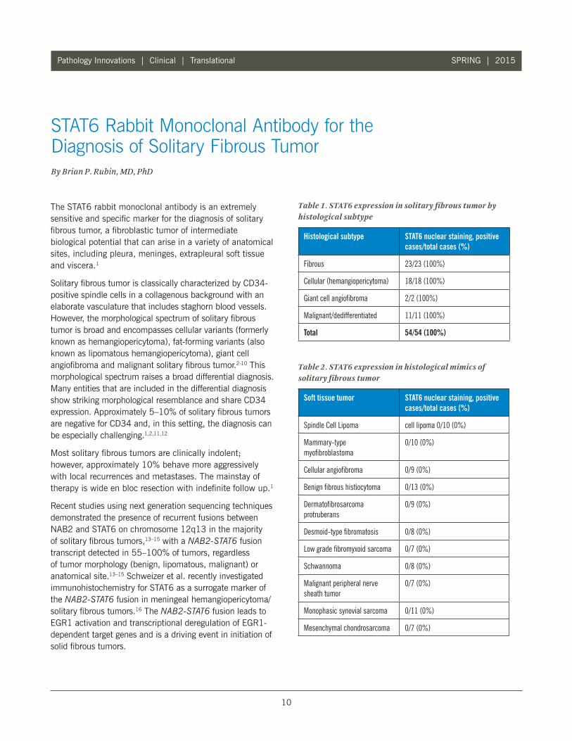
Table 1. STAT6 expression in solitary fibrous tumor by histological subtype
Table 2. STAT6 expression in histological mimics of solitary fibrous tumor
Histological subtype STAT6 nuclear staining, positive cases/total cases (%)
Fibrous 23/23 (100%)
Cellular (hemangiopericytoma) 18/18 (100%)
Giant cell angiofibroma 2/2 (100%)
Malignant/dedifferentiated 11/11 (100%)
Total 54/54 (100%)
Soft tissue tumor STAT6 nuclear staining, positive cases/total cases (%)
Spindle Cell Lipoma cell lipoma 0/10 (0%)
Mammary-type myofibroblastoma
0/10 (0%)
Cellular angiofibroma 0/9 (0%)
Benign fibrous histiocytoma 0/13 (0%)
Dermatofibrosarcoma protruberans
0/9 (0%)
Desmoid-type fibromatosis 0/8 (0%)
Low grade fibromyxoid sarcoma 0/7 (0%)
Schwannoma 0/8 (0%)
Malignant peripheral nerve sheath tumor
0/7 (0%)
Monophasic synovial sarcoma 0/11 (0%)
Mesenchymal chondrosarcoma 0/7 (0%)

11
Pathology Innovations | Clinical | Translational SPRING | 2015
STAT proteins are transcription factors that are normally located in the cytoplasm in latent form and migrate to the nucleus after cytokine exposure and subsequent phosphorylation.17 In cells harboring a NAB2-STAT6 fusion, STAT6 is similarly relocated to the nucleus.14,15
Nuclear expression of STAT6 was detected by immuno-histochemistry in 35 of 37 meningeal hemangiopericytoma and 25 of 25 meningeal solitary fibrous tumors, supporting the idea that meningeal hemangiopericytoma and meningeal solitary fibrous tumor are part of the same histological spectrum of solitary fibrous tumor and not distinct entities.16 Nuclear expression of STAT6 also discriminated solitary fibrous tumor from its various morphological mimics in the meninges, including meningioma, glioblastoma, gliosarcoma, hemangio-blastoma, schwannoma and hemangioma.16
Our study examined the sensitivity and specificity of STAT6 immunohistochemistry in reliably distinguishing solitary fibrous tumors from other soft tissue neoplasms that are in the differential diagnosis. We used a STAT6 rabbit monoclonal antibody (1:100), which had not previously been reported by others, on formalin-fixed, paraffin-embedded whole sections and tissue microarray slides. STAT6 expression was evaluated in 54 solitary fibrous tumors from 26 males and 28 females and 99 soft tissue tumors in the histological differential diagnosis.
Only nuclear staining of STAT6 was considered positive. Distribution of staining was scored as: 0 (no staining), 1+ (1-25%), 2+ (26-50%), 3+ (>50%). Intensity was scored as weak, moderate or strong. Nuclear STAT6 staining was present in all SFT cases tested (54/54, sensitivity 100%), regardless of histology, anatomical site or CD34 status. The majority of cases showed 3+ and strong staining. All tested cases of cellular angiofibroma (0/9), myofibro-blastoma (0/10), spindle cell lipoma (0/10), benign fibrous histiocytoma (0/13), dermatofibrosarcoma protruberans (0/9), low-grade fibromyxoid sarcoma (0/7), schwannoma (0/8), desmoid-type fibromatosis (0/8), monophasic synovial sarcoma (0/11), malignant peripheral nerve sheath tumour (0/7), and mesenchymal chondrosarcoma (0/7) were negative for STAT6 (specificity 100%).
The study further supports the utility of STAT6 immuno-histochemistry as an adjunct in the diagnosis of solitary fibrous tumor.
The study demonstrated that the STAT6 rabbit monoclonal antibody is an extremely sensitive and specific marker for the diagnosis of solitary fibrous tumor, with 100% sensitivity and 100% specificity (no dedifferentiated liposarcomas were tested in this study). Positive nuclear staining for STAT6 was observed in all 54 solitary fibrous tumors, regardless of histology, anatomical site or CD34 status. The results are in concordance with previously published studies of STAT6, but differ from those studies in the antibody that was used. During the period of our validation study, Doyle et al. reported 98% sensitivity in a similar study of 59 solitary fibrous tumors versus other spindle cell neoplasms using a STAT6 rabbit polyclonal antibody.18
In our experience, the STAT6 rabbit monoclonal antibody showed clean nuclear staining with little to no background, and we observed no cytoplasmic staining; consequently, all of our negative cases were easily interpretable and we did not encounter any “false positive” cases.
In practice, the diagnosis of solitary fibrous tumor is usually straightforward; however, occasionally it can present as a diagnostic challenge, for example, in the setting of CD34 negative solitary fibrous tumors and malignant/ dedifferentiated solitary fibrous tumors. Much emphasis has traditionally rested on the expression of CD34 in the diagnosis of solitary fibrous tumor, which supports the diagnosis, but is also shared by several other soft tissue tumors that show morphological similarity, including spindle cell lipoma, mammary- type myofibroblastoma, cellular angiofibroma and dermatofibrosarcoma protuberans. The absence of CD34 expression also does not exclude the diagnosis of solitary fibrous tumor, since a small subset of solitary fibrous tumors is CD34 negative. Other markers such as BCL2 and CD99 are variably used to support the diagnosis of solitary fibrous tumor but are less sensitive than CD34 and equally non-specific.2 In diagnostically challenging cases, particularly on core needle biopsies, immunohistochemistry for STAT6 can be a valuable diagnostic adjunct.

12
Pathology Innovations | Clinical | Translational SPRING | 2015
Ongoing advances in our understanding of the biology of solitary fibrous tumor lends itself to further work on pathway-specific targeted therapies for inoperable or malignant disease.
References:
1. Fletcher CD, Bridge JA, Hogendoorn P, Mertens F, editors. WHO Classification of Tumors of Soft Tissue and Bone. 4th ed. Lyon: IARC, 2013.
2. Goldblum JR, Folpe AL, Weiss SW, editors. Enzinger and Weiss’s Soft Tissue Tumors. 6th ed. Philadelphia: Elsevier, 2013.
3. Brunnemann RB, Ro JY, Ordonez NG, et al. Extrapleural solitary fibrous tumor:Aclinicopathologic study of 24 cases. ModPathol 1999; 12: 1034–42.
4. Cerda-Nicolas M, Lopez-Gines C, Gil-Benso R, et al. Solitary fibrous tumor of the orbit: Morphological, cytogenetic and molecular features. Neuropathology 2006; 26: 557–63.
5. Croxatto JO, Font RL. Hemangiopericytoma of the orbit: A clinicopathologic study of 30 cases. Hum Pathol 1982; 13: 210–8.
6. Folpe AL, Devaney K,Weiss SW. Lipomatous hemangiopericytoma: A rare variant of hemangiopericytoma that may be confused with liposarcoma. Am J Surg Pathol 1999; 23: 1201–7.
7. Guillou L, Gebhard S, Coindre JM. Lipomatous hemangiopericytoma: A fat-containing variant of solitary fibrous tumor? clinicopathologic, immunohistochemical, and ultrastructural analysis of a series in favor of a unifying concept. Hum Pathol 2000; 31: 1108–15.
8. Hasegawa T, Matsuno Y, Shimoda T, et al. Extrathoracic solitary fibrous tumors: Their histological variability and potentially aggressive behavior. Hum Pathol 1999; 30: 1464–73.
9. Dei Tos AP, Seregard S, Calonje E, et al. Giant cell angiofibroma. A distinctive orbital tumor in adults. Am J Surg Pathol 1995; 19: 1286–93.
10. Hanau CA, Miettinen M. Solitary fibrous tumor: Histological and immunohistochemical spectrum of benign and malignant variants presenting at different sites. Hum Pathol 1995; 26: 440–9.
11. Nielsen GP, O’Connell JX, Dickersin GR, Rosenberg AE. Solitary fibrous tumor of soft tissue: A report of 15 cases, including 5 malignant examples with light microscopic, immunohistochemical, and ultrastructural data. Mod Pathol 1997; 10: 1028–37.
12. van de Rijn M, Lombard CM, Rouse RV. Expression of CD34 by solitary fibrous tumors of the pleura, mediastinum, and lung. Am J Surg Pathol 1994; 18: 814–20.
13. Chmielecki J, Crago AM, Rosenberg M, et al. Whole-exome sequencing identifies a recurrent NAB2-STAT6 fusion in solitary fibrous tumors. Nat Genet 2013; 45: 131–2.
14. Robinson DR, Wu YM, Kalyana-Sundaram S, et al. Identification of recurrent NAB2-STAT6 gene fusions in solitary fibrous tumor by integrative sequencing. Nat Genet 2013; 45: 180–5.
15. Mohajeri A, Tayebwa J, Collin A, et al. Comprehensive genetic analysis identifies a pathognomonic NAB2/STAT6 fusion gene, nonrandom secondary genomic imbalances, and a characteristic gene expression profile in solitary fibrous tumor. Genes Chromosomes Cancer 2013; 52: 873–86.
16. Schweizer L, Koelsche C, Sahm F, et al. Meningeal hemangiopericytoma and solitary fibrous tumors carry the NAB2-STAT6 fusion and can be diagnosed by nuclear expression of STAT6 protein. Acta Neuropathol 2013; 125: 651–8.
17. Wurster A, Tanaka T, Grusby MJ. The biology of Stat4 and Stat6. Oncogene 2000; 19: 2577–84.
18. Doyle LA, Vivero M, Fletcher CD, et al. Nuclear expression of STAT6 distinguishes solitary fibrous tumor from histologic mimics. Mod Pathol 2014; 27: 390–5.
19. Doyle LA, Tao DL, Marino-Enriquez A. STAT6 is amplified in a subset of dedifferentiated liposarcoma. Mod Pathol 2014; Jan 24: (Epub ahead of print).

13
Pathology Innovations | Clinical | Translational SPRING | 2015
20. Mandahl N, Orndal C, Heim S, et al. Aberrations of chromosome segment 12q13-15 characterize a subgroup of hemangiopericytomas. Cancer 1993; 71: 3009–13.
21. Martin AJ, Summersgill BM, Fisher C, Shipley JM, Dean AF. Chromosomal imbalances in meningeal solitary fibrous tumors. Cancer Genet Cytogenet 2002; 135: 160–4.
22. Debiec-Rychter M, deWever I, Hagemeijer A, Sciot R. Is 4q13 a recurring breakpoint in solitary fibrous tumors? Cancer Genet Cytogenet 2001; 131: 69–73.
About the Author
Brian B. Rubin, MD, PhD Brian B. Rubin, MD, PhD, is Professor of Pathology and Vice-Chair of Research at the Robert J. Tomsich Pathology & Laboratory Medicine Institute. He is a world-renowned molecular pathologist and researcher with a successful track record and a focused interest in the diagnosis and treatment of sarcomas. Dr. Rubin is author of more than 100 peer-reviewed journal articles and author of numerous reviews and book chapters on sarcomas. He co-authored the WHO Classification of Tumors of Bone and Soft Tissue Tumors (sarcomas) (2002 and 2012). He also has played key roles in the identification of therapeutic targets in gastrointestinal stromal tumor and dermatofibrosarcoma protuberans resulting in FDA approval of imatinib mesylate. His lab is currently focused on identifying therapeutic targets in epithelioid hemangioendothelioma. Contact Dr. Rubin at 216.445.5551 or at [email protected].

14
Pathology Innovations | Clinical | Translational SPRING | 2015
Dear Alumnus,
This is the fourth edition of our commitment to provide you with regular alumni communication. In this issue we bring you an update on L Building renovations.
L Building renovations completed
The Robert J. Tomsich Pathology & Laboratory Medicine Institute (RT-PLMI) is pleased to showcase our newly remodeled areas in the L Building. Hematopathology is now located in larger space on the third floor, with three sign-out rooms, a larger resident area, new laboratories, a conference room and remodeled offices. The previous Heme Path space in L1 now houses the Center for Pathology Education. The Center has a new residents’ and fellows’ headquarters, the MT and Cytotechnologist schools with a Wet Lab, a library and a new conference room with adjoining huddle rooms. The former residents’ headquarters in L2 became our new E-pathology area
that services digital imaging for education and consults both regionally and worldwide. Automated chemistry and hematology and central processing in L1 are entirely remodeled.
We invite you to stop by to tour the newly remodeled L Building. We would be pleased to show you around.
We want to hear from you Please send us your news and accomplishments to be featured in this “Alumni Connect” section in future issues of Pathology Innovations. If you prefer to receive an electronic version, please let us know by providing your preferred email address to [email protected].
Fadi W. Abdul-Karim, MD, MMEd Vice-Chair, RT-PLMI Center for Pathology Education
Jonathan L. Myles, MD, Pathology and Laboratory Medicine Specialty Director
Cleveland Clinic Alumni Association Alumni Connect Steering Committee: Drs. Abdul-Karim and Myles, Daniel Kelly, Kathy Leonhardt, Emily Lopick, Paul Suchy, PhD, and Karl Theil, MD.
Alumni Connect

15
Pathology Innovations | Clinical | Translational SPRING | 2015
Sara Falzarano, MD (left photo), a pathology resident, and Angela Collie, MD, PhD (right photo), who recently completed her dermatopathology fellowship, were among the many Cleveland Clinic residents, faculty and staff members who represented the Robert Tomsich Pathology and Laboratory Medicine Institute in 69 abstracts presented at the USCAP meeting in Boston.
The 2015 USCAP annual meeting, the largest gathering of pathologists in the world, was held in Boston March 21–27. As has been true for many years, the presence of residents, fellows and faculty members from the Robert Tomsich Pathology and Laboratory Medicine Institute was strongly felt.
“We are proud of our residents, fellows and faculty members who contributed to the success of this highly prestigious meeting attended by more than 5,000 pathologists worldwide,” says John R. Goldblum, MD, Chair of the Department of Pathology.
Sixty-nine abstracts were presented from Cleveland Clinic residents, fellows and faculty. In the majority of these abstracts, RT-PLMI residents and fellows served as the first author for these presentations.
In addition to the strong presence in the poster and platform sessions, no other institution was as strongly represented in the meeting’s educational activities. Ten RT-PLMI faculty served as short course directors, which is one of the primary educational forums at the meeting. Three faculty members served as presenters or moderators for the evening subspecialty conferences, and five faculty members served as either moderators or presenters at the various companion society meetings held at the front end of the meeting. A number of faculty members served as scientific platform moderators, and two faculty served
as course directors and/or presenters of USCAP special courses, including Carol Farver, MD, who served as course director and presenter for the residents’ workshop “Leadership, Collaboration and Change in Healthcare - Essential Skills,” and Tarik Elsheikh, MD, who served as course director and presenter for the special course, “Basic Principles in Cytology.” John Goldblum, MD, served as the moderator for the second annual “Hot Topics in Gastrointestinal Pathology” luncheon meeting.
Several individuals also hold key positions within the USCAP or related companion societies. This past year, Dr. Elsheikh served as chair of the USCAP Foundation Committee. Brian Rubin, MD, PhD, and Deborah Chute, MD, are members of the Vogel Award and Stowell-Orbison Award Committees, respectively, and Rish Pai, MD, PhD, and Drs. Elsheikh and Goldblum serve as members of the Innovative Educational Products Development Committee. The Education Committee is the “lifeblood” of the USCAP, and Cleveland Clinic has a major presence on this committee, including Jesse McKenney, MD, who served as the outgoing short course coordinator, Steven Billings, MD, who will be serving as the incoming short course coordinator, and Dr. Farver, who is the newest member of the Education Committee. Dr. Billings is also an important member of the Education Committee’s Unique Live Course Offerings Subcommittee. In addition, Dr. Goldblum served as the USCAP’s President Elect and member of the Board of Directors.
RT-PLMI well represented at USCAP

The Cleveland Clinic Foundation Pathology Innovations Magazine
9500 Euclid Avenue / LL2-1 Cleveland, OH 44195
Pathology Innovations offers information from the medical staff in the Cleveland Clinic’s Robert J. Tomsich Pathology & Laboratory Medicine Institute about its research, services and laboratory technology.
Thomas Daly, MD Medical Editor 216.444.4547
Editorial Board: Thomas Bauer, MD, PhD James Cook, MD, PhD John Goldblum, MD Eric Hsi, MD Lisa Yerian, MD
Kathy Leonhardt Director, Marketing and Communications
Gary Weiland, Editor Willie McAllister, Cover Photo
© 2015 The Cleveland Clinic Foundation
News
Pathology services integrated with regional hospitals
Earlier this year the Robert J. Tomsich Pathology & Laboratory Medicine Institute began performing diagnosis of all biopises and surgical/cytology specimens for Cleveland Clinic patients – no matter their location – by pathologists with subspecialized expertise, including gynecologic pathology.
In the last few years, regional hospital pathology technical services have been successfully consolidated to Cleveland Clinic’s Hillcrest and Fairview hospitals. To complete this progression and move to full subspecialty pathology practice, the technical pathology and gynecologic cytology processing services performed
at Hillcrest and Fairview will be moved to main campus.
Dr. Elsheikh named President of PSC
Tarik Elsheikh, MD, was recently elected to a two-year term as President of the Papanicolaou Society of Pathology (PSC),
an international physician association dedicated to education in cytopathology and small biopsy histology. Society members actively practice cytopathology and surgical pathology at prestigious institutions throughout the United States and worldwide.
Ana Bennett, MD, is the new Director of Pathology Operations.
Mark Melargano, MD, is the new Director of Regional Operations.
Leal Herlitz, MD, is the new Director of Medical Kidney Pathology,
Melissa Piliang, MD, joins Steven Billings, MD, as Co-section Head of Dermatopathology.
RT-PLMI News
The June 1 issue of The Dark Report featured an article, “Lab Test Utilization Delivers Big Gains at Cleveland Clinic,” with Gary Procop, MD.