clinical and pathological aspects of analgesic nephropathy
TRANSCRIPT

Br. J. clin. Pharmac. (1980), 10, 359S-368S
CLINICAL AND PATHOLOGICALASPECTS OF ANALGESIC NEPHROPATHY
R.S. NANRADepartment of Nephrology, Royal Newcastle Hospital, Newcastle, New South Wales 2300,Australia
1 Analgesic nephropathy is part of the analgesic syndrome which has gastrointestinal, haemato-logical, cardiovascular, psychological and psychiatric, and pregnancy and gonadal manifestations;premature ageing may also be a feature.2 Analgesic nephropathy is a form of renal disease characterized by renal papillary necrosis,secondary chronic interstitial nephritis and renal failure with features of predominant tubulo-interstitial dysfunction.3 The percentage of patients with analgesic nephropathy who present with terminal renalfailure is 12%. With appropriate management, 17% of analgesic nephropathy patients improve,50% remain stable and 23% deteriorate. The 6 year cumulative survival is 70%. The majorfactors influencing deterioration are malignant hypertension, persistent proteinuria and smallinitial renal size.4 The risk of renal papillary carcinoma in patients who regularly take analgesics is 8 per 100,000patients per year.
5 Renal papillary necrosis is a consequence of the chronic toxicity of all non-steroidal anti-inflammatory drugs and results from medullary ischaemia secondary to suppression of prosta-glandin E2 synthesis and from direct cellular toxicity.6 Analgesic nephropathy is a preventable form of renal disease and renal failure. It can beprevented by limiting the abuse potential of analgesics rather than by making minor modifications inthe composition of analgesic mixtures.
Introduction
ANALGESIC nephropathy is a form of renal diseasecharacterized by renal papillary necrosis (RPN)which results from the chronic ingestion of non-steroidal anti-inflammatory drugs usually in the formof common analgesic mixtures. The associationbetween this form of renal disease and the abuse ofanalgesic mixtures, usually containing phenacetin,was first recognized by Sptihler & Zollinger inEurope (1953). Analgesic nephropathy has now beenreported from most parts of the world includingcountries such as the Middle East and Japan(Prescott, 1976); the disease, however, is particularlycommon in developed countries such as Australia,Europe, United Kingdom, United States, Canadaand South Africa.
In Australia, the problem of analgesic abuse andanalgesic nephropathy seems to be a majorcommunity problem. Between 4.6% and 45.1% ofdifferent subpopulations in the community consumeanalgesics daily, often for inappropriate reasons(Report from Senate Standing Committee on SocialWelfare, 1977). The community habit was recognizedas early as 1907 (The Lone Hand, 1907) when it was
noted that "what the drink habit is among men inAustralia, the headache powder is among women".Terminal renal failure from analgesic nephropathy isresponsible for 20% of the national dialysis andtransplantation programme (Australia and NewZealand Dialysis and Transplant Registry, 1979) andthis is much higher than figures reported from otherparts of the world - Europe 3.7% (Wing et al.,1978) and Canada 5.5% (Linton, 1972).
Clinical aspects
Analgesic nephropathy is part of a much widerclinical syndrome, the analgesic syndrome (Duggan,1974; Nanra, 1976) (Table 1), which is seen inpatients who chronically abuse analgesics.
Non-renal manifestations ofthe analgesic syndrome
Gastric manifestations occur in more than half of thepatients who abuse analgesics, and peptic ulceration(particularly giant gastric ulcer) has been reported in
0306-5251/80/140359-10 $01.00 (OMacmillan Publishers Ltd 1980359S
360S R.S. NANRA
up to 35%0 of patients (Duggan, 1976). Peptic ulcersoccur only with aspirin-containing analgesic mixturesand are therefore largely absent in the analgesicsyndrome in Europe (Dawborn, Fairley, Kincaid-Smith & King, 1966; Gault, Rudwal, Engles &Dosseter, 1968; Nanra, Stuart-Taylor, De Leon &White, 1978a).
Sixty to ninety per cent of patients have anaemiawhich is mainly related to chronic renal failure andiron deficiency secondary to aspirin-related gastro-intestinal blood loss. However, haemolytic andsideroblastic anaemias have also been described(Dacie & Mollin, 1966; Beutler, 1969; Linton, 1972).Cyanosis, which is associated with methaemo-globinaemia and sulphaemoglobinaemia, is mainlyrelated to a metabolite of phenacetin, p-phenetidine, and a contaminant, p-chloracetanilide(Schnitzer, Smith & Golden, 1965; Gault, Shahidi &Barber, 1974). Methaemoglobinaemia andsulphaemoglobinaemia have become uncommon inrecent years with the withdrawal of phenacetin fromanalgesic mixtures. Splenomegaly has been reportedin about 10% of patients (Fordham & Huffines,1967).
Patients with analgesic nephropathy seem to havepremature and severe atherosclerosis (Nanra, 1976).Ischaemic heart disease has been reported in over athird of patients with analgesic nephropathy(Krishnaswamy, Wallace & Nanra, 1974) and seemsto be related to the severity of hypertension andduration of renal failure. The fact that it is frequentlyobserved in young women is highly significant.Headaches occur in more than 90% of patients
and are the most common reason given for analgesicabuse. Psychological disturbances are common andare reflected in the frequency of associated addictivehabits such as smoking, alcoholism, use of
Table 1 The analgesic syndrome
I Analgesic nephropathy2 Gastric ulcer and complications3 Haematological manifestations
(a) Anaemia(b) Met- and sulphaemoglobinaemia
4 Cardiovascular manifestations(a) Hypertension(b) Ischaemic heart disease(c) Cerebrovascular accidents(d) Peripheral vascular disease
S Psychological and psychiatric manifestations(a) Headaches(b) Dependant personality(c) Dementia
6 Pregnancy and gonadal manifestations(a) Toxaemia of pregnancy(b) Postmaturity(c) Infertility
7 Premature ageing
psychotropic drugs and laxative abuse (Clarkson &Lawrence, 1970; Murray, 1973; Wainscoat & Finn,1974). The personality inadequacies described areintroversion and neuroticism on the background ofdisturbed family and social circumstances (Murray,1974). However, organic features such as migraine,dementia, psychosis, hallucinations andelectroencephalographic abnormalities have all beendescribed (Kasanen & Valleala, 1963; Murray, Green& Adams, 1971; Drtil, 1972).
Clinical and experimental studies suggest thatanalgesic abuse may be associated with a number ofgonadal and pregnancy-related complications; theseinclude postmaturity due to reduced uterine prosta-glandin activity, toxaemia of pregnancy, tera-togenicity, congenital malformations and infertility(Editorial, 1970; Boyd, 1971; Lewis & Schulman,1973; Nanra, 1976).The prematurely aged appearance of analgesic
nephropathy patients has been emphasized (Gault etal., 1968; Nanra, 1976). The multiorgan dysfunction,the relatively severe atherosclerosis and thewidespread presence of the wear-and-tear pigment,lipofuchsin, at a relatively early age may reflectpremature biological ageing (Nanra et al., 1978a).
Analgesic nephropathy
With the exception of a single report in which therewere more males (Nordenfelt & Ringertz, 1961)analgesic nephropathy is a predominantly femaledisease. In a large series reported by Nanra et al.(1978a), the female to male sex ratio was 6.5 to 1 andthis is in accord with other published data (Dawbornet al., 1966; Gault et al., 1968; Duggan, 1974; Bailey,Neale & Little, 1974). Most patients with analgesicnephropathy present between the ages of 30 yr and70 yr, with a peak in the fourth decade. The disease israre under the age of 30 yr and in patients presentingunder that age an alternate diagnosis should beconsidered (Nanra, 1976). Analgesic nephropathymay have a familial tendency (Nordenfelt, 1972).
Analgesic nephropathy is a tubular-interstitialdisorder and features of tubular-medullary dysfunc-tion such as an impaired concentrating and acidifyingcapacity and a sodium-losing state predominate(Steele, Gyory & Edwards, 1969; Cove-Smith &Knapp, 1973; Krishnaswamy, Kuna & Nanra, 1976).A failure to concentrate urine is present in all patientswith analgesic nephropathy, and it is the earliestfunctional defect which is identifiable; thus, nocturiais a prominent and early symptom. Although a defectin urinary acidification may be detected in 250/o ofanalgesic nephropathy patients, frank renal tubularacidosis with a minimum urinarypH greater than 5.7only occurs when the glomerular filtration rate isreduced and is seen in about 10% of such patients(Krishnaswamy et al., 1976). The defect in urinary

ANALGESIC NEPHROPATHY 361S
acidification contributes to the prominent systemicacidosis usually seen with severe renal failure,medullary calcification, renal calculus formation anduraemic bone disease (Blackman, Gibson, Lavan,Learoyd & Posen, 1967; Murphy, 1968; Ingham,Kleerekoper, Stewart & Posen, 1974; Nanra, 1976;De Leon & Nanra, 1976).
Urine abnormalities are common. These includeurinary tract infections (15-6007o) (Gault et al., 1968;Clarkson & Lawrence, 1970; Linton, 1972; Duggan,1974; Bailey et al., 1974), sterile pyuria (95Vo),tubular epithelial celluria, microscopic haematuria(35070) and significant proteinuria (greater than0.3 g daily) (40%o) (Mehta, White & Nanra, 1977).The spectrum of urinary pathogens and their patternof antibiotic sensitivities are similar to those seen inhospitalized patients. Urinary tract infections rarely,if ever, cause renal deterioration except by thedevelopment of mixed infection staghorn calculi,usually with urea splitting organisms, such as Proteusand Pseudomonas. The combination of activeurinary tract infection and potential obstructionfrom calculi or sloughed papillae, or investigativeinstrumentation, is associated with a high risk ofsepticaemia. Urinary tract obstruction may lead tohydronephrosis or pyonephrosis.
Renal tubular celluria is related to the exfoliationof epithelial cells by analgesics (Prescott, 1965) andmay be differentiated from a true sterile pyuria byurinary staining (Prescott & Brodie, 1964). Persistenttubular epithelial celluria may provide the nidus forcalculus formation and is often a reflection ofcontinuing analgesic intake.
Haematuria, which is seen in a third of patients isusually related to continuing analgesic abuse, recentRPN; calculus disease, interstitial cystitis, malignanthypertension and the development of glomerularlesions, or transitional cell tumours of the urothelium.The association between abuse of phenacetin-
containing analgesic mixtures and transitional celltumours of the urothelium has been recognized since1965 (Hultengren, Lagergren & Ljungqvist, 1965)and there are a number of recent reports which haveconfirmed this association (Taylor, 1972; Johansson,Angervall, Bengtsson & Wahlqvist, 1974; Mahony,Storey, Ibanez & Stewart, 1977). In a recent 7 yrstudy at the Royal Newcastle Hospital (Orell, Nanra& Ferguson, 1980), 40 new cases of renal papillarycarcinoma were seen and the overall crude incidencerate was 1.6 tumours per 100,000 population peryear. Forty-nine per cent of the tumours wereassociated with analgesic abuse and analgesicnephropathy and the estimated risk of tumours inanalgesic users was 8 tumours per 100,000 populationper year. The renal papillary carcinoma had a femaleto male ratio of 2.6 to 1 in the 'analgesic group'compared with 1 to 2 in the 'non-analgesic' group.Also the tumour was more often poorly
differentiated and at an advanced stage at the time ofdiagnosis with a tendency to multiple tumours andcoexistent flat carcinoma in situ.
Significant proteinuria has been found in 40%o ofpatients with analgesic nephropathy, and itsincidence and severity have a significant inverserelationship with glomerular filtration rate (Mehta etal., 1977); when the creatinine clearance is less than10 ml/min per 1.73m2 surface area, more than 90%of patients have persistent proteinuria. Renalbiopsies in these patients have revealed glomerularlesions (see pathology).
Hypertension has been reported in 15-70%o ofanalgesic nephropathy patients (Dawborn et al.,1966; Gault et al., 1968; Clarkson & Lawrence, 1970;Linton, 1972; Duggan, 1974; Bailey et al., 1974).Malignant hypertension occurs in 7%o of patientsand the association between sodium and waterdepletion and severe hypertension is of interest(Nanra et al., 1978a). In a recent study in analgesic-fed rats, significant hypertension was found to beassociated with RPN, impaired concentratingcapacity, reduced total body sodium and renal veinprostaglandin E2, and elevated renal vein plasmarenin activity (R.S. Nanra, V. Daniel & M. Howard,unpublished). Severe, resistant or labile hypertensionmay be related to atheromatous renal artery stenosis.Over 85%7o of patients with analgesic nephropathy
present with varying degrees of renal failure. Acuterenal failure associated with recent RPN is oftenassociated with severe systemic acidosis,hypertension, salt and water depletion, oliguria andsepticaemia. However, many patients with analgesicnephropathy and chronic renal failure may berelatively asymptomatic because of the insidiousnature of the disease and may only be recognized as aresult of a routine medical examination. Clinical goutoccurs in 5%o of patients with analgesic nephropathyand normal glomerular filtration rate but theincidence of secondary gout increases to 25%7o whenthere is renal insufficiency (Nanra, Howard & Daniel,1978b).
Diagnosis ofanalgesic nephropathy
The diagnosis of analgesic nephropathy is based on ahistory of significant ingestion of analgesics and thedemonstration of RPN. Analgesic abuse may bedefined as an intake of a minimum of 2 kg aspirin orphenacetin. Patients often consume large quantities(up to 10 g to 30 kg) of aspirin or phenacetin overmany years (Nanra, 1976). Precise quantification ofanalgesic abuse may be difficult because patientstend to conceal or minimize the habit (Gault et al.,1968; Clarkson & Lawrence, 1970). RPN is usuallydemonstrated on an intravenous urogram and inpatients with renal insufficiency, retrograde

362S R.S. NANRA
Figure 1 Retrograde pyelogram showing changes typicalof renal papillary necrosis. 'Ring shadows', medullarycavities and blunted calyces can be seen.
pyelography is necessary for adequate visualizationof calyceal detail. The diagnostic radiologicalfeatures of RPN have been fully described (Lindvall,1960; Hare, 1970; De Leon & Nanra, 1976) andinclude medullary calcification, medullary cavitiesand ring shadows (Figure 1). A 'normal' intravenousurogram does not exclude RPN because it mayrepresent early or mild papillary damage or RPN insitu (Fairley & Kincaid-Smith, 1968; Poynter & Hare,1974). RPN, however, may result from other causeswhich include diabetes mellitus, obstructiveuropathy, sickle-cell disease and acute vascularlesions of the kidneys. Although tuberculosis of thekidney, medullary sponge kidney, pyelogenic cystsand obstructive or postobstructive uropathy shouldbe considered in the differential diagnosis of RPN(Hare, 1970), the most important disease to bedifferentiated is chronic nonobstructive atrophicpyelonephritis secondary to vesicoureteric reflux (DeLeon & Nanra, 1976).RPN is occasionally confirmed by the histological
demonstration of necrotic papillae voided in urine orin medullary tissue obtained by renal biopsy or fromautopsies. When a patient admits to analgesic abuseand the radiology is inconclusive, the diagnosis ofanalgesic nephropathy may be confirmed by the
demonstration of chronic interstitial nephritis onrenal biopsy or by a disproportionate reduction ofthe concentration capacity.
Management
The management of acute severe oliguric renalfailure due to analgesic nephropathy may requirecontrol of severe hypertension with intravenousdiazoxide or clonidine, correction of volumedepletion by normal saline administration,appropriate parenteral doses of frusemide, correctionof electrolyte disturbances and systemic acidosis andthe use of broad spectrum antibiotics if septicaemia issuspected. Acute dialysis may be necessary. At theearliest opportunity, retrograde catheterization andpyelography is usually indicated to excludeobstruction and to define the anatomy of the urinarytract.The long-term management of patients with
analgesic nephropathy should include (1) theavoidance of all non-steroidal anti-inflammatorydrugs as they are potentially nephrotoxic (Nanra &Kincaid-Smith, 1972); (2) the management of themanifestations and complications of renaldysfunction, particularly hypertension and sodiumand water balance; and (3) the careful review of thepatient with view to early detection and treatment ofcomplications such as ischaemic heart disease, silentobstructive uropathy, urinary tract infections,persistent proteinuria (indicating the possibledevelopment of a glomerular lesion) and transitionalcell tumour of the urothelium. Patients should beregularly checked for evidence of continuinganalgesic abuse by screening of the urine for aspirinand phenacetin metabolites (Dubach, 1967; Duggan,1972). Unexplained deterioration of renal functionand persistent microscopic haematuria andproteinuria should be carefully assessed and this mayinclude repeat intravenous urography, urinarycytology, endoscopy with retrograde pyelographyand a renal biopsy. Renal arteriography is indicatedif hypertension control is difficult. Urologicalprocedures are frequently necessary; in a review of232 patients with analgesic nephropathy, 86urological procedures were carried out in 70 patients(Nanra et al., 1978a).
Outcome
Nanra (1978) has analyzed the outcome of 323patients with analgesic nephropathy followed for upto 66 months; 12% of patients died or commencedmaintenance dialysis within 6 months of presentation;17% improved; 50% remained stable; and 23%deteriorated. The 6 yr cumulative survival was 70%0.The major factors associated with deterioration weremalignant hypertension, persistent proteinuria and
ANALGESIC NEPHROPATHY 363S
C
I Ico r > c 1
oC - 0 0 0
a)° a) I ,, o
(3 Cl'fen .u D) < G
C] Xj .Z
(3
Premat
* Headaches
60
ture ageir
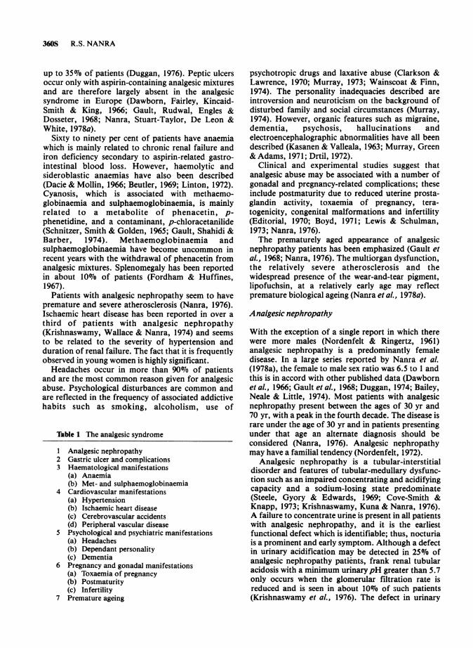
Figure 2 Natural history of a hypothetical patthe analgesic syndrome. The renal manifestatioroccur after the age of 30 years. GIH, UTI arrepresent gastrointestinal haemorrhage, urin;infection and renal pelvic carcinoma, respectiv(
initial renal size and glomerular filtration rate.Overall, 21 Wo of patients died and the main causes ofmortality were ischaemic heart disease, septicaemiaand cerebrovascular accidents. The long-term outlookof analgesic nephropathy patients is optimistic ifpatients stop abusing analgesics. In another studyinvolving 43 patients with severe renal failure, the 5-yrcumulative survival was 73.8 0o (Kincaid-Smith,Nanra & Fairley, 1970).
Analgesic nephropathy patients who are treated bydialysis and transplantation seem to fare poorly incomparison to patients with glomerulonephritis(Nanra, 1977; Nanra et al., 1978a) and this has beenrelated mainly to the aggressive degenerative vasculardisease seen in these patients.
Natural history of the analgesic syndrome
Figure 2 illustrates the natural history of ahypothetical patient with the analgesic syndrome.The habit of analgesic intake commences insecondary school and by school leaving age analgesicconsumption is at adult level (Egger & Champion,1978). The obstetric and gastrointestinal manifesta-tions are usually seen between the ages of 20 yr and40 yr, whereas the renal and other complications tendto occur after the age of 30 years.
Pathological aspects
Macroscopic appearance
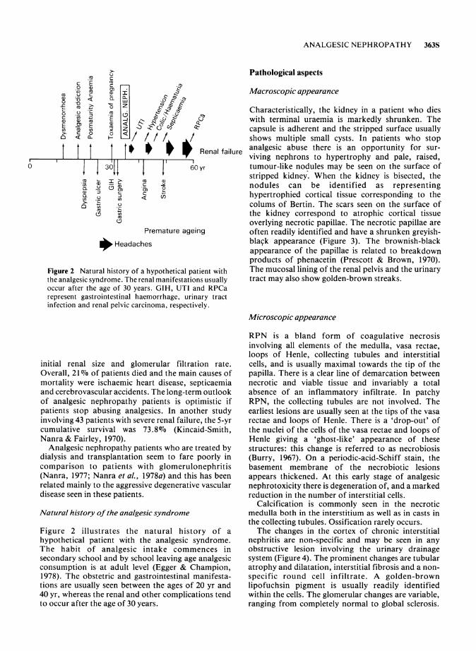
Characteristically, the kidney in a patient who dieswith terminal uraemia is markedly shrunken. Thecapsule is adherent and the stripped surface usuallyshows multiple small cysts. In patients who stop
Renal failure analgesic abuse there is an opportunity for sur-viving nephrons to hypertrophy and pale, raised,
yr tumour-like nodules may be seen on the surface ofstripped kidney. When the kidney is bisected, thenodules can be identified as representinghypertrophied cortical tissue corresponding to thecolums of Bertin. The scars seen on the surface ofthe kidney correspond to atrophic cortical tissueoverlying necrotic papillae. The necrotic papillae are
ig often readily identified and have a shrunken greyish-black appearance (Figure 3). The brownish-blackappearance of the papillae is related to breakdownproducts of phenacetin (Prescott & Brown, 1970).
tient with The mucosal lining of the renal pelvis and the urinaryis usually tract may also show golden-brown streaks.id RPCaary tractely.
Microscopic appearance
RPN is a bland form of coagulative necrosisinvolving all elements of the medulla, vasa rectae,loops of Henle, collecting tubules and interstitialcells, and is usually maximal towards the tip of thepapilla. There is a clear line of demarcation betweennecrotic and viable tissue and invariably a totalabsence of an inflammatory infiltrate. In patchyRPN, the collecting tubules are not involved. Theearliest lesions are usually seen at the tips of the vasarectae and loops of Henle. There is a 'drop-out' ofthe nuclei of the cells of the vasa rectae and loops ofHenle giving a 'ghost-like' appearance of thesestructures: this change is referred to as necrobiosis(Burry, 1967). On a periodic-acid-Schiff stain, thebasement membrane of the necrobiotic lesionsappears thickened. At this early stage of analgesicnephrotoxicity there is degeneration of, and a markedreduction in the number of interstitial cells.
Calcification is commonly seen in the necroticmedulla both in the interstitium as well as in casts inthe collecting tubules. Ossification rarely occurs.The changes in the cortex of chronic interstitial
nephritis are non-specific and may be seen in anyobstructive lesion involving the urinary drainagesystem (Figure 4). The prominent changes are tubularatrophy and dilatation, interstitial fibrosis and a non-specific round cell infiltrate. A golden-brownlipofuchsin pigment is usually readily identifiedwithin the cells. The glomerular changes are variable,ranging from completely normal to global sclerosis.
364S R.S. NANRA
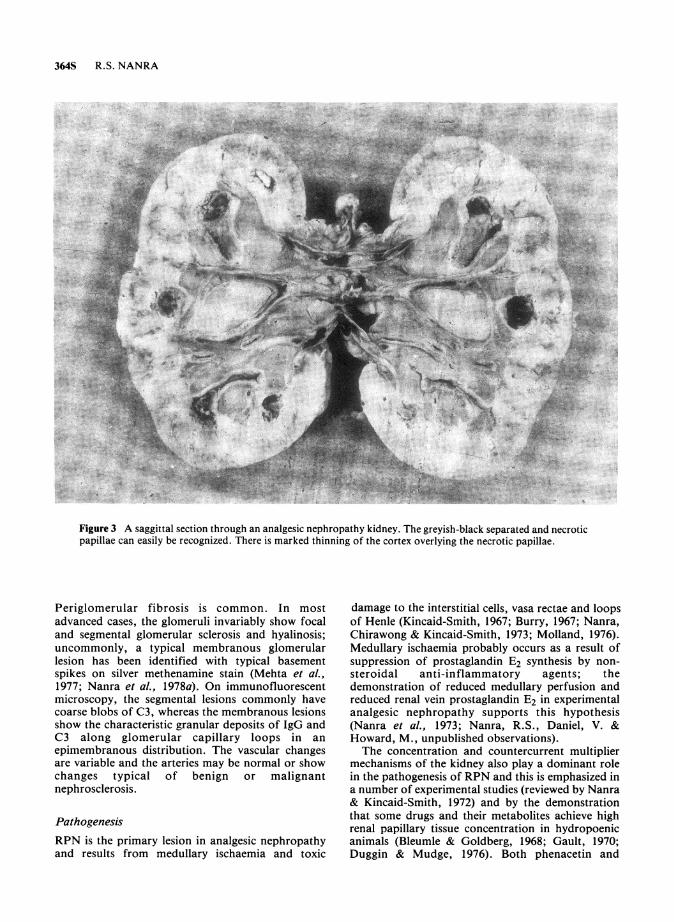
Figure 3 A saggittal section through an analgesic nephropathy kidney. The greyish-black separated and necroticpapillae can easily be recognized. There is marked thinning of the cortex overlying the necrotic papillae.
Periglomerular fibrosis is common. In mostadvanced cases, the glomeruli invariably show focaland segmental glomerular sclerosis and hyalinosis;uncommonly, a typical membranous glomerularlesion has been identified with typical basementspikes on silver methenamine stain (Mehta et al.,1977; Nanra et al., 1978a). On immunofluorescentmicroscopy, the segmental lesions commonly havecoarse blobs of C3, whereas the membranous lesionsshow the characteristic granular deposits of IgG andC3 along glomerular capillary loops in anepimembranous distribution. The vascular changesare variable and the arteries may be normal or showchanges typical of benign or malignantnephrosclerosis.
PathogenesisRPN is the primary lesion in analgesic nephropathyand results from medullary ischaemia and toxic
damage to the interstitial cells, vasa rectae and loopsof Henle (Kincaid-Smith, 1967; Burry, 1967; Nanra,Chirawong & Kincaid-Smith, 1973; Molland, 1976).Medullary ischaemia probably occurs as a result ofsuppression of prostaglandin E2 synthesis by non-steroidal anti-inflammatory agents; thedemonstration of reduced medullary perfusion andreduced renal vein prostaglandin E2 in experimentalanalgesic nephropathy supports this hypothesis(Nanra et al., 1973; Nanra, R.S., Daniel, V. &Howard, M., unpublished observations).The concentration and countercurrent multiplier
mechanisms of the kidney also play a dominant rolein the pathogenesis of RPN and this is emphasized ina number of experimental studies (reviewed by Nanra& Kincaid-Smith, 1972) and by the demonstrationthat some drugs and their metabolites achieve highrenal papillary tissue concentration in hydropoenicanimals (Bleumle & Goldberg, 1968; Gault, 1970;Duggin & Mudge, 1976). Both phenacetin and

ANALGESIC NEPHROPATHY 365S
Figure 4 Renal biopsy showing severe chronic interstitial nephritis. There is severe tubular atrophy, interstitialfibrosis and a round cell infiltrate. One glomerulus shows global sclerosis and another has typical periglomerularfibrosis with an otherwise normal tuft.
paracetamol acutely enhance the efficacy ofantidiuretic hormone in promoting urinaryconcentration (Lozada, Gouaux & Hayes, 1971) andthis may be a significant factor in concentrating toxicmetabolites in the medulla (Nanra et al., 1973).At a cellular level, aspirin interferes with the
tricarboxylic cycle, oxidative phosphorylation andmucopolysaccharide synthesis, covalently acetylatesrenal tissue proteins and depletes cellular glutathione(Smith, 1966; Wheldrake, 1975; Caterson et al.,quoted in Duggin, 1977; Mudge, Gemborys &Duggin, 1978). Paracetamol is the major metaboliteof phenacetin and unlike phenacetin concentratesin the renal medulla. It is believed to be con-
verted to a cytotoxic metabolite which covalentlybinds to tissue proteins and causes damage leadingto glucose-6-phosphate dehydrogenase, glutathioneand dihydronicotinamide adenine dinucleotidephosphate (NADPH) depletion and cellular necrosis.It has been suggested that the synergism between
aspirin and phenacetin or paracetamolnephrotoxicity may be explained on the basis ofinactivation of intracellular enzyme systems andsalicylate inhibition of NADPH supply through thehexose monophosphate shunt (Goldberg, Myers,Peshel, McCarron & Morrison, 1971; Murray &Goldberg, 1975; Mitchell, McMurtry, Statham &Nelson, 1977; Mudge et al., 1978).
The combination of medullary ischaemia andcellular necrosis leads to RPN which has all thehistological features of an infarct (Nanra et al., 1973).The chronic interstitial nephritis is a non specificlesion which results from obstruction to tubules in thenecrotic medulla (Burry, 1967; Kincaid-Smith, 1967;Nanra & Kincaid-Smith, 1972). The golden-brownlipofuchsin-like pigment which is readily seen in thekidney is also widely distributed in most organs ofthe body and is probably a highly oxidized polymerof unsaturated fatty acids related to the oxidanteffect of phenacetin (Pearse, 1960).

366S R.S. NANRA
Aetiological factors
There is considerable clinical, epidemiological andexperimental evidence that chronic analgesicconsumption leads to renal disease and renal failure,and the subject has been extensively reviewed (Haley,1966; Shelley, 1967; Abel, 1971; Nanra & Kincaid-Smith, 1972; Murray & Goldberg, 1975; Nanra et al.,1978a). Animal experiments suggest that RPN mayresult from the chronic toxicity of all non-steroidanti-inflammatory drugs; the results from a numberof studies in rats are summarized in Table 2 (reviewedin Nanra et al., 1978a; R.S. Nanra, V. Daniel& M. Howard, unpublished). In man, analgesicnephropathy is usually associated with abuse ofpopular analgesic mixtures containing proprietarycombinations of aspirin, phenacetin, paracetamol,phenazone (antipyrine), caffeine or codeine.Occasional examples of RPN with individualanalgesics such as aspirin, paracetamol,indomethacin or phenylbutazone have beendescribed but these are uncommon becauseindividual analgesics are rarely addictive. These datahave been recently reviewed elsewhere (Nanra &Kincaid-Smith, 1972; Nanra et al., 1978a).Although the association between analgesic abuse
and RPN is well established, other factors probablyexist and are not fully recognized. A genetic predis-position has recently been given some support by thedemonstration of an increased frequency of theHLA-B12 antigen in analgesic nephropathy(MacDonald et al., 1978). Women seem to be at anincreased risk of renal damage from analgesics;
although the female to male ratio of analgesicabusers in the Australian community is 2 to 1, thefemale to male ratio of analgesic nephropathy isapproximately 7 to 1. Climatic factors and fluidintake have also been incriminated. Of great interestare patients who give a history of massive analgesicabuse but who surprisingly show absent or minimalevidence of tubulomedullary dysfunction.
Conclusions
Analgesic nephropathy is a totally preventable formof progressive renal disease which would beeradicated by legislation to control the availability ofaddicting analgesic mixtures. Community studies arealso required to define the factors that lead to theregular use of pain-killers for inappropriate reasons.Individual analgesics are almost never abused andthere are good reasons to believe that renal damageresults from the synergistic nephrotoxicity of thedifferent components of the analgesic mixtures -aspirin, phenacetin, paracetamol and phenazone.RPN is an expression of nephrotoxicity common toall non-steroidal anti-inflammatory agents and,therefore, modification of the composition ofanalgesic mixtures is unlikely to lead to a significantreduction in analgesic-related renal failure (Nanra etal., 1978a). Even in patients with establishedanalgesic nephropathy, long term survival may beanticipated if patients can stop the abuse ofanalgesics.
In Australia, the unabated sale of analgesic
Table 2 Experimental RPN with individual and compound analgesics
Analgesic *
APC (impure)tAPC (pure)tA+ APAP+CA + salicylamide + CPhenazone + P + C
Dose (mg/kg daily)
900900900840840
Duration (weeks)
8-208-7212-7212-7212-72
AA§pAPAPPhenazoneCPhenylbutazoneIndomethacinMefenamic acid
500200
3,0003,0001,0001501012
100
8-2010-668-208-2012-308-208-2012-308-20
9/277/133/83/71/80/101/92/76/9
*Abbreviations used: A, aspirin; P, phenacetin; APAP, paracetamol; C, caffeine.t Impure = 0.1 7o p-chloracetanilide in P.t Pure = 0.01 No p-chloracetanilide in P.§Cortical scars in 3/13 (23.1 %).
RPN
13/3113/209/167/134/11
(37.5%o)(65.0%)(56.3%)(53.9070)(36.4%)
(33.3%7o)(54.8%)(37.5 o)(42.907o)(12.5%7o)
(1 1.1 o)(28.607o)(66.7%o)

ANALGESIC NEPHROPATHY 367S
mixtures is likely to lead to terminal renal failure in300 new patients per year, with an estimated cost inexcess of A$10,000,000 per annum for theirtreatment (Duggin, 1977). Legislation to limitanalgesic mixtures to prescription might lead to areduction in the incidence of terminal renal failure
References
ABEL, J.A. (1971). Analgesic nephropathy - a review ofthe literature, 1967-1970. Clin. Pharmac. Ther., 12,5837598.
AUSTRALIA AND NEW ZEALAND DIALYSIS AND TRANS-PLANT REGISTRY. (1979).
BAILEY, R.R., NEALE, J.T. & LITTLE, P.J. (1974). Analgesicnephropathy. N.Z. med. J., 79, 1053-1057.
BEUTLER, E. (1969). Drug-induced hemolytic anaemia.Pharmac. Rev., 21, 73-103.
BLACKMAN, J.E., GIBSON, G.R., LAVAN, J.N., LEAROYD,H.M. & POSEN, S. (1967). Urinary calculi and theconsumption of analgesics. Br. med. J., 2, 800-802.
BLEUMLE, L.W. & GOLDBERG, M. (1968). Renal accumula-tion of salicylate and phenacetin: Possible mechanismsin the nephropathy of analgesic abuse. J. clin. Invest.,47,2507-2514.
BOYD, E.M. (1971). Sterility from phenacetin. J. clin.Pharmac., 11, 96-102.
BURRY, A.F. (1967). The evolution of analgesic nephro-pathy. Nephron, 5, 185-201.
CLARKSON, A.R. & LAWRENCE, J.R. (1970). The clinicalfeatures of analgesic nephropathy. In Renal Infectionand Renal Scarring. Ed. Kincaid-Smith, P. & Fairley,K.F. Pp. 375-384. Melbourne: Mercedes Press.
COVE-SMITH, J.R. & KNAPP, M.S. (1973). Sodium handlingin analgesic nephropathy. Lancet, 2, 70-72.
DACIE, J.V. & MOLLIN, D.L. (1966). Siderocytes, sidero-blasts and sideroblastic anaemia. Acta. Med. Scand.,445,237-248.
DAWBORN, J.K., FAIRLEY, K.F., KINCAID-SMITH, P. &KING, W.E. (1966). The association of peptic ulceration,chronic renal disease and analgesic abuse. Quart. J.Med., 35, 69-83.
DE LEON, A.H. & NANRA, R.S. (1976). Radiological changesin analgesic nephropathy and chronic pyelonephritis.Aust. N.Z. J. Med., 6,68.
DRTIL, J. (1972). Psychotic syndromes after long term useof analgesic pyretics. Activ. Nerv. Super. Praha, 14,186-187.
DUBACH, U.C. (1967). p-Aminophenol-Bestimmung in Urinals Routinemethode zur Erfaschung der Phenacetinein-nahme. Dtsch. Med. Wochenschr., 92,211-215.
DUGGAN, J.M. (1972). The phenestix test in the detectionof analgesic abuse. Med. J. Aust., 1, 659-660.
DUGGAN, J.M. (1974). The analgesic syndrome. Aust. N.Z.J. Med., 4,365-372.
DUGGAN,.J.M. (1976). Aspirin in chronic gastric ulcer:an Australian experience. Gut, 17, 378-384.
DUGGIN, G.G. & MUDGE, G.H. (1976). Analgesic nephro-pathy: renal distribution of acetaminophen and itsconjugates. J. Pharmac. exp. Therap., 199, 1-9.
DUGGIN, G.G. (1977). Analgesic induced kidney disease.Aust. J. Pharmaceut. Sci., 6,378-384.
EDITORIAL (1970). Salicylates and malformations. Br.med. J., 1, 642-643.
but the results of this intervention will probably notbe recognized for another decade.
This work was supported by a grant from the NationalHealth and Medical Research Council.
EGGER, G. & CHAMPION, R. (1978). Adolescent Drug andAlcohol Use in New South Wales, 1971-1977. Report78/3, Division of Health Services Research, HealthCommission of New South Wales.
FAIRLEY, K.F. & KINCAID-SMITH, P. (1968). Renal papillarynecrosis with a normal pyelogram. Br. Med. J., 1,156-157.
FORDHAM, C.C. & HUFFINES, W.S. (1967). Splenomegaly,analgesic consumption and renal disease. N. Engl. J.Med., 277,749-751.
GAULT, M.H., RUDWAL, T.C., ENGLES, W.D. & DOSSETER,J.B. (1968). Syndrome associated with the abuse ofanalgesics. Ann. intern. Med., 68, 906-925.
GAULT, M.H. (1970). Demonstration of an intrarenalgradient of 14C-acetylsalicylic acid (ASA) in rabbitsand guinea pigs. Abstr. Ann. Soc. Nephrol., Washing-ton, 28.
GAULT, M.H., SHAHIDI, N.T. & BARBER, V.E. (1974).Methemoglobin formation in analgesic nephropathy.Clin. Pharmac. Ther., 15, 521-527.
GOLDBERG, M., MYERS, C.L., PESHEL, W., McCARRON, D.& MORRISON, A.B. (1971). Mechanism of analgesicabuse nephropathy. J. clin. Invest., 50, 37a.
HALEY, T.J. (1966). Is phenacetin the cause of 'analgesicnephropathy'? New Drugs, 6, 193-202.
HARE, W.S.C. (1970). The radiology of analgesic nephro-pathy. In Renal Infection and Renal Scarring. Ed.Kincaid-Smith, P. & Fairley, K.F. Pp. 409-414.Melbourne: Mercedes Press.
HULTENGREN, N., LAGERGREN, C. & LJUNGQVIST, A.(1965). Carcinoma of the renal pelvis in renal papillarynecrosis. Acta. Chir. Scand., 130, 314-320.
INGHAM, J.P., KLEEREKOPER, M., STEWART, J.H. & POSEN,S. (1974). Symptomatic skeletal disease in non-terminalrenal failure. Med. J. Aust., 2, 873-876.
JOHANSSON, S., ANGERVALL, L., BENGTSSON, U. &WAHLQVIST, L. (1974). Uroepithelial tumours of the renalpelvis associated with abuse of phenacetin-containinganalgesics. Cancer, 33, 743-753.
KASANEN, A. & VALLEALA, P. (1963). Electroencephalo-graphic finding in chronic phenacetin abusers. Acta. Med.Scand., 173, 35-40.
KINCAID-SMITH, P. (1967). Pathogenesis of the renal lesionassociated with the abuse of analgesics. Lancet, 1,859-862.
KINCAID-SMITH, P., NANRA, R.S. & FAIRLEY, K.F. (1970).Analgesic nephropathy: a recoverable form of chronicrenal failure. In Renal Infection and Renal Scarring.Ed. Kincaid-Smith, P. & Fairley, K.F. Pp. 380-390.Melbourne: Mercedes Press.
KRISHNASWAMY, S., WALLACE, D.C. &NANRA, R.S. (1974).Ischaemic heart disease in analgesic nephropathy. Aust.N.Z. Med. J., 4, 426.

368S R.S. NANRA
KRISHNASWAMY, S., KUNA, K. & NANRA, R.S. (1976).Pattern of changes in renal function with analgesicabuse. Aust. N.Z. J. Med., 6, 88.
LEWIS, R.B. & SCHULMAN, J.D. (1973). Influence of acetyl-salicylic acid, an inhibitor of prostaglandin synthesis, onthe duration of human gestation and labour. Lancet, 2,1159-1161.
LINDVALL, N. (1960). Renal papillary necrosis: A roentgeno-graphic study of 155 cases. Acta. Radiol. Stockh., 192,1-153.
LINTON, A.L. (1972). Renal disease due to analgesics. Can.Med. Assoc. J., 107, 749-751.
LOZADA, E.S., GOUAUX, J. & HAYS, R.M. (1971). Thephenylacetamides: potent enhancers of vasopressin.Clin. Res., 19, 539.
MACDONALD, I.M., DUMBLE, L.J., DORAN, T., BASHIR, H.,NANRA, R.S. & KINCAID-SMITH, P. (1978). Increasedfrequency of HLA-B12 in analgesic nephropathy. Aust.N.Z.J.Med., 8,233.
MAHONY, J.F., STOREY, B.G., IBANEZ, R.C. & STEWART,J.H. (1977). Analgesic abuse, renal parenchymal diseaseand carcinoma of the kidney or ureter. Aust. N.Z. J.Med., 7,463-469.
MEHTA, A.R., WHITE, K.H. & NANRA, R.S. (1977). Signifi-cance of proteinuria in analgesic nephropathy. Aust. N. Z.J. Med., 7,200-201.
MITCHELL, J.R., McMURTRY, R.J., STATHAM, C.N. &NELSON, S.D. (1977). Molecular basis of several drug-induced nephropathies. Am. J. Med., 62, 518-526.
MOLLAND, E.A. (1976). Aspirin damage in the rat kidneyin the intact animal and after unilateral nephrectomy.J. Path., 120,43-48.
MUDGE, G.H., GEMBORYS, M.W. & DUGGIN, G.G. (1978).Covalent binding of metabolites of acetaminophen tokidney protein and depletion of renal glutathione. J.Pharmac. exp. Therap., 206,218-226.
MURPHY, K.J. (1968). Calcification of the renal papilla as asign of analgesic nephropathy. Clin. Radiol., 19, 394-399.
MURRAY, R.M., GREENE, J.G. & ADAMS, J.H. (1971).Analgesic abuse and dementia. Lancet, 2, 242-254.
MURRAY, R.M. (1973). The origins of analgesic nephropathy.In Problems on Phenacetin Abuse. Ed. Haschek, H.Pp. 287-292. Vienna: Facta Publications.
MURRAY, R.M. (1974). Personality factors in analgesicnephropathy. Psychol. Med., 4, 69-73.
MURRAY, T. & GOLDBERG, M. (1975). Analgesic abuse andrenal disease. A. Rev. Med., 26, 537-550.
NANRA, R.S. & KINCAID-SMITH, P. (1972). Chronic effectof analgesics on the kidney. Progr. Biochem. Pharmac.,7, 285-323.
NANRA, R.S., CHIRAWONG, P. & KINCAID-SMITH, P.(1973). Medullary ischaemia in experimental analgesicnephropathy: The pathogenesis of renal papillarynecrosis. Aust. N.Z. Med. J., 3, 580-586.
NANRA, R.S. (1976). Analgesic nephropathy. Med. J. Aust.,1, 745-746.
NANRA, R.S. (1977). Recurrent dialysis and transplantationin systemic disorders. In Opuscula Medico-TechnicaLudensia, XX. Australian Dialysis and TransplantationWorkshop. Ed. Thompson, N.M. & Atkins, R.C.Pp. 57. Lund: Rahms.
NANRA, R.S., STUART-TAYLOR, J., DE LEON, A.H. &WHITE, K.H. (1978a). Analgesic nephropathy: etiology,clinical syndrome, and clinicopathologic correlations inAustralia. Kidney Int., 13, 79-92.
NANRA, R.S., HOWARD, M. & DANIEL, V. (1978b).Secondary gout in analgesic nephropathy (AN). Aust.N.Z.J.Med., 8,237.
NANRA, R.S. (1978). Factors which influence the clinicalcourse of analgesic nephropathy. Abstr. seventh Int.Congr. Nephrol., Montreal, G-20.
NORDENFELT, 0. & RINGERTZ, N. (1961). Phenacetintakers dead with renal failure: 27 men and 3 women.Acta. Med. Scand., 170, 385-402.
NORDENFELT, 0. (1972). Deaths from renal failure inabusers of phenacetin containing drugs. Acta. Med.Scand., 191, 11-16.
ORELL, S.R., NANRA, R.S. & FERGUSON, N.W. (1980).Renal pelvic carcinoma in the Hunter Valley. Med. J.Aust. (in press).
PEARSE, A.G.E. (1960). Histochemistry: Theoretical andApplied. Edinburgh and London: Churchill-Livingstone.
POYNTER, J.D. & HARE, W.S.C. (1974). Necrosis in situ:A form of renal papillary necrosis seen in analgesicnephropathy. Radiology, 111, 69-76.
PRESCOTT, L.F. & BRODIE, D.E. (1964). A simpledifferential stain for urinary sediment. Lancet, 2, 940.
PRESCOTT, L.F. (1965). Effects of acetylsalicylic acid,phenacetin, paracetamol and caffeine on renal tubularepithelium. Lancet, 2, 91-96.
PRESCOTT, L.F. (1976). Analgesic nephropathy: the inter-national experience. Aust. N.Z. J. Med., 6, (suppl. 1)44-48.
PRESCOTT, L.F. & BROWN, S.S. (1970). Brown urine: a clueto phenacetin intoxication. Lancet, 1, 253.
REPORT FROM SENATE STANDING COMMITTEE ONSOCIAL WELFARE. (1977). Drug problems in Australia- an intoxicated society? The Parliament of theCommonwealth of Australia, Australian GovernmentPublishing Service, Canberra. Pp. 107-126.
SCHNITZER, B., SMITH, E.B. & GOLDEN, A. (1965). Theeffects of phenacetin and its contaminant on the kidneyof the rat. Am. J. Path., 6, 917-927.
SHELLEY, J.H. (1967). Phenacetin through a lookingglass. Clin. Pharmac. Ther., 8, 427-471.
SMITH, M.J.H. (1966). Metabolic effects of salicylates. InThe Salicylates. Ed. Smith, M.J.H. & Smith, P.K.Pp. 49. New York: Wiley-Interscience.
SPUHLER, 0. & ZOLLINER, H.U. (1953). Die chronische-interstitielle Nephritis. S. Klin. Med., 151, 1-50.
STEELE, T.W., GYORY, A.Z. & EDWARDS, K.D.G. (1969).Renal function in analgesic nephropathy. Br. med. J.,2,213-216.
TAYLOR, J.S. (1972). Carcinoma of the urinary tract andanalgesic abuse. Med. J. Aust., 1, 407-409.
THE LONE HAND. (1907). Headache Cures. Pp. 193-195.Publisher please.
WAINSCOAT, J.S. & FINN, R. (1974). Possible role oflaxatives in analgesic nephropathy. Br. med. J., 4,697-698.
WHELDRAKE, J.F. (1975). The effect of aspirin (acetyl-salicylate) on macromolecular turnover in rat kidneyand liver. Experimentia, 31, 559-569.
WING, A.J., BRUNNER, F.P., BRYNGER, H., CHANTLER,C., DONCKERWOLCKE, R.A., GURLAND, H.J.,HATHWAY, R.A. & JACOBS, C. (1978). Combined reporton regular dialysis and transplantation in Europe,August, 1977. Proc. Eur. Dial. Transplant. Assoc., 12,3-76.