clinical and genetic characteristics of pstpip1-associated
TRANSCRIPT
RESEARCH ARTICLE Open Access
Clinical and genetic characteristics of PSTPIP1-associated myeloid-related proteinemiainflammatory syndromeDan Zhang1, Gaixiu Su1* , Yan Liu2 and Jianming Lai1
Abstract
Objective: To summarise the clinical and genetic characteristics of three children with PSTPIP1-associated myeloid-related proteinemia inflammatory (PAMI) syndrome.
Methods: This study retrospectively analysed the clinical and genetic data of three children with PAMI syndrome inour hospital between April 2018 and January 2020.
Results: One male and two female children were 6 years and 5months, 8 years and 7 months, and 13 years and 3months of age. All three patients had a recurrent blood trilineage hypoplasia and splenomegaly. Patient 1 hadpyoderma gangrenosum, and Ludwig’s angina. Patient 2 had pyogenic arthritis, and pyoderma gangrenosum.Patient 3 had hepatomegaly, pyogenic arthritis, and pulmonary hypertension. Laboratory tests revealed that allthree children had elevated C-reactive protein (CRP) and erythrocyte sedimentation rate (ESR). Patient 1: C-antineutrophilic cytoplasmic antibodies(c-ANCA), positive; antiglobulin test (Coombs), positive. Patient 2: blood zinc,4.38 mg/L (elevated). Patient 3: Antinuclear antibodies (ANA), 1:100, β2 glycoprotein I, positive; Coombs test,positive; RF, 28.3 U/ml (elevated); C3, 0.77 g/L (decreased). Genetic testing showed that all 3 patients had PSTPIP1c.748G > A (p.E250K) spontaneous heterozygous mutations, suggesting the diagnosis of PAMI syndrome. Patient 1was treated with a combination of methylprednisolone and cyclosporine for 8 months. The patient did not developnew skin lesions. The blood count showed mild neutropenia. The spleen was considerably retracted and the CRPbecame normal. Patient 2 was treated with etanercept and methylprednisolone. The patient had no furtherarthralgias and pyoderma gangrenosum showed improvement. The spleen was smaller than before. White bloodcells were shown to be approximately 2–3 × 10⁹/L. The haematocrit, platelets, CRP, and AESR were normal. Patient 3was treated with methylprednisolone, methotrexate, and infliximab 4 times. The patient’s joint symptomsdisappeared gradually and the liver retracted markedly. The pulmonary artery pressure returned to normal.Moreover, Coombs test result was negative. CRP and AESR were lower than before.
Conclusion: PAMI syndrome can manifest as pyogenic arthritis, pyoderma gangrenosum, acne, and trilineagehypoplasia, as well as autoimmune diseases. Glucocorticoid and immunosuppressive therapy are partially effective andcytokine antagonists can be used in refractory cases. Whole-exome genetic testing is helpful to confirm diagnosis.
Keywords: PSTPIP1-associated myeloid-related proteinemia inflammatory syndrome, Tumour necrosis factor antagonist
© The Author(s). 2021 Open Access This article is licensed under a Creative Commons Attribution 4.0 International License,which permits use, sharing, adaptation, distribution and reproduction in any medium or format, as long as you giveappropriate credit to the original author(s) and the source, provide a link to the Creative Commons licence, and indicate ifchanges were made. The images or other third party material in this article are included in the article's Creative Commonslicence, unless indicated otherwise in a credit line to the material. If material is not included in the article's Creative Commonslicence and your intended use is not permitted by statutory regulation or exceeds the permitted use, you will need to obtainpermission directly from the copyright holder. To view a copy of this licence, visit http://creativecommons.org/licenses/by/4.0/.The Creative Commons Public Domain Dedication waiver (http://creativecommons.org/publicdomain/zero/1.0/) applies to thedata made available in this article, unless otherwise stated in a credit line to the data.
* Correspondence: [email protected] of Rheumatology, Capital institute of pediatrics, 2 yabao road,chaoyang district, Beijing, ChinaFull list of author information is available at the end of the article
Zhang et al. Pediatric Rheumatology (2021) 19:151 https://doi.org/10.1186/s12969-021-00636-9

Keypoint
� PAMI syndrome can be manifested as anautoinflammatory disease. It can also show featuresof autoimmune diseases.
� PAMI syndrome is highlighted by intractable declineof leukocyte, which is difficult to treat. Cyclosporinemay be effect for leukopenia. TNFα antagonists areeffective against pyoderma gangrene and pyogenicarthritis, Steriod plays an important role in thetreatment of this disease.
� Clinical and genetic characteristics of PSTPIP1-associated myeloid-related proteinemia inflammatorysyndrome.
BackgroundPSTPIP1-associated myeloid-related proteinemia inflam-matory syndrome (PAMI syndrome), is a rare autoin-flammatory disease caused by pathogenic variants of thePSTPIP1 gene. PAMI syndrome is characterised bychronic systemic inflammation, pyogenic arthritis,hepatosplenomegaly, growth retardation, and may showelevated serum zinc levels and elevated myeloid-relatedprotein (MRP)-8/14 complexes. The pathophysiologicalmechanisms of PAMI syndrome are not fully under-stood. However, the specific mutations in PSTPIP1(p.E250K and p.E257K) are thought to be caused bysignificantly increased binding of immunomodulatoryprotein pyrin due to the charge reversal of ɣ domain.Steriod, immunosuppressants, and biologics have allbeen reported to be effective for this disease. Clinical re-ports of this disease have increased recently due to wide-spread use of whole-exome genetic testing. Threechildren were diagnosed with PAMI syndrome in ourhospital, all with early age of onset. One patient hadpancytopenia and rash, one had pancytopenia and pyo-genic arthritis, and another had arthritis, leukopenia,autoimmune haemolytic anaemia, positive ANA, positiveβ2 glycoprotein, and declined complement. Tumournecrosis factor antagonists were used in two of the threecases, with remarkable clinical efficacy and few sideeffects. We analysed the clinical data and genetic testresults of these three cases to enhance our clinical un-derstanding, diagnosis accuracy, and treatment efficacyof this disease.
MethodsWe retrospectively analysed clinical data and genetictesting results of three children with PAMI syndromediagnosed at the Department of Rheumatology andImmunology, Capital Institute of Pediatrics, betweenApril 2018 and January 2020. One patient was male, andtwo patients were female. We collected their medicalhistory, including age of onset, age at diagnosis, medical
history, physical examination, laboratory tests, andwhole-exome gene testing. Informed consent was ob-tained from the patients’ parents, who signed a writtendocument.
ResultsThe clinical manifestations of the three children aresummarised in Table 1, and the laboratory test data aresummarised in Table 2.Clinical diagnosis and genetic test identified c.748G >
A variant in PSTPIP1 gene of three patients. Thec.748G > A (nucleotide 748 in the coding region changedfrom guanine to adenine) variant causes the p.E250Kmutation (amino acid 250 changed from glutamic acidto lysine), which is a missense mutation. The bioinfor-matics protein function software SIFT, PolyPhen_2, andREVEL predicted it to be deleterious, deleterious, andbenign, respectively.The PSPPIP1 gene follows an autosomal dominant
mode of inheritance and was identified by Sanger inthree family lines, all of which were identified as spon-taneous mutations in probands, thus demonstratingconsistency within their family.
Patient 1Three years ago, an 8-year-old female patient was admit-ted to the hospital with fever, systemic rash, and triline-age hypoplasia. The rash was round, yellow-brownpatches on the trunk and proximal extremities, withbran-like scales on the surface, accompanied by itching.Her spleen was hard and enlarged (10 cm) below ribs.Intravenous human immunoglobulin therapy 2 g/kg wasineffective. Complete blood count monitoring revealed:reduced white blood cell (WBC), 1.68 × 10⁹/L; reducedneutrophil, 0.66 × 10⁹/L; reduced haemoglobin (Hb), 95g/L; reduced red blood cell (RBC), 2.4 × 10¹²/L; reducedreticulocyte, 0.034; reduced platelet (PLT), 63 × 109/L;and elevated C-reactive protein (CRP), up to 111 mg/L.Anti-neutrophil cytoplasmic antibody (ANCA) testedwas positive; antiglobulin test (Coombs) was positive,and bone marrow aspiration suggested trilineage hypo-plasia. The patient was considered to have Ludwig’s
Table 1 Clinical manifestations of three patients
Patient 1 Patient 2 Patient 3
Fever + – +
Failure to thrive – + –
Splenomegaly + + +
Pyogenic arthritis – + +
Pyoderma gangrenosum + + –
Acne – – –
Pulmonary arterial hypertension – – +
Zhang et al. Pediatric Rheumatology (2021) 19:151 Page 2 of 10
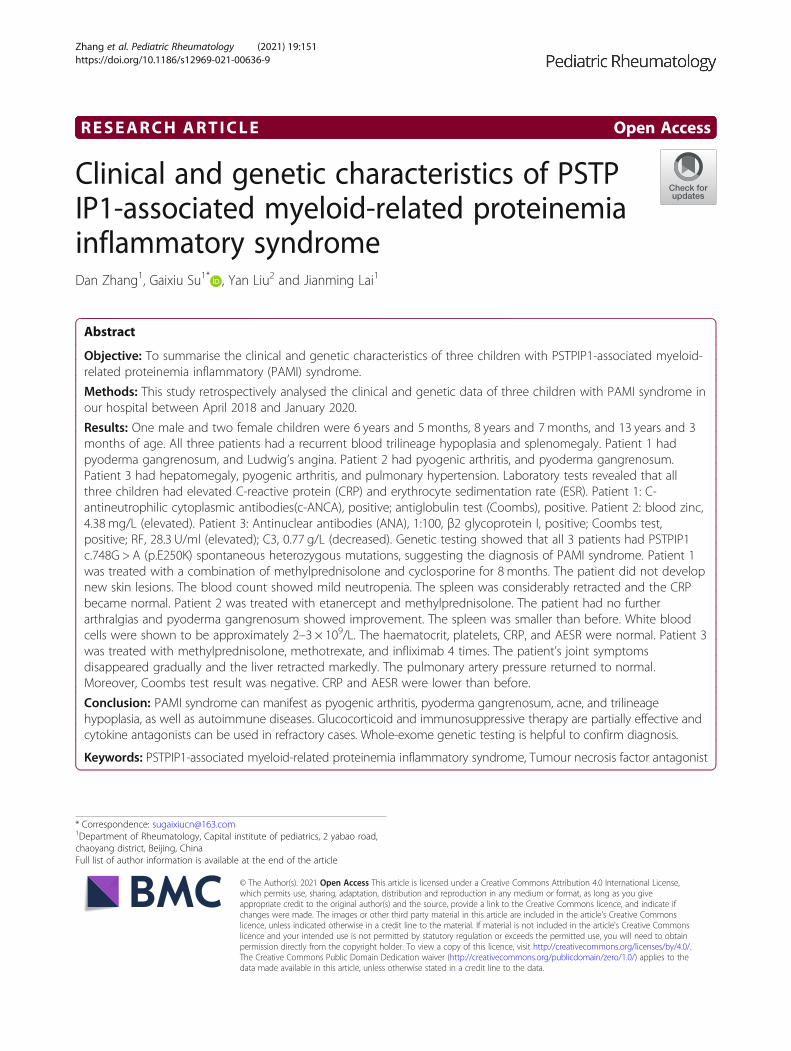
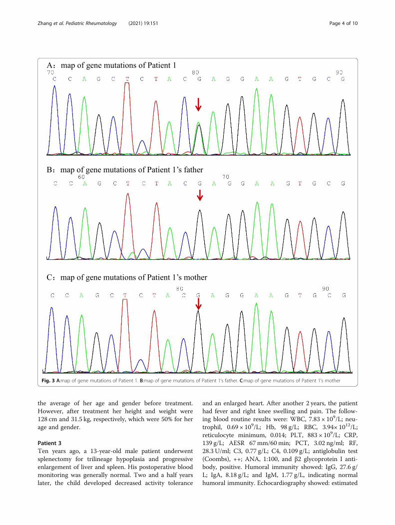
angina two years ago when she had a relapsing fever, leftswelling and painful jaw (Fig. 1), and skin ulceration(Fig. 2). The culture obtained from the exudates waspositive for Staphylococcus aureus. Moreover, the leftjaw was swollen and painful with progressive dysphagia.The whole-exome gene testing was completed and sug-gested a spontaneous PSTPIP1 c.748G > A (p.E250K)(see Fig. 3 for gene mutation profile). Therefore, she wasdiagnosed with PAMI syndrome. Oral Methylpredniso-lone (1 mg/kg/d) and cyclosporine (4 mg/kg/d) were ad-ministered, and the left mandibular swelling graduallydisappeared (Fig. 1). The treatment was followed up fortwo years. Methylprednisolone and cyclosporine was re-duced to 3 mg/d and 75 mg/d, respectively. Only her oldskin lesion existed persistively (Fig. 2). Her blood cellcount and CRP levels returned to normal. Her spleno-megaly recovered.
Patient 2Eight years ago, a 10-year-old female patient underwenta bone marrow aspiration to investigate growth
retardation and splenomegaly, which indicated trilineagehypoplasia. Symptomatic blood transfusion treatmentwas given. However, the patient’s condition did not im-prove. Five years ago, she developed pain in the right hipjoint with limited mobility, which was considered as ahip abscess. The joint cavity was punctured and drained,and the pus was cultured, which was negative. The jointsymptoms improved following the treatment of antibi-otics, component blood transfusion, and intravenous hu-man immunoglobulin. Blood investigation showed: WBC1.87–2.66 × 10⁹/L; reduced neutrophil, 0.42 × 10⁹/L; Hb53–80 g/L; reduced RBC 2.32–2.45 × 10¹²/L; PLT 125–152 × 109/L; CRP 8–69mg/L; AESR 19–140 mm/60 min;SF 520.18 ng/ml; and serum zinc 4.38 mg/L. Whole-exome genetic testing suggested a spontaneous PSTPIP1c.748G > A (p.E250K) mutation (see Fig. 4 for gene mu-tation profile), hence, the diagnosis of PAMI syndromewas made. After administration of infliximab 5mg/kgtwice, the patient developed an allergic reaction. So wediscontinued inflicimab, and started oral colchicine0.125 mg for 1 month, which was ineffective. Therefore,we changed to subcutaneous injection of etanercept (0.8mg/kg) once a week and methylprednisolone 1 mg/kgorally once a day. At 3-year follow-up, the patient didnot have arthralgia any more and splenomegaly im-proved than before. The WBC remained low, between 2and 3 × 10⁹/L. The haematocrit and platelets were nor-mal. In addition, the CRP and AESR returned to normal.Six months before, when methylprednisolone was re-duced from 4mg to 2 mg every other day, the patientdeveloped pyoderma gangrenosum (PG: Figs. 5 and 6),and the secretion culture showed Enterobacter cloacae.Besides, serum zinc level was 6.65 mg/L and faecal cal-protectin level was 72.4 μg/L. we did local debridementand gave her antibiotic. In addition, we increased meth-ylprednisolone to 6 mg/d. after treatment, her PG im-proved considerably (Figs. 5 and 6). The patient’s heightwas 78 cm and weight was 9 kg, which were 3% below
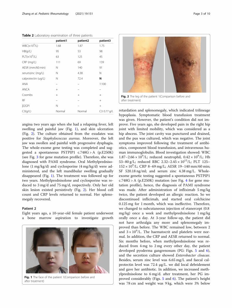
Table 2 Laboratory examination of three patients
patient1 patient2 patient3
WBC(×109/L) 1.68 1.87 1.75
HB(g/L) 95 53 98
PLT(×109/L) 63 125 45
CRP (mg/L) 111 69 139
AESR (mm/60min) N 140 91
serumzinc (mg/L) N 4.38 N
calprotectin (ug/L) N 72.4 N
ANA – – 1:100
ANCA + – –
Coombs + + +
RF – – +
β2GPI N – +
C3(g/L) Normal Normal C3 0.77 g/L
Fig. 1 The face of the patient 1(Comparison before andafter treatment)
Fig. 2 The leg of the patient 1(Comparison before andafter treatment)
Zhang et al. Pediatric Rheumatology (2021) 19:151 Page 3 of 10
the average of her age and gender before treatment.However, after treatment her height and weight were128 cm and 31.5 kg, respectively, which were 50% for herage and gender.
Patient 3Ten years ago, a 13-year-old male patient underwentsplenectomy for trilineage hypoplasia and progressiveenlargement of liver and spleen. His postoperative bloodmonitoring was generally normal. Two and a half yearslater, the child developed decreased activity tolerance
and an enlarged heart. After another 2 years, the patienthad fever and right knee swelling and pain. The follow-ing blood routine results were: WBC, 7.83 × 10⁹/L; neu-trophil, 0.69 × 10⁹/L; Hb, 98 g/L; RBC, 3.94× 10¹²/L;reticulocyte minimum, 0.014; PLT, 883 × 109/L; CRP,139 g/L; AESR 67mm/60min; PCT, 3.02 ng/ml; RF,28.3 U/ml; C3, 0.77 g/L; C4, 0.109 g/L; antiglobulin test(Coombs), ++; ANA, 1:100, and β2 glycoprotein I anti-body, positive. Humoral immunity showed: IgG, 27.6 g/L; IgA, 8.18 g/L; and IgM, 1.77 g/L, indicating normalhumoral immunity. Echocardiography showed: estimated
Fig. 3 A:map of gene mutations of Patient 1. B:map of gene mutations of Patient 1’s father. C:map of gene mutations of Patient 1’s mother
Zhang et al. Pediatric Rheumatology (2021) 19:151 Page 4 of 10

sPAP, 95 mmHg, severe pulmonary hypertension, rightheart enlargement and failure. Contrast-enhanced MRIof the knee showed marked thickening of synovial mem-brane of right knee joint and effusion. Bone marrowaspiration was normal. Aniracetam 2.5 mg qd and tada-lafil 10 mg qd were prescribed as targeted therapy for
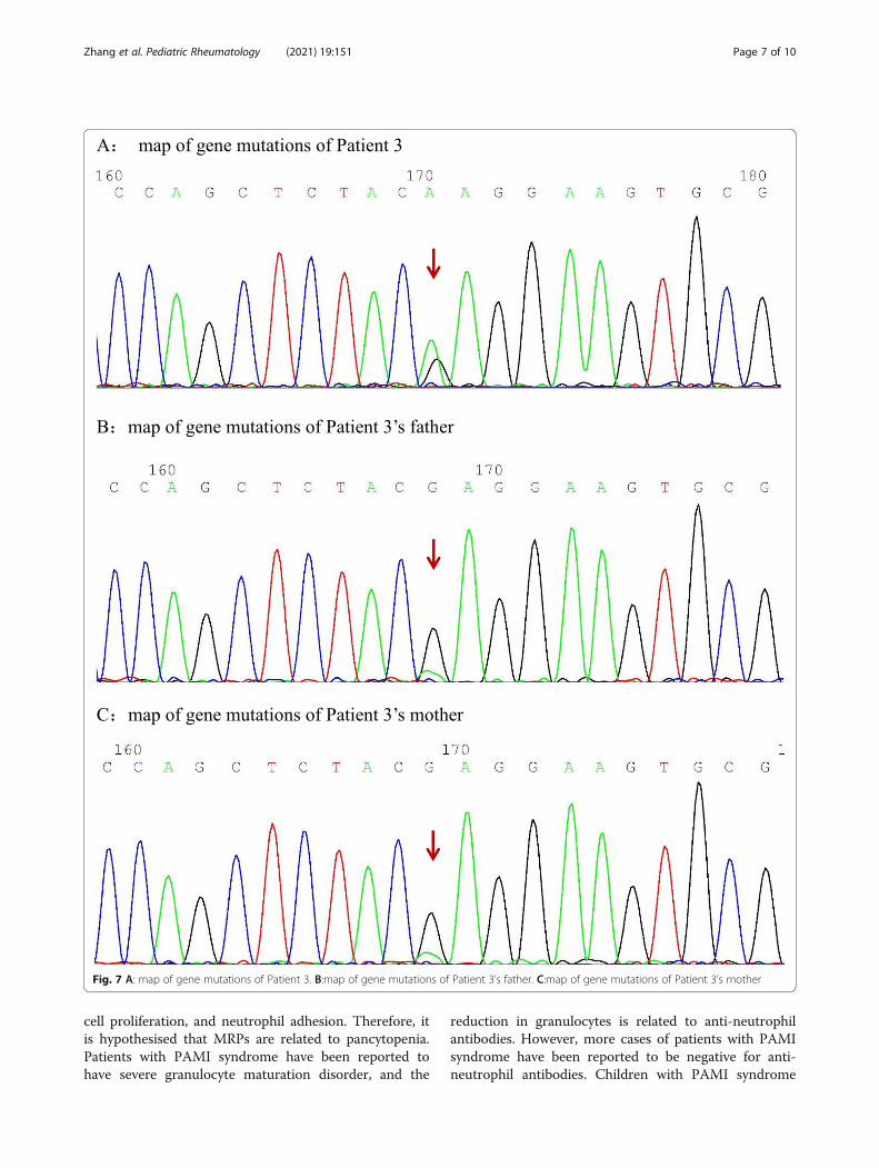
pulmonary hypertension and symptomatic cardiotonicdiuresis. However, her condition did not improve con-siderably. Whole-exome genetic testing was performed,and the results suggested a spontaneous PSTPIP1c.748G > A (p.E250K) (see Fig. 7 for gene mutationprofile) mutation. Therefore, the diagnosis of PAMI
Fig. 4 A:map of gene mutations of Patient 2. B:map of gene mutations of Patient 2’s father. C:map of gene mutations of Patient 2’s mother
Zhang et al. Pediatric Rheumatology (2021) 19:151 Page 5 of 10

syndrome was established. The patient was prescribedoral methylprednisolone 0.5 mg/kg every two weeks,followed by oral methotrexate 10 mg after four weeks,and intravenous infliximab 6mg/kg after eight weeks, to-tally 4 times. The initial treatment of methylpredniso-lone was 20mg once a day. Then the dose was reducedby 4 mg per month until 8 mg QD, following by reduc-tion of 2 mg per month. Infliximab had been used regu-larly throughout the entire treatment. His jointsymptoms disappeared at 2-year follow-up and the hep-atomegaly improved remarkably. The pulmonary hyper-tension disappeared. In addition, Coombs test wasnegative and NT-proBNP was normal, whereas CRP andESR were 57.2 g/L, and 26mm/60min, respectively.
DiscussionPAMI syndrome has been defined as distinct autoinflam-matory disorder with clinical and biochemical features notfound in patients with classical PAPA syndrome [1]. Inaddition to prominent skin inflammation and arthralgia/arthritis, PAMI is characterised by severe chronic systemicinflammation, hepatosplenomegaly, pancytopenia, and
listlessness. Severe course and early onset of disease, hepa-tosplenomegaly, failure to thrive, cytopenia, hyperzince-mia, and extremely high levels of pro-inflammatoryalarmins MRP8/14 separates PAMI syndrome fromPAPA. The mutations of p.E250K and p.E257K result incharge reversal in the y-domain of PSTPIP1 (E→K) andincreased interaction with pyrin compared to p.E250Qmutants. Steriods, immunosuppressants, and biologicshave all been reported to be effective for this disease. Clin-ical reports of this disease have increased recently with thewidespread use of whole-exome genetic testing [2].PSTPIP1 is a proline-serine-threonine phosphatase
interacting protein, which is a tyrosine phosphorylatedprotein involved in cytoskeleton organisation that regu-lates T lymphocyte activation, leukocyte activation, cyto-skeletal organisation, and interleukin-1 release. PSTPIP1mutations are associated with periodic inflammatoryflares reflected by systemic symptoms like fever or anacute phase response and local inflammation of the skin,joints, or other internal organs. At present, 25 sequencevariants have been reported for PSTPIP1 gene [3]. Thepathophysiological mechanisms of PAMI syndrome arenot fully understood, and specific mutations in PSTPIP1(p.E250K and p.E257K) are thought to be caused by sig-nificant increase in the binding of immunomodulatoryprotein pyrin because of the charge reversal of the ɣ do-main. Pyrin has been reported to form an alternativeinflammasome, and mutations in pyrin may lead to anuncontrolled activation of this pathway, resulting in anoverwhelming production of active IL-1β. In a cellularmodel, it has been shown that this process can be trig-gered by the interaction of PSTPIP1 with pyrin. SuchPSTPIP1–pyrin interaction leads to a massive release ofthe pro-inflammatory complex, MRPs, to form a positivefeedback with IL-1, resulting in further exacerbation ofthe autoinflammatory process.MRP, also known as calprotectin, is a member of the
S100 protein family. S100A8 (MRP8) and S100A9(MRP14) form a stable heterodimeric complex and arespatially extended by their C-terminal alpha-helices,which bind as non-covalent bonds to form tetramers.MRP-8/14 is mainly produced in neutrophils, mono-cytes, macrophages, dendritic cells, and vascular smoothmuscle cells. Functionally, the subunit interface of MRP-8/14 has two affinity binding sites for bivalent zinc ions.This property of chelating serum zinc can lead to the de-velopment of hyperzincemia, giving it essential functionssuch as immunomodulation, resistance to bacterial in-fection, promotion of inflammation, and inhibition ofinflammation.Most patients with PAMI syndrome develop pancyto-
penia, especially neutropenia; however, the severityvaries, and the aetiology is unclear. MRPs have beenassociated with the induction of apoptosis, inhibition of
Fig. 5 The elbow of the patient 2 (Comparison before andafter treatment)
Fig. 6 The buttock of the patient 2 (Comparison before andafter treatment)
Zhang et al. Pediatric Rheumatology (2021) 19:151 Page 6 of 10
cell proliferation, and neutrophil adhesion. Therefore, itis hypothesised that MRPs are related to pancytopenia.Patients with PAMI syndrome have been reported tohave severe granulocyte maturation disorder, and the
reduction in granulocytes is related to anti-neutrophilantibodies. However, more cases of patients with PAMIsyndrome have been reported to be negative for anti-neutrophil antibodies. Children with PAMI syndrome
Fig. 7 A: map of gene mutations of Patient 3. B:map of gene mutations of Patient 3’s father. C:map of gene mutations of Patient 3’s mother
Zhang et al. Pediatric Rheumatology (2021) 19:151 Page 7 of 10

tend to have splenomegaly, which is considered apossible cause of pancytopenia.The prominent cutaneous manifestation of PAMI syn-
drome is pyoderma gangraenosum (PG), an inflamma-tory skin disease with histological changes of neutrophilinfiltration. Typical clinical manifestations are single ormultiple skin ulcers with depressions and raisederythematous areas with violet margins, as well as pus-tules, bullae, abscesses, papules, nodules, and ulcers(polymorphic skin lesions, including pustules, bullae, ab-scesses, papules, nodules, and ulcers are characterisedhistologically by a neutrophil-rich inflammatory infil-trate). PG can appear in monogenic autoinflammatorydiseases, namely syndromic PG, and can also be presentin isolated diseases or be associated with systemic dis-eases, such as inflammatory bowel disease, rheumaticdisease, lymphoproliferative disease, and other blooddiseases.The pyogenic arthritis of PAMI syndrome is reported
to be painful and presents as recurrent aseptic monoarti-cular arthritis with a neutrophil-rich infiltrate, whichgenerally occurs in childhood and may also be the firstmanifestation of the disease, possibly leading to jointerosion and destruction.Compared to most autoinflammatory diseases, PAMI
syndrome primarily demonstrates an increase in seruminflammatory markers, such as CRP, erythrocyte sedi-mentation rate, and ferritin.Patient 1 presented with recurrent trilineage hypopla-
sia, splenomegaly, PG, and Ludwig’s angina. Patient 2presented with recurrent trilineage hypoplasia, spleno-megaly, pyogenic arthritis, and growth retardation. Pa-tient 3 presented with intermittent fever, recurrenttrilineage hypoplasia, hepatosplenomegaly, pyogenicarthritis, and pulmonary hypertension. In line with theliterature, all three patients had recurrent trilineagehypoplasia and splenic manifestations, and the bonemarrow aspiration results were negative for haemato-logic malignancies. Two patients had PG and pyogenicarthritis. Ludwig’s angina was present in patient 1. Pul-monary hypertension was present in patient 3, whichwere not reported before. In patient 3, the trilineagehypoplasia recovered completely after splenectomy,which verified the hypothesis reported suggesting thatthe trilineage hypoplasia was associated with hypers-plenism. All three patients had significantly elevatedCRP levels, and patients 2 and 3 had faster AESR, allof which agreed with inflammatory disease. Allpatients were positive in Coombs test, patient 1 hadpositive c-ANCA, and patient 3 had positive ANAand β2GP I with reduced complement C3 levels,which indicated autoimmune disease. All threechildren had spontaneous mutations, which werewild-type, with no PAMI syndrome-related phenotype.
The common clinical manifestations of PAPAsyndrome are pyogenic arthritis, PG, acne, and anaemia[4, 5]. These clinical phenotypes overlap with PAMI syn-drome. However, the prominent manifestation of PAMIsyndrome is pancytopenia. Because a decrease in whiteblood cells and platelets has not been found in patientswith PAPA syndrome. These clinical manifestations canbe used as important factors to distinguish these twodiseases. The currently identified pathogenic mutationsin PAPA syndrome include p.A230T, p.E250Q, p.E256G,p.D246N, and p.D266N, whereas those in PAMI syn-drome are p.E250K and p.E257K. Furthermore, PSTPIP1-associated inflammatory diseases also demonstratepyogenic arthritis, PG, acne, and hidradenitis suppura-tiva syndrome (PAPASH) [6], pyogenic arthritis, PG, andacne-like syndrome (PAPA-like) [7], as well as PG, acne,and ulcerative colitis syndrome (PAC) [8]. The PAPASHpathogenic mutation is p.E277D, the PAPA-like patho-genic mutation is p.G258A, and the PAC pathogenicmutation is p.G403R. Hence, the above disease spectrumcan exhibit a pyogenic arthritis, PG, and acne. Thespecific clinical characteristics such as neutropenia withhepatosplenomegaly, hidradenitis suppurativa, and ul-cerative colitis can help clinicians to diagnose whetherthe patient has PSTPIP1-related autoinflammatory dis-eases, and further genetic testing is needed to clarify thetype of disease [9].There is no standard treatment regimen for PAMI
syndrome; treatment with non-steroidal anti-inflammatory drugs, calcineurin inhibitors, and streoidhas been reported. IL-1 antagonists, which are effectivein PAPA syndrome that is also caused by mutations inthe PSTPIP1 gene, have an unclear therapeutic effect inPAMI syndrome [10]. Among the three children, patient1 was treated with methylprednisolone and cyclosporine.His rash, trilineage hypoplasia, and hepatosplenomegalyimproved considerably, with only mild neutropenia andgranulocyte reduction (approximately 1.5 × 10⁹/L). Pa-tients 2 and 3 were treated with methylprednisolone andtumour necrosis factor antagonists (patient 2 was aller-gic to infliximab; hence, etanercept was administered).for patient 2, trilineage was improved; the inflammatoryindex was lower and the liver and spleen retracted.Moreover, the signs and symptoms of arthritis disap-peared. Pulmonary hypertension in patient 3 returned tonormal. Of the three children, only patient 1 was treatedwith cyclosporine and had the best effect for leukocytesand neutrophils, thus demonstrating the remarkableeffect of cyclosporine on the haematological system.Tumour necrosis factor antagonists had a significanteffect on the recovery of hepatosplenomegaly and jointinflammation. Steroid have contributed to the im-provement of PG and the haematological system. BySearching the Literature, there was a recent report
Zhang et al. Pediatric Rheumatology (2021) 19:151 Page 8 of 10

about hematopoietic stem cell transplantation (HSCT)as a therapeutic option for PAMI syndrome [11]. Fivepatients with PAMI syndrome underwent allogeneicHSCT with myeloablative or reduced-intensity condi-tioning regimens. All 5 patients engrafted; however, 1patient at day + 13 developed hemophagocytic syn-drome, followed by graft rejection at day + 17. After5.5 months, a second HSCT was performed from analternative donor. Another patient at day + 116 devel-oped an intense inflammatory syndrome with signifi-cant serositis and severe mitral and aortic valveregurgitation, controlled with adalimumab, tacrolimus,and prednisone. At the last follow-up, all 5 patientshave predominantly or complete donor chimerismand adequate immune recovery and are free of anyPAMI symptoms. Allogeneic HSCT seems to be aneffective option to cure cytopenia and severe autoin-flammation in PAMI syndrome. Nevertheless, as thenumber of cases is small and the severity of thedisease varies, the response to treatment varies. Thus,more cases of PSTPIP1-specific mutations (p.E250Kand p.E257K) need to be studied to elucidate thepathophysiology and treatment strategies for PAMIsyndrome.
ConclusionIn summary, PAMI is a monogenic autoinflammatorydisease caused by genetic mutations. It can clinicallymanifest as pyogenic arthritis, PG, acne, trilineagehypoplasia, hepatosplenomegaly, and growth retard-ation, as well as autoimmune diseases. Storid andimmunosuppressive therapy is partially effective andcytokine antagonists can be used in refractory cases.Leukopenia is the most severe manifestation and dif-ficult to treat for our 3 cases. Steroid play an import-ant role in the treatment of PAMI. Cyclosporine maybe effective in the treatment of leukopenia. Patientswith pyogenic arthritis with early age of onset, recur-rent trilineage hypoplasia, and associated skin lesionsshould undergo whole-exome genetic testing toachieve early diagnosis and precise treatment.
AbbreviationsPAMI: PSTPIP1-associated myeloid-related proteinemia inflammatory;MRP: myeloid-related protein; CRP: C-reactive protein; ESR: erythrocytesedimentation rate; ANA: antinuclear antibodies; c-ANCA: C-antineutrophiliccytoplasmic antibodies; RF: rheumatoid factor; WBC: white blood cell;Hb: haemoglobin; RBC: red blood cell; PLT: platelet; PG: pyodermagangraenosum; PAPA: pyogenic arthritis, pyoderma gangraenosum, acne,and anaemia; PAPASH: pyogenic arthritis, pyoderma gangraenosum, acne,and hidradenitis suppurativa; PAPA-like: pyogenic arthritis, pyodermagangraenosum, and acne-like; PAC: pyoderma gangraenosum, acne, andulcerative colitis; HSCT: hematopoietic stem cellt transplantation
AcknowledgementsNone.
Authors’ contributionsGaixiu Su and Dan Zhang designed the study. Dan Zhang, Jianming Lai andYan Liu collected the data. Dan Zhang performed the analysis, drafted theinitial manuscript, confirmed revisions, and approved the final manuscript assubmitted. All authors approved the final manuscript as submitted and agreeto be accountable for all aspects of the work.
FundingNo funding or sponsorship was received for this study or publication of thisarticle.
Availability of data and materialsUpon request.
Declarations
Ethics approval and consent to participateNot applicable.
Consent for publicationNot applicable.
Competing interestsThe authors declares that they have no competing interests.
Author details1Department of Rheumatology, Capital institute of pediatrics, 2 yabao road,chaoyang district, Beijing, China. 2Department of Rheumatology, Dalianmunicipal Women and Children’s Medical Center, No.1 and No.3, Sportsnewtown planning road 1,Ganjingzi district, Dalian City, Liaoning, China.
Received: 29 March 2021 Accepted: 30 August 2021
References1. Dai P, Furlong T, Gracie G, Huang ML, Yang T, Wu KHC, et al.
Autoinflammation masquerading as autoimmunity in an adult withheterozygous p.E250K PSTPIP1 mutation. J Clin Immunol. 2019;39(5):519–22.https://doi.org/10.1007/s10875-019-00646-z.
2. Belelli E, Passarelli C, Pardeo M, Holzinger D, De Benedetti F, Insalaco A.Haematological involvement associated with a mild autoinflammatoryphenotype, in two patients carrying the E250K mutation of PSTPIP1. ClinExp Rheumatol. 2017;Suppl 108(6):113–5.
3. WJM J, Grobarova V, Leleux J, Jongeneel L, van Gijn M, van Montfrans JM,et al. Proline-serine-threonine phosphatase interacting protein 1 (PSTPIP1)controls immune synapse stability in human T cells. J Allergy Clin Immunol.2018;142(6):1947–55. https://doi.org/10.1016/j.jaci.2018.01.030.
4. Fathalla BM, Al-Wahadneh AM, Al-Mutawa M, Kambouris M, El-Shanti H.A novel de novo PSTPIP1 mutation in a boy with pyogenic arthritis,pyoderma gangrenosum, acne (PAPA) syndrome. Clin Exp Rheumatol.2014;32(6):956–8.
5. Martinez-Rios C, Jariwala MP, Highmore K, Duffy KW, Spiegel L, Ronald M.Laxer, Jennifer Stimec, Imaging findings of sterile pyogenic arthritis,pyoderma gangrenosum and acne (PAPA) syndrome: differential diagnosisand review of the literature. Pediatr Radiol. 2019;49(1):23–36. https://doi.org/10.1007/s00247-018-4246-1.
6. Marzano AV, Trevisan V, Gattorno M, Ceccherini I, De Simone C, Crosti C.Pyogenic arthritis, pyoderma gangrenosum, acne, and hidradenitissuppurativa (PAPASH): a new autoinflammatory syndrome associated with anovel mutation of the PSTPIP1 gene. JAMA Dermatol. 2013;149(6):762–4.https://doi.org/10.1001/jamadermatol.2013.2907.
7. Geusau A, Mothes-Luksch N, Nahavandi H, Pickl WF, Wise CA, Pourpak Z,et al. Identification of a homozygous PSTPIP1 mutation in a patient with aPAPA-like syndrome responding to canakinumab treatment. JAMADermatol. 2013;149(2):209–15. https://doi.org/10.1001/2013.
8. Zeeli T, Padalon-Brauch G, Ellenbogen E, Gat A, Sarig O, Sprecher E.Pyoderma gangrenosum, acne and ulcerative colitis in a patient with anovel mutation in the PSTPIP1 gene. Clin Exp Dermatol. 2015;40(4):367–72.https://doi.org/10.1111/ced.12585.
9. Klötgen HW, Beltraminelli H, Yawalkar N, van Gijn ME, Holzinger D, BorradoriL. The expanding spectrum of clinical phenotypes associated with PSTPIP1
Zhang et al. Pediatric Rheumatology (2021) 19:151 Page 9 of 10

mutations: from PAPA to PAMI syndrome and beyond. Br J Dermatol. 2018;178(4):982–3. https://doi.org/10.1111/bjd.16136.
10. Hashmi SK, Bergstrom K, Bertuch AA, Despotovic JM, Muscal E, Xia F, et al.PSTPIP1-associated myeloid-related proteinemia inflammatory syndrome: Arare cause of childhood neutropenia associated with systemic inflammationand hyperzincemia. Pediatr Blood Cancer. 2019;66(1):e27439. https://doi.org/10.1002/pbc.27439.
11. Laberko A, Burlakov V, Maier S, Abinun M, Skinner R, Kozlova A, et al. HSCTis effective in patients with PSTPIP1-associated myeloid-related proteinemiainflammatory (PAMI) syndrome. J Allergy Clin Immunol. 2020;S0091–6749(20):31764–4. https://doi.org/10.1016/j.jaci.2020.11.043.
Publisher’s NoteSpringer Nature remains neutral with regard to jurisdictional claims inpublished maps and institutional affiliations.
Zhang et al. Pediatric Rheumatology (2021) 19:151 Page 10 of 10