clinical and experimental applications of nir-led photobiomodulation
TRANSCRIPT

121
Photomedicine and Laser SurgeryVolume 24, Number 2, 2006© Mary Ann Liebert, Inc.Pp. 121–128
Clinical and Experimental Applications of NIR-LEDPhotobiomodulation
KRISTINA D. DESMET, B.S.,1 DAVID A. PAZ, B.S.,2 JESSE J. CORRY, M.D.,2JANIS T. EELLS, Ph.D.,1 MARGARET T.T. WONG-RILEY, Ph.D.,3 MICHELE M. HENRY, B.S.,4
ELLEN V. BUCHMANN, B.S.,2 MARY P. CONNELLY, B.S.,2 JULIA V. DOVI, Ph.D.,2HUAN LING LIANG, M.D.,3 DIANE S. HENSHEL, Ph.D.,5 RONNIE L. YEAGER, M.S.,5DEBORAH S. MILLSAP, M.S.,5 JINHWAN LIM, M.S.,5 LISA J. GOULD, M.D., Ph.D.,6
RINA DAS, Ph.D.,7 MARTI JETT, Ph.D.,7 BRIAN D. HODGSON, D.D.S.,8 DAVID MARGOLIS, M.D.,9and HARRY T. WHELAN, M.D.2
ABSTRACT
This review presents current research on the use of far-red to near-infrared (NIR) light treatment in variousin vitro and in vivo models. Low-intensity light therapy, commonly referred to as “photobiomodulation,” useslight in the far-red to near-infrared region of the spectrum (630–1000 nm) and modulates numerous cellularfunctions. Positive effects of NIR–light-emitting diode (LED) light treatment include acceleration of woundhealing, improved recovery from ischemic injury of the heart, and attenuated degeneration of injured opticnerves by improving mitochondrial energy metabolism and production. Various in vitro and in vivo models ofmitochondrial dysfunction were treated with a variety of wavelengths of NIR-LED light. These studies wereperformed to determine the effect of NIR-LED light treatment on physiologic and pathologic processes. NIR-LED light treatment stimulates the photoacceptor cytochrome c oxidase, resulting in increased energy metab-olism and production. NIR-LED light treatment accelerates wound healing in ischemic rat and murinediabetic wound healing models, attenuates the retinotoxic effects of methanol-derived formic acid in rat mod-els, and attenuates the developmental toxicity of dioxin in chicken embryos. Furthermore, NIR-LED lighttreatment prevents the development of oral mucositis in pediatric bone marrow transplant patients. The ex-perimental results demonstrate that NIR-LED light treatment stimulates mitochondrial oxidative metabolismin vitro, and accelerates cell and tissue repair in vivo. NIR-LED light represents a novel, noninvasive, thera-peutic intervention for the treatment of numerous diseases linked to mitochondrial dysfunction.
1Department of Clinical Laboratory Sciences, University of Wisconsin–Milwaukee, Milwaukee, Wisconsin.2Department of Neurology, Medical College of Wisconsin, Milwaukee, Wisconsin.3Department of Cell Biology, Neurobiology, and Anatomy, Medical College of Wisconsin, Milwaukee, Wisconsin.4Department of Ophthalmology, Medical College of Wisconsin, Milwaukee, Wisconsin.5School of Public and Environmental Affairs, Indiana University, Bloomington, Indiana.6Department of Plastic Surgery, Medical College of Wisconsin, Milwaukee, Wisconsin.7Department of Molecular Pathology, Walter Reed Army Institute of Research, Silver Spring, Maryland.8Department of Dentistry, Children’s Hospital of Wisconsin, Milwaukee, Wisconsin.9Department of Hematology and Oncology, Medical College of Wisconsin, Milwaukee, Wisconsin.
14258c04.PGS 5/2/06 9:39 AM Page 121

122 DeSmet et al.
INTRODUCTION
LOW-INTENSITY LIGHT THERAPY, commonly referred to as“photobiomodulation,” by light in the far-red to near-
infrared (NIR) region of the spectrum (630–1000 nm) modu-lates numerous cellular functions. Clinical and experimentalapplications of photobiomodulation have expanded over thepast 30 years.1 Low-power lasers and light-emitting diodes(LED) are well-accepted therapeutic tools in the treatment ofinfected, ischemic, and hypoxic wounds, along with other softtissue injuries.2–5 Positive effects of photobiomodulation in-clude acceleration of wound healing, improved recovery fromischemic injury in the heart, and attenuated degeneration in theinjured optic nerve.4,6,7
At the cellular level, photobiomodulation can modulate fi-broblast proliferation, attachment and synthesis of collagenand procollagen, promote angiogenesis, and stimulate macro-phages and lymphocytes by improving energy metabolismwithin the mitochondria. In addition, photobiomodulation hasdemonstrated the ability to promote the production of growthfactors, such as keratinocyte growth factor (KGF), transform-ing growth factor (TGF), and platelet-derived growth factor(PDGF).5,8–10
Optimal wavelengths and energy densities necessary fortherapeutic interventions have been characterized. Wave-lengths within the far-red to near-infrared range (630–1000nm) along with a minimal energy density of 4 J/cm2 have beenproven effective at stimulating biological processes.4,10–13
Lasers are limited in their ability to deliver monochromaticfar-red to -NIR light. Combined wavelengths cannot easily bereproduced with lasers, and the beam width makes it difficultto treat large areas. Moreover, lasers emit a fair amount of heat,which has the potential to produce tissue damage. An effectivealternative to lasers are LED arrays, which were initially de-veloped by NASA for experimental plant growth in space.LED arrays produce light in the far-red to NIR at optimalwavelengths and energy densities. The arrays can be con-structed in various sizes to accommodate large areas and do notemit any heat, which eliminates the danger of additional tissuedamage. Light emitted by LED arrays at optimal wavelengthspenetrates skin and tissue to a depth of approximately 23cm.4,12–14 Further, NIR-LED light therapy has been deemed anonsignificant risk by the FDA and has been approved for usein humans.
NIR-LED PHOTOBIOMODULATIONSTIMULATES THE PHOTOACCEPTOR
CYTOCHROME C OXIDASE
The mechanism by which far-red to NIR light produces itsbiological effects remains to be elucidated. There is a growingbody of evidence that suggests that one primary effect is thestimulation of mitochondrial cytochromes, which in turn initi-ate secondary cell-signaling pathways.1,11,16–18 The overall re-sult of photobiomodulation is increased energy metabolismand improved cell viability.18
Within mammalian tissues, there are three major photoac-ceptor molecules: hemoglobin, myoglobin, and cytochrome c
oxidase.18 Of these three, cytochrome c oxidase is the only onethat is involved in energy metabolism and production, as itcomprises complex IV of the electron transport chain locatedwithin the mitochondria. Thus, cytochrome c oxidase has beenpostulated as the photoacceptor molecule for the biological ef-fects of photobiomodulation.
The evidence to support cytochrome c oxidase as the pri-mary photoacceptor has been steadily growing. Cellular prolif-eration studies comparing the action spectrum following laserirradiation compared to the absorption spectra of possible pho-toacceptor molecules have suggested cytochrome c oxidase asthe primary photoacceptor.16 In addition, it has been demon-strated that up to 50% of NIR light is absorbed by mitochon-drial chromophores, including cytochrome c oxidase.12,13 Instudies using primary cultured neurons and tetrodotoxin(TTX)—a voltage-dependent sodium channel blocker that im-pedes neuronal impulses, decreases ATP demand, and down-regulates cytochrome c oxidase—NIR-LED light treatment hasbeen shown to reverse the toxic effects of TTX. This is accom-plished by reverting levels of cytochrome c oxidase back tocontrol levels in TTX-exposed NIR-LED light–treated neuronsand up-regulating the enzyme’s activity in NIR-LEDlight–treated control neurons.17 Furthermore, the action andabsorption spectra in the far-red to NIR wavelengths, com-pared to the action and absorption spectra of cytochrome c oxi-dase activity and ATP content in neurons exposed to TTX thatreceived NIR-LED light treatment, parallel each other18 (Fig.1). NIR-LED light treatment has also partially restored cy-tochrome c oxidase activity in primary cultured neurons ex-posed to 10–100 µM potassium cyanide (KCN), significantlyreduced cell death in neurons exposed to 300 µM KCN, signif-icantly restored ATP levels in neurons treated with 10 µMKCN, and enhanced the effect of photobiomodulation by pre-treating neurons with NIR-LED light prior to exposure to10–100 µM KCN in vitro.
NIR-LED PHOTOBIOMODULATIONACCELERATES WOUND HEALING
IN VITRO AND IN VIVO
There is a growing need for safe and efficacious therapeuticintervention for the treatment of chronic wounds. Hyperbaricoxygen therapy (HBO) is a common treatment for ischemic,hypoxic, and infected wounds, but it is not appropriate for allpatients.4 HBO therapy is contraindicated in patients who havechronic medical conditions and are claustrophobic. Access to afacility equipped with HBO may also be a problem.4 NIR-LEDphotobiomodulation can serve as an alternative to HBO.
The process of wound healing occurs in three phases: first, asubstrate is laid down; second, cell proliferation occurs; andthird, remodeling of the tissue takes place. Photobiomodula-tion exerts its biological effect during the proliferative phase ofwound healing. In vitro experimentation utilizing NIR-LEDlight treatments at various wavelengths has shown to signifi-cantly increase cell growth in a variety of cell lines, includingmurine fibroblasts, rat osteoblasts, rat skeletal muscle cells,and normal human epithelial cells.4 Accelerated wound healingfollowing photobiomodulation has also been demonstrated in a
14258c04.PGS 5/2/06 9:39 AM Page 122

Applications of NIR-LED Photobiomodulation 123
number of in vivo models, including toads, mice, rats, guineapigs, and swine.19,20 In an in vivo rat model of ischemicwounds, a decrease in wound size and acceleration of woundclosure has been demonstrated in rats treated with 880-nmNIR-LED light.4 Human studies using NIR-LED light therapyhave demonstrated greater amounts of epithelialization forwound closure and accelerated healing of skin grafts.2,21
To determine if NIR-LED light treatment can improve im-paired healing, we used a murine model of diabetic healing,which is characterized by a delayed re-epithelialization.22
Polyvinyl acetal (PVA) sponges were implanted subcuta-neously in the dorsum of genetically diabetic mice (BKS.Cg-m+/+ Leprdb). The mice were subsequently treated with 670-nmNIR-LED light, and wounds were harvested for RNA analysis.
Microarray analysis revealed that basement membrane andtissue regenerating genes were significantly up-regulated inmice that received NIR-LED light treatments as compared tocontrols. Integrins, nidogens, laminin, actin, and kinesin motorproteins were up-regulated. All of these proteins are necessaryat specific time points for wound-induced epithelial cell migra-tion and differentiation.22 Up-regulation of these genes is onepossible mechanism by which NIR-LED photobiomodulationcan accelerate wound closure. Semaphorins/collapsins are an-other group of genes that were significantly up-regulated inmice receiving NIR-LED light treatments. Specifically, murinesemaphorin H is involved in the inhibition of sensory periph-eral nerve ingrowth. Murine semaphorin H, along with othersemaphorin/collapsin proteins, is involved in pain manage-ment. Pain has been shown to slow the healing process by the
recruitment of inflammatory cells to the site of injury.22 De-creasing pain via NIR-LED light could aid in the accelerationof wound closure.
Genes that were down-regulated in NIR-LED light–treatedmice include cytokine receptors, interleukin-1, interleukin-10,and macrophage inflammatory protein–2. A decrease in thesegenes encoding for proteins associated with the inflammatoryresponse results in a decrease in pain, which in turn increasesthe ability of tissue-regenerating proteins to facilitate woundclosure. Another group of genes that were down-regulated in re-sponse to NIR-LED light treatment were those encodingproapototic proteins. Activator of apoptosis harakiri (HRK),programmed cell death 1 protein precursor (PDCD-1; PD-1),and receptor-interacting protein (RIP) were all down-regulated.
NIR-LED PHOTOBIOMODULATION AS AN EFFECTIVE THERAPEUTIC TOOL
FOR THE PREVENTION OF ORALMUCOSITIS IN PEDIATRIC BONE
MARROW TRANSPLANT PATIENTS
Chemotherapy and/or radiation therapy is administered priorto bone marrow transplant (BMT). Mucositis, especially oralmucositis (OM), is a common debilitating side effect of thistreatment. The development of these ulcerations causes severepain, compromises the ability of the patient to eat and drinkindependently, and can lead to infection and to increased mor-bidity.23–25 Since NIR-LED photobiomodulation accelerates
Rel
ativ
e A
bsor
banc
e or
Act
ivit
y
Cytochrome Oxidase Spectrum (Oxidized)
Cytochrome Oxidase Activity (% Control)
ATP Content (% Control)
1.25
1
0.75
0.5
0.25
0600 700 800 900 1000 1100
Wavelength
FIG. 1. Action and absorption spectra in the far-red to near-infrared (NIR) region of the spectrum for cytochrome c oxidase ascompared to the relative cytochrome c oxidase activity and ATP content in tetrodotoxin (TTX)–exposed neurons treated withNIR–light-emitting diode (LED) light at varying wavelengths expressed as percent of controls.
14258c04.PGS 5/2/06 9:39 AM Page 123
124 DeSmet et al.
wound healing and increases cell proliferation, this treatmentwas used in an attempt to treat pediatric BMT patients prophy-lactically to prevent the development of oral mucositis.26
The first clinical trial of NIR-LED light treatment as a pre-ventative treatment for the development of OM was performedat the Children’s Hospital of Wisconsin in Milwaukee, Wiscon-sin. Thirty-two pediatric patients receiving myeloablative ther-apy were treated with 670-nm NIR-LED light once a day for14 days post-BMT at an energy density of 4 J/cm2. Patients re-ceived NIR-LED light treatment on the left extraoral epithe-lium and sham treatment on the right. Subsequent to the lighttreatment, patients were asked to rate left and right buccal painas compared to throat pain, which served as an untreated con-trol. NIR-LED light treatment produced a significant reductionin left and right buccal pain (48% and 39%, respectively) whencompared to throat pain. In addition, the incidence of OM inthis patient population was decreased, with only 53% of pa-tients developing OM, when compared to historical epidemio-logical data, which suggests that 70–90% of the patientpopulation receiving BMT should have developed OM(Fig. 2). The results of this clinical trial demonstrate that NIR-LED light treatment may be an effective preventive counter-measure to the development of OM in cancer patients. Thisstudy served as the foundation for the current multi-centered,double-blinded trial underway.
NIR-LED PHOTOBIOMODULATION AS TREATMENT FOR RETINAL
TOXICITY IN VIVO
Mitochondrial dysfunction plays a central role in the patho-genesis of numerous retinal and neurodegenerative diseases, in-cluding age-related macular degeneration, Leber’s hereditaryoptic neuropathy, and Parkinson’s and Alzheimer’s disease.27
Furthermore, mitochondrial dysfunction has been shown to playan integral role in the development of retinal toxicity resultingfrom methanol intoxication.28,29 The neurotoxic agent inmethanol intoxication is the metabolite formic acid. Formic acidis a mitochondrial toxin that specifically inhibits cytochrome coxidase in the retina and optic nerve, resulting in blindness.30,31
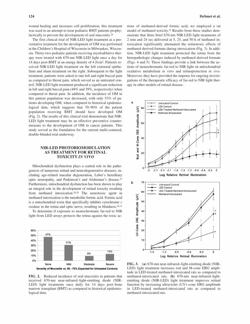
To determine if exposure to monochromatic far-red to NIRlight from LED arrays protects the retina against the toxic ac-
tions of methanol-derived formic acid, we employed a ratmodel of methanol toxicity.32 Results from these studies dem-onstrate that three brief 670-nm NIR-LED light treatments of2 min and 24 sec delivered at 5, 25, and 50 h of methanol in-toxication significantly attenuated the retinotoxic effects ofmethanol-derived formate during intoxication (Fig. 3). In addi-tion, NIR-LED light treatment protected the retina from thehistopathologic changes induced by methanol-derived formate(Figs. 4 and 5). These findings provide a link between the ac-tions of monochromatic far-red to NIR light on mitochondrialoxidative metabolism in vitro and retinoprotection in vivo.Moreover, they have provided the impetus for ongoing investi-gations of the therapeutic efficacy of far-red to NIR light ther-apy in other models of retinal disease.
FIG. 2. Reduced incidence of oral mucositis in patients thatreceived 670-nm near-infrared–light-emitting diode (NIR-LED) light treatments once daily for 14 days post–bonemarrow transplant (BMT) as compared to historical epidemio-logical data.
FIG. 3. (a) 670-nm near-infrared–light-emitting diode (NIR-LED) light treatment increases rod and M-cone ERG ampli-tude in LED-treated methanol-intoxicated rats as compared tomethanol-intoxicated rats. (b) 670-nm near-infrared–light-emitting diode (NIR-LED) light treatment improves retinalfunction by increasing ultraviolet (UV)–cone ERG amplitudein LED-treated methanol-intoxicated rats as compared tomethanol-intoxicated rats.
b
a
14258c04.PGS 5/2/06 9:39 AM Page 124

Applications of NIR-LED Photobiomodulation 125
The prolonged effect of three brief NIR-LED light treat-ments in mediating the retinoprotective actions in methanol in-toxication suggests that 670-nm NIR-LED light treatmentinduces a cascade of signaling events, which is initiated by theinitial absorption of light by cytochrome c oxidase. These sig-naling events may include the activation of immediate earlygenes, transcription factors, cytochrome oxidase subunit geneexpression, and a host of other pathways related to increasedoxidative metabolism. Preliminary gene expression studiesin control untreated, methanol intoxicated, and NIR-LEDlight–treated methanol-intoxicated rodents were performed. At
least 80 genes are involved in subsequent biological processesresulting from methanol intoxication and NIR-LED light treat-ment. Of these, at least 26 genes are up-regulated in methanol-intoxicated rats. These same genes are down-regulated inNIR-LED light–treated methanol intoxicated rats, as comparedto methanol-intoxicated rats. NIR-LED light regulates the ex-pression of a number of genes that control important cellularfunctions and include DNA repair proteins, antioxidant en-zymes, molecular chaperones, protein biosynthesis enzymes,trafficking and degradation proteins, along with cell growthand maintenance proteins.
FIG. 4. Near-infrared–light-emitting diode (NIR-LED) light treatment protects the retina from morphologic changes resultingfrom methanol intoxication. Untreated control (A), LED control (B), methanol-intoxicated (C), and LED-treated methanol-intoxicated (D) rats.
14258c04.PGS 5/2/06 9:39 AM Page 125

126 DeSmet et al.
NIR-LED PHOTOBIOMODULATIONATTENUATES DIOXIN-INDUCED
DEVELOPMENTAL TOXICITY
Dioxin (2,3,7,8-tetrachlorodibenzo-p-dioxin) is the mostacutely toxic of a group of chemicals known collectively aspolycyclic halogenated aromatic hydrocarbons (PHAHs), andis used as the model chemical to investigate the mechanism ofaction of the larger chemical class. The PHAHs are potent de-velopmental toxins that cause increased embryo mortality aswell as sub-lethal changes in the morphological patterning ofthe skeleton and of multiple organs, including the heart and thebrain.33,34 Dioxin, acting in part through activation of a tran-scription factor (ARNT), is known to affect the expression of anumber of genes. These genes encode for proteins that play arole in cell-cell and cell-extracellular matrix interactions, cellsignaling, cytoskeleton-related proteins, proteins associatedwith cell cycle regulation, and the homeostasis and metabolism
of many xenobiotics and hormones.35–37 Further, dioxin haslong been known to induce cellular oxidative stress and in-crease production of free radicals.38 It is through a combinationof these mechanisms that dioxin and the PHAHs are believedto increase the incidence of birth defects, and a variety of can-cers and hormonally linked dysfunctions in humans andwildlife. In addition, late embryo mortality, which is typical ofbirds exposed to higher levels of dioxin, has long been hypoth-esized to be due to cellular stress decreasing available energyneeded for the animal to peck out of the shell.39
To determine the effect of 670-nm NIR-LED light therapyon dioxin-induced developmental toxicity, a chicken (Gallusgallus) embryo model was employed. Domestic chickens havebeen investigated as an animal model for vertebrate embryonicdevelopment for over a century.40 The embryonic developmentof chicken is well characterized anatomically, physiologically,biochemically, and in terms of the molecular cues that controlthe developmental process. Moreover, chicken embryos are
FIG. 5. Near-infrared–light-emitting diode (NIR-LED) light treatment protects the photoreceptor ultrastructure from the retino-toxic effects of methanol intoxication. Untreated control (A), LED control (B), methanol-intoxicated (C), and LED-treatedmethanol-intoxicated (D) rats.
14258c04.PGS 5/2/06 9:39 AM Page 126

Applications of NIR-LED Photobiomodulation 127
sensitive to many developmental toxins and are therefore anideal laboratory model. For this study, domestic chicken eggswere divided into the following treatment groups: no-inject,sunflower oil vehicle, and 2,20,200 ppt dioxin. All of thesegroups contained untreated control eggs and 670-nm NIR-LEDlight–treated eggs resulting in an energy density of 4 J/cm2 at24-h intervals. Results from these experiments indicate thatdaily light treatment throughout embryonic development is notdetrimental to the health of the embryo.41 Further, daily NIR-LED light treatment reduced dioxin-induced mortality of chickembryos by 40% as well as the incubation time before the em-bryo start to hatch (initial pip time).42,43 Thus, NIR-LED lighttreatment obviates at least some of the adverse developmentalimpacts of a model xenobiotic.
CONCLUSION
Experimental results demonstrate that NIR-LED light treat-ment stimulates mitochondrial oxidative metabolism in vitro,and accelerates cell and tissue repair in vivo. NIR-LED lightrepresents a novel, noninvasive, therapeutic intervention forthe treatment of numerous diseases linked to mitochondrialdysfunction, including age-related macular degeneration,Leber’s hereditary optic neuropathy, and Parkinson’s and Alz-heimer’s disease.
ACKNOWLEDGMENTS
This work was supported by the Defense Advanced Re-search Projects Agency (DARPA; grants N66001-01-1-8969,N66001-03-18906, and N66001-04-1-8923), the NationalAeronautics and Space Administration (NASA; grants NAS8-99015, NAS8-97277, and NNM 05AB8C), the Chad BaumannResearch Foundation Endowment, and Bleser Foundation En-dowed Professorship.
REFERENCES
1. Karu, T. (1998). The Science of Low Power Laser Therapy. Lon-don: Gordon and Breach.
2. Conlan, M.J., Rapley, J.W., and Cobb, C.M. (1996). Biostimula-tion of wound healing by low-energy laser irradiation. J. Clin.Periodontol. 23, 492–496.
3. Sommer, A.P., Pinheiro, A.L., Mester, A.R., et al. (2001). Biostim-ulatory windows in low-intensity laser activation: lasers, scannersand NASA’s light-emitting diode array system. J. Clin. Laser Med.Surg. 19, 29–33.
4. Whelan, H.T., Smits, R.L., Buchmann, E.V., et al. (2001). Effectsof NASA light-emitting diode irradiation on wound healing. J.Clin. Laser Med. Surg. 19, 305–314.
5. Yu, W., Naim, J.O., and Lanzafame, R.J. (1997). The effect of laserirradiation on the release of bFGF from 3T3 fibroblasts. J. Clin.Laser Med. Surg. 20, 55–63.
6. Oron, U., Yaakobi, T., Oron, A., et al. (2001). Attenuation of in-farct size in rats and dogs after myocardial infarction by low-energy laser irradiation. Lasers Surg. Med. 28, 204–211.
7. Assia, E.M., Rosner, M., Belkin, M., et al. (1989). Temporal pa-rameters of low-energy laser irradiation for optimal delay of post-traumatic degeneration of optic nerve. Brain Res. 476, 205–212.
8. Mester, A.R., Nagylueskay, S., Mako, E., et al. (1998). Experimen-tal immunological study with radiological application of low-power laser, in: Laser in Medicine. W. Waidelich (ed.). Berlin:Springer-Verlag, pp. 502–512.
9. Mester, E., and Jaszsagi-Nargy, E. (1973). The effects of laser radi-ation on wound healing and collagen synthesis. Studia Biophys.Band 35, 227–230.
10. Lubart, R., Wollman, Y., Friedman, H., et al. (1992). Effects of vis-ible and near-infrared lasers on cell culture. J. Photochem. Photo-biol. 12, 305–310.
11. Karu, T. (2003). Low-Power Laser Therapy. Biomedical PhotonicsHandbook. Boca Raton, FL: CRC Press.
12. Beuvoit, B., Kitai, T., and Chance, B. (1994). Correlation betweenthe light scattering and the mitochondrial content of normal tissuesand transplantable rodent tumors. Anal. Biochem. 226, 167–174.
13. Beuvoit, B., Evans, S.M., Jenkins, T.M., et al. (1994). Contributionof the mitochondrial compartment to the optical properties of therat liver: a theoretical and practical approach. Biophys. J. 6,2501–2510.
14. Chance, B., Nioka, S., Kent, J., et al. (1988). Time-resolved spec-troscopy of hemoglobin and myoglobin in resting and ischemicmuscle. Anal. Biochem. 174, 698–707.
15. Wong-Riley, M.M.T., Liang, H.L., Eells, J.T., et al. (2005). Photo-biomodulation directly benefits primary neurons functionally inac-tivated by toxins. J. Biol. Chem. 280, 4761–4771.
16. Karu, T. (1999). Primary and secondary mechanisms of action ofvisibile to near-IR radiation on cells. J. Photochem. Photobiol.Biol. 49, 1–17.
17. Wong-Riley, M.M.T., Bai, X., Buchmann, E., et al. (2001). Light-emitting diode treatment reverses the effect of TTX on cytochromeoxidase in neurons. Neuroreport 12, 3033–3037.
18. Wong-Riley, M.M.T., Huan, L.L., Eells, J.T., et al. (2005). Photo-biomodulation directly benefits primary neurons functionally inac-tivated by toxins. J. Biol. Chem. 6, 4761–4771.
19. Bibikova, A., and Oron, U. (1995). Regeneration in denervatedtoad (Bufo viridis) gastroenemius muscle and the promotion of theprocess by low-energy laser irradiation. Anat. Rec. 241, 123–128.
20. Al-Watban, F.A. (1997). Laser acceleration of open skin woundclosure in rats and its dosimetric dependence. Lasers Life Sci. 7,237–247.
21. Miller, M., and Truhe, T. (1993). Lasers in dentistry: an overview.JADA 124, 32–35.
22. Whelan, H.T., Buchmann, E.V., Dhokalia, A., et al. (2003). Effect ofNASA light-emitting diode irradiation on molecular changes forwound healing in diabetic mice. J. Clin. Laser Med. Surg. 21, 67–74.
23. Schubert, M.M., Sullivan, K.M., and Truelove, E.L. (1986). Headand neck complications of bone marrow transplantation, in: Headand Neck Management of the Cancer Patient. D.E. Peterson, E.G.Elias, and S.T. Sonis (eds.). The Hauge: Martinus Nijhoff, pp.401–427.
24. Kolbinson, D.A., Schubert, M.M., Flournoy, N., et al. (1988).Early oral changes following bone marrow transplantation. OralSurg. Oral Med. Oral Pathol. 66, 130–138.
25. Dreizen, S., McCredie, K.B., Dicke, K.A., et al (1979). Oral com-plications of bone marrow transplantations; in adults with acuteleukemia. Postgrad. Med. 66, 187–194.
26. Whelan, H.T., Connelly, J.F., Hodgson, B.D., et al. (2002). NASAlight-emitting diodes for the prevention of oral mucositis in pedi-atric bone marrow transplant patients. J. Clin. Laser Clin. Med. 20,319–324.
27. Carelli, V., Ross-Cisneros, F.N., and Sadun, A.A. (2002). Opticnerve degeneration and mitochondrial dysfunction: genetic and ac-quired neuropathies. Neurochem. Int. 40, 573–584.
28. Seme, M.T., Summerfelt, P.M., Henry, M.M., et al. (1999). For-mate-induced inhibition of photoreceptor function in methanol in-toxication. J. Pharmacol. Exp. Ther. 289, 361–370.
14258c04.PGS 5/2/06 9:39 AM Page 127

128 DeSmet et al.
29. Seme, M.T., Summerfelt, P.M., Henry, M.M., et al. (2001). Differ-ential recovery of retinal function after mitochondrial inhibition bymethanol intoxication. Ophthalmol. Visual Sci. 42, 834–841.
30. Nicholls, P. (1975). Formate as an inhibitor of cytochrome c oxi-dase. Biochem. Biophys. Res. Commun. 67, 610–616.
31. Nicholls, P. (1976). The effect of formate in cytochrome aa3 andan electron transport in the intact respiratory chain. Biochem. Bio-phys. Acta 430, 13–29.
32. Eells, J.T., Henry, M.M., Summerfelt, P., et al. (2003). Therapeuticphotobiomodulation for methanol-induced retinal toxicity. Proc.Natl. Acad. Sci USA 100, 3439–3444.
33. Henshel, D.S., Hehn, B., Vo, M.T., et al. (1993). A short-term testfor dioxin teratogenicity using chicken embryos. In: ASTM STP1216: Second Symposium on Environmental Toxicology and RiskAssessment. J. Hughes et al. (eds.). Philadelphia: ASTM, pp.159–174.
34. Henshel, D.S. (1998). Developmental neurotoxic effects of dioxinand dioxin-like compounds on domestic and wild avian soecies.Environ. Toxicol. Chem. 17, 88–98.
35. Karachi, M., Hashimoto, S., Obata, A., et al. (2002). Identificationof 2,3,7,8-tetrachlorodibenzo-p-dioxin-responsive genes in mouseliver by serial analysis of gene expression. Biochem. Biophys.Res. Commun. 292, 368–377.
36. Martinez, J.M., Afshari, C.A., Bushel, P.R., et al. (2002). Differen-tial toxicogenomic responses to 2,3,7,8-tetrachlorodibenzo-p-dioxin in malignant and nonmalignant human airway epithelialcells. Toxicol. Sci. 69, 409–423.
37. Zeytun, A., McKallip, R.J., Fisher, M., et al. (2002). Analysis of2,3,7,8-tetrachlorodibenzo-p-dioxin–induced gene expression pro-file in vivo using pathway-specific cDNA arrays. Toxicology 178,241–260.
38. Hassoun, E.A., Li, F., Abushaban, A., et al. (2001). Productionof superoxide anion, lipid peroxidation and DNA damage in the
hepatic and brain tissues of rats after subchronic exposure tomixtures of TCDD and its congeners. J. Appl. Toxicol. 21,211–219.
39. McLaughlin, J., Jr., Marliac, J.P., Verrett, M.J., et al. (1963). Theinjection of chemicals into the yolk sac of fertile eggs prior to in-cubation as a toxicity test. Toxicol. Appl. Pharmacol. 5, 760–771.
40. Henshel, D.S. (1997). An argument for the chicken embryo as amodel for the developmental toxicological effects of the polyhalo-genated aromatic hydrocarbons (PHAHs), in: Environmental Toxi-cology and Risk Assessment: Biomarkers and Risk Assessment.ASTM STP 1306, 5th vol. D.A. Bengtson and D.S. Henshel (eds.).Philadelphia: American Society for Testing and Materials, pp.219–229.
41. Yeager, R.L., Franzosa, J.A., Millsap, D.S., et al. (2005). Effects of670-nm phototherapy on development. Photomed. Laser Surg. 23,268–272.
42. Yeager, R.L., Franzosa, J.A., Millsap, D.S., et al. (2006). Survivor-ship and mortality implications of developmental 670-nm pho-totherapy—dioxin co-exposure. Photomed. Laser Surg. 24, 29–32.
43. Yeager, R.L., Franzosa, J.A., Millsap, D.S., et al. (2006). Embryonicgrowth and hatching implications of developmental 670-nm pho-totherapy—dioxin co-exposure. Photomed. Laser Surg. (in press).
Address reprint requests to:Dr. Harry T. Whelan
Department of NeurologyMedical College of Wisconsin
8701 Watertown Plank Rd.Milwaukee, WI 53226
E-mail: [email protected]
14258c04.PGS 5/2/06 9:39 AM Page 128