citethis: metallomics,2012,4,156–165 paperkjansen/biology/hemoglobin/2012... · zn induced...
TRANSCRIPT
156 Metallomics, 2012, 4, 156–165 This journal is c The Royal Society of Chemistry 2012
Cite this: Metallomics, 2012, 4, 156–165
Zn induced structural aggregation patterns of b-amyloid peptides byfirst-principle simulations and XAS measurements
Paolo Giannozzi,a Karl Jansen,b Giovanni La Penna,*c Velia Minicozzi,d
Silvia Morante,d Giancarlo Rossid and Francesco Stellatoe
Received 1st September 2011, Accepted 25th November 2011
DOI: 10.1039/c2mt00148a
We show in this paper that in the presence of Zn ions a peculiar structural aggregation pattern of
b-amyloid peptides in which metal ions are sequentially coordinated to either three or four
histidines of nearby peptides is favored. To stabilize this configuration a deprotonated imidazole
ring from one of the histidines forms a bridge connecting two adjacent Zn ions. Though present
in zeolite imidazolate frameworks, remarkably in biological compounds this peculiar
Zn–imidazolate–Zn topology is only found in enzymes belonging to the Cu,Zn-superoxide
dismutase family in the form of an imidazolate bridging Cu and Zn. The results we present are
obtained by combining X-ray absorption spectroscopy experimental data with detailed
first-principle molecular dynamics simulations.
1 Introduction
Alzheimer’s disease (AD) belongs to a family of heterogeneouspathologies (more than 20), generically termed amyloidosis, inwhich endogenous proteins and/or peptides switch from thephysiological soluble configuration to a pathological fibrillarinsoluble state.1,2
It is generally believed that metals play an important role inthe development of the disease on the basis of the observationthat senile plaques in AD patients have been generally foundto display an increased concentration of transition metals(with Cu, Zn and Fe being the most abundant).3,4 Moreoverthere is solid evidence that a breakdown in metal tra!ckingregulation has a significant impact on the development of age-related neurodegenerative pathologies.5,6
Cu and Zn have been the most studied metal ions (see ref. 7–9and references therein). Their relevance has been also confirmedby the experimental observation that treatment with Cu and Znchelators inhibits accumulation of b-amyloid (Ab) peptideswhich are the main proteinaceous components of the amyloidbrain deposition detected in AD patients.10 A further interesting
fact is that Zn is seen to induce Ab aggregation more e"ectivelythan Cu.11,12
From a structural point of view to date most of the experi-mental results13,14 and numerical simulations15,16 converge on theconclusion of a stable intra-molecular Cu coordination. Thesituation appears to be much more complicated for Zn2+–Ab-peptide complexes, and several Zn2+ coordination modes havebeen proposed.17–19
Recent EPR data (see ref. 20 and references therein) andXAS21 measurements, carried out in the related case of theprion protein, have shown that there is a competition forpeptide binding between the two ions, thus suggesting theexistence of a mechanism for fine regulation of metal bindingto prevent cell damage.In this general framework it appears to be of the utmost
importance to understand and clarify whether and how Cu2+
and Zn2+ di"er in their way of interacting with amyloidogenicpeptides in general, and Ab-peptides in particular.21
For Zn, NMR investigations13 have suggested a variety ofinter-molecular metal binding modes involving, besides otherligands, di"erent numbers of histidines. The existence ofpeculiar inter-molecular binding modes was confirmed by aXAS study of the Zn2+–Ab(13–21) complex22 in which pairsof peptides appear to be cross-linked by a Zn2+ bridge bindingtwo histidines belonging to di"erent peptides. In more recentNMR studies also intra-peptide coordination modes in whichZn2+ binds three histidines and either the N-terminus19 or theGlu11 residue,
23 have been proposed. The variability of Zn2+
coordination modes (according to e.g. concentration, samplepreparation, etc.) was also confirmed by XAS data on theZn2+–Ab(1–40) complex.7,8 The structure suggested by XASanalysis showed a high histidine crowding around Zn, with at
aUniversity of Udine, Department of Chemistry, Physics andEnvironment, via del Cotonificio 108, 33100 Udine, Italy
b John von Neumann Institute for Computing—DESY Zeuthen,Platanen allee 6, 15738 Zeuthen, Germany
cNational Research Council, Institute for Chemistry of Organo-metallic Compounds, via Madonna del Piano 10, 50019 Sestofiorentino (Firenze), Italy. E-mail: [email protected];Fax: +39 0555225203; Tel: +39 0555225264
dDepartment of Physics, University of Roma ‘‘Tor Vergata’’ andNational Institute of Nuclear Physics, Section of Roma ‘‘Tor Vergata’’,via della Ricerca Scientifica, 00133 Roma, Italy
eCentre for Free-Electron Laser Science—DESY, Notkestrasse 85,22607 Hamburg, Germany
Metallomics Dynamic Article Links
www.rsc.org/metallomics PAPER

This journal is c The Royal Society of Chemistry 2012 Metallomics, 2012, 4, 156–165 157
least three His sidechains coordinated to Zn. In order to reacha definite conclusion about the Zn coordination the localstructural information extracted from XAS experiments hadto be complemented with other spectroscopical and biochemicaldata. The four-histidines Zn coordination was found compatiblewith all the available data, while the three-histidines coordinationwas excluded (see the discussion in ref. 8). Since along theAb-peptide sequence only three histidines are present, thisgeometrical arrangement hints at a Zn2+-promoted inter-peptideaggregation mode. A four-histidines Zn binding mode, like theone described above, is rather peculiar as it was not describedamong proteins endowed with a mononuclear Zn site before2003.24 A recent survey of metal ion coordination in the PDB25
found one case of Zn coordination to four His sidechainsbelonging to the same protein monomer (PDB code 1PB026)over 5854 PDB entries containing at least one Zn atom. Oneother case of Zn coordination by four His sidechains involvessidechains belonging to di"erent monomers (the dinuclear hemeactivator protein HAP1, and PDB codes 1HWT and 1QP927).
Another peculiarity of Zn–Ab-peptide binding is thatexperimental data suggest that the four involved histidinesare the two consecutive His13 and His14 residue pairs comingfrom two nearby Ab-peptides. This is a very special arrange-ment as structural databases report one single instance where ametal (actually Cu) is bound to two histidines that are one nextto the other along the protein sequence.28
There are instead many cases where Cu is coordinated tofour histidine sidechains belonging to the same monomer, butall of them concern proteins belonging to the wide class ofCu,Zn-superoxide dismutases (Cu,Zn-SOD). Actually a fullyZn-substituted Cu,Zn-SOD enzyme (encoded by the humanSOD1 gene) has been shown to be at least as stable as the wild-type heterodinuclear protein.29
The coordination of Zn by four neutral imidazolyl groups isinfrequent even within non-protein coordination complexes. Asearch for this coordination in the Cambridge structural data-base (CSD)30 yields, with the exception of tetraimidazole Zn2+
perchlorate,31 only structures in which the imidazolyl group iseither deprotonated (imidazolate) orN-methyl protected. In thefirst case each imidazolate group coordinates two Zn ions(see CSD code IMIDZB0132) leading to the formation of anextended network, a process that indeed can only be preventedby N-methylation.
Recently the special properties of Zn–imidazolate–Znbridge topology have found a wide area of applications inthe zeolite imidazolate frameworks (ZIF).33 ZIF structure isreminiscent of that of zeolites, with zinc and imidazolate in theformer taking approximately the positions that aluminiumand silicate have in the latter. In both cases extended, regularand stable structures with large cavities are formed. Notice-ably, Zn–imidazolate compounds can be obtained in thepresence of weak bases, like dimethyl-formammide, thusshowing that the acidity of NH in the imidazolyl group isassisted by the formation of the Zn–imidazolate aggregate.
One might be tempted to conclude that the peculiar Znbinding mode suggested by recent experimental data is ahallmark of peptide aggregation. Although they certainly givea significant indication in this direction, XAS data alone arenot enough to reach such a conclusion. In fact, it is certainly
true that the strength of XAS resides in its sensitivity to shortrange order as it allows us to reliably identify at atomicresolution the average structure around the metal absorberfor systems in any state of aggregation. But the limitations ofthis spectroscopy are precisely that the structural informationone can get is what results from averaging over the di"erentpossibly existing structures in which the metal absorber maybe coordinated and is furthermore confined to within a sphereof a few Angstroms around the absorber (say 5–6 A). On theother hand, in order to fully understand the complicatedpeptide aggregation mechanism assisted by metals, one mustbe able to reconstruct the structure of the system at distanceslarger than 6 A from the absorber, as it looks that a largeoligomeric or polymeric structure gets formed.In this work we show that the configurations of the Zn–Ab
complexes that are able to simultaneously fit the experimentalfindings (see ref. 8) and the results coming from numericalsimulations are quite complicated. Ab-peptides are cross-linked by Zn ions, with one Zn bonded by two pairs ofadjacent histidines (His13 and His14) from two peptides, anda second Zn is necessary to stabilize the system, replacing theproton in the NH group of one of the involved imidazolylsidechains. This pattern can repeat itself thus including a largenumber of monomers. Indeed, our extensive first-principle(Car–Parrinello molecular dynamics) and semi-empirical(tight-binding methods and Born–Oppenheimer moleculardynamics) combined simulations support the picture of aZn-assisted deprotonation of (at least) one of the histidinesidechains of the peptide in the formation of aggregatedcomplexes.
2 Methods
In this section we describe the construction of the four modelsof Ab-peptide–Zn complexes we have built to fit the experi-mental XAS data. The construction is based on generalstructural information and more or less sophisticated numericalmethods (ranging from empirical modeling of single and multiplepeptide compounds to small truncated first-principle simulations,the latter being dealt with at the level of the density functionaltheory). The geometry of the resulting metal site is optimized(i.e. fitted) against the available XAS data. The w(k) data arefitted employing the EXCURV98 package.34
The necessity of using this two-fold strategy (i.e. large scalemodeling plus data fitting) comes from the fact that we do notsimply want to reproduce Zn-K-edge XAS spectra (for which,as we said, the knowledge of the atomic geometry in a smallsphere of radius, say, 5–6 A is enough), but we also want toidentify stable Zn–Ab-peptide organized structures (for whichthe knowledge of the geometry of the sample at large isrequired). This is necessary in order to be able to answer thekey question of whether and how Zn can promote the firststeps of Ab-peptide aggregation processes.The starting point of all the successive modeling steps is the
construction, through interactive molecular manipulationsperformed with the VMD graphical interface,35 of configura-tions of a complex formed by two Ab(1–16) chains plus aZn2+ ion.
158 Metallomics, 2012, 4, 156–165 This journal is c The Royal Society of Chemistry 2012
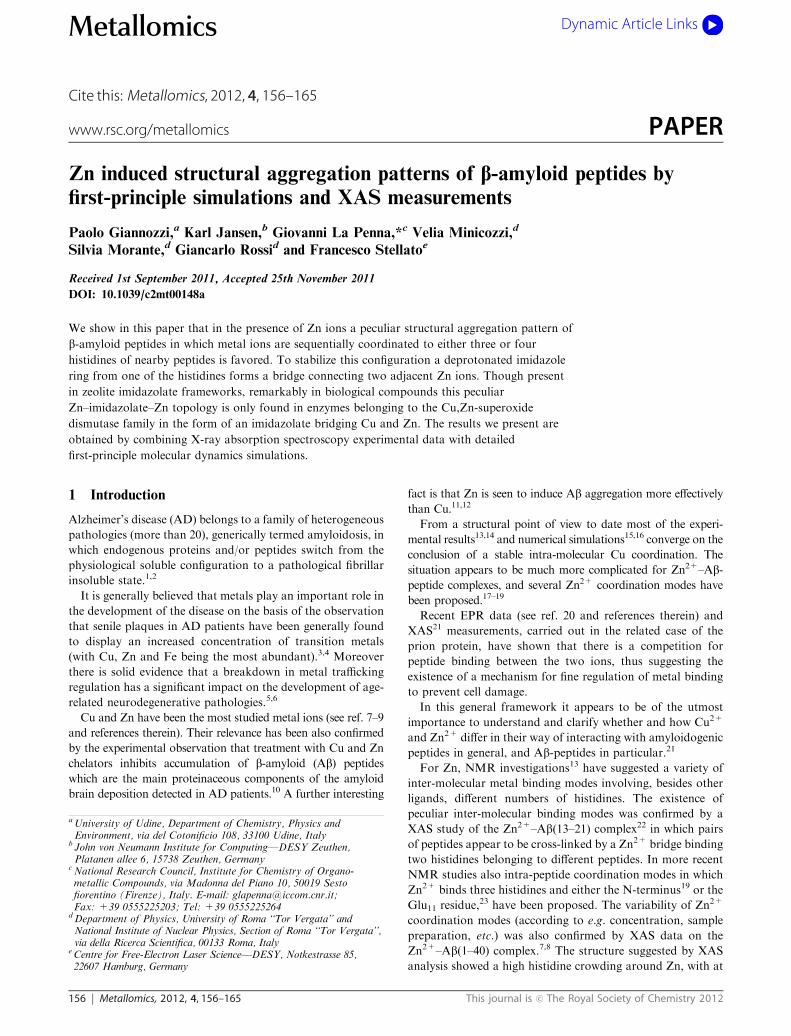
The two identical Ab(1–16) chains, with charged N- andC-termini, were designed (see Fig. 1, Step 1) with F and Cbackbone dihedral angles set at 1801 and the other geometricalparameters taken from standard amino acid geometries. Asshown in the figure, the two Ab chains, from now on referredto as chain A and B, respectively, are assembled in anantiparallel way with the two Nd atoms of His(A)
14 and His(B)14
bonded to a single Zn atom. The energy of this initialconfiguration, computed with the empirical PARM94 Amberforce-field36 (as for the protein) and the parameters reported inref. 17 and 37 (as for the Zn coordination), was minimizedusing the program NAMD38 (see Fig. 1, Step 2).
To relax the above structure a Monte Carlo randomwalk39 (MCRW) was used, where all the dihedral angles inthe two peptide chains were randomly changed (Step 3), withthe exception of those belonging to the two His14 sidechainsthat were held fixed to keep the Nd(His(A)
14 )–Zn–Nd(His(B)14 )motif blocked in space. The virtue of this random walksampling is that no configurations of the Zn-bridgingdimer with ‘‘bad’’ contacts between pairs of atoms are everaccepted.w
Along the trajectory, one particular configuration (model S1,see Step 4a) was picked up in which Zn is closer than 3 A fromthe Ne of His13 in both monomers. The reason for this choice isthat among the others the resulting four-fold Ne(His(A)
13 )–Nd(His(A)14 )–Ne(His(B)13 )–Nd(His(B)14 ) Zn-coordinationz is, accordingto NMR data23 and Car–Parrinello (CP-MD) simulations,17 themost stable one.
A second model (S2 in the following) was constructed whereZn is coordinated to three histidine imidazole rings (instead offour, as in S1) in a 3N1O configuration (with the oxygencoming from the main chain of one of the bound histidines).This model was built to test whether a three-imidazole Zngeometry can be a valid alternative to the four-imidazole Zncoordination mode advocated in the literature to explain andfit the spectral features of the XAS Zn–Ab data.8 The con-struction of model S2 went on in the following way. Amongthe set of configurations produced by MCRW, one in whichZn is coordinated to Nd of His(A)
13 , His(A)14 , His(B)14 and O of His
(A)14 , was selected (Step 4b).y The reason for binding the oxygenof His14 is that the resulting geometry mimics a sort of ‘‘half-histidine’’ structure in the sense that from the XAS point ofview only half of the pathways available to the photo-electroncompared to the situation in which the metal is bound to animidazole ring are now allowed.The Zn-bridged Ab(1–16) dimers (S1 and S2) built as we
explained, were truncated down to models a"ordable by semi-empirical and first–principle methods (Step 5). In particularthe segments 1–10 of both peptide chains were removed andthe Glu11 N-terminal was capped with the usual acetyl group.Then the standard minimization of energy in the empirical
force-field was performed (Step 6).Each truncated dimer was merged into an orthorhombic cell
whose dimensions were determined in order for the periodicreplicas to be separated by 0.5 nm. The cell is filled with 376TIP3P40 water molecules. The water is thermalized andbrought to 300 K increasing the temperature in steps of100 K from 0 K to 300 K by four empirical MD trajectoriesof 10 ps each.41 During the thermalization procedure peptideand Zn atoms were kept fixed in space. In the last 300 K stepthe pressure was held constant at 1 bar (compressibility ofbulk water at room conditions was used).The final solvated configurations obtained in Step 6 were
then subjected to the tight-binding semi-empirical (TB) calcu-lations42 described below to further relax the whole system. TBcalculations were performed by employing the density func-tional tight-binding method implemented in the DFTB+code.42 The parameters for organic compounds containingZn were used.43 The G-point condition (K= 0) was adopted inall the electronic structure calculations. After energy minimi-zation, to bring the system at room temperature a first constantenergy Born–Oppenheimer MD (BO-MD) simulation step of200 fs was performed, which led the system to an averagetemperature of 46 ! 4 K. The system was progressively heatedup to the desired temperature of 300 K by using the same type ofthermostat of the empirical MD simulations41 (Step 7). At eachtemperature BO-MD simulations were carried out for 200 fs witha time-step of 1 fs.Since, as also suggested by the analysis of XAS data, Zn is
preferably penta-coordinated, the oxygen of a water moleculeis brought at a binding distance from the metal as a fifth Znligand. The resulting configurations of the S1 and S2 modelswere finally subjected to a full quantum-mechanical step usingCP-MD44 (Step 8) in order to check for the stability of the Zn
Fig. 1 Flow-chart of the construction of models S1 and S2. See text
for the meaning of symbols and details.
w In the system we are considering, which has 124 dihedral angles, thenumber of attempted torsional moves was 106, with an acceptance rateof about 1/2.z Recall that the Nd (His(A)
14 )-Zn-Nd (His(B)14 ) motif was held fixed in theMCRW step.
y The Nd coordination is comparatively more frequent in MCRWbecause, when a histidine detaches, it rebinds the Zn preferably viaNd.
This journal is c The Royal Society of Chemistry 2012 Metallomics, 2012, 4, 156–165 159
coordination. It is indeed a well recognized fact that classicallyunnoticed instabilities are easily spotted and amplified to avisible magnitude by even very short (of the order of a fewpicoseconds) quantum-mechanical simulations of the CP-MDtype.45 From now on, with the word stability we mean theresistance of Zn coordination to thermal fluctuations at roomconditions (liquid water at T = 300 K).
To carry out the necessary quantum-mechanical CP-MDsimulations, the parallel version of the Quantum ESPRESSOpackage,46 which incorporates Vanderbilt ultra-soft pseudo-potentials47,48 and the Perdew–Burke–Ernzerhof exchangecorrelation functional,49 was employed. Electronic wave func-tions were expanded in plane waves up to an energy cuto" of25 Ry, while a 250 Ry cuto" was used for the expansion of theaugmented charge density in the proximity of the atoms, asrequired in the ultrasoft pseudopotential scheme.49 Simula-tions were carried out according to the general protocoldescribed in ref. 50.
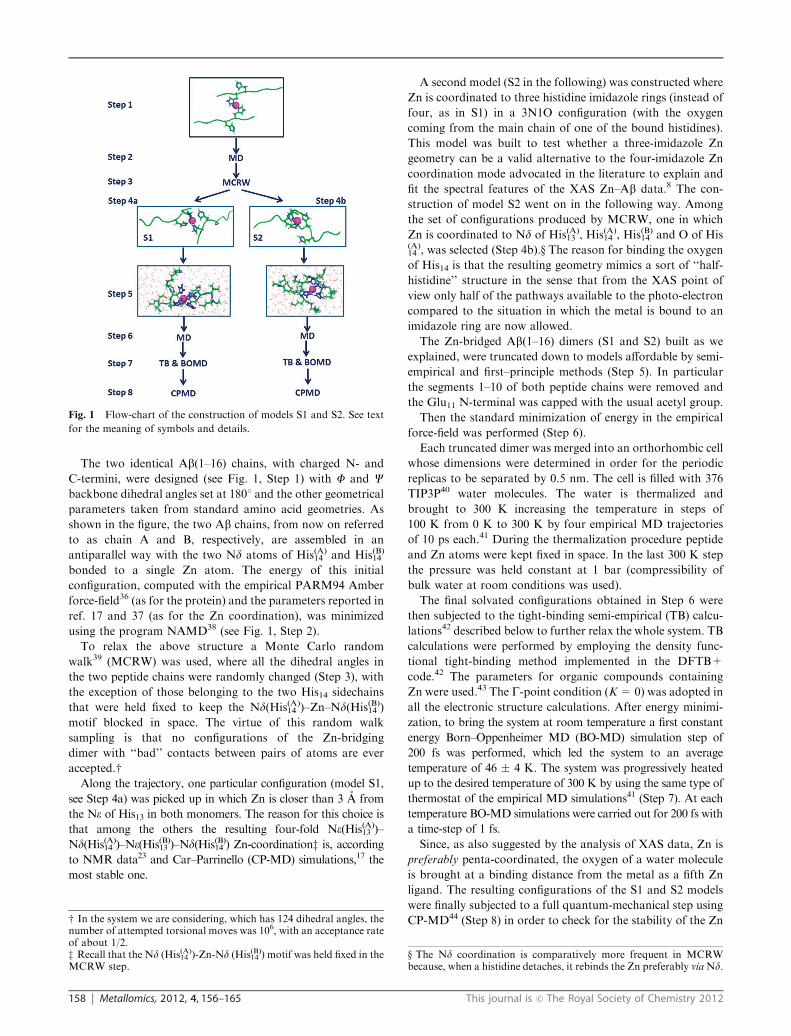
A third model, S3, was designed, slightly modifying modelS1, by substituting the He atom of His(A)
14 with a second Zn ion.In the following we will refer to the two Zn atoms as Zna andZnb. Znb is bound to the Ne of His(A)
14 and two oxygens fromthe Glu11 sidechain. To keep the system in the CP-MDsimulation cell neutral, one water molecule, relatively far fromthe peptide, was replaced by a chlorine atom.
A final fourth model, S4, is obtained by adding a thirdpeptide fragment (chain C, truncated down to a 11–16 frag-ment, as it was done for chains A and B), with Ne (His(C)13 ) andNd (His(C)14 ) atoms in the same positions as the two watermolecules that are bound to the second Zn site (Znb site) inmodel S3 at the end of CP-MD simulation (see Table 1 in theResults section). Since the whole system is now neutral, thechlorine atom is removed from bulk water. The two systemswere successively slowly heated up to 300 K using the same TBand BO-MD steps described above for S1 and S2 (see Fig. 1,Step 7). Due to the impossible large dimension of system S4,only S3 has been subjected to CP-MD simulations (Step 8).
In Fig. 2 for the reader’s convenience we summarize how, inthe four di"erent models we have described, Zn ions are boundto the histidines of the various Ab monomers successivelycoming into play.
3 Results
The salient features of the evolution of the S1, S2, S3 and S4model configurations in the long process of system construc-tion and thermalization are summarized in the flow-chart ofFig. 1 and Table 1, respectively.In the second column of Table 1 (starting configuration) we
list the Zn coordination modes of the four systems before theCP-MD (for models S1, S2 and S3) and BO-MD (for modelS4) simulations. At the beginning of these last simulationsteps, Zn in model S1 and Zna in models S3 and S4 arecoordinated to four nitrogens (from four histidines) and oneoxygen from a water molecule. In model S2, Zn is coordinatedto three nitrogen from three histidines, one oxygen from awater molecule and a second oxygen from the main chain ofHis(A)
14 . In model S3, an extra Zn, denoted by Znb, is addedsolely to check whether its presence is able to stabilize the fourhistidine coordination of Zna. It is coordinated to one nitrogenfrom the histidine bridging the two Zn ions and two oxygensfrom the Glu11 sidechain. In model S4, which is intended torepresent a hypothetic, perhaps more realistic situation, a thirdpeptide chain is added to stabilize the whole compound. In thissystem Znb is coordinated to three nitrogens from threehistidines and one oxygen from Glu11. These four ‘‘starting’’configurations are depicted in the left column of Fig. 3.The structural modifications that occur when we perform
the last CP-MD (for models S1, S2 and S3) and BO-MD (formodel S4) simulation steps are summarized in the third andfourth columns of Table 1.Heating the systems from 0 to 250 K (third column), one
notices that both in the S1 and S2 models Zn loses one ligandand ends up in a tetrahedral 3N1O coordination. In fact, itloses the nitrogen of a histidine in the case of S1 and theoxygen of His14 in the case of S2. For what concerns model S3,it is very remarkable that the unlikely three-fold coordinationof the extra Znb readily becomes a more realistic tetra-coordination owing to the oxygen of a water molecule thatis attracted within its coordination shell (2.5 A). Interestinglyenough the Zn coordination of both metal sites in model S4remains unchanged.
Table 1 For each system in the first column, we report in the secondcolumn the atoms that lie within 2.5 A from Zn (i.e. inside the Zn-coordination shell) before the CP-MD or BO-MD simulations. Thesubscripts (His, Glu, W) denote the residue/molecule to which theatom belongs (W stands for water). In S3 and S4 models the two Znatoms are indicated by Zna and Znb. In the third and fourth columnsthe structural variations observed at the end of the simulations attemperatures up to 250 K and at 300 K, respectively, are reported.‘‘Minus’’ indicates that a ligand leaves the Zn coordination sphere,while ‘‘plus’’ that it enters it
Name Starting configuration
Zn-binding modification
0–250 K 300 K
S1 Zn: 4NHis + 1OW "1NHis NoneS2 Zn: 3NHis + 1OHis + 1OW "1OHis NoneS3 Zna: 4NHis + 1OW Znb: +1OW Znb: "1OGlu + 1OW
Znb: 1NHis + 2OGlu
S4 Zna: 4NHis + 1OW None NoneZnb: 3NHis + 1OGlu
Fig. 2 A schematic view of the Zn site in the four models we have
constructed. Histidines connected by blue bands belong to the same
peptide. The staple comprising His14 and Zn in the S2 model is there to
recall that, besides three imidazoles, the oxygen of the His14 main chain
is also bound to the metal.

160 Metallomics, 2012, 4, 156–165 This journal is c The Royal Society of Chemistry 2012
In the last column, we report the final modifications that takeplace when the systems are brought to room temperature. We seethat the only thing that happens is that in model S3 one of thetwo Znb coordinated Glu11 oxygens is replaced by the oxygen ofa second water molecule attracted in the coordination shell.
In Fig. 3 the configurations before (left panel) and after(right panel) the last CP-MD (for models S1, S2 and S3) andBO-MD (for model S4) simulation steps are compared.
All the final models shown in the right column of Fig. 3(see the last column of Table 1) have been tested against XASdata by fitting their geometrical parameters.z The results arereported in Fig. 4 where the red solid line represents theexperimental spectrum (common to all panels) and the dottedblack lines represent the fitted curves. The results of the fits tothe Znb site in the S3 model, where the metal is coordinated toone histidine, have not been reported. Indeed since this extra
Zn was only introduced to stabilize the S3 tetrahedral Znacoordination, its coordination mode is of not much structuralinterest.The comparison displayed in Fig. 4 shows that di"erent
models with either three or four Zn-coordinated histidines canbe constructed which are able to satisfactorily fit the XAS data ofref. 8. This finding shows that XAS data alone are not enough tofully resolve the metal site coordination structure. Indeed, inref. 8 besides XAS data a number of other spectroscopicinformation were used to elucidate this issue. Here we show thatnumerical simulations can be employed to uniquely identifystable metal–Ab-peptide configurations, which also allow us todetermine the long-range molecular arrangement of the system.In order to explore possible e"ects of the detected long-
range coordination topology on the structure within the 1–10residue region of Ab peptides identified in the various first-principle and semiempirical approaches described above,empirical models including the entire 1–16 regions of the threepeptide chains of models S1, S2 and S4 were also simulated atT = 300 K. The structure of the 11–16 region, as well as thepositions of the Zn atoms, were kept fixed as they appear inthe final configuration displayed on the right hand side ofFig. 3. In Fig. 5 (left) the initial configurations (with the whole1–10 regions in the all-trans configuration attached to thecorresponding 11–16 peptides) of S1, S2 and S4 models are
Fig. 3 Left column: starting (energy minimized) configurations of
models S1, S2, S3 and S4 (from top to bottom). Right column: final
configurations (at 300 K) after CP-MD (for models S1, S2 and S3) and
BO-MD (for model S4) simulation steps. Zn atoms are shown in orange.
Only His, Glu and Lys sidechains are displayed. Blue and red spheres
refer to Ca atoms of N- and C-termini, respectively. H atoms are not
displayed, except when they belong to water molecules. The VMD
program35 was employed for molecular manipulations and drawings.
Fig. 4 Experimental (red full line) and fitted (black dotted line) XAS
data. The analysis of XAS data is made by starting with the fitting
geometry obtained at the end of CPMD (or BO-MD) simulation steps.
The fit to the uninteresting (see text) Znb one histidine site in model S3
is not reported.
z Note that concerning the initial geometry of the four models we usedfor XAS data fitting, we have actually taken the configurationobtained after a further minimization step intended to bring thesimulated system nearer to the actual experimental conditions wheresamples were frozen down to 20 K (liquid nitrogen).

This journal is c The Royal Society of Chemistry 2012 Metallomics, 2012, 4, 156–165 161
displayed, and are compared (right) with the configurationsobtained after a 10 ps classical simulation. All simulationswere carried out in vacuum and with a small cut-o" (0.5 nm)for non-bonding interactions to prevent unphysical much toolarge hydrophobic forces.
Comparison of the final structures one obtains for the threemodels is fairly interesting. It clearly shows that in models S1and S2 the probability of mutual interactions between chainsA and B is low, while the 1–10 residue regions have morechances to collapse onto the Zn-jointed 11–16 regions of thecorresponding chain. In contrast, in model S4 the chance ofinteractions is enhanced by the N-terminal chain crowding(three chains instead of two) and the relative positions ofchains A and C. In order to get more insight on the S4 model,the configuration displayed in Fig. 5 bottom-right was mergedinto an orthorhombic box of 1739 empirical water molecules(see Methods) and simulated for 500 ps at T = 300 K. Thismore detailed empirical model confirms what was found in
vacuum, namely that quite strong interactions, persistent forthe whole duration of the simulation, are able to stabilize themutual wrapping of chains A and C.The configuration obtained after 500 ps at T = 300 K is
displayed in Fig. 6, with the same orientation as in Fig. 5bottom-right. The mutual arrangement of chains A and Cshows a propensity for a sort of double helix, where the two1–10 regions are extended left-handed helices, and slightlyshifted along the sequence. Each residue in chain A is approxi-mately facing a next sequence residue of chain C. Thisarrangement, induced by the mutual orientation of the 11–16regions in chains A and C, is imposed by Zn coordination, andit is sealed by the stable salt-bridge between Arg(C)5 andGlu(A)
3 (Cz (Arg(C)5 )–Cd (Glu(A)3 ) = 3.7 ! 0.3 A), in turn helped
by the sequence shift between the two chains. Interestingly,always as a consequence of this residue sequence shift,Nd (His(A)6 ) comes out to be relatively close to Znb (6 A onaverage, with a minimal value of 3.6 A), thus indicating apossible further mechanism pointing to an increase of Zn inducedhistidine crowding propensity.
4 Discussion
In this section, we will discuss in more detail the interactionsstabilizing the crowding of histidine sidechains around Zn inthe models we have proposed.Let us start by examining how the structure of the S1, S3 and
S4 models have ‘‘evolved’’ along their construction history.
Fig. 5 Starting (left panel) all-trans N-termini configurations and
final (right panel) MD configurations at T = 300 K, after t = 10 ps,
for models S1, S2 and S4, extended to full Ab (1–16) peptides (see text
for details). Zn atoms are displayed in orange, atoms in the 11–16
regions are displayed with sticks, hydrogen atoms are not shown.
N-termini are represented as ribbons.
Fig. 6 Final MD (t = 500 ps, T = 300 K) configuration of the S4
model extended to the full Ab (1–16) peptide, solvated in water (see
text for details). Zn atoms are displayed in orange, atoms are displayed
with sticks, hydrogen atoms are not shown. Chain A is in red, B is in
gray and C is in blue. Bonds in the 11–16 residue regions are thicker
than those in the 1–10 regions.
162 Metallomics, 2012, 4, 156–165 This journal is c The Royal Society of Chemistry 2012
We do not discuss S2 as in this model Zn is coordinated to onlythree histidines. Naturally a special attention will be devoted towhat happened at the end of the simulations (either CP-MD orBO-MD), i.e. when the systems are brought to roomtemperature.
In the case of the S1 model, the initial (step 1, Fig. 1)
antiparallel orientation of the two peptides making the dimer
is largely relaxed already in the first empirical (MD and
MCRW) simulation runs. Further important structuralmodifications occur during the final CP-MD step. In fact,
comparing the S1 structures (first row of Fig. 3) at the
beginning (left) and at the end (right) of the simulation, one
notices that, although the salt-bridge between Lys(A)16 and Glu
(B)11 sidechains is maintained throughout the whole simulation,
the salt-bridge between the opposite termini (Lys(B)16 –Glu(A)11 )
never forms. The reason is that, due to the distortion of thepeptide chain forced by the Zn binding to two adjacent
histidines, only one of the two possible salt-bridges can form.
But the key feature of this model is that the bond between
His(A)14 and Zn is broken during the first stage of the CP-MD
thermalization (see Table 1) leaving a three histidine coordi-
nated Zn. Most probably in the breaking of the His(A)14 bond an
important role is played by Glu(A)11 , since the latter remains
very near to the He of His(A)14 throughout the whole CP-MD
simulation (first row from top of Fig. 3). This information
definitely could have not been inferred from XAS data alone.To try to repair this undesired feature, in model S3 a second
Zn was added. As one can see from Table 1, Zna remains
coordinated to four histidine sidechains. Furthermore, unlikethe case of S1 discussed above, Glu(A)
11 now forms a stable bond
with Znb, thus reducing its interaction with His(A)14 and the
propensity to form a salt-bridge with Lys(B)16 .The resulting Zna and Znb coordination modes are rather
di"erent. As we said, Zna remains coordinated to four histidinesidechains, while Znb is coordinated to one histidine and a
glutamic acid. Such a coordination would not provide a good
fit to XAS spectral data, as the measured XAS signal isdetermined by the ‘‘e"ective (average) number’’ of histidine per
Zn ion (in this case something between 2 and 2.5).In model S4 a third peptide chain, besides the extra Znb, is
introduced. Now both the Lys(A)16 –Glu(B)11 salt-bridge (as in models
S1 and S3) and the Znb–Glu(A)11 bond (as in model S3) are formed.
Furthermore, we notice that Glu(A)11 remains in proximity of
Lys(C)16 .By analyzing the S4 model configuration evolution during
the TB BO-MD simulation at 300 K, one notices that the
Zna–His–Znb bridge favors a stable topology for oligomeric
assemblies, in which the three peptides are significantly rotated
with respect to the initial antiparallel arrangement (see the
bottom-right snapshot in Fig. 3 and the bottom of Fig. 5). The
Zna–His–Znb bridge, while allowing the binding of Glu(A)11
sidechain to the second Znb ion, forbids the formation of the
Lys(B)16 –Glu(A)11 salt-bridge but, at variance with what happens
in the S1 case, an array of Glu#X$11 " Lys
#X0$16 salt-bridges can be
easily formed, without significant distortion of the Zna four-
histidine sidechain coordination.The stability of the S4 system is related on the one hand to the
formation of a Zn–His–Zn bridge assisted by a deprotonated
imidazole ring (imidazolate), and on the other hand to the largeangle occurring between peptides A and C when the peptide B isintercalated between the two. This angle is in fact definitely largerthan that formed by chains A and B when they are interactingwith a single Zn ion (like in models S1 and S2). This intercalatedstructure ‘‘survives’’ the BO-MD simulation at 300 K owing tothe formation of a Glu(A)11 –Znb binding with Znb forcing thenecessary arrangement of the His13–His14 pair of peptide Cwithin the Zn coordination sphere.Naturally a crucial test of the phenomenological reliability
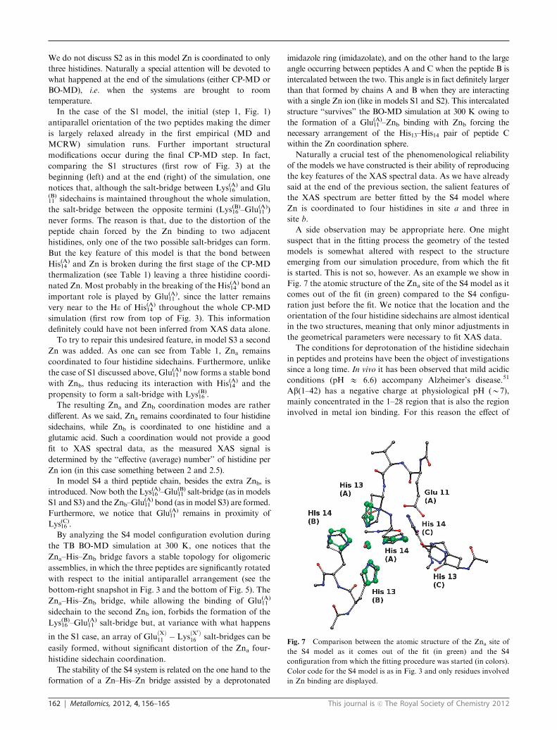
of the models we have constructed is their ability of reproducingthe key features of the XAS spectral data. As we have alreadysaid at the end of the previous section, the salient features ofthe XAS spectrum are better fitted by the S4 model whereZn is coordinated to four histidines in site a and three insite b.A side observation may be appropriate here. One might
suspect that in the fitting process the geometry of the testedmodels is somewhat altered with respect to the structureemerging from our simulation procedure, from which the fitis started. This is not so, however. As an example we show inFig. 7 the atomic structure of the Zna site of the S4 model as itcomes out of the fit (in green) compared to the S4 configu-ration just before the fit. We notice that the location and theorientation of the four histidine sidechains are almost identicalin the two structures, meaning that only minor adjustments inthe geometrical parameters were necessary to fit XAS data.The conditions for deprotonation of the histidine sidechain
in peptides and proteins have been the object of investigationssince a long time. In vivo it has been observed that mild acidicconditions (pH E 6.6) accompany Alzheimer’s disease.51
Ab(1–42) has a negative charge at physiological pH (B7),mainly concentrated in the 1–28 region that is also the regioninvolved in metal ion binding. For this reason the e"ect of
Fig. 7 Comparison between the atomic structure of the Zna site of
the S4 model as it comes out of the fit (in green) and the S4
configuration from which the fitting procedure was started (in colors).
Color code for the S4 model is as in Fig. 3 and only residues involved
in Zn binding are displayed.

This journal is c The Royal Society of Chemistry 2012 Metallomics, 2012, 4, 156–165 163
pH on amyloid deposit formation was monitored in vitro,52
also in the presence of metal ions.11 In this last study it isshown that the same mild acidic conditions observed in vivoincrease the propensity for in vitro aggregation of Ab(1–40/42)in the presence of di"erent metal ions. In the absence of metalions, the Ab(1–40/42) aggregation becomes irreversible at pH= 5.5, a result somehow expected since the "2 charge of the1–28 region can be partially neutralized by histidine protona-tion. Nevertheless, at pH = 6.6 aggregation with no metalions is slower than in the presence of ions. As for the particularcase of Zn, the binding to Ab(1–40) does not occur at pHbelow 6.0, but the level of aggregation induced by Zn is almostinvariant in the 6.2–8.5 pH range. Therefore, the conditionsfor Zn induced oligomerization are expected to be constant inthe same pH range.
An interesting observation suggested by our simulationresults is the similarity of the structure of the most stableamong the models we have investigated, namely S4, with thatof enzymes belonging to the Cu,Zn-SOD family. Indeed theCu,Zn-SOD family contains enzymes with structures displayinghigh crowding of histidine sidechains around both, Cu and Zn,metal sites. In particular Cu is bound to four histidines, like Znain the S4 model, while Zn is coordinated to three histidines andone Asp, in a configuration which corresponds to that of Znb inthe S4 model, with Asp replaced by the almost equivalent Gluresidue. The histidine crowding in SOD enzymes is helped by thepresence of two metal ions bridged by the histidine sidechain inthe unusual form of an imidazolate anion. The spectroscopicsignatures of the active site of human Cu,Zn-SOD, with thedeprotonated form of His63 (human SOD sequence) bridging Cuand Zn, are invariant in the pH range 5.0–10.5, while the samesignatures become strongly pH dependent when His63 is mutatedto Ala.53 The dependence of the Cu(II)/Cu(I) standard reductionpotential in human Cu,Zn-SOD is consistent with the presence ofthe bridging form of His63 in the pH range 5–9.54 These results,together with a large amount of structural data,55 show that aconstrained geometry for the sites hosting metal ions inCu,Zn-SOD strongly assists the stability of the imidazolate anionin a pH range including mild acidic conditions.
One may object to this conclusion that it has been recentlyshown by29 that, after fully replacing Cu with Zn in the humanSOD1 enzyme, the Zn ion located in the Cu site (ZnCu,equivalent to our Zna) loses the bond with the bridgedhistidine. It should be noted, however, that when the bridginghistidine detaches at its place an oxygen from a sulfate bindsZn. Thus in our opinion the system studied in that papercannot be directly compared to our S4 model, because of thestrong influence on the metal environment coming from anionbinding to metal sites. Fortunately, however, there exist in theliterature data on the Cu,Zn-SOD with Cu in the reduced formand in the absence of metal binding anions. This system isquite interesting to compare with ours because the reduced Cuis isoelectronic to Zn. Indeed in ref. 56 and 57 where the Cureduced SOD was crystalized the stability of the Cu–His–Znbridge was shown to be preserved.
The similarity of the Zn coordination topology in model S4with that of Cu,Zn-SOD is particularly remarkable if oneobserves that our construction of the S4 assembly as a modelof Zn induced Ab oligomers is completely independent from
all the reported Cu,Zn-SOD structures. The topology of theinitial S4 model was dictated by the binding of Glu(A)
11 , His(A)14
and the two water molecules to Znb, as it has been obtained inthe final S3 structure (see the S3 right panel in Fig. 3). Therelative position of Glu(A)
11 with respect to His(A)14 , very similar
to that of Asp81 with respect to His61 in Cu,Zn-SOD (bovineSOD sequence), is the building block for the stable binding ofchain C to Znb and for the stabilization of the S4 oligomer.
5 Conclusions
The electrostatic interactions between positively andnegatively charged residues largely contribute in determiningthe mutual orientation of two approaching peptides and arethus important in providing the seeds for oligomer formationand their aggregation into larger assemblies. However, in thepresence of transition metal ions, the coordination chemistryof the latter competes with the less specific electrostatic inter-actions between the peptide chain. In this work, by combiningXAS and first-principle simulation results, we are able to takeinto account, in a consistent way, both types of interactionsthat span significantly di"erent length scales. In fact, although,as already remarked in the Introduction, the short rangesensitivity of the XAS technique allows us to identify at atomicresolution the structure around the absorber (in our case Zn)within a sphere of 5–6 A, this knowledge is not enough to fullyunderstand the role played by the metal in the complicatedpeptide aggregation mechanism. For this purpose, also thereconstruction of the system structure at distances larger than6 A is necessary. This is exactly what we provided with ourfirst-principle, semiempirical and empirical simulations.Indeed, the simulation results we have presented in the
previous sections have lead us to the conclusions which wemay summarize as follows.(1) We have been able to construct stable Zn–Ab-peptide
sites (models S3 and S4 in Fig. 2, respectively) in which twopeptides are bridged by a Zn ion (Zna) in a four histidinecoordination mode.(2) In the case of the S3 model a second Zn (Znb) is
somewhat ad hoc introduced to ensure the stability of theconfiguration under CP-MD simulations. The four histidinecoordinated Zna site is compatible with experimental results.(3) A more realistic structure (model S4) has been con-
structed by adding a third peptide (peptide C in Fig. 2) to thesystem in a topology dictated by Glu(A)
11 , His(A)14 and water
coordination to Zn in model S3. The S4 model, besidesyielding a very good fit to XAS data, provides the geometricand chemical constraints that make both Zn sites structurallystable (see last row in Table 1).Together with the remark that similar Zn–imidazolate–Zn
coordination is observed in Zn imidazolate frameworks (ZIF)and in the Cu,Zn-SOD enzyme even at the mild acidic pHcompatible with the acidosis observed in AD, the interestingconclusion we can draw is that in all these instances, i.e. ZIF,Cu,Zn-SOD and S4 model, the crowding of imidazolyl groupsis helped by the presence of two metal ions bridged by theimidazolyl group in the unusual form of an imidazolate.The relevance of the work presented in this paper comes
from the observation that only on the basis of the quality of

164 Metallomics, 2012, 4, 156–165 This journal is c The Royal Society of Chemistry 2012
the XAS fits displayed in Fig. 4 it would be impossible tochoose among the various models we have considered and thusidentify the structure of the first b-amyloid aggregates that canpossibly form in the presence of metals. A careful study of thestability of small oligomeric assemblies is necessary. The aimof this paper was precisely to present a combined empiricaland first-principle computational strategy by which themechanical stability of di"erent models can be studied. Theapplication of this approach to the case of Ab-peptides inthe presence of Zn has lead us to the identification of a verypeculiar arrangement of Zn ions and peptides whose stabilitylargely depends on the formation of a Zn–His–Zn bridgeassisted by an unusual deprotonated imidazole ring (imidazo-late), very much like the one it is found in Cu,Zn-SOD even atmild acidic conditions.
Acknowledgements
All the computational work has been performed within theDEISA Extreme Computing Initiative (DECI), projectBiCaPS (2008–2009) and the HCH03 project of the Johnvon Neumann Institute for Computing (2009–2010). Partialfinancial support from INFN TO61 project and PRIN project20083Y34Y7 is acknowledged.
References
1 S. B. Prusiner, N. Engl. J. Med., 2001, 344, 1516–1526.2 M. B. Pepys, Philos. Trans. R. Soc. London, Ser. B, 2001, 356,203–210.
3 M. A. Lovell, J. D. Robertson, W. J. Teesdale, J. L. Campbell andW. R. Markesbery, J. Neurol. Sci., 1998, 158, 47–52.
4 M. A. Smith, P. L. Harris, L. M. Sayre and G. Perry, Proc. Natl.Acad. Sci. U. S. A., 1997, 94, 9866–9868.
5 A. I. Bush, Trends Neurosci., 2003, 26, 207–214.6 K. J. Barnham, C. L. Masters and A. I. Bush, Nat. Rev. DrugDiscovery, 2004, 3, 205–214.
7 F. Stellato, G. Menestrina, M. Della Serra, C. Potrich,W. Tomazzolli, R. Meyer-Klaucke and S. Morante, Eur. Biophys.J., 2006, 35, 340–351.
8 V. Minicozzi, F. Stellato, M. Comai, M. Dalla Serra, C. Potrich,W. Meyer-Klaucke and S. Morante, J. Biol. Chem., 2008, 283,10784–10792.
9 S. Morante, Curr. Alzheimer Res., 2008, 5, 508–524.10 C. J. Maynard, A. I. Bush, C. L. Masters, R. Cappai and
Q.-X. Lin, Int. J. Exp. Pathol., 2005, 86, 147–159.11 C. S. Atwood, R. D. Moir, X. Huang, R. C. Scarpa,
N. M. Bacarra, D. M. Romano, M. A. Hartshorn, R. E. Tanziand A. I. Bush, J. Biol. Chem., 1998, 273, 12817–12826.
12 C. S. Atwood, R. C. Scarpa, X. Huang, R. D. Moir, W. D. Jones,D. P. Fairlie, R. E. Tanzi and A. I. Bush, J. Neurochem., 2000, 75,1219–1233.
13 C. D. Syme and J. H. Viles, Biochim. Biophys. Acta, ProteinsProteomics, 2006, 1764, 246–256.
14 P. Faller and C. Hureau, Dalton Trans., 2009, 1080–1094.15 Y. Mantri, M. Fioroni and M. H. Baik, JBIC, J. Biol. Inorg.
Chem., 2009, 13, 1197–1204.16 D. F. Ra"a, R. Gomez-Balderas, P. Brunelle, G. A. Rickard and
A. Rauk, JBIC, J. Biol. Inorg. Chem., 2005, 10, 887–902.17 S. Furlan and G. La Penna, Phys. Chem. Chem. Phys., 2009, 11,
6468–6481.18 T. Miura, K. Suzuki, N. Kohata and H. Takeuchi, Biochemistry,
2000, 39, 7024–7031.19 J. Danielsson, R. Pierattelli, L. Banci and A. Graslund, FEBS J.,
2007, 274, 46–59.20 E. D. Walter, D. J. Stevens, M. P. Visconte and G. L. Millhauser,
J. Am. Chem. Soc., 2007, 129, 15440–15441.
21 F. Stellato, A. Spevacek, O. Proux, G. L. Millhauser andS. Morante, Eur. Biophys. J., 2011, 40, 1259–1270.
22 J. Dong, J. E. Shokes, R. A. Scott and D. G. Lynn, J. Am. Chem.Soc., 2006, 128, 3540–3542.
23 S. Zirah, S. A. Kozin, A. K. Mazur, A. Blond, M. Cheminant,I. Segalas-Milazzo, P. Debey and S. Rebu"at, J. Biol. Chem., 2006,281, 2151–2161.
24 S. Karlin and Z.-Y. Zhu, Proc. Natl. Acad. Sci. U. S. A., 1997, 94,14231–14236.
25 M. B. Peters, Y. Yang, B. Wang, L. Fusti-Molnar, M. N. Weaverand K. M. J. Merz, J. Chem. Theory Comput., 2010, 6,2935–2947.
26 A. Teplyakov, G. Obmolova, P. P. Khil, A. J. Howard,R. D. Camerini-Otero and G. L. Gilliland, Proteins: Struct.,Funct., Genet., 2003, 51, 315–318.
27 D. King, L. Zhang, L. Guarente and R. Marmorstein, Nat. Struct.Biol., 1999, 6, 64–71.
28 X. Siebert, B. A. Eipper, R. E. Mains, S. Prigge, N. J. Blackburnand L. M. Amzel, Biophys. J., 2005, 89, 3312–3319.
29 R. W. Strange, S. V. Antonyuk, M. A. Hough, P. A. Doucette,J. S. Valentine and S. S. Hasnain, J. Mol. Biol., 2006, 356,1152–1162.
30 F. H. Allen, Acta Crystallogr., Sect. B: Struct. Sci., 2002, 58,380–388.
31 C. Bear, K. A. Duggan and H. Freeman, Acta Crystallogr., Sect.B: Struct. Crystallogr. Cryst. Chem., 1975, 31, 2713–2715.
32 H. Xiaochun, L. Dan, T. Yexiang and C. Xiaoming, Acta Scient.Nat. Univ. Sunyatseni, 1998, 37, 55–56.
33 R. Banerjee, A. Phan, B. Wang, C. Knobler, H. Furukawa,M. O’Kee"e and O. M. Yaghi, Science, 2008, 319, 939–943.
34 N. Binsted, EXCURV98, CCLRCDaresbury Laboratory, Warrington,Cheshire, UK, 1998.
35 W. Humphrey, A. Dalke and K. Schulten, J. Mol. Graphics, 1996,14, 33–38, http://www.ks.uiuc.edu/Research/vmd.
36 W. D. Cornell, P. Cieplak, C. I. Bayly, I. R. Gould, K. M. J. Merz,D. M. Ferguson, D. C. Spellmeyer, T. Fox, J. W. Caldwell andP. A. Kollman, J. Am. Chem. Soc., 1995, 117, 5179–5197.
37 Y.-P. Pang, K. Xu, J. El Yazal and F. G. Prendergast, Protein Sci.,2000, 9, 1857–1865.
38 J. C. Phillips, R. Braun, W. Wang, J. Gumbart, E. Tajkhorshid,E. Villa, C. Chipot, R. D. Skeel, L. Kale and K. Schulten,J. Comput. Chem., 2005, 26, 1781–1802, http://www.ks.uiuc.edu/Research/namd.
39 G. La Penna, S. Morante, A. Perico and G. C. Rossi, J. Chem.Phys., 2004, 121, 10725–10741.
40 W. L. Jorgensen, J. Chandrasekhar, J. D. Madura, R. W. Impeyand M. J. Klein, J. Chem. Phys., 1983, 79, 926–935.
41 H. J. C. Berendsen, J. P. M. Postma, W. F. van Gunsteren, A. DiNola and J. R. Haak, J. Chem. Phys., 1984, 81, 3684–3690.
42 B. Aradi, B. Hourahine and T. Frauenheim, J. Phys. Chem. A,2007, 111, 5678–5684, http://www.dftb-plus.info.
43 N. H. Moreira, G. Dolgonos, B. Aradi, A. L. da Rosa andT. Frauenheim, J. Chem. Theory Comput., 2009, 5, 605–614.
44 R. Car and M. Parrinello, Phys. Rev. Lett., 1985, 55, 2471–2474.45 V. Minicozzi and S. Morante, Int. J. Quantum Chem., 2010, 110,
656–680.46 P. Giannozzi, S. Baroni, N. Bonini, M. Calandra, R. Car,
C. Cavazzoni, D. Ceresoli, G. L. Chiarotti, M. Cococcioni,I. Dabo, A. Dal Corso, S. de Gironcoli, S. Fabris, G. Fratesi,R. Gebauer, U. Gerstmann, C. Gougoussis, A. Kokalj, M. Lazzeri,L. Martin-Samos, N. Marzari, F. Mauri, R. Mazzarello, S. Paolini,A. Pasquarello, L. Paulatto, C. Sbraccia, S. Scandolo,G. Sclauzero, A. P. Seitsonen, A. Smogunov, U. Paolo andR. M. Wentzcovitch, J. Phys.: Condens. Matter, 2009,21, 395502, http://www.quantum-espresso.org.
47 D. Vanderbilt, Phys. Rev. B: Condens. Matter, 1990, 41,7892–7895.
48 P. Giannozzi, F. De Angelis and R. Car, J. Chem. Phys., 2004, 120,5903–5915.
49 J. P. Perdew, K. Burke and M. Ernzerhof, Phys. Rev. Lett., 1996,77, 3865–3868.
50 F. Guerrieri, V. Minicozzi, S. Morante, G. Rossi, S. Furlan andG. La Penna, JBIC, J. Biol. Inorg. Chem., 2009, 14, 361–374.
51 C. M. Yates, J. Butterworth, M. C. Tennant and A. Gordon,J. Neurochem., 1990, 55, 1624–1630.
This journal is c The Royal Society of Chemistry 2012 Metallomics, 2012, 4, 156–165 165
52 C. Soto, M. C. Branes, J. Alvarez and N. C. Inestrosa,J. Neurochem., 1994, 63, 1191–1198.
53 J. A. Graden, L. M. Ellerby, J. A. Roe and J. S. Valentine, J. Am.Chem. Soc., 1994, 116, 9743–9744.
54 H. A. Azab, L. Banci, M. Borsari, C. Luchinat, M. Sola andM. S. Viezzoli, Inorg. Chem., 1992, 31, 4649–4655.
55 L. M. Ellerby, D. E. Cabelli, J. A. Graden and J. S. Valentine,J. Am. Chem. Soc., 1996, 118, 6556–6561.
56 W. R. Rypniewski, S. Mangani, B. Bruni, P. L. Orioli, M. Casatiand K. S. Wilson, J. Mol. Biol., 1995, 251, 282–296.
57 M. Ferraroni, W. R. Rypniewski, B. Bruni, P. Orioli andS. Mangani, JBIC, J. Biol. Inorg. Chem., 1998, 3, 411–422.