chronic pulmonary aspergillosis complicating sarcoidosis · chronic pulmonary aspergillosis...
TRANSCRIPT
Chronic pulmonary aspergillosiscomplicating sarcoidosis
Yurdagül Uzunhan1,2, Hilario Nunes1,2, Florence Jeny1,2, Maxime Lacroix1,3,Sophie Brun4, Pierre-Yves Brillet1,3, Emmanuel Martinod5,Marie-France Carette6, Diane Bouvry1,2, Caroline Charlier7,8,Fanny Lanternier7,8, Carole Planès1,2, Abdellatif Tazi9, Olivier Lortholary7,8,Robert P. Baughman10 and Dominique Valeyre1,2
Affiliations: 1Paris 13 University, EA2363, Sorbonne Paris Cité, Bobigny, France. 2Assistance Publique –Hôpitaux de Paris, Avicenne Hospital, Pulmonary Dept, Bobigny, France. 3Assistance Publique – Hôpitaux deParis, Avicenne Hospital, Radiology Dept, Bobigny, France. 4Assistance Publique – Hôpitaux de Paris, AvicenneHospital, Mycology and Parasitology Dept, Bobigny, France. 5Assistance Publique – Hôpitaux de Paris,Avicenne Hospital, Thoracic Surgery Dept, Bobigny, France. 6Assistance Publique – Hôpitaux de Paris, TenonHospital, Radiology Dept, Paris, France. 7Paris Descartes University, Sorbonne Paris Cité, Assistance Publique –Hôpitaux de Paris, Necker-Enfants Malades Hospital, Necker Pasteur Center for Infectious Diseases and TropicalMedicine, Paris, France. 8Institut Pasteur, National Reference Center for Invasive Mycoses and Antifungals,CNRS URA3012, Paris, France. 9Paris Diderot University, Sorbonne Paris Cité, Assistance Publique – Hôpitauxde Paris, Saint-Louis Hospital, Pulmonary Dept, Paris, France. 10Interstitial Lung Disease and SarcoidosisClinic, Dept of Internal Medicine, University of Cincinnati, Cincinnati, OH, USA.
Correspondence: Yurdagül Uzunhan, Hôpital Avicenne, 125 rue de Stalingrad, Bobigny, France.E-mail: [email protected]
@ERSpublicationsCPA occurs in advanced pulmonary sarcoidosis, and survival is predicted by CPI, PH and fibrosisextent http://ow.ly/R3w830b2HvR
Cite this article as: Uzunhan Y, Nunes H, Jeny F, et al. Chronic pulmonary aspergillosis complicatingsarcoidosis. Eur Respir J 2017; 49: 1602396 [https://doi.org/10.1183/13993003.02396-2016].
ABSTRACT Chronic pulmonary aspergillosis (CPA) complicating sarcoidosis (SA) is associated withhigh mortality, and there is a lack of clarity regarding the relative contributions of SA or CPA.
This was a retrospective single-centre study on CPA-SA.In total, 65 patients (44 men), aged 41.4±13.5 and 48.3±11.9 years at the time of SA and CPA
diagnoses, respectively, were included between 1980 and 2015. Of these, 64 had fibrocystic SA, most oftenadvanced, with composite physiological index (CPI) >40 (65% of patients) and pulmonary hypertension(PH) (31%), and 41 patients (63%) were treated for SA (corticosteroids or immunosuppressive drugs).Chronic cavitary pulmonary aspergillosis (CCPA) was the most frequent CPA pattern. Regardingtreatment, 55 patients required long-term antifungals, 14 interventional radiology, 11 resection surgery andtwo transplantation. Nearly half of the patients (27; 41.5%) died (mean age 55.8 years); 73% of the patientsachieved 5-year survival and 61% 10-year survival. Death most often resulted from advanced SA and rarelyfrom haemoptysis. CPI, fibrosis extent and PH predicted survival. Comparison with paired healthycontrols without CPA did not show any difference in survival, but a higher percentage of patients hadhigh-risk mould exposure.
CPA occurs in advanced pulmonary SA. CPA-SA is associated with high mortality due to theunderlying advanced SA rather than to the CPA. CPI, fibrosis extent and PH best predict outcome.
Copyright ©ERS 2017
This article has supplementary material available from erj.ersjournals.com
Received: July 11 2016 | Accepted after revision: Feb 18 2017
Conflict of interest: Disclosures can be found alongside this article at erj.ersjournals.com
https://doi.org/10.1183/13993003.02396-2016 Eur Respir J 2017; 49: 1602396
ORIGINAL ARTICLESARCOIDOSIS AND ASPERGILLOSIS
IntroductionChronic pulmonary aspergillosis (CPA) may complicate fibrocystic pulmonary sarcoidosis (SA) [1, 2]. Theburden of CPA-SA has been estimated at 3–12% of cases [2], and SA might be the underlyingpredisposing condition for CPA in 7.1% of cases [3]. In the literature, CPA-SA is reported to have a highmortality rate [4–13], with no CPA categorisation and no clear information on the respectivecontributions of CPA and SA to death [2]. Studies have implicated corticosteroid and immunosuppressivetreatments in CPA-SA, as well as underlying pulmonary fibrocystic lesions and smoking habit [11].
We report a series of 65 patients with CPA-SA seen in a tertiary centre for SA, who were monitored withthe support of a French infectious disease reference centre for invasive mycoses. Comparisons were madewith a healthy control (HC) group with a similar phenotype except for CPA. Our goals were to: 1)investigate CPA categorisation and SA phenotyping at diagnosis, 2) look for CPA-SA risk factors, 3)describe the effect of antifungals on CPA outcome, 4) assess survival, 5) specify the respective contributionof CPA and SA on mortality, and 6) look for predictive outcome factors.
MethodsThis retrospective study was conducted in Avicenne Hospital Pulmonology Dept with institutional reviewboard approval (CLEA-R13).
Patient selectionThe database of patients admitted to the pulmonary department was examined. SA and CPA diagnoseswere matched for the 1980–2015 period. The included patients met the following criteria. SA diagnosismade according to the 1999 statement on SA [14]. Evidence of noncaseating granulomas was required.CPA diagnosis based on the presence of: 1) at least one pulmonary cavity on thoracic imaging with orwithout one or more fungal balls in a cavity, and 2) positive blood anti-Aspergillus immunoglobulin Gantibodies or 3) cultures/histology implicating Aspergillus spp. [1] or showing hyphae compatible withAspergillus.
The HC group was made up of patients with fibrocystic SA without current or subsequent CPA treated inthe same department, paired 1:1 with the patient group according to the date of the fibrocystic lungdiagnosis (difference <5 years).
Clinical, pulmonary function, haemodynamics and biological characteristics at CPA diagnosisEntry into the study was the date of CPA diagnosis. The following information was collected: age, sex,geographical origin, smoking habit, high-risk occupational exposure to air moulds and dusts, and presenceof comorbid or past conditions. Extrapulmonary manifestations were recorded [15].
Pulmonary function tests included spirometry (n=64), diffusing capacity of the lung for carbon monoxide(DLCO) (n=64) and room-air blood gases (n=42). Forced vital capacity (FVC), forced expiratory volume in1 s (FEV1) and DLCO were expressed as a percentage of predicted values [16, 17].
Doppler echocardiography was performed on all patients within 6 months of CPA diagnosis. Pulmonaryhypertension (PH) was defined by estimated systolic pulmonary artery pressure >40 mmHg.
Available serum C-reactive protein (n=35) and serum angiotensin-converting enzyme (sACE) (n=48) levelswere recorded.
Computed tomography analysisFor further information on the computed tomography analysis, see the supplementary material. Computedtomography scans were reviewed in concert by two authors (PYB, a radiologist with 15 years of experiencein interstitial lung disease, and YU) at CPA diagnosis.
Fibrotic lesions were classified into four patterns according to ABESEHRA et al. [18]. Total disease extent wasrounded to the nearest 5%, and the relative proportions of the individual patterns contributing to the totaldisease extent were estimated to the nearest 5%. Main pulmonary artery diameter (MPAD) and ascendingaorta diameter (AAD) had been assessed in high-resolution computed tomography (HRCT) scans at CPAdiagnosis.
CPA categorisation was applied according to DENNING et al. [1].
Composite physiological index and Walsh’s algorithmThe composite physiological index (CPI) was calculated [19] and the prognosis algorithm of WALSH et al.[20] applied.
https://doi.org/10.1183/13993003.02396-2016 2
SARCOIDOSIS AND ASPERGILLOSIS | Y. UZUNHAN ET AL.
Fungal investigationsFor further information on the fungal investigations, see the supplementary material. Tests for serumAspergillus-specific IgG were carried out for 64 patients. Fungal culture was performed on sputum (n=47)or bronchoscopy aspirate (n=44).
Risk factors for CPAWe looked for risk factors for CPA by comparing the CPA-SA and HC groups. Attention was paid togeographical origin, smoking habit, previous pneumothorax, SA treatments and the presence of diabetesmellitus at time of diagnosis of fibrocystic SA.
Patients’ mould exposure was also assessed (patients’ employment was mentioned in the files). Weconsidered exposures and job titles that have been associated with aspergillosis in prior studies [21];construction work, farming, gardening, florist shop work and forestry work were considered high-risk jobsfor fungal spores [22].
Treatments and outcomePatients were monitored through regular visits and tests at time intervals depending on clinicalrequirements. The physician in charge prescribed treatment. Every event was recorded, and we looked forfactors predictive of survival. Survival in the CPA-SA group was compared with the HC group.
For the CPA follow-up evaluation, we compared CT before and after 6–12 months of antifungals using thecriteria of GODET et al. [23]. The following data were collected: number and volume of cavities, numberand maximal diameter of fungus balls, maximal thickness of cavity wall and maximal pleural thickening.Maximal diameter of the largest cavities, alveolar consolidation, lobar collapse, ground-glass attenuationand nodules were also assessed. According to GODET et al. [23], maximal thickness of cavity wall andmaximal pleural thickening are the most relevant CT variants for assessing response to treatment.
Statistical analysisQuantitative values are reported as mean and SDs, and qualitative data as numbers (percentages).Information regarding vital status and causes of death were obtained by reviewing medical records and bycontacting referral physicians or general practitioners. Survival was calculated from inclusion, i.e. date ofCPA diagnosis until the end of the follow-up period.
We used STATA software (version 13; StataCorp, College Station, Texas, USA) for statistical analyses. Weperformed group comparisons using the t-test, Wilcoxon rank sum, Chi-squared statistics and Fisher’sexact test where relevant. Univariate analysis based on the proportional hazards model was used with thelog-rank test. All significant parameters were entered into the multivariate Cox proportional hazardsmodel. Survival curves were based on the Kaplan–Meier method and compared using the log-rank test.For all statistical analyses, p=0.05 was considered significant.
ResultsSelection of patientsFrom our database of 3137 patients with SA, 80 patients with pulmonary CPA-SA recorded betweenJanuary 1980 and December 2015 were identified; of these, 15 patients were excluded (insufficient medicaldata (n=5), no confirmed SA (n=4), incomplete CPA criteria (n=6)). The remaining 65 patients (2% of theSA population) made up the final study population.
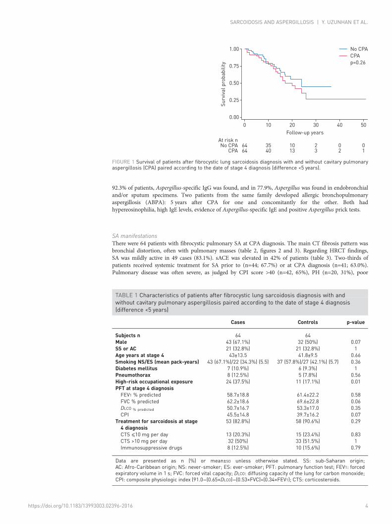
The HC group (64 patients) was similar to the CPA-SA group in age, FVC, FEV1, DLCO and CPI (figure 1and table 1).
Presentation at CPA diagnosisPopulation characteristicsThe population characteristics are summarised in table 2. The diagnosis of SA preceded the diagnosis ofCPA for 55 patients (85%), while CPA and SA diagnoses were made concomitantly in 10 cases (15%).
CPA presentationOf the 65 patients, 24 (37%) presented haemoptysis at CPA diagnosis, 26 (40%) had weight loss (mean3.5 kg) and 29 (45%) reported fatigue. CPA presentation could be categorised in most patients (figure 2).The most frequent phenotypes were chronic cavitary pulmonary aspergillosis (CCPA) (n=27) and simpleaspergilloma (n=17). There was one case of subacute invasive aspergillosis (SAIA). Some overlaps betweenCCPA and aspergillomas or between aspergillomas and chronic fibrosing pulmonary aspergillosis andsometimes between all three entities were noted, while some phenotypes were not classifiable due toconfusing SA lesions (n=10). The biological data concerning CPA diagnosis are summarised in table 3. In
https://doi.org/10.1183/13993003.02396-2016 3
SARCOIDOSIS AND ASPERGILLOSIS | Y. UZUNHAN ET AL.
92.3% of patients, Aspergillus-specific IgG was found, and in 77.9%, Aspergillus was found in endobronchialand/or sputum specimens. Two patients from the same family developed allergic bronchopulmonaryaspergillosis (ABPA): 5 years after CPA for one and concomitantly for the other. Both hadhypereosinophilia, high IgE levels, evidence of Aspergillus-specific IgE and positive Aspergillus prick tests.
SA manifestationsThere were 64 patients with fibrocystic pulmonary SA at CPA diagnosis. The main CT fibrosis pattern wasbronchial distortion, often with pulmonary masses (table 2, figures 2 and 3). Regarding HRCT findings,SA was mildly active in 49 cases (83.1%). sACE was elevated in 42% of patients (table 3). Two-thirds ofpatients received systemic treatment for SA prior to (n=44; 67.7%) or at CPA diagnosis (n=41; 63.0%).Pulmonary disease was often severe, as judged by CPI score >40 (n=42, 65%), PH (n=20, 31%), poor
00.00
0.25
0.50
Surv
ival
pro
babi
lity
0.75
1.00
10 20Follow-up years
30 40 50
64 35 10 2 0 064
No CPACPA
At risk n
40 13 3 2 1
p=0.26
No CPACPA
FIGURE 1 Survival of patients after fibrocystic lung sarcoidosis diagnosis with and without cavitary pulmonaryaspergillosis (CPA) paired according to the date of stage 4 diagnosis (difference <5 years).
TABLE 1 Characteristics of patients after fibrocystic lung sarcoidosis diagnosis with andwithout cavitary pulmonary aspergillosis paired according to the date of stage 4 diagnosis(difference <5 years)
Cases Controls p-value
Subjects n 64 64Male 43 (67.1%) 32 (50%) 0.07SS or AC 21 (32.8%) 21 (32.8%) 1Age years at stage 4 43±13.5 41.8±9.5 0.66Smoking NS/ES (mean pack-years) 43 (67.1%)/22 (34.3%) (5.5) 37 (57.8%)/27 (42.1%) (5.7) 0.36Diabetes mellitus 7 (10.9%) 6 (9.3%) 1Pneumothorax 8 (12.5%) 5 (7.8%) 0.56High-risk occupational exposure 24 (37.5%) 11 (17.1%) 0.01PFT at stage 4 diagnosisFEV1 % predicted 58.7±18.8 61.4±22.2 0.58FVC % predicted 62.2±18.6 69.6±22.8 0.06DLCO % predicted 50.7±16.7 53.3±17.0 0.35CPI 45.5±14.8 39.7±16.2 0.07
Treatment for sarcoidosis at stage4 diagnosis
53 (82.8%) 58 (90.6%) 0.29
CTS ⩽10 mg per day 13 (20.3%) 15 (23.4%) 0.83CTS >10 mg per day 32 (50%) 33 (51.5%) 1Immunosuppressive drugs 8 (12.5%) 10 (15.6%) 0.79
Data are presented as n (%) or mean±SD unless otherwise stated. SS: sub-Saharan origin;AC: Afro-Caribbean origin; NS: never-smoker; ES: ever-smoker; PFT: pulmonary function test; FEV1: forcedexpiratory volume in 1 s; FVC: forced vital capacity; DLCO: diffusing capacity of the lung for carbon monoxide;CPI: composite physiologic index (91.0−(0.65×DLCO)−(0.53×FVC)+(0.34×FEV1); CTS: corticosteroids.
https://doi.org/10.1183/13993003.02396-2016 4
SARCOIDOSIS AND ASPERGILLOSIS | Y. UZUNHAN ET AL.
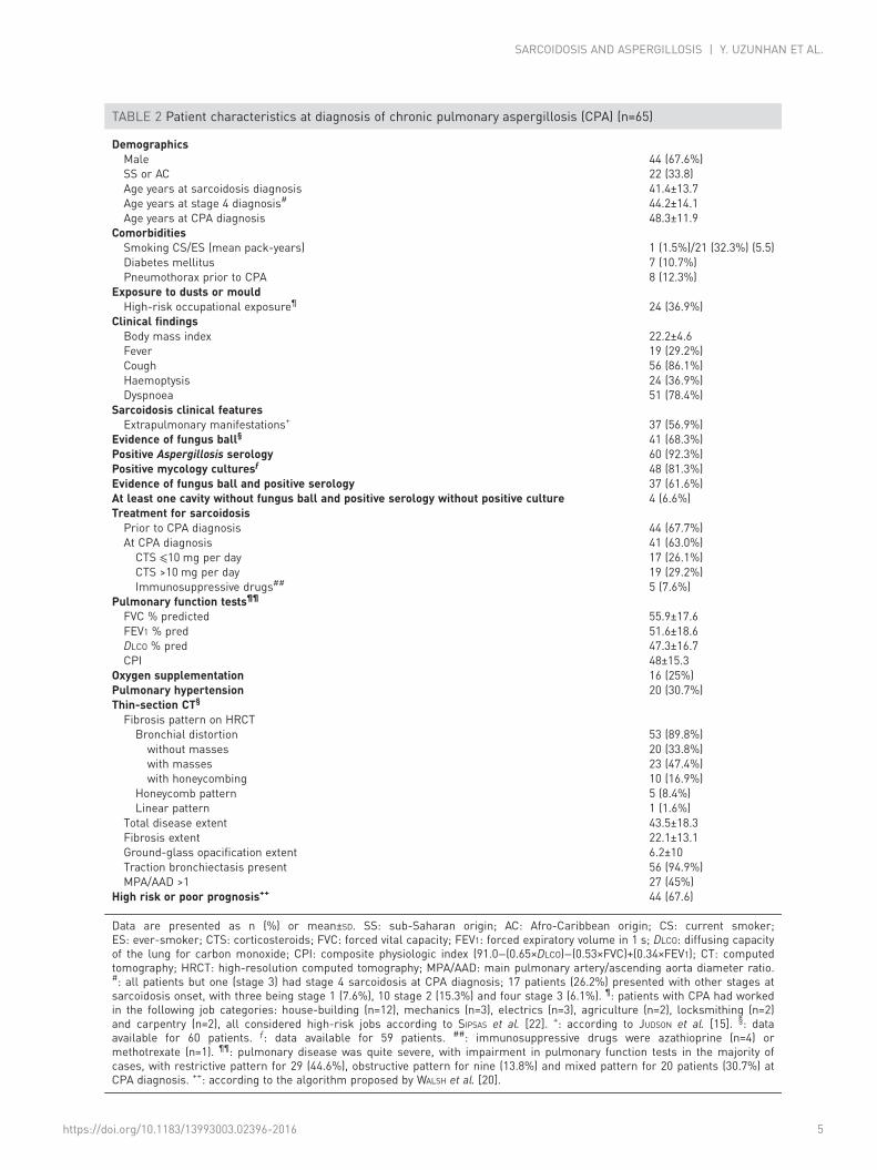
TABLE 2 Patient characteristics at diagnosis of chronic pulmonary aspergillosis (CPA) (n=65)
DemographicsMale 44 (67.6%)SS or AC 22 (33.8)Age years at sarcoidosis diagnosis 41.4±13.7Age years at stage 4 diagnosis# 44.2±14.1Age years at CPA diagnosis 48.3±11.9
ComorbiditiesSmoking CS/ES (mean pack-years) 1 (1.5%)/21 (32.3%) (5.5)Diabetes mellitus 7 (10.7%)Pneumothorax prior to CPA 8 (12.3%)
Exposure to dusts or mouldHigh-risk occupational exposure¶ 24 (36.9%)
Clinical findingsBody mass index 22.2±4.6Fever 19 (29.2%)Cough 56 (86.1%)Haemoptysis 24 (36.9%)Dyspnoea 51 (78.4%)
Sarcoidosis clinical featuresExtrapulmonary manifestations+ 37 (56.9%)
Evidence of fungus ball§ 41 (68.3%)Positive Aspergillosis serology 60 (92.3%)Positive mycology culturesƒ 48 (81.3%)Evidence of fungus ball and positive serology 37 (61.6%)At least one cavity without fungus ball and positive serology without positive culture 4 (6.6%)Treatment for sarcoidosisPrior to CPA diagnosis 44 (67.7%)At CPA diagnosis 41 (63.0%)CTS ⩽10 mg per day 17 (26.1%)CTS >10 mg per day 19 (29.2%)Immunosuppressive drugs## 5 (7.6%)
Pulmonary function tests¶¶
FVC % predicted 55.9±17.6FEV1 % pred 51.6±18.6DLCO % pred 47.3±16.7CPI 48±15.3
Oxygen supplementation 16 (25%)Pulmonary hypertension 20 (30.7%)Thin-section CT§
Fibrosis pattern on HRCTBronchial distortion 53 (89.8%)without masses 20 (33.8%)with masses 23 (47.4%)with honeycombing 10 (16.9%)
Honeycomb pattern 5 (8.4%)Linear pattern 1 (1.6%)
Total disease extent 43.5±18.3Fibrosis extent 22.1±13.1Ground-glass opacification extent 6.2±10Traction bronchiectasis present 56 (94.9%)MPA/AAD >1 27 (45%)
High risk or poor prognosis++ 44 (67.6)
Data are presented as n (%) or mean±SD. SS: sub-Saharan origin; AC: Afro-Caribbean origin; CS: current smoker;ES: ever-smoker; CTS: corticosteroids; FVC: forced vital capacity; FEV1: forced expiratory volume in 1 s; DLCO: diffusing capacityof the lung for carbon monoxide; CPI: composite physiologic index (91.0−(0.65×DLCO)−(0.53×FVC)+(0.34×FEV1); CT: computedtomography; HRCT: high-resolution computed tomography; MPA/AAD: main pulmonary artery/ascending aorta diameter ratio.#: all patients but one (stage 3) had stage 4 sarcoidosis at CPA diagnosis; 17 patients (26.2%) presented with other stages atsarcoidosis onset, with three being stage 1 (7.6%), 10 stage 2 (15.3%) and four stage 3 (6.1%). ¶: patients with CPA had workedin the following job categories: house-building (n=12), mechanics (n=3), electrics (n=3), agriculture (n=2), locksmithing (n=2)and carpentry (n=2), all considered high-risk jobs according to SIPSAS et al. [22]. +: according to JUDSON et al. [15]. §: dataavailable for 60 patients. ƒ: data available for 59 patients. ##: immunosuppressive drugs were azathioprine (n=4) ormethotrexate (n=1). ¶¶: pulmonary disease was quite severe, with impairment in pulmonary function tests in the majority ofcases, with restrictive pattern for 29 (44.6%), obstructive pattern for nine (13.8%) and mixed pattern for 20 patients (30.7%) atCPA diagnosis. ++: according to the algorithm proposed by WALSH et al. [20].
https://doi.org/10.1183/13993003.02396-2016 5
SARCOIDOSIS AND ASPERGILLOSIS | Y. UZUNHAN ET AL.
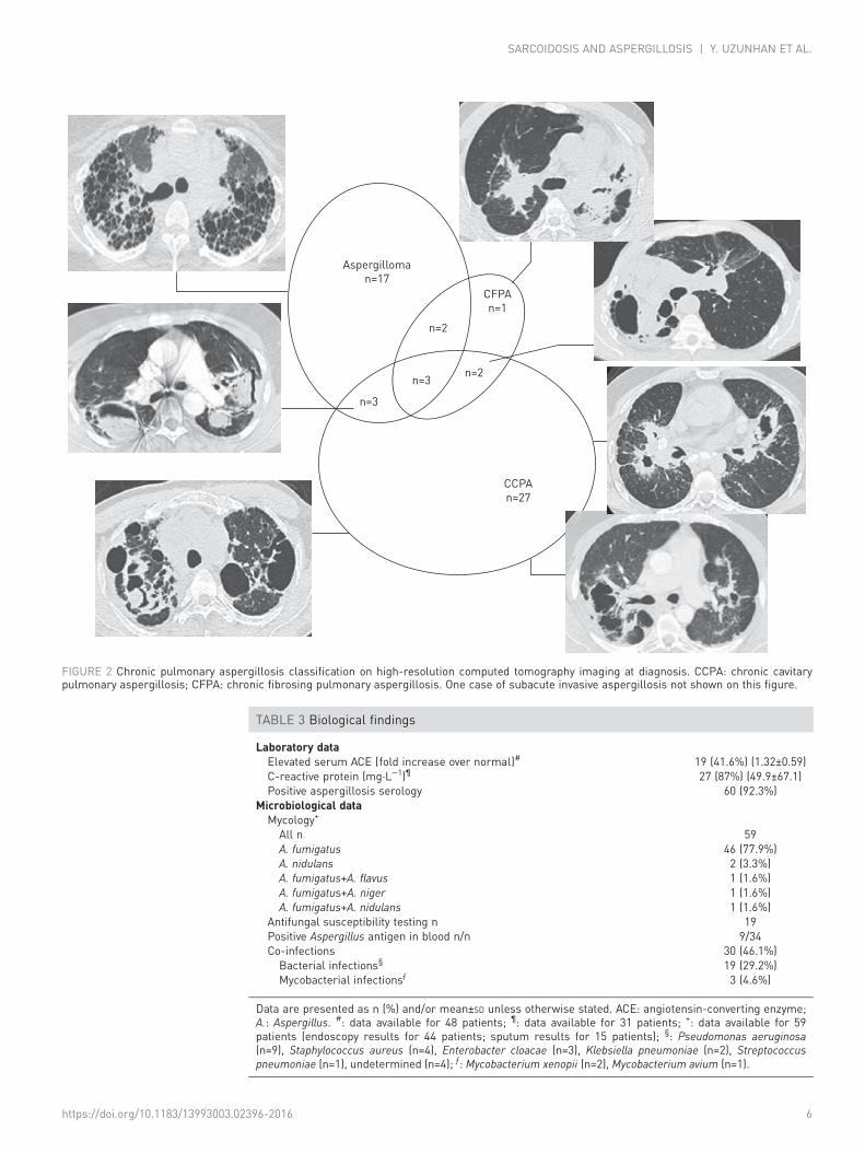
Aspergilloman=17
CCPAn=27
CFPAn=1
n=2
n=2n=3
n=3
FIGURE 2 Chronic pulmonary aspergillosis classification on high-resolution computed tomography imaging at diagnosis. CCPA: chronic cavitarypulmonary aspergillosis; CFPA: chronic fibrosing pulmonary aspergillosis. One case of subacute invasive aspergillosis not shown on this figure.
TABLE 3 Biological findings
Laboratory dataElevated serum ACE (fold increase over normal)# 19 (41.6%) (1.32±0.59)C-reactive protein (mg·L−1)¶ 27 (87%) (49.9±67.1)Positive aspergillosis serology 60 (92.3%)
Microbiological dataMycology+
All n 59A. fumigatus 46 (77.9%)A. nidulans 2 (3.3%)A. fumigatus+A. flavus 1 (1.6%)A. fumigatus+A. niger 1 (1.6%)A. fumigatus+A. nidulans 1 (1.6%)
Antifungal susceptibility testing n 19Positive Aspergillus antigen in blood n/n 9/34Co-infections 30 (46.1%)Bacterial infections§ 19 (29.2%)Mycobacterial infectionsƒ 3 (4.6%)
Data are presented as n (%) and/or mean±SD unless otherwise stated. ACE: angiotensin-converting enzyme;A.: Aspergillus. #: data available for 48 patients; ¶: data available for 31 patients; +: data available for 59patients (endoscopy results for 44 patients; sputum results for 15 patients); §: Pseudomonas aeruginosa(n=9), Staphylococcus aureus (n=4), Enterobacter cloacae (n=3), Klebsiella pneumoniae (n=2), Streptococcuspneumoniae (n=1), undetermined (n=4); ƒ: Mycobacterium xenopii (n=2), Mycobacterium avium (n=1).
https://doi.org/10.1183/13993003.02396-2016 6
SARCOIDOSIS AND ASPERGILLOSIS | Y. UZUNHAN ET AL.
survival prognosis according to the algorithm of WALSH et al. [20] (n=44, 66%), or oxygensupplementation (n=16, 25%) (table 2).
CPA risk factorsThere was no difference between the CPA-SA and HC groups in geographical origin, smoking habit,pneumothorax in the past, diabetes or treatment for SA (figure 1 and table 1). There was a trend towardsa higher proportion of males in the CPA population, and the proportion of high-risk mould-exposure jobswas significantly higher in the CPA-SA than in the HC group (figure 1 and table 1).
CPA treatment and outcomeAt CPA diagnosis, besides receiving treatment targeting SA, patients received oxygen supplementation(n=16, 25%) and drugs to treat pulmonary hypertension (n=6, 9%).
During the course of CPA, patients were treated for CPA or haemoptysis. Major haemoptysis wasconsidered to be due mainly to CPA (figure 4). However, haemoptysis might have been due to
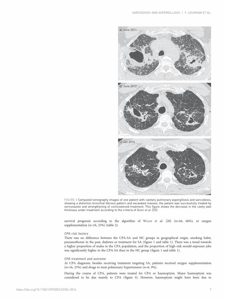
a)
b)
c)
June 2011
June 2012
Jan 2016
FIGURE 3 Computed tomography images of one patient with cavitary pulmonary aspergillosis and sarcoidosis,showing a distortion bronchial fibrosis pattern and excavated masses; the patient was successfully treated byvoriconazole and strengthening of corticosteroid treatment. This figure shows the decrease in the cavity wallthickness under treatment according to the criteria of GODET et al. [23].
https://doi.org/10.1183/13993003.02396-2016 7
SARCOIDOSIS AND ASPERGILLOSIS | Y. UZUNHAN ET AL.
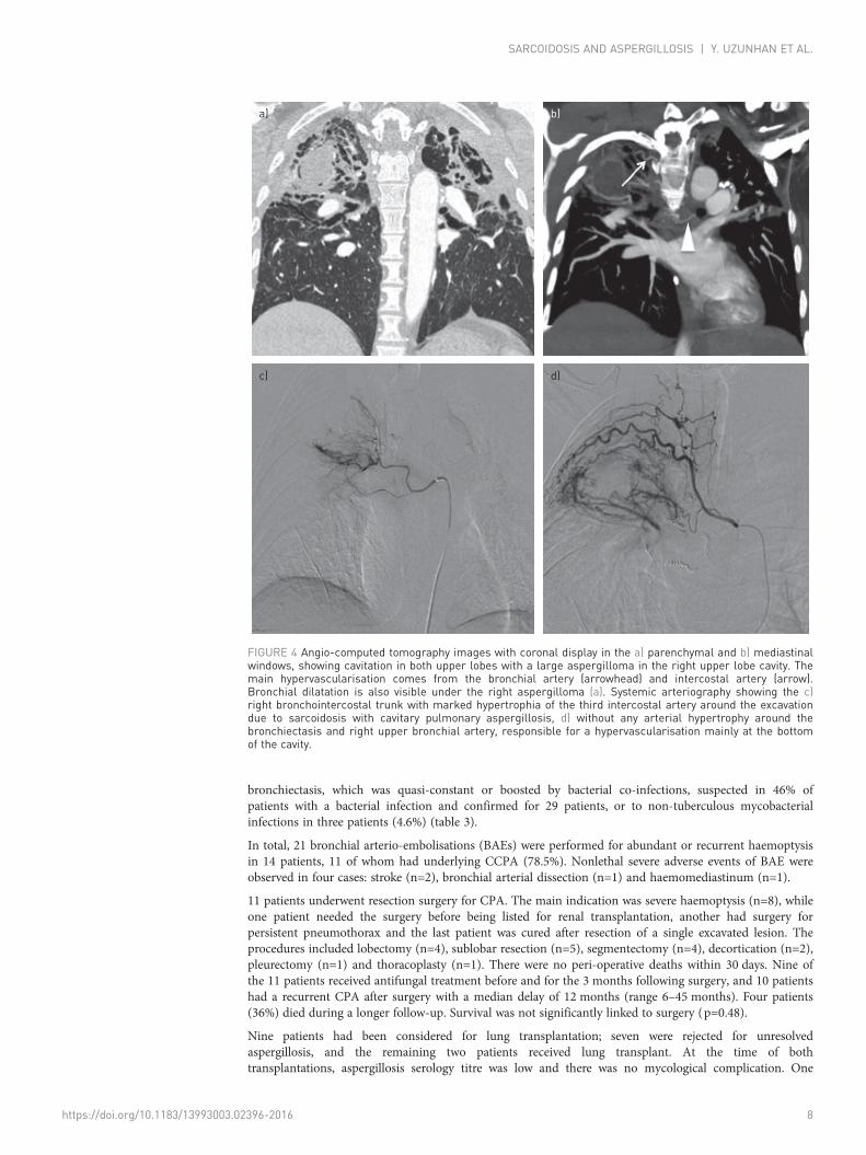
bronchiectasis, which was quasi-constant or boosted by bacterial co-infections, suspected in 46% ofpatients with a bacterial infection and confirmed for 29 patients, or to non-tuberculous mycobacterialinfections in three patients (4.6%) (table 3).
In total, 21 bronchial arterio-embolisations (BAEs) were performed for abundant or recurrent haemoptysisin 14 patients, 11 of whom had underlying CCPA (78.5%). Nonlethal severe adverse events of BAE wereobserved in four cases: stroke (n=2), bronchial arterial dissection (n=1) and haemomediastinum (n=1).
11 patients underwent resection surgery for CPA. The main indication was severe haemoptysis (n=8), whileone patient needed the surgery before being listed for renal transplantation, another had surgery forpersistent pneumothorax and the last patient was cured after resection of a single excavated lesion. Theprocedures included lobectomy (n=4), sublobar resection (n=5), segmentectomy (n=4), decortication (n=2),pleurectomy (n=1) and thoracoplasty (n=1). There were no peri-operative deaths within 30 days. Nine ofthe 11 patients received antifungal treatment before and for the 3 months following surgery, and 10 patientshad a recurrent CPA after surgery with a median delay of 12 months (range 6–45 months). Four patients(36%) died during a longer follow-up. Survival was not significantly linked to surgery (p=0.48).
Nine patients had been considered for lung transplantation; seven were rejected for unresolvedaspergillosis, and the remaining two patients received lung transplant. At the time of bothtransplantations, aspergillosis serology titre was low and there was no mycological complication. One
a) b)
c) d)
FIGURE 4 Angio-computed tomography images with coronal display in the a) parenchymal and b) mediastinalwindows, showing cavitation in both upper lobes with a large aspergilloma in the right upper lobe cavity. Themain hypervascularisation comes from the bronchial artery (arrowhead) and intercostal artery (arrow).Bronchial dilatation is also visible under the right aspergilloma (a). Systemic arteriography showing the c)right bronchointercostal trunk with marked hypertrophia of the third intercostal artery around the excavationdue to sarcoidosis with cavitary pulmonary aspergillosis, d) without any arterial hypertrophy around thebronchiectasis and right upper bronchial artery, responsible for a hypervascularisation mainly at the bottomof the cavity.
https://doi.org/10.1183/13993003.02396-2016 8
SARCOIDOSIS AND ASPERGILLOSIS | Y. UZUNHAN ET AL.
patient died after 24 months because of chronic graft dysfunction, while the other was still alive 8 yearsafter transplantation.
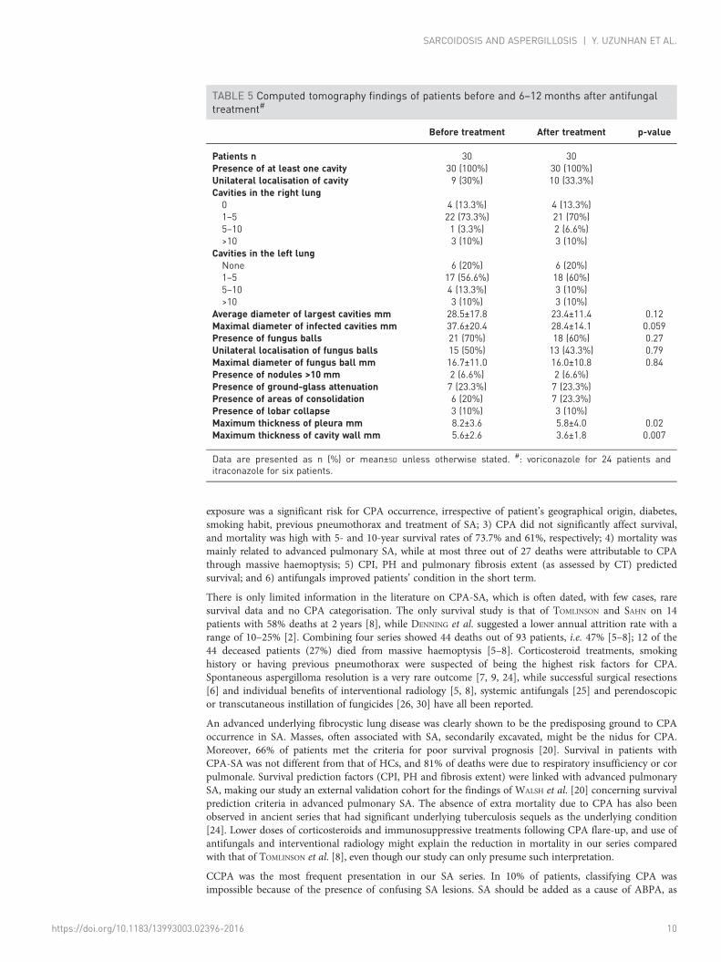
Fifty-five patients received antifungal treatments for more than 6 weeks. Antifungal treatment relied onazoles, mostly on voriconazole (n=38) and itraconazole (n=23) (table 4). Serum concentration wasmonitored for 24 patients during azole therapy. Because of adverse effects (mostly liver toxicity withcytolysis (20%) and voriconazole-associated phototoxicity (7.2%)), these drugs were discontinued forone-third of patients. The treatment was withdrawn after good clinical/radiological response, at least 6–12 months later, but resumed later, possibly because of a new CPA flare-up. Corticosteroids were reducedduring CPA flare-up and increased again during SA exacerbation. The case presented in figure 3 illustratesthe result obtained by voriconazole followed by a stronger SA treatment, allowing both control of CPAand reduction of the underlying SA lesions. One patient with ABPA responded well to voriconazole.Cough and general symptoms often improved with antifungals. The effect of antifungals (voriconazole in24 patients and itraconazole in 6) could be assessed in 30 patients who underwent antifungal therapy forat least 6 months, by comparison of CT scans taken before and after 6–12 months of treatment (table 5).The maximal thickness of the cavitation wall (p=0.007) and pleura (p=0.02) decreased significantly.
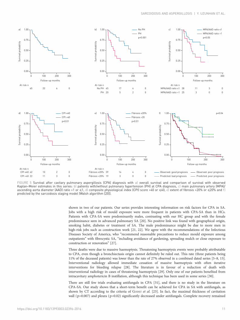
Survival, mechanisms of death and predictive outcome factorsSurvivalPatients were followed up for 7.5±6.2 years; 73% achieved 5-year and 61% achieved 10-year survival (figure 5).At the end of the study, 37 patients were alive (two of whom had undergone lung transplantation), onepatient was lost to follow-up and 27 (41.5%) were dead. Mean age at death was 55.8 years (range 31–79 years). Survival did not differ with date of CPA diagnosis (before or after 2000). Survival from the dateof diagnosis of fibrocystic lesions was not different between patients with CPA-SA and HCs (p=0.26)(figure 1 and table 1).
Causes of deathFor two patients, the cause of death was not ascertained. Respiratory failure was the most common causeof death (n=13; 48.1%). Nine patients died from right heart failure (33.3%) and three died from severehaemoptysis (11.1%). Death in these three cases was probably due to CPA, as the review of bronchialarteriography in nine out of 14 patients who underwent BAE found bronchial hypertrophy in the area ofcavities related to CPA in all cases (figure 4). For 22 patients (34% of the total; 81% of patients who died),death was attributable to the underlying advanced SA alone.
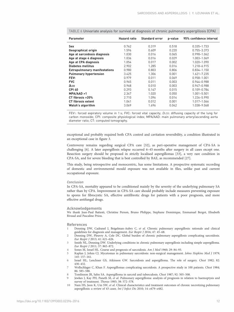
Survival prediction factorsAs shown in table 6, the following parameters at CPA diagnosis were significantly associated with a higherdeath risk on univariate analysis: age (p=0.002), FVC (p=0.003), DLCO (p=0.003), CPI >40 (p=0.015), CTfibrosis extent (p=0.001), MPAD/AAD ratio on HRCT (p=0.05), pulmonary hypertension (p=0.001),Walsh algorithm (p=0.04) and diabetes (p=0.016). The independent predictive indicators of mortality atCPA diagnosis were PH (p=0.003), CPI (p=0.004) and fibrosis extent (p=0.002) (figure 5).
DiscussionOur study reports the largest cohort on CPA-SA to date, and provides several answers to importantunsolved questions: 1) advanced pulmonary fibrocystic SA, often with a bronchial distortion patternassociated with masses, was almost always an underlying basis for CPA-SA; 2) a job with high-risk mould
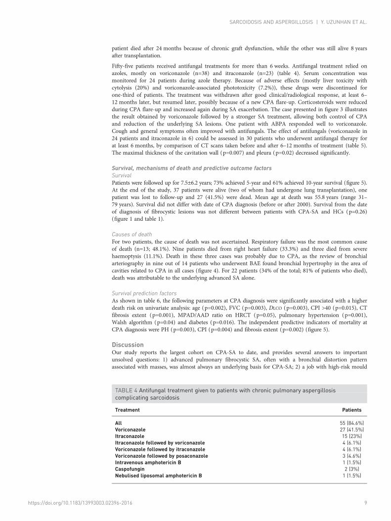
TABLE 4 Antifungal treatment given to patients with chronic pulmonary aspergillosiscomplicating sarcoidosis
Treatment Patients
All 55 (84.6%)Voriconazole 27 (41.5%)Itraconazole 15 (23%)Itraconazole followed by voriconazole 4 (6.1%)Voriconazole followed by itraconazole 4 (6.1%)Voriconazole followed by posaconazole 3 (4.6%)Intravenous amphotericin B 1 (1.5%)Caspofungin 2 (3%)Nebulised liposomal amphotericin B 1 (1.5%)
https://doi.org/10.1183/13993003.02396-2016 9
SARCOIDOSIS AND ASPERGILLOSIS | Y. UZUNHAN ET AL.
exposure was a significant risk for CPA occurrence, irrespective of patient’s geographical origin, diabetes,smoking habit, previous pneumothorax and treatment of SA; 3) CPA did not significantly affect survival,and mortality was high with 5- and 10-year survival rates of 73.7% and 61%, respectively; 4) mortality wasmainly related to advanced pulmonary SA, while at most three out of 27 deaths were attributable to CPAthrough massive haemoptysis; 5) CPI, PH and pulmonary fibrosis extent (as assessed by CT) predictedsurvival; and 6) antifungals improved patients’ condition in the short term.
There is only limited information in the literature on CPA-SA, which is often dated, with few cases, raresurvival data and no CPA categorisation. The only survival study is that of TOMLINSON and SAHN on 14patients with 58% deaths at 2 years [8], while DENNING et al. suggested a lower annual attrition rate with arange of 10–25% [2]. Combining four series showed 44 deaths out of 93 patients, i.e. 47% [5–8]; 12 of the44 deceased patients (27%) died from massive haemoptysis [5–8]. Corticosteroid treatments, smokinghistory or having previous pneumothorax were suspected of being the highest risk factors for CPA.Spontaneous aspergilloma resolution is a very rare outcome [7, 9, 24], while successful surgical resections[6] and individual benefits of interventional radiology [5, 8], systemic antifungals [25] and perendoscopicor transcutaneous instillation of fungicides [26, 30] have all been reported.
An advanced underlying fibrocystic lung disease was clearly shown to be the predisposing ground to CPAoccurrence in SA. Masses, often associated with SA, secondarily excavated, might be the nidus for CPA.Moreover, 66% of patients met the criteria for poor survival prognosis [20]. Survival in patients withCPA-SA was not different from that of HCs, and 81% of deaths were due to respiratory insufficiency or corpulmonale. Survival prediction factors (CPI, PH and fibrosis extent) were linked with advanced pulmonarySA, making our study an external validation cohort for the findings of WALSH et al. [20] concerning survivalprediction criteria in advanced pulmonary SA. The absence of extra mortality due to CPA has also beenobserved in ancient series that had significant underlying tuberculosis sequels as the underlying condition[24]. Lower doses of corticosteroids and immunosuppressive treatments following CPA flare-up, and use ofantifungals and interventional radiology might explain the reduction in mortality in our series comparedwith that of TOMLINSON et al. [8], even though our study can only presume such interpretation.
CCPA was the most frequent presentation in our SA series. In 10% of patients, classifying CPA wasimpossible because of the presence of confusing SA lesions. SA should be added as a cause of ABPA, as
TABLE 5 Computed tomography findings of patients before and 6–12 months after antifungaltreatment#
Before treatment After treatment p-value
Patients n 30 30Presence of at least one cavity 30 (100%) 30 (100%)Unilateral localisation of cavity 9 (30%) 10 (33.3%)Cavities in the right lung0 4 (13.3%) 4 (13.3%)1–5 22 (73.3%) 21 (70%)5–10 1 (3.3%) 2 (6.6%)>10 3 (10%) 3 (10%)
Cavities in the left lungNone 6 (20%) 6 (20%)1–5 17 (56.6%) 18 (60%)5–10 4 (13.3%) 3 (10%)>10 3 (10%) 3 (10%)
Average diameter of largest cavities mm 28.5±17.8 23.4±11.4 0.12Maximal diameter of infected cavities mm 37.6±20.4 28.4±14.1 0.059Presence of fungus balls 21 (70%) 18 (60%) 0.27Unilateral localisation of fungus balls 15 (50%) 13 (43.3%) 0.79Maximal diameter of fungus ball mm 16.7±11.0 16.0±10.8 0.84Presence of nodules >10 mm 2 (6.6%) 2 (6.6%)Presence of ground-glass attenuation 7 (23.3%) 7 (23.3%)Presence of areas of consolidation 6 (20%) 7 (23.3%)Presence of lobar collapse 3 (10%) 3 (10%)Maximum thickness of pleura mm 8.2±3.6 5.8±4.0 0.02Maximum thickness of cavity wall mm 5.6±2.6 3.6±1.8 0.007
Data are presented as n (%) or mean±SD unless otherwise stated. #: voriconazole for 24 patients anditraconazole for six patients.
https://doi.org/10.1183/13993003.02396-2016 10
SARCOIDOSIS AND ASPERGILLOSIS | Y. UZUNHAN ET AL.
shown in two of our patients. Our series provides interesting information on risk factors for CPA in SA.Jobs with a high risk of mould exposure were more frequent in patients with CPA-SA than in HCs.Patients with CPA-SA were predominantly males, contrasting with our HC group and with the femalepredominance seen in advanced pulmonary SA [20]. No positive link was found with geographical origin,smoking habit, diabetes or treatment of SA. The male predominance might be due to more men inhigh-risk jobs such as construction work [21, 22]. We agree with the recommendations of the InfectiousDiseases Society of America, who “recommend reasonable precautions to reduce mould exposure amongoutpatients” with fibrocystic SA, “including avoidance of gardening, spreading mulch or close exposure toconstruction or renovation” [27].
Three deaths were due to massive haemoptysis. Threatening haemoptysis events were probably attributableto CPA, even though a bronchiectasis origin cannot definitely be ruled out. This rate (three patients being11% of the deceased patients) was lower than the rate of 27% observed in a combined dated series [5–8, 15].Interventional radiology allowed immediate cessation of massive haemoptysis with often iterativeinterventions for bleeding relapse [28]. The literature is in favour of a reduction of death withinterventional radiology in cases of threatening haemoptysis [29]. Only one of our patients benefited fromintracavitary amphotericin B instillation, although this technique has been used in some series [30].
There are still few trials evaluating antifungals in CPA [31], and there is no study in the literature onCPA-SA. Our study shows that a short-term benefit can be achieved for CPA in SA with antifungals, asshown by CT according to the criteria of GODET et al. [23]. In fact, the maximal thickness of cavitationwall (p=0.007) and pleura (p=0.02) significantly decreased under antifungals. Complete recovery remained
0
0.00
0.25
0.50
Surv
ival
pro
babi
lity 0.75
1.00b)
100 200
Follow-up months
300
45 17 4 0
20
No PH
PH 5 2 0
At risk n
p=0.001
No PH
PH
0
0.00
0.25
0.50
Surv
ival
pro
babi
lity 0.75
1.00a)
100 200
Follow-up months
300
65 22 6 0At risk n
0
0.00
0.25
0.50
Surv
ival
pro
babi
lity 0.75
1.00c)
100 200
Follow-up months
300
28 11 3 0
25
MPA/AAD ratio ≤1
MPA/AAD ratio >1 3 0 0
At risk n
p=0.05
MPA/AAD ratio ≤1
MPA/AAD ratio >1
0
0.00
0.25
0.50
Surv
ival
pro
babi
lity 0.75
1.00e)
100 200
Follow-up months
300
39 14 4 0
19
Fibrosis ≤20%
Fibrosis >20% 3 0 022CPI >40
CPI ≤40
11 3 0
At risk n
p=0.01
Fibrosis ≤20%
Fibrosis >20
p=0.01
CPI ≤40
CPI >40
0
0.00
0.25
0.50
Surv
ival
pro
babi
lity 0.75
1.00d)
100 200
Follow-up months
300
42 10 2 0At risk n
0
0.00
0.25
0.50
Surv
ival
pro
babi
lity 0.75
1.00f)
100 200
Follow-up months
300
Observed: poor prognosis
Predicted: poor prognosis
p=0.04
Observed: good prognosis
Predicted: bad prognosis
FIGURE 5 Survival after cavitary pulmonary aspergillosis (CPA) diagnosis with a) overall survival and comparison of survival with observedKaplan–Meier estimates in this series: b) patients with/without pulmonary hypertension (PH) at CPA diagnosis, c) main pulmonary artery (MPA)/ascending aorta diameter (AAD) ratio >1 or ⩽1, d) composite physiological index (CPI) score >40 or ⩽40, e) extent of fibrosis >20% or ⩽20% and f)predicted by the sarcoidosis staging model (Walsh algorithm [20]).
https://doi.org/10.1183/13993003.02396-2016 11
SARCOIDOSIS AND ASPERGILLOSIS | Y. UZUNHAN ET AL.
exceptional and probably required both CPA control and cavitation reversibility, a condition illustrated inan exceptional case in figure 3.
Controversy remains regarding surgical CPA care [32], as peri-operative management of CPA-SA ischallenging [6]. A later aspergillosis relapse occurred 6–45 months after surgery in all cases except one.Resection surgery should be proposed in strictly localised aspergillomas [33], a very rare condition inCPA-SA, and for severe bleeding that is best controlled by BAE, as recommended [27].
This study, being retrospective and monocentric, has some limitations. A prospective systematic recordingof domestic and environmental mould exposure was not available in files, unlike past and currentoccupational exposure.
ConclusionIn CPA-SA, mortality appeared to be conditioned mainly by the severity of the underlying pulmonary SArather than by CPA. Improvement in CPA-SA care should probably include measures preventing exposureto spores for fibrocystic SA, effective antifibrotic drugs for patients with a poor prognosis, and moreeffective antifungal drugs.
AcknowledgementsWe thank Jean-Paul Battesti, Christine Person, Bruno Philippe, Stephane Dominique, Emmanuel Bergot, ElisabethRivaud and Pascaline Priou.
References1 Denning DW, Cadranel J, Beigelman-Aubry C, et al. Chronic pulmonary aspergillosis: rationale and clinical
guidelines for diagnosis and management. Eur Respir J 2016; 47: 45–68.2 Denning DW, Pleuvry A, Cole DC. Global burden of chronic pulmonary aspergillosis complicating sarcoidosis.
Eur Respir J 2013; 41: 621–626.3 Smith NL, Denning DW. Underlying conditions in chronic pulmonary aspergillosis including simple aspergilloma.
Eur Respir J 2011; 37: 865–872.4 Sones M, Israel HL. Course and prognosis of sarcoidosis. Am J Med 1960; 29: 84–93.5 Kaplan J, Johns CJ. Mycetomas in pulmonary sarcoidosis: non-surgical management. Johns Hopkins Med J 1979;
145: 157–161.6 Israel HL, Lenchner GS, Atkinson GW. Sarcoidosis and aspergilloma. The role of surgery. Chest 1982; 82:
430–432.7 Wollschlager C, Khan F. Aspergillomas complicating sarcoidosis. A prospective study in 100 patients. Chest 1984;
86: 585–588.8 Tomlinson JR, Sahn SA. Aspergilloma in sarcoid and tuberculosis. Chest 1987; 92: 505–508.9 Jewkes J, Kay PH, Paneth M, et al. Pulmonary aspergilloma: analysis of prognosis in relation to haemoptysis and
survey of treatment. Thorax 1983; 38: 572–578.10 Nam HS, Jeon K, Um SW, et al. Clinical characteristics and treatment outcomes of chronic necrotizing pulmonary
aspergillosis: a review of 43 cases. Int J Infect Dis 2010; 14: e479–e482.
TABLE 6 Univariate analysis for survival at diagnosis of chronic pulmonary aspergillosis (CPA)
Parameter Hazard ratio Standard error p-value 95% confidence interval
Sex 0.762 0.319 0.518 0.335–1.733Geographical origin 1.596 0.609 0.220 0.755–3.373Age at sarcoidosis diagnosis 1.030 0.016 0.065 0.998–1.062Age at stage 4 diagnosis 1.036 0.016 0.029 1.003–1.069Age at CPA diagnosis 1.054 0.017 0.002 1.020–1.090Diabetes mellitus 2.902 1.285 0.016 1.218–6.915Extrapulmonary manifestations 0.980 0.803 0.806 0.834–1.150Pulmonary hypertension 3.425 1.306 0.001 1.621–7.235FEV1 0.979 0.011 0.069 0.958–1.001FVC 0.965 0.011 0.003 0.944–0.988DLCO 0.968 0.010 0.003 0.947–0.988CPI 40 0.293 0.147 0.015 0.109–0.784MPA/AAD >1 2.347 1.020 0.050 1.001–5.501CT fibrosis >20% 2.710 1.096 0.014 1.226–5.990CT fibrosis extent 1.041 0.012 0.001 1.017–1.066Walsh’s algorithm 3.069 1.696 0.042 1.038–9.068
FEV1: forced expiratory volume in 1 s; FVC: forced vital capacity; DLCO: diffusing capacity of the lung forcarbon monoxide; CPI: composite physiological index; MPA/AAD: main pulmonary artery/ascending aortadiameter ratio; CT: computed tomography.
https://doi.org/10.1183/13993003.02396-2016 12
SARCOIDOSIS AND ASPERGILLOSIS | Y. UZUNHAN ET AL.
11 Pena TA, Soubani AO, Samavati L. Aspergillus lung disease in patients with sarcoidosis: a case series and review ofthe literature. Lung 2011; 189: 167–172.
12 Nardi A, Brillet PY, Letoumelin P, et al. Stage IV sarcoidosis: comparison of survival with the general populationand causes of death. Eur Respir J 2011; 38: 1368–1373.
13 Shlobin OA, Nathan SD. Management of end-stage sarcoidosis: pulmonary hypertension and lung transplantation.Eur Respir J 2012; 39: 1520–1533.
14 Statement on sarcoidosis. Joint Statement of the American Thoracic Society (ATS), the European RespiratorySociety (ERS) and the World Association of Sarcoidosis and Other Granulomatous Disorders (WASOG) adoptedby the ATS Board of Directors and by the ERS Executive Committee, February 1999. Am J Respir Crit Care Med1999; 160: 736–755.
15 Judson MA, Costabel U, Drent M, et al. The WASOG Sarcoidosis Organ Assessment Instrument: an update of aprevious clinical tool. Sarcoidosis Vasc Diffuse Lung Dis 2014; 31: 19–27.
16 Cotes JE, Chinn DJ, Quanjer PH, et al. Standardization of the measurement of transfer factor (diffusing capacity).Eur Respir J 1993; 6: Suppl. 16, 41–52.
17 Quanjer PH, Tammeling GJ, Cotes JE, et al. Lung volumes and forced ventilatory flows. Eur Respir J 1993; 6:Suppl. 16, 5–40.
18 Abehsera M, Valeyre D, Grenier P, et al. Sarcoidosis with pulmonary fibrosis: CT patterns and correlation withpulmonary function. AJR Am J Roentgenol 2000; 174: 1751–1757.
19 Wells AU, Desai SR, Rubens MB, et al. Idiopathic pulmonary fibrosis: a composite physiologic index derived fromdisease extent observed by computed tomography. Am J Respir Crit Care Med 2003; 167: 962–969.
20 Walsh SL, Wells AU, Sverzellati N, et al. An integrated clinicoradiological staging system for pulmonarysarcoidosis: a case-cohort study. Lancet Respir Med 2014; 2: 123–130.
21 Caira M, Candoni A, Verga L, et al. Pre-chemotherapy risk factors for invasive fungal diseases: prospectiveanalysis of 1,192 patients with newly diagnosed acute myeloid leukemia (SEIFEM 2010-a multicenter study).Haematologica 2015; 100: 284–292.
22 Sipsas NV, Kontoyiannis DP. Occupation, lifestyle, diet, and invasive fungal infections. Infection 2008; 36:515–525.
23 Godet C, Laurent F, Bergeron A, et al. Computed tomography assessment of response to treatment in chronicpulmonary aspergillosis. Chest 2016; 150: 139–147.
24 Aspergilloma and residual tuberculous cavities—the results of a resurvey. Tubercle 1970; 51: 227–245.25 Keir GJ, Garfield B, Hansell DM, et al. Cyclical caspofungin for chronic pulmonary aspergillosis in sarcoidosis.
Thorax 2014; 69: 287–288.26 Guleria R, Pande JN. Endoscopic instillation of fungicide to treat aspergilloma. Lancet 1996; 348: 621.27 Patterson TF, Thompson GR 3rd, Denning DW, et al. Practice Guidelines for the Diagnosis and Management of
Aspergillosis: 2016 update by the Infectious Diseases Society of America. Clin Infect Dis 2016; 63: e1–e60.28 Fartoukh M, Parrot A, Khalil A. Aetiology, diagnosis and management of infective causes of severe haemoptysis in
intensive care units. Curr Opin Pulm Med 2008; 14: 195–202.29 Fartoukh M, Khoshnood B, Parrot A, et al. Early prediction of in-hospital mortality of patients with hemoptysis:
an approach to defining severe hemoptysis. Respiration 2012; 83: 106–114.30 Kravitz JN, Berry MW, Schabel SI, et al. A modern series of percutaneous intracavitary instillation of
amphotericin B for the treatment of severe hemoptysis from pulmonary aspergilloma. Chest 2013; 143: 1414–1421.31 Agarwal R, Vishwanath G, Aggarwal AN, et al. Itraconazole in chronic cavitary pulmonary aspergillosis: a
randomised controlled trial and systematic review of literature. Mycoses 2013; 56: 559–570.32 Farid S, Mohamed S, Devbhandari M, et al. Results of surgery for chronic pulmonary Aspergillosis, optimal
antifungal therapy and proposed high risk factors for recurrence—a National Centre’s experience. J CardiothoracSurg 2013; 8: 180.
33 Lejay A, Falcoz PE, Santelmo N, et al. Surgery for aspergilloma: time trend towards improved results? InteractCardiovasc Thorac Surg 2011; 13: 392–395.
https://doi.org/10.1183/13993003.02396-2016 13
SARCOIDOSIS AND ASPERGILLOSIS | Y. UZUNHAN ET AL.