chronic peripheral oedema: the critical role of the ... · system in tissue fluid balance. ......
TRANSCRIPT

Oedema is an excess of interstitial fluid and is animportant sign of ill health in clinical medicine. Itmay occur in the lungs (pulmonary oedema), theabdominal cavity (ascites) and other body cavities(synovial, pericardial and pleural effusions) but inthis article only peripheral (subcutaneous) oedema isdiscussed.
In medical practice peripheral oedema tends to getpigeonholed according to possible systemic or periph-eral causes eg heart failure, nephrotic syndrome,venous obstruction or lymphoedema. This viewpointfails to appreciate the many dynamic physiologicalforces contributing to oedema development and inparticular the central role of the lymphatic drainagesystem in tissue fluid balance. Consequently, the clin-ician’s approach to chronic oedema is often misguidedand the necessary medical intervention inappropriate,for example, empirical use of diuretics. In this articlewe propose a system for managing peripheral oedema,which is based on physiological principles, that canthen guide treatment.
Why is chronic oedema important?
Besides being a physical sign of a potentially fatal systemic condition such as heart failure, chronicoedema impairs local cell nutrition due to increasedinterstitial diffusion distances of oxygen and nutri-ents so tissue viability can become compromised.Swollen limbs can be painful, giving rise to impairedmobility as well as a predisposition to infection andblistering progressing to ulceration.
Chronic oedema is a common problem in the com-munity particularly for district nurses. A recent epi-demiological study in South West London estimateda crude prevalence of 1.33/1000 population rising toone in 200 people over the age of 65. 29% of theoedema cases had experienced cellulitis in the pre-ceding year with one quarter of these cases requiringadmission. Oedema caused time off work in morethan 80% of sufferers and employment status wasaffected in 9%. Quality of life suffered, with cleardeficits in many domains of the well-validated SF-36questionnaire.1
Understanding oedema
Oedema develops when the microvascular (capillaryand venules) filtration rate exceeds lymph drainagefor a sufficient period, either because the filtrationrate is high or because lymph flow is low or a combi-nation of the two. Filtration rate is governed by theStarling principle of fluid exchange, which isdescribed succinctly and quantitatively by theStarling equation for flow across a semipermeablemembrane:
Jv = Lp S [(Pc) – Pi – σ (πp – πi)]
where Jv = capillary filtration rate
Lp = hydraulic conductance of capillary
wallS = surface areaP = pressure within capillary (c) or
interstitium (i)σ = osmotic reflection coefficient of
capillary wallπ = osmotic pressure of plasma (p) or
interstitial fluid (i)
� REVIEWS
448 Clinical Medicine Vol 4 No 5 September/October 2004
Peter S MortimerMD FRCP, Professorof Dermatological
Medicine
J Rodney LevickDPhil MRCP,
Professor ofPhysiology
St George’sHospital MedicalSchool, London
Clin Med2004;4:448–53
Chronic peripheral oedema:
the critical role of the lymphatic system
Peter S Mortimer and J Rodney Levick
Key Points
Lymph transport, not venous capillary reabsorption, is the main processresponsible for interstitial fluid drainage
All oedema is due to an imbalance between capillary filtration and lymphdrainage
Elevation and diuretics reduce venous pressure and consequentlycapillary filtration but do not improve lymph drainage
Lymph flow is stimulated by tissue movement and in particular exercise
In theory an increase in lymph transport, if achievable, should benefit allforms of oedema
Calcium channel blocking agents should be contraindicated in thepresence of any peripheral oedema unless there is no alternativetreatment
Diuretics should only be prescribed long-term in circumstances of saltand water overload, eg heart failure, venous disease, and not on anempirical basis for any oedema
Movement through active or passive exercise should always be thepreferred option for reducing leg oedema due to decompensatedlymph drainage, with elevation practised during the periods of rest

In simple terms filtration of fluid from capillary into intersti-tium is driven by the hydraulic (water) pressure gradient acrossthe wall (Pc – Pi) and is opposed by the osmotic pressure gradient (πp – πi), which is the suction force keeping fluid in the circulation. Strictly speaking, πi and Pi are the forces imme-diately downstream of the semi permeable membrane ratherthan the global interstitial values.2
Other factors important in determining filtration
a) Lp, which refers to the ease with which water can cross thecapillary wall
b) S, which is the surface area for filtration, and
c) σ, which refers to the ability of the capillary wall to preventleakage of plasma protein from the circulation.
The Starling equation provides a logical approach for classi-fying oedema that is due to increased filtration:
a) Raised capillary pressure; because capillary pressure is moresusceptible to changes in venous pressure rather thansystemic (arterial) blood pressure as post-capillary resistanceis much lower than pre-capillary resistance. Peripheralvenous pressure is raised in:
• Right ventricular failure
• Salt and water overload (overtransfusion)
• Venous obstruction
• Venous reflux (chronic venous disease) eg followingdeep vein thrombosis, primary varicose veins
• Dependency (the effect of gravity)
• Certain drugs, especially calcium-channel blockingagents.
b) Reduced plasma osmotic pressure (COP) essentially meanshypoalbuminaemia, which can arise from:
• Malnutrition
• Intestinal disease (malabsorption or protein loss)
• Nephrotic syndrome
• Hepatic failure – failure to synthesise albumin due toliver disease or chronic inflammatory states.
c) Increased capillary permeability, when inflammation causesa breakdown in the endothelial barrier facilitating thepassage of both plasma proteins and water across thecapillary wall.3 In addition, vasodilatation causes a rise incapillary pressure (and blood flow).
The importance of lymph transport rather thanvenous capillary reabsorption for interstitial fluiddrainage
Accumulation of capillary filtrate in the tissue spaces is avoidedmainly through lymph drainage and not, as was previouslythought, through reabsorption by the venous capillaries.Traditionally it was taught that the arterial end of capillaries filtered fluid while the venous end reabsorbed the bulk of fluid
filtered. This view is not supported by modern evidence, whichdemonstrates that in most vascular beds there is a net but dwin-dling filtration along the entire length of well-perfused capil-laries. It is true that pressure in venous capillaries does fall belowplasma COP. However, when direct measurements of Pi and πi
in muscle, mesentery or warm skin at heart level are madedirectly these are far from negligible, contrary to popular belief.The sum of all Starling forces is not an absorptive forcein venous capillaries. It is a slight filtration force. Data from12 tissues confirm that venular blood pressure exceeds the sumof pressures opposing filtration.4
Exchange vessels (mainly capillaries and post-capillary venules)can reabsorb fluid for a short period if the Starling pressures aredisturbed. For example, following haemorrhage precapillaryvasoconstriction, coupled with a drop in both arterial andvenous pressures, will reduce capillary pressure sufficiently forabsorption to develop transiently. Very soon, however, the re-absorption of interstitial water raises the protein concentration inthe pericapillary space, so raising the local osmotic pressure there(πI). Consequently reabsorption stops and slight filtration isrestored. For reasons explained in Levick and Mortimer’s 1999paper,4 sustained absorption of fluid into the microcirculationis a normal feature of intestinal mucosal capillaries, renal per-itubular capillaries and lymph node capillaries but not peripheraltissues.
Since the old concept of sustained fluid absorption by venouscapillaries is no longer tenable, this places the major responsi-bility for drainage of interstitial fluid in the hands of the lym-phatic system. The latter is now perceived to be of even greaterimportance to tissue volume homeostasis than was formerlyrealised.5
Restraining factors against oedema
Several factors ‘buffer’ the capillary filtration rate, and thus tendto prevent oedema formation.
Elevation of interstitial fluid pressure
Stiffness of tissues resists swelling. A small increase in interstitialfluid volume in a stiff tissue (low compliance) will cause a rela-tively large increase in interstitial pressure (Pi) which then opposesfiltration eg subfascial muscle compartments. Conversely a tissuesuch as eyelid skin is loose and compliant and Pi will not increaseso much for a given volume increase. Therefore, oedema con-tinues to form and accumulate to large volumes. Conversely,placing a bandage or rigid stocking around a leg will reduce com-pliance (increase stiffness) and resist stretch. Consequently, Pi willincrease more steeply for a given amount of interstitial volumeincrease, and the increased Pi will oppose filtration.
Fall in interstitial COP (πi )
An increase in filtration rate will dilute the interstitial proteinconcentration and consequently reduce the osmotic pressureimmediately outside the semipermeable membrane (πi). The
Chronic peripheral oedema: the critical role of the lymphatic system
Clinical Medicine Vol 4 No 5 September/October 2004 449

resulting increase in the osmotic pressure gradient (πp – πI) willraise the suction force, keeping fluid with the blood compart-ment. This is probably of major importance in the protectionagainst pulmonary, as well as peripheral oedema.
Increased lymph flow
Increases in interstitial fluid pressure and volume stimulatelymph flow.5 Transport of interstitial fluid into and along initiallymphatics is a complex, poorly understood process dependenton intermittent changes in tissue (interstitial) pressure frommovement (active and passive exercise), massage, local arterialpulsation and, in more central tissues, breathing. Lymph flow inthe larger more proximal-collecting lymphatics is generated byactive lymphatic pumping. Collecting lymphatics contract andare mainly responsible for pumping lymph up the leg againstgravity to the inguinal lymph nodes. Successive segments of col-lecting lymphatic vessel behave like mini hearts in series and thecontractile cycle bears striking similarities to the cardiac cycle.Sympathetic input influences the pumping rate while the dias-tolic filling (lymph supply from upstream initial lymphatics)largely controls the force of contraction. Flow in collecting lym-phatics is only as good as the supply from initial lymphatics.Influx of calcium ions is important for smooth muscle contrac-tion in the walls of the collecting lymphatics.6 Therefore calciumchannel blocking agents are likely to work at least in part on thelymphatic in causing peripheral oedema.
The important point is that it is the lymph vessels that returnthe capillary filtrate back to the blood stream via the lymphnodes and eventually the thoracic duct. This completes theextravascular circulation of fluid and protein and maintainstissue volume homeostasis. If lymphatic drainage is impaired inthe face of normal filtration, oedema will occur (lym-phoedema). Lymph flow should respond to increases in capil-lary filtration and so prevent oedema. By failing to compensatefully for increased capillary filtration and so permitting swelling,the lymphatic is to some extent failing in its duty in all types ofoedema. This could help explain differences in the degree of legoedema seen in patients with venous disease and also in right-sided heart failure. It is possible that ‘true’ lymphatic failure,namely reduced lymph transport, does eventually occur in cir-cumstances of high capillary filtration when high lymph flowcannot be sustained.
The leg has additional buffering mechanisms to resistoedema. These include:
a) postural vasoconstriction in a dependent leg resulting fromactivation of the veni-arteriolar response
b) a rise in plasma colloid osmotic pressure in the venules of adependent leg in response to increased concentration ofplasma proteins following increased microvascular filtration
c) activation of the calf muscle pump, which not only reducesvenous pressure by increasing venous return, but alsoincreases lymph transport.7
Diagnosis
Chronic oedema can occur in any subcutaneous tissue – face,trunk, breast, genitalia, but more often than not it develops in alimb, usually the lower limb. For the purpose of simplicity thefollowing scheme deals with lower limb oedema but the princi-ples stand for any site. Oedema that is symmetrical eg equalbetween right and left legs, is more likely to be of systemicorigin, for example, right-sided heart failure or hypopro-teinaemia. Oedema that is asymmetrical, eg worse in the leftthan the right, may still have a systemic contribution but is likelyto be due predominantly to peripheral pathology ie impairedlocal venous or lymph drainage. Oedema that is unilateral egone leg only is likely to result from pathology arising within thelimb or adjoining quadrant of the trunk.
The first consideration in assessing peripheral oedema mustbe to exclude serious systemic causes such as heart failure. Thisis not always straightforward. Careful examination of thejugular venous pressure is crucial, but the reliability of a clinicaldiagnosis is poor and investigations including a 12-lead ECGand measurement of B-type natriuretic peptide may be neces-sary.8 Other explanations for raised venous pressure eg venousobstruction, chronic venous disease and prolonged periods ofdependency should be sought and plasma proteins measured.Cancer uncommonly presents with peripheral oedema unlessadvanced at diagnosis but relapsed cancer may frequently be a cause of limb swelling. Therefore cancer should always be considered and investigated appropriately. In advanced cancernumerous factors may contribute to oedema, including bothvenous and lymphatic obstruction, hypoalbuminaemia andimmobility.
Distinguishing lymphoedema from oedema due toincreased filtration (filtration oedema)
It is important to distinguish lymphoedema from filtrationoedema because the treatment is different. Currently there areno drug or surgical interventions effective in treating lym-phoedema. Diuretics do little and may be harmful if used longterm.
When taking a history there are certain features of the oedemathat can help distinguish an oedema due to impaired lymphdrainage from an oedema arising from overwhelming filtration.Here we refer to oedema arising from an increased capillary rateas a filtration oedema and oedema arising from a fundamentalfailure in lymph transport as lymphoedema. Oedema whereboth mechanisms contribute will be referred to as oedema ofmixed origin.
Pitting
It is often said that lymphoedema does not pit but this is nottrue. Pitting is invariably present but sustained pressure for some20 seconds may be necessary owing to the firmer (and thicker)nature of the skin and subcutaneous tissues. In establishedlymphoedema the skin may double in thickness particularly at
Peter S Mortimer and J Rodney Levick
450 Clinical Medicine Vol 4 No 5 September/October 2004
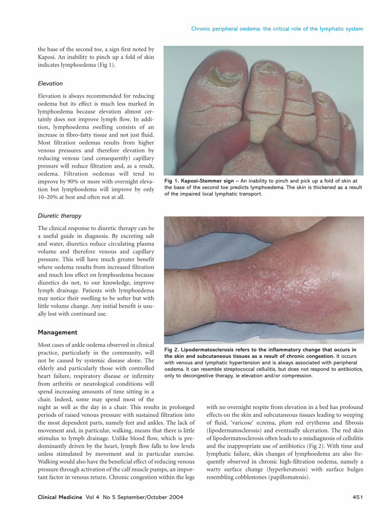
the base of the second toe, a sign first noted byKaposi. An inability to pinch up a fold of skinindicates lymphoedema (Fig 1).
Elevation
Elevation is always recommended for reducingoedema but its effect is much less marked inlymphoedema because elevation almost cer-tainly does not improve lymph flow. In addi-tion, lymphoedema swelling consists of anincrease in fibro-fatty tissue and not just fluid.Most filtration oedemas results from highervenous pressures and therefore elevation byreducing venous (and consequently) capillarypressure will reduce filtration and, as a result,oedema. Filtration oedemas will tend toimprove by 90% or more with overnight eleva-tion but lymphoedema will improve by only10–20% at best and often not at all.
Diuretic therapy
The clinical response to diuretic therapy can bea useful guide in diagnosis. By excreting saltand water, diuretics reduce circulating plasmavolume and therefore venous and capillarypressure. This will have much greater benefitwhere oedema results from increased filtrationand much less effect on lymphoedema becausediuretics do not, to our knowledge, improvelymph drainage. Patients with lymphoedemamay notice their swelling to be softer but withlittle volume change. Any initial benefit is usu-ally lost with continued use.
Management
Most cases of ankle oedema observed in clinicalpractice, particularly in the community, willnot be caused by systemic disease alone. Theelderly and particularly those with controlledheart failure, respiratory disease or infirmityfrom arthritis or neurological conditions willspend increasing amounts of time sitting in achair. Indeed, some may spend most of thenight as well as the day in a chair. This results in prolongedperiods of raised venous pressure with sustained filtration intothe most dependent parts, namely feet and ankles. The lack ofmovement and, in particular, walking, means that there is littlestimulus to lymph drainage. Unlike blood flow, which is pre-dominantly driven by the heart, lymph flow falls to low levelsunless stimulated by movement and in particular exercise.Walking would also have the beneficial effect of reducing venouspressure through activation of the calf muscle pumps, an impor-tant factor in venous return. Chronic congestion within the legs
with no overnight respite from elevation in a bed has profoundeffects on the skin and subcutaneous tissues leading to weepingof fluid, ‘varicose’ eczema, plum red erythema and fibrosis(lipodermatosclerosis) and eventually ulceration. The red skinof lipodermatosclerosis often leads to a misdiagnosis of cellulitisand the inappropriate use of antibiotics (Fig 2). With time andlymphatic failure, skin changes of lymphoedema are also fre-quently observed in chronic high-filtration oedema, namely awarty surface change (hyperkeratosis) with surface bulgesresembling cobblestones (papillomatosis).
Chronic peripheral oedema: the critical role of the lymphatic system
Clinical Medicine Vol 4 No 5 September/October 2004 451
Fig 1. Kaposi-Stemmer sign – An inability to pinch and pick up a fold of skin atthe base of the second toe predicts lymphoedema. The skin is thickened as a resultof the impaired local lymphatic transport.
Fig 2. Lipodermatosclerosis refers to the inflammatory change that occurs inthe skin and subcutaneous tissues as a result of chronic congestion. It occurswith venous and lymphatic hypertension and is always associated with peripheraloedema. It can resemble streptococcal cellulitis, but does not respond to antibiotics,only to decongestive therapy, ie elevation and/or compression.

Improving lymph flow by physical means
One approach that is theoretically of value in all forms ofperipheral oedema is an improvement in lymph drainage. As explained previously, all oedema arises either because of aprimary failure in lymph drainage (lymphoedema) or becauseincreased capillary filtration overwhelms increased lymphdrainage (which under-compensates). Therefore, means thatimprove lymph transport are desirable when lymph drainage isfailing. Unfortunately no drug therapy is effective at increasinglymph flow. Lymph drainage can be improved by using simplephysiological principles to stimulate lymph flow.
As already mentioned movement and exercise, by inducingalternating changes in interstitial fluid pressure, increase initiallymphatic filling, and hence lymph flow. This may also promoteflow in conducting lymphatics whose contractility has failed.Patients with lymphoedema often notice that walking reducesswelling. The addition of a bandage or equivalent form of outerpressure on the leg will enhance the effect of movement. Theidea is not to squeeze fluid out of the limb with force likesqueezing toothpaste out of a tube, but to create an outer collarto the leg against which the calf and foot muscles can press toimprove pumping. A short stretch bandage will generate higherinterstitial pressure during muscle contractions to stimulatelymph flow but result in low tissue pressure during muscle relax-ation which then permits lymph vessel refilling. Such treatmenthas the added benefit of lowering venous pressure in the leg andso reducing filtration. Care is needed during the first applicationof any bandage in case rapid fluid shifts back into the circulationcause pulmonary oedema in anyone with heart failure. Care alsohas to be taken in circumstances where arterial supply and cuta-neous sensation are compromised.
‘Decongestion’ of the legs may reverse more or less all the co-morbidity from the swelling, particularly the skin changes.Once swelling has been reduced, control needs to be maintainedthrough encouragement of movement and exercise whilewearing appropriately fitted support hosiery. In the elderly andthose patients with hand arthritis or disability, the applicationand removal of hosiery can be problematic but technique canhelp and aids to application do exist.
Elevation
Elevation of the legs is often chosen over exercise for infirmpatients who spend long periods in a chair. Unfortunately, legelevation is only of partial benefit unless the trunk is reclinedbecause there remains a considerable venous pressure increasefrom heart to legs when sitting upright. Only by lying com-pletely flat (or, better still, with ankles above heart level) willvenous pressure be reduced sufficiently to reduce oedema.Elevation acts by reducing local capillary pressure and thus filtration, allowing lymph drainage to outpace the reduced cap-illary filtration rate. Some degree of leg movement throughactive or passive muscle exercise would always be the preferredoption for reducing leg oedema, with elevation practised duringthe periods of rest.
Pneumatic compression pumps
In theory, pneumatic compression pumps would offer addi-tional benefit for patients spending considerable time in a chair.Unfortunately there is no evidence that such pumps improvelymph flow and they may simply displace fluid.9 Once again,care has to be taken in case of rapid fluid shifts. The equipmentis not prescribable so patients may have to purchase it them-selves. It is often the patients in most need who can ill afford thecost.
Diuretics
Too often, diuretics are prescribed for oedema on an empiricalbasis without due thought to underlying pathophysiology.Diuretics should only be prescribed in circumstances of salt andwater retention, such as happens in heart failure and nephroticsyndrome. Arguably, they should not be prescribed in any othercircumstances particularly in the long term where adverse effectsincluding electrolyte imbalance can be harmful. Indeed, thedevelopment of ‘idiopathic oedema of women’ has been largelyattributed to diuretic abuse.10 Rebound oedema on diureticwithdrawal can be an exacerbating factor in many forms ofchronic peripheral oedema.
Lymphoedema clinics
Until recently it has been a long held view that nothing can bedone for lymphoedema and patients have been told to put upwith it despite the morbidity and risk of life-threatening septi-caemia through cellulitis. In Europe in the early 1980’s treat-ment was introduced which did not use drugs or surgery butused physical methods designed to stimulate lymph transport.This decongestive lymphatic therapy for limb lymphoedemaemployed isotonic muscle exercise (not strength building butexercise that involved lengthening and shortening of muscles) asthe cornerstone, with compression (bandages or hosiery) toenhance the effect of exercise.11 A specific form of massage(manual lymphatic drainage therapy) was used concurrently tostimulate lymph drainage from the root of the limb andadjoining quadrant of the trunk to normally draining lymphaticbasins. The massage was therefore intended to ‘clear the wayahead’ for limb lymph drainage and avoid congestion above thelevel of compression. In moderate to severe lymphoedema anintensive period of treatment using multi-layer bandaging, exer-cises and manual lymphatic drainage is used to reduce swollenlimbs so that subsequent maintenance treatment with hosieryand exercise is more effective at controlling the condition.12
These combination treatments have a sound physiologicalbasis but the quality of evidence proving efficacy is limited. Arandomised, controlled parallel group clinical trial comparingmulti-layer bandaging followed by hosiery versus hosiery alonedemonstrated significantly greater limb volume reduction withbandaging and hosiery at 24 weeks.13
Since the mid-1980s an increasing number of lymphoedemaclinics have been established throughout the UK.14 Most are
Peter S Mortimer and J Rodney Levick
452 Clinical Medicine Vol 4 No 5 September/October 2004

cited in palliative care or oncology units in response to Calmandirectives for provision of cancer services. These clinics arestaffed by a range of healthcare professionals, mainly led bynurses and physiotherapists, who have specifically trained inlymphoedema therapy. They have proved a very useful resourcefor management of chronic peripheral oedema of mixed aeti-ology not just cancer-related lymphoedema. Unfortunately, asnon-cancer cases are probably in the majority, demand is nowexceeding available resources. The patients’ association, theLymphoedema Support Network, perceiving great benefit fromthese clinics has been lobbying Parliament with success. It has sofar achieved an Adjournment Debate15 and well supported EarlyDay Motions.
One weakness in the provision of care for chronic oedema is the availability of suitably trained vascular, and more specifically, lymphatic, physicians. Lymphoedema has been traditionally seen by vascular surgeons yet in the UK very little,if any, lymphatic surgery is performed. As explained in thisarticle, most cases of chronic peripheral oedema are moreappropriately managed medically.
Conclusion
Chronic peripheral oedema is seen commonly in medical practice but the controlling role of the lymphatic in its develop-ment is not appreciated and consequently management is often inappropriate. It is important to consider and treat serious underlying systemic conditions such as heart failure,nephrotic syndrome and cancer but once these are excluded alymphoedema-directed treatment programme should be introduced.
Contact details
The Lymphoedema Support NetworkBritish Lymphology SocietySt Luke’s CryptSydney StreetLondon SW3 6NHTelephone: 020 7351 4480Email: [email protected]: www.lymphoedema.org/lsn
References
1 Moffatt C, Franks PJ, Doherty DC, Williams Af et al. Lymphoedema: anunderestimated health problem. QJM 2003;96:731–8.
2 Levick JR. Revision of the Starling principle: new views of tissue fluidbalance. J Physiol 2004;557.3:704.
3 McDonald DM, Thurston G, Baluk P. Endothelial gaps as sites forplasma leakage in inflammation. Microcirculation 1999;6:7–22.
4 Levick JR, Mortimer PS. Fluid ‘balance’ between microcirculation andinterstitium in skin and other tissues: revision of the classical filtration-reabsorption scheme. In: Messmer K (ed) Microcirculation in chronicvenous insufficiency. Prog Appl Microcirc 1999;23:42–62.
5 Levick JR, McHale NG. The physiology of lymph production andpropulsion. In Browse NL, Burnand KG, Mortimer PS (eds). Diseases ofthe lymphatics. London: Arnold, 2003:44–64.
6 Toland HM, McCloskey KD, Thornbury KD, McHale NG, HollywoodMA. Calcium activated chloride current in sheep lymphatic smoothmuscle. Am J Physiol 2000;279:1327–35.
7 Aukland K. Why don’t our feet swelling in the upright position? NewsPhysiol Sci 1994;9:214–19.
8 National Institute for Clinical Excellence. Chronic heart failure: man-agement of chronic heart failure in adults in primary and secondary care.London: NICE, 2003.
9 Miranda F Jr, Perez MC, Castigloni ML Juliano Y et al. Effect of sequen-tial pneumatic compression on both leg lymphoedema volume and onlymph transport as semi-quantitatively evaluated by lympho-scintigraphy. Lymphology 2001;34:135–41.
10 Macgregor GA, Markandu ND, Roulston JE, Jones JC, de WardernerHE. Is ‘idiopathic’ oedema idiopathic? Lancet 1979;i:397–400.
11 Bates DO, Stanton AWB, Levick JR, Mortimer PS. The effect of hosieryon interstitial fluid pressure and arm volume fluctuations in breastcancer related arm oedema. Phlebology 1995;10:46–50.
12 Ko DSC, Lerner R, Klose G, Cosimi AB. Effective treatment of theextremities. Arch Surg 1998;133:452–8.
13 Badger CMA, Peacock JL, Mortimer PS. A randomised controlled par-allel group clinical trial comparing multi-layer bandaging followed byhosiery versus hosiery alone in the treatment of patients withlymphoedema of the limb. Cancer 2000;88:2832–7.
14 British Lymphology Society. Directory of treatment centres, 2003.www.lymphoedema.org/bls.
15 Hansard. 10 December 2002: Column 64WH.
Chronic peripheral oedema: the critical role of the lymphatic system
Clinical Medicine Vol 4 No 5 September/October 2004 453