chronic lymphocytic leukemia - diva portal
TRANSCRIPT

ACTAUNIVERSITATIS
UPSALIENSISUPPSALA
2020
Digital Comprehensive Summaries of Uppsala Dissertationsfrom the Faculty of Medicine 1684
Chronic lymphocytic leukemia
Studies from genetics to epidemiology with focus onthe impact of different treatments
MATTIAS MATTSSON
ISSN 1651-6206ISBN 978-91-513-1016-9urn:nbn:se:uu:diva-420378

Dissertation presented at Uppsala University to be publicly examined in H:sonHolmdahlsalen, Akademiska sjukhuset, Ing. 100, Uppsala, Wednesday, 25 November 2020at 13:00 for the degree of Doctor of Philosophy (Faculty of Medicine). The examinationwill be conducted in English. Faculty examiner: Professor Clemens Wendtner (Klinik fürHämatologie, Onkologie, Immunologie, Palliativmedizin, Infektiologie und TropenmedizinMünchen Klinik Schwabing Akademisches Lehrkrankenhaus Ludwig-Maximilians-Universität München).
AbstractMattsson, M. 2020. Chronic lymphocytic leukemia. Studies from genetics to epidemiologywith focus on the impact of different treatments. Digital Comprehensive Summaries ofUppsala Dissertations from the Faculty of Medicine 1684. 74 pp. Uppsala: Acta UniversitatisUpsaliensis. ISBN 978-91-513-1016-9.
The progress in our understanding of the biology and pathophysiology of chronic lymphocyticleukemia (CLL), as well as the development of new treatments, necessitates additionalresearch on; (i) the impact of different therapies within subgroups of CLL patients, (ii) solidepidemiological data on the prevalence of CLL and on comorbidities within the CLL population,and (iii) new means of prognostication, as the value of traditional prognostic markers is uncertainwhen applied to new treatments.
In paper I we studied the efficacy of chemo(immuno)therapy in stereotyped subsets #1 and#2. We could demonstrate that the improvement in survival seen over time in CLL in general,was not observed in these two subgroups. This suggests that alternative treatment options shouldbe explored in these patients, and that subset assignment can be used as a predictive tool.
In paper II we could demonstrate a significant rise (56%) in the prevalence of CLL in Swedenfrom 2000 to 2015. We then developed a model to estimate the future prevalence of CLL.Applying this, we estimated a further increase in the absolute number of CLL patients withapproximately 70% over the next 20 years, a rise with important health-economic impact.
In paper III we showed that 32% of all CLL patients were diagnosed with at least onecardiovascular disease (CVD) within 10 years before diagnosis, as well as 37% before startof treatment. Of these, 81% had ≥3 concomitant CVD diagnoses. Within 5 years after startof treatment, an additional 28% of patients (without previous CVD) were diagnosed with aCVD. This is particularly important considering the known cardiovascular side-effects of BTK-inhibitors.
In paper IV we studied clonal dynamics in 10 patients with high-risk CLL during treatmentwith ibrutinib, with a long-term clinical follow-up. Seven out of 10 displayed major clonal shiftsand 5 of these experienced disease progression, which was not seen in the 3 patients withoutclonal shifts. We suggest further studies of clonal shifts as a new means of prognostication inpatients treated with BTK-inhibitors.
We conclude that; (i) CLL patients of subsets #1 and #2 do not benefit of “old” treatmentsand should be explored for alternatives, (ii) the prevalence in CLL is higher than previouslydescribed with an expected continuing rise, (iii) the burden of cardiovascular comorbidities inCLL is high, and (iv) the occurrence of clonal shifts during ibrutinib treatment suggests inferioroutcome.
Keywords: Chronic lymphocytic leukemia, genetics, epidemiology
Mattias Mattsson, Department of Immunology, Genetics and Pathology, Experimental andClinical Oncology, Rudbecklaboratoriet, Uppsala University, SE-751 85 Uppsala, Sweden.
© Mattias Mattsson 2020
ISSN 1651-6206ISBN 978-91-513-1016-9urn:nbn:se:uu:diva-420378 (http://urn.kb.se/resolve?urn=urn:nbn:se:uu:diva-420378)

“Knowledge is proud that he has learned so much. Wisdom is humble that he knows not more.”
William Cowper


List of Papers
This thesis is based on the following papers, which are referred to in the text by their Roman numerals. I: Baliakas P*, Mattsson M*, Hadzidimitriou A, Minga E, Agathangelidis A, Sutton LA, Scarfo L, Davis Z, Yan XJ, Plevova K, Sandberg Y, Vojdeman FJ, Tzenou T, Chu CC, Veronese S, Mansouri L, Smedby KE, Giudicelli V, Nguyen-Khac F, Panagiotidis P, Juliusson G, Anagnostopoulos A, Lefranc MP, Trentin L, Catherwood M, Montillo M, Niemann CU, Langerak AW, Pospisilova S, Stavroyianni N, Chiorazzi N, Oscier D, Jelinek DF, Shanafelt T, Darzentas N, Belessi C, Davi F, Ghia P, Rosenquist R, Stamatopoulos K. No improvement in long-term survival over time for chronic lymphocytic leukemia patients in stereotyped subsets #1 and #2 treated with chemo(immuno)therapy. Haematologica. 2018 Apr;103(4):e158-e161. PMID: 29269523 *Equal first authors II: Mattsson M, Sandin F, Kimby E, Höglund M, Glimelius I. Increasing prevalence of chronic lymphocytic leukemia with an estimated future rise: A nationwide population-based study. Am J Hematol. 2020 Feb;95(2):E36-E38. PMID: 31725930 III: Larsson K*, Mattsson M*, Ebrahim F, Glimelius I, Höglund M. High prevalence and incidence of cardiovascular disease in chronic lymphocytic leukaemia: a nationwide population-based study. Br J Haematol. 2020 Jun 9. Online ahead of print. PMID: 32515008 *Equal first authors IV: Mattsson M*, Ljungström V*, Pandzic T, Mansouri L, Hamberg-Levedahl K, Young E, Baliakas P, Rosenquist R. Clonal evolution patterns in high-risk chronic lymphocytic leukemia treated with ibrutinib. Submitted *Equal first authors Reprints were made with permission from the respective publishers.

Related Papers published during the PhD-period
Bhoi S, Baliakas P, Cortese D, Mattsson M, Engvall M, Smedby KE, Juliusson G, Sutton LA, Mansouri L. (2016) UGT2B17 expression: a novel prognostic marker within IGHV-mutated chronic lymphocytic leukemia? Haematologica. 2016 Feb;101(2):e63-5. Epub 2015 Nov 20. PMID: 26589911 Baliakas P, Mattsson M, Stamatopoulos K, Rosenquist R. (2016) Prognostic indices in chronic lymphocytic leukaemia: where do we stand how do we proceed? J Intern Med. 2016 Apr;279(4):347-57. Epub 2015 Dec 28.PMID: 26709197 Bhoi S, Ljungström V, Baliakas P, Mattsson M, Smedby KE, Juliusson G, Rosenquist R, Mansouri L. (2016) Prognostic impact of epigenetic classification in chronic lymphocytic leukemia: The case of subset #2 Epigenetics. 2016 Jun 2;11(6):449-55. Epub 2016 Apr 29. PMID: 27128508 Winqvist M, Asklid A, Andersson PO, Karlsson K, Karlsson C, Lauri B, Lundin J, Mattsson M, Norin S, Sandstedt A, Hansson L, Österborg A. (2016) Real-world results of ibrutinib in patients with relapsed or refractory chronic lymphocytic leukemia: data from 95 consecutive patients treated in a compassionate use program. A study from the Swedish Chronic Lymphocytic Leukemia Group Haematologica. 2016 Dec;101(12):1573-1580. Epub 2016 May 19. PMID: 27198718 Mattsson M, Scarfò L BTK-inhibitors: Focus on Ibrutinib and similar Agents. (2018) Resistance of Targeted Therapies Excluding Antibodies for Lymphomas Volym 17: Resistance to targeted Anti-Cancer Therapeutics. Red. A. Ferreri. Springer, 2018 ISBN 978-3-319-75183-2 (printed), 978-3-319-75184-9 (e-book)-

Primo D, Scarfò L, Xochelli A, Mattsson M, Ranghetti P, Espinosa AB, Robles A, Gorrochategui J, Martínez-López J, de la Serna J, González M, Gil AC, Anguita E, Iraheta S, Munugalavadla V, Quéva C, Tannheimer S, Rosenquist R, Stamatopoulos K, Ballesteros J, Ghia P. (2018) A novel ex vivo high-throughput assay reveals antiproliferative effects of idelalisib and ibrutinib in chronic lymphocytic leukemia Oncotarget. 2018 May 25;9(40):26019-26031. eCollection 2018 May 25. PMID: 29899839 Baliakas P, Moysiadis T, Hadzidimitriou A, Xochelli A, Jeromin S, Agathangelidis A, Mattsson M, Sutton LA, Minga E, Scarfò L, Rossi D, Davis Z, Villamor N, Parker H, Kotaskova J, Stalika E, Plevova K, Mansouri L, Cortese D, Navarro A, Delgado J, Larrayoz M, Young E, Anagnostopoulos A, Smedby KE, Juliusson G, Sheehy O, Catherwood M, Strefford JC, Stavroyianni N, Belessi C, Pospisilova S, Oscier D, Gaidano G, Campo E, Haferlach C, Ghia P, Rosenquist R, Stamatopoulos K; European Research Initiative on CLL (ERIC). (2019) Tailored approaches grounded on immunogenetic features for refined prognostication in chronic lymphocytic leukemia Haematologica. 2019 Feb;104(2):360-369. Epub 2018 Sep 27. PMID: 30262567 Sylvan SE, Asklid A, Johansson H, Klintman J, Bjellvi J, Tolvgård S, Kimby E, Norin S, Andersson PO, Karlsson C, Karlsson K, Lauri B, Mattsson M, Sandstedt AB, Strandberg M, Österborg A, Hansson L. (2019) First-line therapy in chronic lymphocytic leukemia: a Swedish nation-wide real-world study on 1053 consecutive patients treated between 2007 and 2013. Haematologica. 2019 Apr;104(4):797-804. Epub 2018 Nov 22. PMID: 30467205 Winqvist M, Andersson PO, Asklid A, Karlsson K, Karlsson C, Lauri B, Lundin J, Mattsson M, Norin S, Sandstedt A, Rosenquist R, Späth F, Hansson L, Österborg A; Swedish CLL Group. Long-term real-world results of ibrutinib therapy in patients with relapsed or refractory chronic lymphocytic leukemia: 30-month follow up of the Swedish compassionate use cohort Haematologica. 2019 May;104(5):e208-e210. 198820. Epub 2018 Dec 4. PMID: 30514799

Brieghel C, da Cunha-Bang C, Yde CW, Schmidt AY, Kinalis S, Nadeu F, Andersen MA, Jacobsen LO, Andersen MK, Pedersen LB, Delgado J, Baumann T, Mattsson M, Mansouri L, Rosenquist R, Campo E, Nielsen FC, Niemann CU. (2020) The number of signaling pathways altered by driver mutations in chronic lymphocytic leukemia impacts disease outcome Clin Cancer Res. 2020 Mar 15;26(6):1507-1515. Epub 2020 Jan 9. PMID: 31919133 Condoluci A, Terzi di Bergamo L, Langerbeins P, Hoechstetter MA, Herling CD, De Paoli L, Delgado J, Rabe KG, Gentile M, Doubek M, Mauro FR, Chiodin G, Mattsson M, Bahlo J, Cutrona G, Kotaskova J, Deambrogi C, Smedby KE, Spina V, Bruscaggin A, Wu W, Moia R, Bianchi E, Gerber B, Zucca E, Gillessen S, Ghielmini M, Cavalli F, Stussi G, Hess MA, Baumann TS, Neri A, Ferrarini M, Rosenquist R, Forconi F, Foà R, Pospisilova S, Morabito F, Stilgenbauer S, Döhner H, Parikh SA, Wierda WG, Montserrat E, Gaidano G, Hallek M, Rossi D. (2020) International prognostic score for asymptomatic early-stage chronic lymphocytic leukemia Blood. 2020 May 21;135(21):1859-1869. PMID: 32267500 Svanberg R, Ostrowski SR, Nasserinejad K, Kersting S, Dobber JA, Mattsson M, Tran HTT, Levin MD, Mous R, Kater AP, Niemann CU. (2020) Changes in primary and secondary hemostasis in patients with CLL treated with venetoclax and ibrutinib. Leuk Lymphoma. 2020 Aug 31:1-10. Online ahead of print. PMID: 32865439

Contents
Chronic lymphocytic leukemia ..................................................................... 15
The changing perception of chronic lymphocytic leukemia ..................... 15
Epidemiology - the lack of data on prevalence ......................................... 16
Diagnosis of CLL– simple and reproducible ............................................ 17
The importance of immunogenetics and genetics in CLL ........................ 17
Immunogenetics – the story of the B-cell receptor ................................... 17 IGHV gene mutational status ............................................................... 19 Stereotyped subsets .............................................................................. 20 Clinical impact of the IGHV gene mutational status ............................ 21
The genetic hierarchy of CLL ................................................................... 21 Fluorescence in situ hybridization (FISH) ........................................... 21 Chromosome banding analysis ............................................................. 23 Sequencing – Next-generation sequencing .......................................... 23 Clonal evolution – Darwinism at work ................................................ 25
Prognostication and prediction – does really one fit all? .......................... 27
Treatment of CLL .......................................................................................... 31
Chemotherapy, antibodies and chemoimmunotherapy – an evolving story .................................................................................................................. 31
Treating CLL with TP53-aberrations ....................................................... 34
Allogeneic stem cell transplantation ......................................................... 34
Paradigm shift – new treatments ............................................................... 35 Targeting signals to survive – the Bruton’s tyrosine kinase ................. 36 Targeting phosphatidyl-inositol-3-kinases ........................................... 39 BCL2 inhibition – restoring the apoptotic machinery .......................... 40 CAR-T cell therapy .............................................................................. 41 PD1 and PD-L1 inhibitors .................................................................... 41
Summary of treatment and questions to be answered ............................... 41
Aims of the thesis .......................................................................................... 43
Patients and methods ..................................................................................... 44
Patient material ......................................................................................... 44
Methods .................................................................................................... 45
Statistical analyses .................................................................................... 46
Results and discussion ................................................................................... 47
Paper I ....................................................................................................... 47 Main findings and conclusions ............................................................. 47 Limitations ............................................................................................ 49
Paper II ...................................................................................................... 49 Main findings and conclusions ............................................................. 49 Limitations ............................................................................................ 52
Paper III .................................................................................................... 52 Main findings and conclusions ............................................................. 52 Limitations ............................................................................................ 54
Paper IV .................................................................................................... 54 Main findings and conclusions ............................................................. 54 Limitations ............................................................................................ 57
Concluding remarks ...................................................................................... 58
Acknowledgments ......................................................................................... 60
References ..................................................................................................... 63

Abbreviations
ADCC Antibody dependent cell-mediated cytotoxicity AE Adverse event AID Activation induced cytidine deaminase Allo SCT Allogeneic stem cell transplantation AML Acute myeloid leukemia ATM Ataxia telangiectasia BCL2 B-cell lymphoma 2 BCR B-cell receptor BH3 BCL2 homology 3 BIRC3 Baculoviral IAP repeat containing 3 BR Bendamustine and rituximab BTK Bruton’s tyrosine kinase CAR-T Chimeric antigen receptor T-cells CBA Chromosome banding analysis CCF Cancer cell fraction CD Clusters of differentiation CDC Complement dependent cytotoxicity CDR Complementarity determining region 3 CHOP Cyclophosphamide, adriamycin, oncovin, and
prednisone ChR Chlorambucil and rituximab CI Cumulative incidence CIT Chemoimmunotherapy CLL-IPI Chronic lymphocytic leukemia –
international prognostic index CLL Chronic lymphocytic leukemia CMV Cytomegalovirus CNA Copy-number aberrations CO Chlorambucil and obinutuzumab COP Cyclophosphamide, oncovin and prednisone CR Complete remission CVD Cardiovascular disease CYP3A4 Cytochrome P450 3A4 DNA Deoxyribonucleic acid

EBMT European Society for Blood and Marrow Transplantation EGFR Epidermal growth factor receptor EGR Early growth response 2 FC Fludarabine and cyclophosphamide FCR Fludarabine, cyclophosphamide and rituximab FISH Fluorescence in situ hybridization GC Germinal center GCLLSG German CLL study group GP General practitioner GVHD Graft versus host disease HR Hazard ratio ICD International statistical classification of diseases and related health problems ICN1 Intracellular cleaved form of Notch1 IdR Idelalisib and rituximab IG Immunoglobulin IGHD Immunoglobulin heavy delta IGHJ Immunoglobulin heavy joining IGHV Immunoglobulin heavy variable IPS-E International prognostic score in early stage CLL IR Ibrutinib and rituximab ITK Interleukin 2 inducible T-cell kinase iwCLL International workshop on CLL LYN Lck/Yes novel tyrosine kinase MAF Mutation annotation format MAP3K Mitogen activated protein kinase MBL Monoclonal B-cell lymphocytosis MCL Mantle cell lymphoma M-CLL Mutated chronic lymphocytic leukemia MDS Myelodysplastic syndrome miRNA micro ribonucleic acid MPN Myeloproliferative neoplasia MRD Minimal residual disease MYD88 Myeloid differentiation primary response 88 NF-𝜅B Nuclear factor kappa-light-chain-enhancer of
activated B cells NGS Next-generation sequencing NOTCH1 Notch homolog 1 gene ORR Overall response rate OS Overall survival PCR Polymerase chain reaction PD1 Programmed cell death protein 1 PDL1 Programmed cell death protein ligand 1 PEST Polypeptide sequence rich in proline (P), glutamic acid
(E), serine (S), and threonine (T)

PFS Progression-free survival PI3K Phosphatidyl-inositol-3-kinases PLC𝛾2 Phospholipase-gamma-2 RNA Ribonucleic acid RPS15 Ribosomal Protein S15 SF3B1 Splicing factor 3b subunit 1 SHM Somatic hypermutation sIgM Surface Immunoglobulin M SNV Single nucleotide variants SRC SRC proto-oncogene, non-receptor tyrosine kinase SYK Spleen tyrosine kinase TEC Transient erythroblastopenia of childhood TP53 Tumor protein p53 TTFT Time to first treatment TTNT Time to next treatment U-CLL Unmutated chronic lymphocytic leukemia VAF Variant allele frequency VDJ Variable, joining, diversity VO Venetoclax and obinutuzumab VR Venetoclax and rituximab WES Whole-exome sequencing WGS Whole-genome sequencing WHO World health organization


15
Chronic lymphocytic leukemia
The changing perception of chronic lymphocytic leukemia Chronic lymphocytic leukemia (CLL) was previously perceived by many as an incurable and slowly progressive disease, mainly affecting elderly men, and with few treatment options at hand. Newly diagnosed patients were informed that they suffered from a relatively benign disease with a tendency of slow progression. This despite that a significant proportion of the patients (25%) are below the age of 65 years at diagnosis [1], the majority of patients ultimately need treatment, and that the CLL disease and its complications are the cause of death in the majority of patients [2]. Initial investigation, follow-up and treatment of CLL patients was uniform, despite the obvious significant differences between patients regarding rate of disease progression, response to treatment and survival. During the last two decades we have experienced an unprecedented progress in the understanding of the underlying disease biology in CLL leading to the characterization of a number of prognostic and, in some cases, predictive biomarkers, some of which have been implemented in clinical routine. In parallel, we have seen the development and implementation of new treatments with different mechanisms of action. These treatments are now rapidly replacing the use of chemotherapeutic agents. In many instances these new treatments have proven to have higher efficacy and less, but also different, toxicities. Due to this development, the care of CLL patients has become more challenging, but also more rewarding. The therapeutic goals have in many instances been revised. Progress has led to improvement in both progression-free survival (PFS) for patients in need of treatment, as well as overall survival (OS) [2, 3]. Nevertheless, CLL is still regarded as an incurable disease, though curative treatment may be a realistic possibility in the near future.
This progress will, providing that the incidence of CLL remains stable [1], lead to an increase in the prevalence of the disease with health-economic consequences, although reliable data on actual disease prevalence and future predictions have been lacking. Furthermore, this progress highlights the need

16
to individualize treatment based on the molecular profile and clinical features in each patient. This thesis is aimed at addressing a broad range of questions, such as the significance of genetic and immunogenetic features for prognosis and choice of therapy, the impact that the paradigm shift to more targeted treatments will have on the prevalence of the disease, as well as the spectrum of cardiovascular comorbidity among CLL patients.
Epidemiology - the lack of data on prevalence CLL is the most common leukemia in Sweden, with an annual incidence of 5.3/100 000 without significant changes over-time [1]. This is in contrast to many other lymphomas that showed a continuous increase in age-standardized incidence during the 1980s and 1990s and reached a plateau in the 2000s [4]. The disease is more prevalent in men than in women with a ratio of 1.6:1. Women have, for unknown reasons, a more favorable prognosis than men [1]. The risk of acquiring CLL increases with age and the median age at diagnosis in Sweden is 72 years [1], which is similar to many other hematological malignancies, e.g. acute myeloid leukemia (AML), myelodysplastic syndromes (MDS), myeloproliferative neoplasias (MPN) and multiple myeloma, and the disease does not exist in children. Although CLL is the most common leukemia, published population-based data on prevalence and changes in prevalence over time are scarce [4-7]. There is a striking difference in the risk of developing CLL between populations of different ethnical background, with the highest incidence in Caucasian populations [8]. This difference also persists in ethnic groups that emigrate, indicating a genetic susceptibility to acquire CLL rather than environmental causes [9]. Strengthening this notion is the aggregation of CLL in some families, and that the risk of developing CLL is 5-7 times higher in first-degree relatives to patients with CLL compared to others [10]. Sensitive flow cytometry methods have also revealed a high incidence (15-17%) of clonal B-cells with a CLL phenotype in first-degree relatives to CLL patients [11, 12]. Monoclonal B-cell lymphocytosis (MBL) [13, 14] is characterized by the existence of a small B-cell clone, but not fulfilling the criteria for CLL or any other B-cell malignancy. MBL precedes the development of CLL in most if not all patients and can be separated into high-count MBL (>0.5x10⁹ clonal B-cells/L) and low-count MBL (<0.5x10⁹ clonal B-cells/L). This distinction has clinical relevance as high-count MBL has an 1-2% estimated annual risk
17
of developing into CLL, while the risk for low-count MBL to develop into CLL does not seem to differ from the age-matched healthy population[15].
Diagnosis of CLL– simple and reproducible CLL is a disease of morphologically mature but functionally defective B-lymphocytes with both an increased proliferation rate and defective apoptosis [16-18]. The diagnosis of CLL is, in the majority of cases, straightforward and defined according to WHO [19] and iwCLL criteria [20] as: >5x10⁹ clonal B-lymphocytes/L with mature morphology and a characteristic phenotype, CD5+, CD10- ,CD23+, CD20+dim, CD200+ carrying either kappa or lambda light chains on the cell surface. The differential diagnoses are mainly other B-cell lymphoproliferative disorders, especially mantle cell lymphoma (MCL). Due to the stringent diagnostic criteria for the diagnosis of CLL, coupled with reliable diagnostic methods without major changes over the last 30 years, the reliability of epidemiological data collected over time is high compared to many other hematological malignancies.
The importance of immunogenetics and genetics in CLL Two major breakthroughs in the research on CLL were made just around the millennial shift. The first was the publication of two simultaneous papers in 1999 describing the prognostic importance of the mutational status of the immunoglobulin heavy variable (IGHV) genes expressed by the B-cell receptor (BCR) [21, 22]. The second was the publication in 2000 of the pivotal paper describing the prognostic importance of 4 specific genetic aberrations, namely deletion of chromosomes 17p [del(17p)], 11q [del(11q)], 13q [del(13q)] and trisomy 12 (+12) detected by fluorescence in situ hybridization (FISH) [23]. Considering their paramount importance for the understanding of the biology, prognostication and treatment of CLL, the fields of immunogenetics and genetics of the disease are described in more detail below.
Immunogenetics – the story of the B-cell receptor Each B-lymphocyte carries on its surface a unique immunoglobulin (IG) expressed by the BCR [24, 25]. The unique diversity in the Ig conformation results from a complex process during B-cell development involving the
18
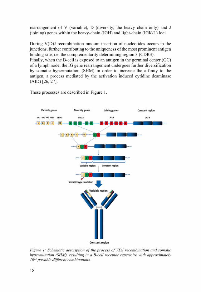
rearrangement of V (variable), D (diversity, the heavy chain only) and J (joining) genes within the heavy-chain (IGH) and light-chain (IGK/L) loci. During V(D)J recombination random insertion of nucleotides occurs in the junctions, further contributing to the uniqueness of the most prominent antigen binding-site, i.e. the complementarity determining region 3 (CDR3). Finally, when the B-cell is exposed to an antigen in the germinal center (GC) of a lymph node, the IG gene rearrangement undergoes further diversification by somatic hypermutation (SHM) in order to increase the affinity to the antigen, a process mediated by the activation induced cytidine deaminase (AID) [26, 27]. These processes are described in Figure 1.
Figure 1: Schematic description of the process of VDJ recombination and somatic hypermutation (SHM), resulting in a B-cell receptor repertoire with approximately 1012 possible different combinations.

19
The binding of an antigen to the IG component of the BCR leads to the formation of the signalosome in which the SRC-kinase LYN phosphorylates CD79A and CD79B, leading to phosphorylation of the tyrosine kinase SYK. This in turn propagates signaling through phosphorylation of the tyrosine kinase BTK (Bruton’s tyrosine kinase) and PLCg2 (Phospholipase-gamma-2) [28]. The signal is further propagated downstream of the signalosome through a cascade involving, among others, phosphatidyl-inositol-3 (PI3K), ultimately leading to activation of transcription factors including NF-kappa-B, as depicted in Figure 2.
Figure 2: Simplified scheme of BCR-signaling in B-cells. From: Mattsson, M and Scarfò, L, BTK Inhibitors: Focus on Ibrutinib and Similar Agents. Resistance of Targeted Therapies Excluding Antibodies for Lymphomas, Springer 2018: p. 1-22. [29] (reprinted by permission) The final results of BCR signaling are changes in gene expression that regulate proliferation, migration and apoptosis.
The response to signaling through the BCR in normal B-cells is heterogeneous and depends on the density of surface Immunoglobulin M (sIgM) and duration and strength of signaling.
IGHV gene mutational status As mentioned in the introduction, two pivotal papers published in 1999 described the prognostic importance of the IGHV gene mutational status [21, 22]. Approximately 60% of patients exhibited CLL cells that had undergone SHM of the clonotypic IGHV genes, and were designated IGHV-mutated CLL (M-CLL), while patients with CLL cells that had not gone through SHM of the IGHV genes (40%) were termed IGHV-unmutated CLL (U-CLL).
20
In general, M-CLL patients have a more favorable outcome compared to U-CLL patients that follow more aggressive disease courses with rapid disease progression and active disease in need of treatment.
In addition, U-CLL patients are more often in need of relapse treatment after receiving first-line treatment with chemotherapy or chemoimmunotherapy (CIT). These differences in response to therapy are also manifested in the significant differences in OS observed between the two subgroups when using these treatments.
The cut-off in the distinction between M-CLL and U-CLL was set at 98% identity to the germline, a distinction based on clinical data and not reflecting a true biological cut-off. One reason behind the survival difference observed, is that M-CLL and U-CLL differ in the strength of the signaling through the BCR, where the former has a weak or mitigated response and the latter a stronger response to BCR stimulation. The result of BCR signaling in M-CLL and U-CLL is also different, with BCR-signaling in M-CLL leading to anergy and in U-CLL to proliferation [30]. The prognostic impact of IGHV gene mutational status was challenged in 2002, with the discovery that patients utilizing the IGHV3-21 gene had an as equally poor prognosis as U-CLL, despite that the majority belonged to the M-CLL subgroup [31]. At the same time, it was discovered that a significant proportion of patients utilizing the IGHV3-21 gene also carried highly similar VH CDR3 sequences as well as identical light chains, providing a strong evidence for antigen involvement during CLL development. Further research could prove that a significant proportion of CLL patients belonging to both the M-CLL and U-CLL subgroups showed identical or semi-identical VH CDR3 within their BCR. As the probability that this event would happen by chance is extremely small (estimated to 10-12) this strongly implies some sort of selection, presumably antigen-driven [32].
Stereotyped subsets Today, it is established that more than 40% of the CLL patients can be classified into different subgroups, termed stereotyped subsets, with cases belonging to each subset carrying quasi-identical or stereotyped BCRs on their surface. Approximately 12% of the patients belong to one of 19 major subsets [33]. Importantly, mounting evidence demonstrates that patients assigned to a specific subset share similar biological characteristics and prognosis [34].
A classic example is stereotyped subset #2 which consists of patients utilizing the IGHV3-21/IGLV3-21 genes. It is the largest subset and constitutes approximately 3% of all CLL cases and 5.5% of those in need of

21
treatment [33, 35]. Assignment to subset #2 has been shown to be associated with inferior prognosis, despite that few of these patients carry TP53-aberrations (see below). Another subset with adverse prognosis is subset #1 (IGHV1/5/7/IGKV1(D)-39) which is the largest subset within U-CLL. In contrast, patients belonging to subset #4 (IGVH4-34/IGKV2-30) have a very favorable outcome with median time to first treatment exceeding 10 years [36].
Clinical impact of the IGHV gene mutational status Although the prognostic impact of assigning patients to the M-CLL and U-CLL subgroups has been extensively studied over the years, the IGHV gene mutational status has not until recently been recommended in clinical routine. Solid data are now accumulating that the IGHV mutational status has a predictive role and should be taken into consideration when selecting treatment in many cases. This is also reflected by the updated iwCLL guidelines, as well as Swedish National CLL guidelines, where it is now recommended to analyze the IGHV mutational status in routine clinical practice [20].
The genetic hierarchy of CLL While the prognostic importance of different genetic abnormalities in CLL has been known for a long time [37], classical chromosome banding analysis (CBA) has been difficult to perform in CLL, due to the inherent problems in culturing CLL cells and obtaining metaphases.
Fluorescence in situ hybridization (FISH) By applying fluorescence in situ hybridization (FISH), in the seminal paper by Döhner et al in 2000 [23], CLL patients could be classified into 5 subgroups with different survival based on 4 different chromosomal aberrations, i.e. del(17p), del(11q), del(13q) and +12 (the fifth group represents those without any aberration detected). Using FISH, at least one of these aberrations can be detected in up to 80% of patients with CLL. According to the Döhner hierarchical model, patients with del(17p) and del(11q) exhibit a significantly worse prognosis compared to patients with isolated del(13q), while cases negative for any of these 4 abnormalities (‘normal FISH’) or harboring +12 have an intermediate prognosis. Of these aberrations, del(17p) was associated with a particularly dismal prognosis with a median survival of only 32 months, mainly due to inferior response to chemotherapy as well as CIT. This explained by the fact that the

22
deletion leads to inactivation of the TP53 gene, a gene of key importance for cell cycle control. This gene can also be inactivated by mutations that can be detected by sequencing. Most common is del(17p) coexisting with a TP53-mutation on the other allele (60%), approximately 30% of patients have biallelic TP53-mutations, while 10% have an isolated del(17p)[38]. TP53-aberrations, i.e. del(17p) and/ or TP53 mutation, occur in 4 to 8% of patients at diagnosis but, due to clonal evolution, in up to 30 to 40% of patients with relapsed/refractory disease [39-41]. Based on more recent deep-sequencing data, the occurrence of even small subpopulations carrying a TP53 mutation seems to be associated with inferior outcome [42] Patients carrying del(11q) also have an inferior prognosis when treated with chemotherapy and CIT. The deletion causes loss of the ATM gene, a gene that is central in DNA damage response; approximately 30-40% of these patients also have an ATM mutation on the other allele [43, 44]. del(11q) occurs in about 10-15% of patients at diagnosis with a rise to 30% at relapse [45]. While the typical clinical picture in these patients is bulky lymphadenopathy and a good initial response to treatment, they generally experience a fast relapse and progressive disease. While patients with trisomy 12 often have an atypical immunophenotype , the prognosis appears to a large extent be governed by the IGHV mutational status and is not influenced by the presence of other prognostic markers, as no genes of pathogenic importance have so far been identified on chromosome 12[34, 46, 47]. The largest group of patients are those carrying del(13q), which is found in up to 60% of all CLL, with 35-40% carrying it as the sole aberration [23, 48]. Del(13q) as a sole aberration is associated with favorable prognosis with a median survival of 133 months according to the Döhner et al study [23]. The deletion leads to the loss of two micro-RNAs, miR15A and miR16A [49], subsequently leading to the upregulation of the BCL2 protein, an antiapoptotic protein located in the mitochondrial membrane. The inhibition of BCL2 using the BH3-mimetic venetoclax is today used in clinical practice and further described in the treatment chapter. Nota bene, while the prognostic significance of these genetic aberrations has been extensively studied and validated in patients treated with chemotherapy or CIT, the prognostic impact in patients treated with BTK-inhibitors or BCL2-inhibitors is much less known.
23
Chromosome banding analysis The previous difficulties with obtaining sufficient metaphases to perform CBA have now been overcome by the addition of novel mitogens (e.g. CpG oligonucleotide and IL2)[50, 51]. Using the modern culturing protocols, complex karyotype, defined as ³3 or ³5 aberrations has been associated with an inferior prognosis [48, 52-55]. In a recent publication including more than 5,000 cases, patients with ³5 aberrations were demonstrated to have a particularly dismal outcome, while an inferior prognosis was only observed in patients with 3 or 4 aberrations in association with TP53 aberrations [54].
Indeed, the presence of a complex karyotype has been shown to be an even stronger predictor of outcome than TP53-aberrations in relapsed/refractory (R/R) CLL patients treated with the BTK-inhibitor ibrutinib[53].
Recently, the combination of complex karyotype, genetic aberrations (in particular TP53-aberrations) and IGHV mutational status has been suggested as a novel hierarchical model to improve prognostication[54]. There is now work ongoing to reach consensus on the definition of complex karyotype, develop and validate the best methods to detect it, and prospectively study its clinical impact. Until then, the presence or not of a complex karyotype is not recommended to be used in the clinical decision making.
Sequencing – Next-generation sequencing Technical progress in sequencing and bioinformatics has made it possible to further explore the genome in CLL. Older techniques such as Sanger sequencing are now being complemented or in most cases replaced by next-generation sequencing (NGS). This is a field of fast and continuous development, with the possibilities to sequence either a few specific genes or perform whole-exome sequencing (WES) or whole-genome sequencing (WGS). Thus, it is now possible both to the scan a large part of the genome for mutations (WES/WGS) or to detect with high sensitivity specific mutations present only in low proportion of tumor cells (targeted NGS). This progress has led to the discovery of more than 2,000 genes found to be recurrently mutated in CLL[56, 57]. To date, more than 40 driver genes or potential driver genes have been associated with CLL. The majority of these occur at a low frequency (<1-5%) with only a few (ATM, NOTCH1, SF3B1 and TP53) reported in more than 5% of the patients [56-58]. In addition to TP53 and ATM aberrations, mutations in NOTCH1, SF3B1, BIRC3, EGR2 and RPS15 have a negative prognostic impact in CLL, while the impact of mutations in MYD88 is still uncertain[59, 60]. The main pathways affected by these mutations are DNA-damage response, NOTCH1-signaling, RNA-

24
splicing, NF-kB signaling, BCR-signaling, toll-like receptor signaling and chromatin modification[61]. Recent data also suggests that the number of pathways affected by driver mutations have an impact on prognosis [62]. The consequence of NOTCH1 mutations (predominantly a 2 bp deletion) is the accumulation of the intracellular domain (ICN1), followed by constitutive activation of the NOTCH pathway [63]. NOTCH1 mutations occur in about 10% of newly diagnosed patients and up to 20% in those with advanced disease[58, 64]. They are associated with trisomy 12, assignment to stereotyped subset #8, U-CLL and an elevated risk of Richter transformation [41, 64-70]. The clinical picture resembles 11q deletion, i.e. patients often have a short time to first treatment and a short time to progression after chemotherapy or CIT [39, 71-73]. NOTCH1 mutations have also been associated with low expression of CD20 and no benefit from the addition of anti-CD20-antibodies [74, 75], but this has to date not changed clinical practice or treatment guidelines in Sweden. SF3B1 mutations have been associated with aberrant mRNA splicing of a number of genes involved in DNA-damage response and NOTCH-signaling; however, the exact pathogenic mechanisms of these mutations in CLL are still unknown. SF3B1 mutations are found in 5-17% of patients [76, 77] and are associated with shorter time to first treatment and OS. They are highly enriched (45%) within subset #2 [39, 68, 70, 78, 79] and also associated with del(11q) and ATM mutations[39]. Mutations in BIRC3 are correlated with a very poor prognosis. The BIRC3 protein is involved in the MAP3K-non-canonical NF-kB pathway and BIRC3 mutations lead to constitutive activation of this pathway [70]. Mutations in BIRC3 are rarely detected at diagnosis (2-4%) but accumulates with treatment and have been found in 24% of R/R CLL patients in one study [58, 76, 77, 80]. Interestingly, they are mutually exclusive to 17p deletion/TP53 mutations but associated with deletion 11q and trisomy 12 [41]. Mutations in MYD88 lead to constitutive NF-kB activation and are found in more than 90% of patients with Waldenstrom’s macroglobulinemia[81]. Mutations in this gene are also found in CLL, but at a lower frequency (2-5%), with an enrichment in patients with M-CLL and without major differences in outcome in relation to wildtype patients [59, 60]. Mutations in the transcriptional factor EGR2 are associated with a very poor outcome, similar to TP53-aberrant CLL, and were found in 3.8% of the patients in a large cohort of CLL patients. They were associated with advanced-stage disease, U-CLL, ATM lesions and TP53 mutations. Of notice

25
was the dismal outcome for patients with concomitant EGR2 and TP53 mutations [82]. Finally, mutations in the gene coding for the ribosomal protein RPS15 have been found to be enriched after FCR treatment, with 20% of patients harboring this mutation at relapse. RPS15 mutations are associated with TP53 aberrations and a more clinically aggressive disease [69].
Figure 3: The frequency of copy number alterations and mutations, and the different pathways affected from: Fabbri and Dalla-Favera, Nature Reviews, Cancer. Vol. 16 2016. (reprinted by permission)
Clonal evolution – Darwinism at work Clonal evolution is a crucial event in progression, relapse and resistance to treatment in malignancies, present also in CLL[83] . Broadly, the genetic aberrations identified in CLL can be separated into those occurring at a clonal level (clonal driver mutations), e.g. deletion 13q and trisomy 12, and mutations occurring at a subclonal level (subclonal drivers), e.g. TP53 and SF3B1 mutations. With treatment administered and the cells exposed to evolutionary pressure, there is a selection and expansion of subclones not sensitive to treatment or with a growth advantage in relation to other cells (Figure 4). This is associated with treatment failure and a worse outcome [84-89]. The latter is underscored by the accumulation post-treatment of mutations associated with an inferior outcome, such as TP53, BIRC3 and NOTCH1.

26
Figure 4: Illustration of the concept of clonal evolution. A: Continuous treatment e.g. BTKi. B: Intermittent treatment e.g. CIT. The red circles depict cells resistant to the administered treatment Using FISH, which has a low sensitivity to detect subclonal changes, it has been shown that clonal evolution occurs in more than 25% of untreated patients after >5 years follow-up [90]. With more sensitive techniques, such as NGS, more detailed information on changes in tumor cell composition has been acquired [57, 91]. When using chemotherapy or CIT, the treatment is usually limited in time, and the goal is in most cases to reach as deep remission as possible, with patients obtaining a complete remission (CR) with only a small amount of minimal residual disease (MRD) or no MRD at all (MRD negativity). This is followed by monitoring and retreatment when (or if) the disease relapses, and the patient fulfills the established criteria for initiation of treatment. This approach has been associated with the creation of “evolutionary bottlenecks” with the emergence of resistant clones when the disease recurs (Figure 4B). With modern treatments, i.e. BCR-inhibitors (BCRi) and BCL2-inhibitors, the risk of clonal evolution and progress during or after treatment is largely unknown. The preferred treatment of today among these new drugs, is the use of the BCRi ibrutinib. Treatment with single-drug ibrutinib is very effective in ameliorating the patient´s signs and symptoms of disease, but it very rarely leads to a CR, and in even fewer cases leads to an MRD negativity[92, 93]. Thus, the patients usually have a prevailing high level of tumor cells, albeit these cells are in a quiescent state (Figure 4A). This has raised the question whether these remaining tumor cells might be associated with a risk of clonal evolution during long-term treatment.
27
Another new concept of treatment is that of time-limited treatment with BCL2-inhibitors alone or in different combinations (BCRi +/-BCL2i+/- CD20-antibodies). These treatments have a greater potential of inducing CR and MRD negativity, but if there is a risk of creating “evolutionary bottlenecks” and inducing resistance is largely unknown [94, 95]. Recent data from one centre has indicated that the propensity of early clonal shifts (within 6 months after start of treatment), and the presence of subclonal drivers, might be of prognostic importance in patients receiving treatment on a continuous basis with ibrutinib [94]. There are also data indicating that patients with ongoing clonal shifts before treatment have a higher risk of progressive disease [96].
Prognostication and prediction – does really one fit all? Already in 1975, the first system for prognostication in CLL was published, namely the Rai staging system [97]. This was followed by a similar prognostic score, the Binet staging system, in 1981 [98]. These staging systems separate patients into 3 main prognostic subgroups based on easy and accessible clinical and laboratory parameters, namely the presence (or absence) of lymphadenopathy, hepatomegaly and splenomegaly as well as anemia and/or thrombocytopenia. Over time, the practical utility of these staging systems has diminished due to the fact that the majority of patients (approximately 3/4) are today diagnosed in early clinical stages, i.e. Binet A and Rai 0-I. Among these patients, the Rai and Binet systems cannot help in further identifying patients with high risk of progression and in need of starting treatment. In addition, Rai and Binet staging do not contribute any predictive information at the time of treatment initiation, i.e. they cannot help in selecting the best treatment for the individual patient. However, despite that their importance has decreased, they are still used in routine clinical practice and are also a part of the CLL-IPI (see below). The expanding knowledge on the biology of CLL has identified a large number of different prognostic variables. This has led to the development of new prognostic indices based on different combination of genetic and phenotypic factors as exemplified in Figure 5, with variables that can be classified into host-related, clinical, laboratory, genetic and phenotypic factors.

28
Figure 5: Different variables used in the prognostication of CLL and their use in prognostic scores/indices. From; Baliakas, P, Mattsson, M et al., Prognostic indices in chronic lymphocytic leukaemia: where do we stand how do we proceed? J Intern Med, 2016. 279(4): p. 347-57.[99] (reprinted by permission) The recently developed CLL-international prognostic index (CLL-IPI) [100] is based on five variables, i.e. the age of the patient (≤65 vs >65 years), the presence of TP53-aberrations, IGHV status, β2-microglobulin level and clinical stage (Binet A or Rai 0 vs. Binet B or C or Rai I-IV), and separates patients into 4 risk groups (low, intermediate, high and very-high risk).
The CLL-IPI was developed as a tool for prediction of OS after start of treatment, but has also been shown to predict time to first treatment (TTFT) among patients without indication for treatment [101].
This system, as well as other new prognostic scores and indices, was developed and validated in populations treated with chemotherapy and/or CIT, and has not yet been largely tested on populations treated with BCRi or BCL2 inhibitors [102, 103]. Due to this, none of the new prognostic scoring systems have yet been broadly introduced in routine clinical practice. Recently, a new prognostic index, the International prognostic score in early stage CLL (IPS-E), was developed. This score aims at assessing the risk for

29
newly diagnosed patients, without treatment indication at diagnosis, to develop a need for treatment within 5 years.
This index is based on three parameters; the IGHV mutational status (U-CLL=1 point), presence or not of palpable lymph nodes (presence=1 point) and the absolute lymphocyte count (>15x109/L= 1 point). The proportion of patients developing need of treatment 5 years after diagnosis was determined to be 8%, 28% and 61% in the low-risk group (score 0), intermediate-risk group (score 1) and high-risk group (score 2-3), respectively [104]. From a practical point of view, the use of prognostic and predictive markers should be viewed in the context of what time-point the assessment is made, especially when applying time-limited treatments as described above. The different time-points when the patient is assessed can be designated as “decision points”[99]. The prognostication of a patient with newly diagnosed CLL without treatment indication differs profoundly from the patient that has developed need for treatment, and even more from the patient that has been treated with one or more lines of treatments. Thus, the ideal would be the use of different systems based on the different “decision points” (Figure 6).
Figure 6: Depiction of the different “decision-points” during CLL-evolution. From; Baliakas, P., Mattsson, M et al., Prognostic indices in chronic lymphocytic leukaemia: where do we stand how do we proceed? J Intern Med, 2016. 279(4): p. 347-57. (reprinted by permission) In the future, we will need to identify more predictive markers, meaning factors that give information on the outcome of different therapies and therefore guide the choice of treatment. Disregarding host- and treatment-related factors, the only truly predictive factor in use during the last decades has been the presence or not of TP53 aberrations [74, 105, 106].

30
When continuous, indefinite treatment with BCRi is applied, there are also different clinical situations when the patient is assessed. That is; before treatment, during stable disease (but with residual tumor cells) or at a time of suspected progression. At start of treatment, the primary question is to assess the likelihood for the patient to respond to treatment. During stable disease, one would ideally want to monitor the evolution of the disease and predict the risk of progression, while at the time of suspected or manifest progression, the aim is to evaluate the cause of resistance (e.g. BTK and/or PLCγ2 mutations) and find the optimal new treatment to be applied. While the IGHV mutational status has an established impact on prognosis, there has also been an interest to assess its predictive capacity. In 2016, follow-up data from the German CLL8 trial as well as from the MD Anderson Cancer Center demonstrated that the long-term outcome of FCR treatment (fludarabine, cyclophosphamide and rituximab) differed significantly between M-CLL and U-CLL patients, with shorter PFS and OS for those with U-CLL [107, 108].
More recently, data from several large randomized trials (described on page 36-37) demonstrated superior PFS for ibrutinib, alone or in combinations, compared to CIT in U-CLL patients, whereas no significant differences in PFS were seen in M-CLL patients. These data have resulted in national/international guidelines recommending the IGHV mutational status to be analyzed in routine clinical practice, as it may guide in the choice of treatment [20]. The importance of being able to choose treatments with a high efficacy is evident, especially with the expanding number of different treatments at hand. In addition, it becomes more and more important to be able to evaluate the risk of adverse events associated with the different treatments. As the majority of CLL patients are elderly and are expected to have a high burden of co-morbidities, knowledge of the tolerability and the risks associated with different treatments is of paramount importance.

31
Treatment of CLL
At diagnosis, approximately 15% of patients are in need of treatment, and with time approximately 60% of patients will eventually require treatment [1, 109] However, therapy for CLL is not be initiated in patients with asymptomatic disease as several trials have failed to prove the value of such a strategy [110-113]. Criteria for starting treatment are outlined in Table 1[20]. Table 1: Criteria for initiating treatment in CLL.
Developing or worsening anemia and/or thrombocytopenia due to bone-marrow infiltration Constitutional symptoms Night sweats ³1 month and/or fever 38°C for ³2 weeks without infection Weight-loss ³10% in 6 months Significant fatigue Autoimmune anemia or thrombocytopenia resistant to conventional treatments Massive lymphadenopathy (³10 cm) Massive splenomegaly (³6 cm below the costal margin) Progressive or symptomatic lymphadenopathy or splenomegaly Lymphocyte doubling time £6 months or rise with ³50% in £2 months (not to be used a sole criterium)
Chemotherapy, antibodies and chemoimmunotherapy – an evolving story Over a very long period of time the backbone of CLL treatment was the alkylating agent chlorambucil, introduced after the publication of a pivotal paper by David Galton in 1955[114]. The use of this drug has since then been widespread until recent years, and it has also been used as comparator in many trials studying new treatments. The use of combination chemotherapy regimens such as COP (cyclophosphamide+oncovin+prednisone) and CHOP (cyclophosphamide+ adriamycin+oncovin+prednisone), did not lead to any progress compared to

32
chlorambucil [112]. Adding corticosteroids to chlorambucil is in general not beneficial [115]. In 1991, the first trial was published with the purine analog fludarabine in CLL, with subsequent studies showing higher overall response rate (ORR) and CR rates as well as longer response duration compared to chlorambucil [116, 117]. These results were confirmed in subsequent phase III trials, but none of those could show an improvement in OS [118]. Similar results were seen with other purine analogues, i.e. cladribine and pentostatin [119], but further development favored fludarabine, which became the most commonly used purine analogue in CLL. Fludarabine was then combined with cyclophosphamide in the FC-regimen. In three large simultaneous trials, the FC-combination was proven superior to both single agent chlorambucil and single agent fludarabine [120-122] with respect to ORR, duration of response and CR rate. Disappointingly, this improvement in response was not translated into a prolonged OS. The first treatment that definitely could prove a survival advantage compared to previous treatments, was the FCR-regimen, which included the monoclonal anti-CD20-antibody rituximab in addition to the FC-treatment.
Rituximab is a humanized monoclonal antibody targeting the CD20-antigen that is expressed on both precursor and mature B-lymphocytes, leading to both complement dependent cytotoxicity (CDC) as well as antibody-dependent cellular cytotoxicity (ADCC) [123]. CD20 is expressed weakly on the surface of CLL cells and initial trials with rituximab as monotherapy showed only modest single agent efficacy in CLL, requiring the use of very high doses of the drug. A phase II trial using FCR as first line treatment was published in 2005 [124] and showed an impressive CR rate of 70%. It was followed in 2010 by the pivotal publication of the CLL8-trial [105], with a CR-rate of 44% in the FCR-treated patients vs. 22% in the FC-treated group and a progression free survival at 3-years of 65% vs. 45%, respectively. Follow-up data from the CLL8-trial has highlighted the adverse impact of TP53-aberrations, as well as the prognostic impact of reaching MRD-negativity. In addition, long term follow-up data revealed that M-CLL patients had a superior outcome compared to U-CLL patients, with a plateau in the survival curve in the former group [74, 107, 125]. Despite the efficacy of FCR, its use is limited to younger and fit patients, primarily due to myelosuppression leading to infectious complications. Another concern is also the risk of secondary malignancies including MDS, AML and Richter transformation, affecting 13% of the patients in the follow-

33
up of the CLL8-trial [107]. Due to this, a large proportion of the patients were still treated with chlorambucil upfront as they were elderly and/or unfit and thus not suitable for fludarabine-based regimens. The interest was then focused on an old drug - bendamustine - a drug with both alkylating and purine analog properties [126]. In 2001-2002 two phase II trials proved its efficacy and tolerability in CLL [127, 128]. A phase III study on bendamustine vs. chlorambucil [129] published in 2009 reported a CR rate of 31% vs. 2% and PFS of 22 months vs. 8 months, respectively. Despite this, no difference in OS was observed between the two treatments when updated results were published in 2012 [130]. As the next step, bendamustine was combined with rituximab (BR) in the same fashion as FCR. A phase III trial (GCLLSG CLL10) comparing BR to FCR was published in 2016 [131]. The results proved FCR to be more effective than BR, with a PFS of 55 months vs. 42 months, but with significantly more neutropenia and infections in the patients treated with FCR. These adverse events (AE) were so pronounced in patients above the age of 65 years that it outweighed the positive effects of treatment with FCR. The ensuing general recommendation following this trial was that FCR retained its role as first-line treatment in fit patients below the age of 65, whereas BR was recommended for unfit patients, or those above the age of 65. In parallel with studies on these rituximab-combinations, several trials have studied whether the addition of rituximab and other anti-CD20-antibodies to chlorambucil could improve the results in older/unfit patients without inducing intolerable toxicity. Two new CD20-antibodies were used in these trials - ofatumumab and obinutuzumab. Ofatumumab is a type-I human monoclonal antibody targeting CD20 at another epitope than rituximab [132]. It was approved for single drug use in CLL refractory to fludarabine and alemtuzumab or fludarabine-refractory CLL with bulky disease [133, 134]. It was later studied in combination with chlorambucil and compared to chlorambucil alone in the COMPLEMENT-1 trial [135]. In February 2019, ofatumumab was withdrawn from the market for the use in CLL by the pharmaceutical company, instead focusing on its use in multiple sclerosis. Obinutuzumab is a type-II humanized antibody targeting CD20 with a stronger ADCC and weaker CDC in comparison to type-I antibodies such as rituximab and ofatumumab [136]. The first phase-I obinutuzumab trial was published in 2014 [137]. Follow up studies included the pivotal phase III CLL11-trial from the German CLL study group (GCLLSG) [138, 139]. This three-arm trial compared chlorambucil vs. chlorambucil+rituximab (ChR) vs.

34
chlorambucil+obinutuzumab (CO) with PFS of 11 months, 16 months and 17 months, respectively. With long-term follow up data, a difference in OS was seen between the group treated with chlorambucil alone compared to ChR and CO, with the latter two regimens resulting in better OS. The addition of obinutuzumab resulted in more infusion-related reactions and neutropenia, but without more infections than ChR.
This study definitively established that the addition of an anti-CD20-antibody to chlorambucil is superior to chlorambucil used alone, and that it is feasible even in elderly and/or patients with high co-morbidity.
Treating CLL with TP53-aberrations The inferior effect of chemotherapy and CIT in patients with TP53-aberrations was evident in all trials using these treatments. In search for better alternatives for these patients, progress was made with the antibody alemtuzumab. Alemtuzumab is a humanized antibody targeting the CD52-antigen, which is present on the surface of B-cells, T-cells as well as NK-cells, macrophages and monocytes [140]. Alemtuzumab was granted approval for the use in CLL in 2001 after the positive findings in phase I and II trials [141]. Due to its broad effects on the immune system, the use of alemtuzumab remained limited, and it was mainly used in patients with TP53-aberrations where it was proven effective[142-145]. Until the introduction of BCR-inhibitors and BCL2-inhibitors the treatment of choice for patients with TP53-aberrations was alemtuzumab, or for the relatively few young and fit patients, allogeneic stem cell transplantation [146, 147].
Today, alemtuzumab is not in routine use for treatment of CLL due to the introduction of BCR-inhibitors and BCL2-inhibitors. These new treatment modalities have shown superior efficacy and outcome in patients with TP53-aberrations compared to chemotherapy, CIT and alemtuzumab[148, 149]. Today, all patients with TP53-aberrations are recommended treatment with these modern therapies.
Allogeneic stem cell transplantation Allogeneic stem cell transplantation (allo SCT) has been regarded as the only treatment with curative potential in CLL. The use of it has been hampered by the significant risk of treatment-related mortality and chronic graft versus host disease (GVHD)[150]. In 2007 a consensus EBMT document was published with criteria for when and how to use allo SCT in CLL. The advice was to use reduced intensity conditioning regimens, and the indications were CLL with TP53-aberration or patients refractory to, or relapsing within 2 years after treatment with CIT [147].

35
After the introduction of BCR-inhibitors and BCL2-inhibitors, the use of allo SCT in CLL has dropped rapidly. Today, there is no consensus on when to perform allo SCT, and the current advice is that high-risk patients must be assessed individually regarding the indication for this treatment [150].
Paradigm shift – new treatments The successive refinement of CIT described above was followed by a decisive paradigm-shift when novel treatments with BCR-inhibitors and BCL2-inhibitors were introduced. Treatments with chemotherapy and CIT have restricted modes of action when targeting malignant cells. The new treatments introduced have expanded the ways to interfere with the different survival mechanism used by the CLL cell. These different targets, as well as potential new targets for the treatment of CLL are outlined in Figure 7.
Figure 7: Overview of the different targets for approved treatments in CLL (red bold) and possible future treatments (red – light). The picture depicts a CLL cell in the lymph node microenvironment . Abbreviations; BTK=Bruton’s tyrosine kinase, CD=clusters of differentiation, DNA=deoxyribonucleic acid, FDC=follicular dendritic cell, IMIDs=immunomodulatory imide drugs, LYN=Lck/Yes novel tyrosine kinase, NLC=nurse like cell, PI3K= phosphatidyl-inositol-3-kinase, PD1= programmed cell death protein, PDL1=programmed cell death protein ligand 1, PLC𝛾2=phospholipase-gamma-2, sIg=surface immunoglobulin, SYK= spleen tyrosine kinase.

36
Targeting signals to survive – the Bruton’s tyrosine kinase In 1952, Dr. Ogden Bruton described the disease X-linked hypogammaglobulinemia, a disease characterized by B-cell lymphopenia and severe hypogammaglobulinemia[151].
In the 1980s, the gene coding for the Bruton’s tyrosine kinase (BTK) was identified on the X chromosome [152]. The cloning of the gene revealed a cytoplasmic tyrosine kinase downstream of the BCR, with PLCg2 as its primary substrate. This knowledge opened the possibility of blocking BCR signaling as a potential treatment option for autoimmune diseases and B-cell malignancies.
Ibrutinib – the first BTK-inhibitor In 1999 the first BTK-inhibitor was developed in the laboratory but not used further [153]. The first clinically useful BTK-inhibitor was introduced in 2010 following the development of the BTK-inhibitor PCI-3275, subsequently named ibrutinib [154].
In 2013, Byrd et al published a pivotal phase I study based on data from 56 patients with B-cell malignancies treated with escalating doses of ibrutinib and showing remarkably high response rates, few adverse events and with the optimal daily dose of 420 mg [155]. The first phase III trial with ibrutinib (RESONATE) compared ibrutinib with ofatumumab in a population of heavily pretreated patients with relapsed/refractory CLL, showing a significant better PFS and OS for the ibrutinib-treated patients despite a short median follow-up time of 12 months [156]. Notably, patients with TP53-aberrations seemed to respond well to ibrutinib. RESONATE-17, a single-arm study with ibrutinib including 145 patients carrying TP53-aberrations confirmed this, with ORR and OS at 24 months of 83% and 75% respectively, which was superior to historical controls [148]. The use of ibrutinib in the setting of primary treatment was studied in the RESONATE-2 trial[157]. This phase III trial randomized patients above the age of 65, without TP53-aberration, to receive single agent therapy with either ibrutinib or chlorambucil. The first data were published in 2015 with long-term follow-up published in 2020[158], proving a benefit for patients treated with ibrutinib both regarding PFS and OS at 24 months (ibrutinib 95%; chlorambucil 84%). Based on these data ibrutinib was approved for use both in the primary and relapsed setting in CLL patients with or without TP53-aberration. A number of clinical trials have thereafter studied ibrutinib in different combinations. In R/R CLL, phase III data have been published on the

37
combination of ibrutinib+BR vs. BR alone in the HELIOS-trial [159], showing superiority of the combination treatment. Unfortunately, this trial did not include an arm with single-dose ibrutinib, and the outcome of the BR+ibrutinib treatment seems to be comparable to results from treatment with ibrutinib alone in other trials. When used in primary treatment, recent data have shown that first-line treatment with ibrutinib alone, or in combination therapies, have resulted in superior PFS compared to CIT. Important to notice in these trials is that the differences in PFS are restricted to patients with U-CLL, whereas no significant differences have been described in M-CLL patients.
In the elderly, the ALLIANCE-trial compared ibrutinib, ibrutinib+rituximab (IR) and bendamustine+rituximab (BR) in previously untreated patients >65 years of age, resulting in a PFS at 2 years of 87%, 88% and 74% respectively[160]. Interestingly, this study showed no benefit of the addition of rituximab to ibrutinib.
Another trial in elderly patients, the iLLUMINATE-trial, compared ibrutinib+obinutuzumab with chlorambucil+obinutuzumab, with an estimated PFS at 30 months of 79% and 31% respectively[161].
Finally, in the NCI E1912-trial, focusing on a younger and “fit” population, the same outcome was seen in a cohort of previously untreated patients treated with either FCR or ibrutinib+rituximab, with a PFS at 3 years of 73% and 89% respectively [162].
Adverse events of BTK-inhibitors As the concept of BTK-inhibitor treatment is different from CIT, so is the spectrum of AEs. The major AEs with ibrutinib are diarrhea, bleeding tendency due to inhibition of platelet aggregation, hypertension and an elevated risk of developing atrial flutter (AF) [163]. The pathophysiology of the cardiovascular effects of ibrutinib are largely unknown, although data suggests off-target effects on TEC-kinases leading to effects on the PI3K-Akt pathway to be of importance [164]. A meta-analysis of published trials has shown that treatment with ibrutinib is associated with a near 3-fold increased risk of hypertension, and a more than 4-fold increased risk of AF, in comparison to the comparator arms [165]. The risk of cardiovascular side-effects is also higher in patients with pre-existing cardiovascular risk-factors [166-168]. As the CLL population in general is old, with a potential high comorbidity, these cardiovascular effects are important to account for. In addition, the potent effect of ibrutinib on platelet aggregation can be a problem if patients are to be treated with anticoagulation, which is often indicated due to AF. In clinical trials with ibrutinib, the use of oral vitamin-K antagonists (e.g. warfarin) has been a criterion for exclusion. These side effects warrant more

38
data to be collected on cardiovascular morbidity and mortality in CLL patients. Comorbidities and polypharmacy may also cause problems with drug interactions as ibrutinib is metabolized through the CYP3A4-system. Lastly, the long-term effects of treatment with ibrutinib are yet not known, as the longest published follow-up data are after five years of treatment.
Duration of treatment A concern regarding ibrutinib treatment is that only a few patients reach CR, with even fewer reaching MRD negativity. As a consequence, the majority of patients have a high number of residual CLL cells left, this despite normalized blood counts and relief of symptoms.
Due to this, the treatment with ibrutinib is at the moment advised to be indefinite. This might entail clonal evolution, as described in Figure 4, and the potential risk of patients developing resistance to treatment, progression and Richter transformation [29, 169-171]. Richter transformation seems to be an early event during treatment with ibrutinib, with decreasing risk over time, and possibly an effect the inclusion of heavily pretreated, high-risk patients in the initial trials[172]. In contrast, the risk of developing resistance to ibrutinib treatment seems to be unchanged over time.
Resistance to ibrutinib Resistance to ibrutinib has so far been found to be caused by mutations in two genes in the majority of cases [173, 174]. One is a mutation in the BTK gene, most commonly C481S, that causes an amino-acid substitution (cysteine to serine), leading to a conformational change in the binding-site for ibrutinib which prevents binding of the drug[175]. The other mechanism of resistance are mutations of PLCγ2 which is the major substrate for BTK. These mutations lead to constitutive activation of PLCγ2 and bypasses the block of BCR-signaling caused by ibrutinib. The emergence of these resistance mutations often precedes clinical resistance with many months. In addition to the clinical problems that may occur during treatment with ibrutinib, the monthly cost of treatment is high. This in combination with its continuous use as well as the increasing prevalence of CLL, raises concerns regarding the impact on health economy.
New BTK-inhibitors Due to the broad kinase activity of ibrutinib, work is ongoing to develop BTK-inhibitors that are more specific, with stronger binding to BTK. Acalabrutinib is a second generation BTK-inhibitor currently approved in the US for the treatment of CLL and mantle cell lymphoma[176]. Several phase

39
III trials studying acalabrutinib as monotherapy or in different combinations have recently been published[177-179]. Acalabrutinib binds stronger to BTK and is more selective than ibrutinib with less activity on other kinases such as EGFR, ITK and TEC[180]. As described above, the inhibition of these kinases is associated with side effects such as diarrhea, platelet dysfunction and atrial flutter. The hope is that this more selective activity will be associated with less AEs. Zanubrutinib (BGB-3111) and tirabrutinib (ONO/GS-4059) are other second generation BTK-inhibitors currently tested in clinical trials. To overcome the resistance to BTKi-treatment caused by BTK mutations, there is also an ongoing development of BTK-inhibitors (e.g. vecabrutinib, fenebrutinib) that bind non-covalently to the enzyme and potentially have an effect even in the case of a conformational mutation in the drug-binding pocket [181].
Targeting phosphatidyl-inositol-3-kinases The phosphatidyl-inositol-3-kinases (PI3K) is a group of kinases involved in BCR-signaling [182] that play an important role in many cancers including B-cell malignancies. The PI3Ks can be classified into three classes, of which most interest has been focused on class I. The PI3Ks in class I can be further subdivided into four isoforms: alpha, beta, gamma and delta. Extensive research has been invested into studying both pan-PI3K-inhibitors, dual PI3K-inhibitors as well as more specific PI3K-inhibitors. In 2010 and 2011, promising pre-clinical data on the PI3K-d inhibitor CAL101, subsequently named idealisib, was reported [183]. In 2014, Furman et al published a first phase-III trial in which patients with R/R CLL, and deemed not being able to receive further chemotherapy or CIT, were randomized to receive either single-drug rituximab (R) or the combination of idelalisib 150 mg twice daily + rituximab (IR). The results showed an ORR of 13% (R) vs. 81% (IdR) and an OS at 12 months of 80% (R) vs. 92% (IdR) [184], that led to the approval of idealisib for treatment of R/R CLL. The design of this trial was criticized for using single-drug rituximab as comparator, as this treatment is not very effective in CLL Subsequent trials using idelalisib in treatment-naïve patients with non-Hodgkin lymphomas were stopped due to a high number of AEs related to immune disturbances (e.g. transaminitis, enterocolitis, pneumonitis), possibly related to an imbalance between T-cell subsets leading to autoimmunity [185]. There was also a high incidence of infectious complications including pneumocystis jirovecii pneumonia and cytomegalovirus (CMV) reactivation [185]. Due to this, the drug is presently only approved for use in the relapse setting.

40
A second generation of PI3K-inhibitors are also under development. Based on data from the phase III DUO trial, the dual PI3K-delta and gamma inhibitor duvelisib was approved for use in R/R CLL in the US [186].
BCL2 inhibition – restoring the apoptotic machinery Overexpression of the anti-apoptotic, mitochondrial BCL2 protein is a hallmark in many cancers, including CLL. The overexpression may lead to tumor progression and resistance to drugs that depend on an intact apoptotic machinery. The BH3-mimetic venetoclax binds and inactivates BCL2 leading to a restoration of apoptosis [187].
In 2016, the first phase I/II-trial proving the efficacy of venetoclax in R/R CLL was published [188]. The drug was also proved effective in patients with TP53-aberrations as well as those refractory to BCR-inhibitors[189, 190]. The subsequent phase III MURANO trial led to the approval of venetoclax+rituximab (VR) in R/R CLL [191, 192]. Importantly, this trial also established the feasibility of fixed-duration therapy with venetoclax. Patients with R/R CLL were randomized to receive either 2 years of treatment with VR (6 cycles of rituximab) or 6 cycles of BR. The rationale behind adding rituximab to venetoclax was to overcome potential resistance to venetoclax mediated by changes in the microenvironment [193]. The VR treatment was superior to BR both regarding PFS and OS. Furthermore, 63% of the patients treated with VR reached MRD-negativity in peripheral blood, which was associated with a favorable outcome. The CLL14-trial explored time-limited, first-line treatment of CLL patients with comorbidities, randomizing between venetoclax+obinutuzunmab (VO) and chlorambucil+obinutuzumab [149]. The VO treatment showed a superiority in PFS after 24 months that led to the approval of the VO-combination in first-line treatment of CLL. The most common AEs of venetoclax therapy are GI-disturbances and neutropenia.
Due to its potency, there is a high risk for rapid tumor reduction causing tumor lysis syndrome (TLS). This has led to the recommendation to slowly ramp-up the dose over a period of 5 weeks when initiating treatment [194]. As is the case for BCRi, there are also patients developing resistance to venetoclax. A number of different resistance mechanisms have been described including clonal shifts, reprogrammed mitochondrial function, mutations leading to overexpression of MCL1 as well as impaired binding of the drug due to BCL2 mutations [195-198]

41
No long-term follow-up data on the different treatments with venetoclax are available yet.
CAR-T cell therapy The use of CAR-T cell therapy has evoked a huge interest in the treatment of many malignancies. Its efficacy in CLL was proven early on in development, although further use has been somewhat hampered by the development of efficient targeted therapies as described above, as well as the results with inferior efficacy of CAR-T in CLL, compared with many other malignancies [199, 200]. The latter is possibly in part due to the defective function of T-cells seen in patients with CLL (T-cell exhaustion) [201]. Interestingly, the possible use of treatment with ibrutinib together with CAR-T cells is now being explored. This is based on the effect of ibrutinib on ITK (interleukin-2 inducible kinase) and its possible potential to overcome the T-cell exhaustion [202, 203].
PD1 and PD-L1 inhibitors Although preclinical data suggest that PD1 and PD-L1 expression play a part in suppressing the immune reaction towards the malignant cells in CLL [204], clinical data are scarce. A phase II trial with pembrolizumab in CLL, including 16 patients with R/R CLL and 9 with Richter transformation, showed no responses in the cohort of R/R CLL, while 4 out of 9 patients with transformation showed an objective response [205].
Summary of treatment and questions to be answered The treatment of CLL, both first-line and in the relapse setting, is currently undergoing rapid and profound changes, with non-chemotherapy treatments at large replacing the “old” treatments with chemotherapy and CIT. Reflecting this, there are currently (August 2020) 77 ongoing CLL trials with venetoclax and 342 trials with ibrutinib registered at clinicaltrials.gov.
As described in this chapter, it is evident that the understanding of the genetics and immunogenetics in CLL has had a pivotal impact on the development of novel treatments. Progress has also been made in the fields of epigenetics and the lymph node microenvironment. The importance of the latter illustrated by the clinical effect of the immunomodulatory drug lenalidomide in patients with CLL [206-208]. As the progress is fast and ongoing, guidelines both regarding first-line therapy as well as therapy of relapsed disease, need to be updated with short intervals to be kept up to date. As often, progress is followed by many new questions to be answered;

42
• What is the optimal timing and best combinations of the different treatments when using CIT, BCR-inhibitors, BCL2 inhibitors and anti-CD20-antibodies?
• Is there a future role for allo SCT in CLL? • Will treatments targeting the immune system, such as CAR-T therapy
and immunomodulatory drugs lead to further progress? • Should treatment be limited in time or indefinite? • Will the goal of treatment be relief of symptoms, as deep response as
possible, or even cure? • How do we balance the effect vs. risk of AEs, especially in the elderly
and in patients with comorbidities? • How can we develop robust prognostic and predictive tools, as well
as means of monitoring treatment, when using modern drugs? • How to prevent, detect and handle the occurrence of drug resistance? • What will the societal consequences be of the rapidly increasing costs
of treatment in combination with the rise in prevalence?

43
Aims of the thesis
The overall aim of this thesis was to address clinically relevant questions in CLL by collecting, analyzing and interpreting data using a broad approach, ranging from epidemiologic studies on a population level, to in-depth studies on genetics and immunogenetics in individual patients. The specific aims of each paper were: Paper I To explore whether the improved OS generally observed in CLL over time, mainly due to the introduction of CIT, is also valid for the poor-prognostic group of patients belonging to stereotyped subsets #1 and #2. Paper II To acquire solid population-based data on the actual prevalence of CLL and the changes in prevalence over time in Sweden. As the next step, based on these data, as well as data from randomized controlled trials comparing old and new treatments, to construct a model that could be used to estimate the future prevalence of CLL, taking into consideration the impact on survival of new treatment modalities. Paper III Within an unselected population-based cohort, describe the disease burden of cardiovascular disease (CVD) in patients with CLL, both at the time of diagnosis as well as at the time of initiation of treatment. Furthermore, to investigate the cumulative incidence of new CVD during the first 5 years after start of treatment with chemotherapy or CIT. Paper IV To investigate the occurrence and clinical impact of clonal shifts and subclonal drivers in patients with high-risk CLL treated with the BTK-inhibitor ibrutinib. This by applying WES and genomic arrays to sequential samples collected over a longer period of time, and to correlate these data with long-term clinical outcome.

44
Patients and methods
Patient material In Paper I we studied a multicentre cohort including in total 3504 CLL patients from 15 institutions in 9 countries in Europe and the US. Selected for the analysis were all patients that had received at least one line of treatment between May 1980 and February 2014. Basic demographics, data on treatment initiation and immunogenetic data was available for all patients, while FISH data was available for 53%. Patients were stratified into two groups based on the time of start of primary treatment, i.e. 1st of January 2006 and 1st of February 2014. These time points chosen based on the changes in clinical practice and treatment guidelines that had been implemented. The cut-off January 2006 was selected as the introduction of CIT into clinical practice started at this time. February 2014 was chosen based on the approval of ibrutinib in the US, as well as that the drug was made accessible in Europe on compassionate-use. To perform the study published in Paper II and Paper III we collected epidemiological data retrieved from several Swedish health registries. From the National Cancer Register (National Board of Health and Welfare) all individuals with a diagnosis of CLL from 1958 until the end of 2015 were collected. Data from the Swedish Cause of Death register and the Register of the Total Population and Population Changes were collected from Statistics, Sweden, as well as data on immigration and emigration. To be able to make statistical calculations on the future prevalence of CLL in Sweden (Paper II), we also needed to construct a purpose-built model. In this process it was necessary to retrieve data on treatment and survival from the Swedish CLL register, started 1st of January 2007. In the model, we also incorporated survival data and hazard ratios (HR) from randomized controlled trials comparing old and new treatments, identified by performing a detailed literature search.
The final model used is presented in detail in Paper II. In Paper III, we used the same epidemiological data as described above, restricting us to all patients diagnosed with CLL in Sweden between 1st January 2007 and 31st of December 2010 (n=2078) from the National Cancer Register and using treatment data from the Swedish CLL register. In addition,

45
data on cardiovascular diagnoses were collected from the Swedish National Patient Register. The time point 2007 was chosen in order to have access to treatment data on all patients from the Swedish CLL register. The cohort was used to study the history of CVD from 10 years before and up to the time of diagnosis, as well as at the time of start of treatment. To study the cumulative incidence (CI) of new CVD during the first 5 years after start of chemotherapy or CIT, the 828 patients starting treatment between 1st of January 2007 and 31st of December 2016 were followed from start of treatment until first CVD, death or censored. From this analysis, 56 patients that had been treated with ibrutinib either as first-line or at relapse were excluded, as we wanted to have a study population not exposed to ibrutinib due to the cardiovascular side-effects of this drug. In Paper IV 10 unselected, consecutive patients with high-risk CLL treated with ibrutinib with available samples from the local U-CAN biobank were included. The median time from pre-treatment sample to analyzed treatment sample was 14 months (range, 11-39 months). Germline DNA collected from buccal washes and tumor samples from blood and/or bone-marrow at start of treatment and during follow-up was available for all patients. Detailed clinical data from the time-point of CLL diagnosis to the time point when the patients were deceased or censored was accessible for all patients with a total follow-up time of 60 months (range, 23-67 months)
Methods The different techniques applied in the papers are described in detail in each paper and/or supplement included in the thesis. In Paper I, to determine the IGHV mutational status and subset assignment, PCR amplification and sequence analysis of IGHV-IGHD-IGHJ rearrangements were performed on either genomic DNA (gDNA) or complementary DNA (cDNA). The resulting sequence data were analysed using validated online tools. Only productive rearrangements were evaluated. Information was extracted regarding IG gene usage, VH CDR3-IMGT length and amino acid sequence composition and SHM status. The assignment to a stereotyped subset depends on predefined criteria; (i) the amino-acid length of the VH CDR3, (ii) the composition of the VH CDR3, and (iii) the usage of IGHV genes belonging to the same clan [33, 209]. FISH analysis on patients included in Paper I was applied to determine the presence of del(17p), del(13q), del(11q) or trisomy 12 using standard protocols.
46
In Paper IV we performed WES on paired tumor/normal samples from all patients. The mean sequencing depth was 107x for all tumor samples and 95x for the germline controls. Single nucleotide variants (SNVs) and small indels were detected with a 10% variant allele frequency (VAF) cutoff. Genomic array analysis was also performed on all tumor and normal samples in Paper IV according to standard protocols. Copy-number alterations (CNAs) were detected using the rawcopy analysis suite. Evidence of complex karyotype was defined as ≥3 and ≥5 CNAs. Finally, to be able to follow clonal shifts over-time in patients in Paper IV, we calculated the cancer cell fraction (CCF) in each sample, followed by a longitudinal cluster analysis. The calculation of CCF was carried out using the ABSOLUTE algorithm as described in the paper. The CCF adjusted MAF files were aggregated and analyzed using PhylogicNDT to follow clonal shifts over time. Significant clonal shifts were defined as a change in mean CCF for a mutation cluster of ≥0.1 from the pre-treatment sample to the last available time point, as previously described by Landau et al [94].
Statistical analyses In all papers, quantitative variables were analyzed using means and medians as well as min-max values. Descriptive statistics were applied on discrete parameters including counts and frequency distributions (percentages). In Paper I associations between categorical variables were assessed using the Chi-2 test. To assess OS, we applied the Kaplan-Meier method, while the log-rank test was used to assess differences in survival.Tests were two-sided and statistical significance was defined as a P-value <0.05. Statistical analysis was performed using the Statistica Software v.10 (StatSoftInc, Tulsa, OK, USA). In paper II, future relative survival was estimated based on the mean of the relative survival observed during the years 2011-2015. The estimates of relative survival and predictions of future (expected) mortality rates were used to predict OS among living CLL patients at the end of 2015, and also among predicted future cases (2016-2060), based on a stable incidence of the disease. Relative survival was estimated up to 10 years after diagnosis, and then extrapolated by assuming a constant excess mortality rate after 10 years, equal to the average between 8 and 10 years after diagnosis. A flexible parametric model, with age at diagnosis included as a restricted cubic spline, was applied to estimate future relative survival for all ages [210]. This enabled us to calculate the prevalence, as well as the absolute number of patients for each year to come. Statistical analysis was done using R (R Core Team 2019).

47
Results and discussion
Paper I No improvement in long-term survival over time for chronic lymphocytic leukemia patients in stereotyped subsets #1 and #2 treated with chemo(immuno)therapy
Main findings and conclusions When comparing the two cohorts, cohort A treated 1980-2005 and cohort B treated 2006-2014, we could not find any significant differences in baseline characteristics, including critical prognostic variables such as age, sex, IGHV mutational status and genetic aberrations. This implies that the two groups were comparable in terms of important clinical and biological features as well as demographics. Nevertheless, the median OS was significantly different between the cohorts, with an improved survival in the later time-period (Figure 8A). This is in accordance with previously published data showing an improvement in survival in CLL during the time span of the study [2].
We could also demonstrate a significant improvement in OS in major subgroups, such as M-CLL, U-CLL, patients <55 years of age, patients with del(13q) or trisomy 12, male and female patients. Notably, a significant improvement in OS was seen in the high-risk group of patients with del(11q) (Figure 8B) as well as in elderly patients, many of whom were probably not fit for intensive treatment.
The exception to the finding of improved survival over time was observed in 3 specific subgroups; (i) patients assigned to subset #1, (ii) patients assigned to subset #2, and (iii) patients with del(17p) (Figure 8C).

48
Figure 8: Kaplan-Meier curves displaying OS within different risk-groups separated into cohort A (1980-2005) and cohort B (2006-2014). A: All cases. B: Patients with del(11q) C: Patients with del(17p) D: Patients assigned to subset #1 without del(17p). E: Patients assigned to subset #2 without del(17p). While the finding of lack of improvement in survival in patients with del(17p) is in line with previous data [105] and highlights the well-described resistance to both chemotherapy and CIT that is associated with dysfunctional TP53, the similar absence of improvement in OS observed for cases assigned to subset #1 (median OS, 6.6 years versus 8.3 years in groups A and B respectively, p=0.31) and subset #2 (median OS: 7.3 years versus 10.7 years in groups A and B, respectively, p=0.14) is novel.
Importantly, this lack of improvement in survival was also the case when analyzing our data on subset #1 and #2 excluding all cases with del(17p) (Figure 8D and E). This implies that patients assigned to subset #1 and #2 have had no benefit of the improvement in treatments with chemotherapy and CIT over time. Therefore, we suggest that these subsets should be candidates for inclusion in clinical trials in which novel drugs or combinations of drugs are tested. This finding could also be a basis for further research in these subsets, studying the pathobiological background to their apparent inherent resistance to treatment with chemotherapy and CIT. Although we know that patients in different stereotyped subsets share common pathobiological features and outcome [36], this knowledge has not yet had an impact on choice of treatment (that is, to have a predictive value). Our study can hopefully be used as a basis for future stratification of patients to different treatments based on subset assignment.
After the publication of our data, the prognostic impact of stereotypy was assessed by analyzing clinical trial cohorts within the GCLLSG, demonstrating that subset #2 was as an independent prognostic factor for both

49
earlier TTFT and TTNT. The authors concluded that subset #2 assignment should be used for future risk stratification [211].
Limitations The main limitations of paper I is the retrospective nature of data and the lack of detailed information regarding treatments administered to the individual patients. This prevents an in-depth analysis on the effect of different specific therapies on outcome. A more detailed analysis on the impact of addition of, for example anti-CD20-antibodies, on survival in different age-groups, cytogenetic risk-groups and M-CLL vs. U-CLL would have been of great interest. Another limitation is the relatively low number of cases assigned to subsets #1 and #2. This despite that subset #1 and #2 are the two largest stereotyped subsets, comprising approximately 5-8% of all CLL patients. However, this does not preclude that important clinical applications can be drawn from these findings. In comparison, when analyzing patients before first line treatment, these two subsets have a similar prevalence as TP53-aberrations, which has been the most important predictive marker in CLL for many years.
Paper II Continuous increase in prevalence of chronic lymphocytic leukemia with an estimated future rise - a nationwide population-based study from Sweden
Main findings and conclusions The findings in this paper were based on data from the Swedish Cancer register, the Swedish CLL register, the Swedish Cause of Death register and the Register of the Total Population and Population Changes. We calculated the actual prevalence of CLL in Sweden in 2015 to be 52.0/100,000 inhabitants (5124 cases). This represents the first true population-based calculation on the prevalence of CLL and is a higher figure than most previously published data that were all based on estimates of selected cohorts of different sizes [5, 7, 212-214]. We could also demonstrate a continuous rise of 73% in the absolute number of CLL patients in Sweden between 2000 (n=2954) and 2015 (n=5124) (Figure 9). This corresponding to a 70% rise in the prevalence of the disease during this period of time.

50
Figure 9: The changes of the absolute number of CLL cases and the prevalence/100 000 inhabitants between the year 1980 and 2015. Using data from the Swedish CLL register and from published randomized clinical trials, we could also show that new treatments were implemented within a short period of time (less than 5 years) after the publication of pivotal new data and guidelines. We proceeded to calculate the estimated rise in prevalence in the future, assuming unchanged incidence. When performing this analysis, we compared two scenarios; (i) unchanged survival, and (ii) improved survival due to the implementation of new treatments. In the case of unchanged survival, the estimated future rise in the number of CLL patients was estimated to 30% at 10 years and 51% at 20 years. In the case of improved survival, the increase in absolute numbers was calculated to 35% (n=6920) at 10 years and 70% (n=8724) at 20 years. The calculated actual prevalence of CLL at these time-points was 63.1/100 000 and 74.9/100 000, respectively (Figure 10).

51
Figure 10: The estimated future rise in absolute number of patients and the prevalence (number of patients/100 000 inhabitants) of CLL in Sweden applying two different scenarios; unchanged survival and improved survival. Despite that our study was not designed primarily for health-economic calculations, we performed crude calculations on what the economic consequences could be in case of the calculated rise in the prevalence of CLL. These calculations were based on published data on the yearly extra direct and indirect costs of the CLL disease. Based on German data, a country comparable to Sweden regarding treatment availability and choice, assuming several other parameters to remain unchanged, the extra yearly cost due to the estimated increase of CLL cases over the next 10 or 20 years using our “best case” scenario, would be approximately US$ 7 200 000 and US$ 15 000 000, respectively. This calculation was made without considering any extra costs due to higher drug prices or decreased costs for outpatient/inpatient care.
52
Limitations The data used to calculate the previous and current prevalence of CLL are retrospective, and the quality of these data depend on the standard of the registries used. As the Swedish healthcare registries have previously been proven to be of high quality, we believe that this is a minor limitation[215]. Inherent to all estimations of future prevalence is the difficulties and insecurities associated with the prediction of the development and introduction of new therapies, as well as their impact on survival. This could lead to both under- and overestimation of the future prevalence. As the incidence of CLL is different world-wide with a high incidence in Caucasians and a low incidence in Asians, extrapolating of our results to non-Western countries should be done with caution.
Paper III High prevalence and incidence of cardiovascular disease in chronic lymphocytic leukemia: a nationwide population-based study
Main findings and conclusions In a cohort of 2078 patients diagnosed with CLL in Sweden between 2007 and 2010, we observed that 32% had been diagnosed with a CVD within 10 years prior to the diagnosis of CLL, and an even higher proportion of patients (37%) had been diagnosed with a CVD within 10 years before initiating treatment for CLL. Importantly, we could find a very high disease burden in patients with a history of CVD, with 81% of patients diagnosed with ≥3 different CVDs. The distribution of different diagnoses of CVD in the two cohorts is illustrated in Figure 11.

53
Figure 11: Percentage of patients diagnosed with different cardiovascular diseases (CVDs) during a time period of 10 years before diagnosis (blue, n=2078) or start of treatment (orange, n=828). All CVDs are shown in the column far left. Diagnoses of cardiac arrest, ventricular tachycardia and peripheral vascular disease were omitted due to low numbers. We then studied the cumulative incidence (CI) of CVD after the initiation of treatment with chemotherapy or CIT and identified a high cumulative incidence of new CVD. Among patients without any previous CVD at start of treatment, the 3-year CI was found to be 21% and the 5-year CI to be 28%, with hypertension and atrial fibrillation as the most prevalent diagnoses. We conclude that in an unselected population-based cohort of CLL patients, the disease burden of CVD is high both at the time of diagnosis and start of treatment. Furthermore, among patients without CVD at start of treatment there is a high incidence of CVD within the first 5 years after treatment initiation. As the treatment of CLL today is rapidly shifting towards the use of BTKis, this high comorbidity is of particular interest and concern. These drugs have significant effects on the cardiovascular system, leading to an increased risk of cardiovascular AEs, most notable atrial flutter and hypertension[165].
54
There also seem to be an elevated risk of severe ventricular arrhythmias when treatment with BTKi are applied [216]. Importantly, these risks are enhanced in patients with a previous history of CVD[166-168]. In addition, due to their cardiovascular morbidities, these patients often have an indication for treatment with anticoagulants, a treatment that entails bleeding risks due to the effect on platelet aggregation that is observed with BTKi.
Limitations This study is based on data from different national health registries and we cannot exclude that CVD diagnoses are missed or incorrectly registered. As the Swedish registries used are known to be of a high quality and also have a very high coverage for the time period of our study, we deem the risk of this affecting our results as low[1]. However, the data on CVDs before diagnosis and start of treatment does not include data from general practitioners (GPs), as no registration of CVDs was done by GPs during the time-span of data collection. Consequently, the registered numbers of patients with CVD at these time-points might be underestimated, especially regarding diagnoses such as mild hypertension. As no cohort of age- and sex-matched controls was included, the study does not answer the question as to whether CVDs are more prevalent among patients with CLL at diagnosis, or at time of start of treatment, compared to the normal population. Neither does the study give an answer whether the CI of CVD is higher in patients starting treatment, compared to those managed with the ‘watch and wait’ strategy.
Paper IV Clonal evolution patterns in high-risk chronic lymphocytic leukemia treated with ibrutinib.
Main findings and conclusions In the first part of this paper, by performing WES on pre-treatment samples and samples obtained approximately one year after start of ibrutinib treatment, we describe the mutational landscape in a cohort of 10 high-risk CLL patients, including the analysis of whether the mutational load/pattern changed over time. Based on our data, we found that the mutational load was at an expected level similar to previously described high-risk CLL cohorts[57, 58, 94]. Furthermore, the number of non-synonymous mutations did not change over time during treatment with ibrutinib; 385 (median, 26 per sample) non-synonymous mutations were detected pre-treatment and 338 (median, 27.5 per

55
sample) at follow-up. In all samples, we could also detect one or more driver or putative driver mutation previously described in CLL, but there was no significant change in the number of drivers or putative drivers over time (pretreatment, n=20, follow-up n=22) In conclusion, these findings indicate that the treatment with ibrutinib per se does not induce exonic mutations. By combining WES with genomic array data, we proceeded with the analysis of potential clonal changes within each tumor over-time, defined as a change in CCF of >0.1 between the pre-treatment and follow-up sample. Significant clonal shifts were found to occur over-time in 7/10 patients, while 3/10 had a genetically stable disease without clonal shifts. The presence of one or more driver mutation at the subclonal level was detected in 5/10 patients. Examples of data on clonal shifts from two patients, the first with significant and the second without significant clonal shifts, are shown in Figure 12 and 13.
Figure 12: Illustration of data from a patient with significant clonal shifts during treatment with Ibrutinib. A: Line diagram illustrating changes in CCF of different clones over time. B: Pie chart illustrating clonal abundance. C: Phylogenetic tree of the same clones

56
Figure 13: Illustration of data from a patient without significant clonal shifts during treatment with Ibrutinib. A: Line diagram illustrating changes in CCF of different clones over time. B: Pie chart illustrating clonal abundance. C: Phylogenetic tree of the same clones These findings were correlated with detailed clinical data collected during a long follow-up period (median 60 months, range 23-67) after start of treatment with ibrutinib. Based on this, 5 out of the 7 patients with significant clonal shifts experienced disease progression or transformation during follow-up; 3 out of these also had an adverse outcome.
In contrast, none of the 3 patients with a clonally stable disease experienced disease progression or adverse outcome. We could also observe that the 5 patients with subclonal driver mutations all belonged to the group with significant clonal shifts, and that the 3 patients with adverse outcome during follow-up all had detectable sublonal driver mutations. Important to point out when interpreting these data, is that the follow-up samples were collected at a time of clinical response to treatment, with the disease in a clinical steady-state without signs of progression. In summary, while treatment with ibrutinib appeared to have no effect on the mutational frequency in high-risk CLL, our data highlights the potential clinical relevance of monitoring for presence of clonal changes and subclonal driver mutations when estimating risk of progression and outcome. If these data can be reproduced in larger cohorts of patients, analysis of clonal stability, at a time point when the disease is considered to be in a steady-state, might be applicable in the clinical setting, e.g. in prediction of treatment outcome in patients receiving continuous treatment with BTKi. Further on, it might even be applied as a predictive tool when selecting between time-

57
limited, intensive combination treatment or indefinite treatment with BTKi (as is standard today).
Limitations The major limitation of this study is the limited number of patients included. This precludes statistical calculations and solid conclusions to be drawn from our data. The patient cohort also consists of patients with high-risk disease and no patients undergoing primary treatment with standard-risk disease were included. Due to this, reliable inferences cannot be made regarding these data in this latter patient category.

58
Concluding remarks
Despite major progress in our understanding of this fascinating, albeit sometimes elusive disease, until recently the only truly predictive biological marker in CLL has been the presence or absence of TP53-aberrations. Other factors deciding the choice of treatment have been patient and/or treatment related i.e. age, renal function, line of treatment and response to previous treatments. Surprisingly, there has also been a lack of unselected and truly population-based data regarding the prevalence of the disease, as well as data on comorbidities in CLL. To develop predictive markers, two prerequisites need to be fulfilled, (i) the discovery and understanding of biological disease markers, and (ii) the development of treatments with different modes of action.
Explained and exemplified in another way; first we have to identify and characterize clinically relevant subgroups of CLL, e.g. subset assignment. Then, we need to have a tool-box with different treatments, e.g. CIT, BCRi and BCL2i, to study in clinical trials regarding their efficacy in different subsets. Only at this point can we choose the right treatment for the right patient. Today these prerequisites are fulfilled. We have an extensive knowledge of the disease biology of CLL, as well as a number of new treatment options with completely different modes of action. With this, we are getting closer to realizing a long-standing goal: personalized medicine. However, a lot of work remains to be done to identify the most effective treatment for each patient. In the different papers presented in this thesis, we have tried to contribute small pieces to this vast puzzle. In Paper I we show that patients assigned to subsets #1 and #2 have not benefitted from the successive progress in survival seen over-time when using chemo(immuno)therapy. This motivates future research focusing on the effect of alternative treatment modalities (e.g. targeted treatments) for these patients. Subset assignment could then serve a new predictive tool. Paper II presents population-based data on the current and historic prevalence of CLL. We also estimate the future prevalence of CLL, based on a model

59
accounting for improvements in survival. This enables a fact-based discussion of the implications of personalized medicine: the very positive and welcome impact on our patients lives on the one hand and the rapidly rising costs of treating CLL and other malignancies on the other. Reliable epidemiologic data like the one presented will help answer the critical question of how to best use our resources. Paper III focuses on CLL patients and cardiovascular comorbidities in a population-based cohort. BTK-inhibitors have substantial effects on the cardiovascular system, as well as on platelet aggregation. In addition, they have pharmacokinetic properties and risks of drug interactions that are of clinical importance. These side effects have to be accounted for when choosing treatment. However, solid data on cardiovascular comorbidity in CLL have been lacking. In our cohort, more than 1/3 of all CLL patients were diagnosed with at least one CVD already at diagnosis, and even more (37%) before start of treatment. The majority of these patients displayed a high number of concomitant CVDs (81% with ≥3 diagnoses). These findings are of importance when planning the future care and choosing the right treatment strategies for these patients. Returning to the development of predictive tools in Paper IV, we describe possible new ways to predict treatment outcome in patients treated with ibrutinib. By assessing the occurrence of significant clonal shifts as well as the presence of subclonal driver mutations, we suggest that this prediction is possible already at the time of a seemingly stable disease. While we acknowledge that these are very early data on a small cohort of patients, we hope that our findings will prompt further research in this area. All papers collected in this thesis have very different approaches, but one unifying objective: they aim to study CLL in a clinically meaningful way. In doing so, this thesis presents clinical data as well as genetic and immunogenetic features, ranging from the individual patient to large scale epidemiological cohorts. Hopefully, the facts and findings presented can contribute to further research and development in CLL.

60
Acknowledgments
When looking back, one of the most important lessons (maybe the most important…) I have learned when working on my thesis, is how dependent I am of others.
No man or woman is an island, and the support in learning the craft of scientific work that I have received from so many of you is obvious and has been crucial.
That said, this thesis would not have been written without all the patience and support given to me by my family, friends and colleagues in all aspects of life - I thank you all! I would like to express my gratitude to my supervisor Prof. Richard Rosenquist for taking on the task of guiding an “old clinician” through this work. This has been done with great patience and tenacity, as well an understanding of the difficulties that can follow when combining clinical and scientific work.
To my co-supervisor Dr. Panagiotis Baliakas: The list of most contacted people in my cellphone has 6 names on it; my wife, children, parents and yours! I think this says all about your friendship and support. Also thank you, and apologizes to, Stavroula for disturbing you with my calls and e-mails to Panos on weekends and holidays.
To my other co-supervisor, Assoc. prof. Martin Höglund. I see you as one of the beacons of clinical hematology in Sweden. Sharing the same office, I appreciate your patience in answering all the questions asked over the desk, as well as your wise answers. Dr. Viktor Ljungström, thank you for taking so much of your time to explain many of the facts of analytical work to me. Your knowledge combined with your educational skills are impressive. Assoc. prof. Ingrid Glimelius thank you for your clear-minded thoughts on pitfalls and possibilities. Dr. Karin Larsson your persistence and endurance in both work and running is impressive. Keep up the good work!
Prof. Eva Kimby, from the start of the Swedish CLL group in 2004, and during our work together, I have been inspired by your passionate interest in CLL. Research nurse Kerstin Hamberg-Levedahl, thank you for your meticulous work in looking after all the logistics and practical aspects necessary to carry out much of our work.

61
Statisticians Fredrik Sandin, RCC Uppsala-Örebro and Fereshte Ebrahim, RCC Stockholm-Gotland thank you for your statistical support and for solving miscellaneous statistical problems. At RCC Stockholm-Gotland Marie Abrahamsson, head of the support team of the Swedish CLL register, thank you for long lasting and good cooperation. At the Department of Immunology, Genetics and Pathology; Tatjana Pandzic and Emma Young, thank you for all the time and work you have put into the analytical work incorporated in parts of the thesis. I thank Lesley Ann Sutton for helping me find the right English words (all the mistakes in the thesis are mine…). Larry Mansouri, I appreciate your help in retrieving samples from the U-CAN biobank. Thank you Sujata Bhoi and Diego Cortese for welcoming me into your group. I also thank our co-workers in Thessaloniki, Greece, Prof. Kostas Stamatopoulos and Dr. Aliki Xochelli as well as in Milan, Italy, Prof. Paolo Ghia and Dr. Lydia Scarfò. The Department of Hematology in Uppsala is characterized by good spirits and mutual respect for each other. It is a good place to work (and laugh).
Thank you; Emma Bergfelt Lennmyr your positive attitude is contagious, Kristina Carlson, for all your help in solving clinical problems – you are also one of the beacons of Swedish hematology, Honar Cherif for your positive leadership and friendship, Elisabeth Ejerblad our register work goes on, Anna Eriksson the time of AML will come, Helene Hallböök my guide in the labyrinth of ALL, Sören Lehmann for your scientific advice, Daniel Moreno Berggren as discussions on democracy are as important as work, Torbjörn Karlsson my advisor in literature, Gunnar Larfors for keeping up a good leadership, Ulla Olsson-Strömberg for our cooperation on starting clinical trials, Sara Rosengren for showing the face of a good clinician, Stina Söderlund my fellow in mastocytosis, Tobias Tolf for pursuing the clinical trials and Albin Österroos running alongside Karin for the future. I also want to send many thanks to former colleagues Bengt Smedmyr and Hans Hägglund, for believing in me and giving me the opportunity to come to Uppsala. At the Department of Oncology, thank you Hans Hagberg for all your advice and inspiration. To all the staff at 101A and Hematologimottagningen at Akademiska sjukhuset; you are too many to name - I thank you all.

62
My background as a clinician has been important during this work. I thank my former colleagues in Karlstad, Björn Hedlund and Ingmar Nilsson for the years working together. You made me become a hematologist!
I also send thanks to all my other former colleagues and co-workers in Karlstad.
The Swedish CLL-group and all Swedish hematologists are to be thanked for fruitful cooperation and the data they have contributed to this thesis. Naturally, my thoughts at this time go to all the patients that I have met over the years. In truth, you are the inspiration to this work! To my friends Urban and Hülya, Peter and Eva, Anders and Sophie, Magnus and Annika, I thank you for your friendship and hope that I can reward it with being better at keeping contact with you in the future. My two brothers, Staffan and Håkan with families. You know me by heart, and I always enjoy seeing you all. I look forward to us coming together again. To my parents Göran and Anna-Stina; you have always been there to support me at all times, giving me reassurance and sharing all your love and wisdom. I could not have wished for better parents! Gustav, Oskar and Selma. You are so close to my heart. Seeing you make me happy and proud. I am looking forward to be there with you in the future to come. To Nina my love. You are the one who have been there all the time. I hope you know how grateful I am to have you in my life!

63
References
1. RCC, R.C., Kronisk lymfatisk leukemi. Årsrapport nr. 3. 2007-2016. https://www.cancercentrum.se/samverkan/cancerdiagnoser/blod-lymfom-myelom/kronisk-lymfatisk-leukemi-kll/kvalitetsregister/rapporter/. 2019.
2. da Cunha-Bang, C., et al., Improved survival for patients diagnosed with chronic lymphocytic leukemia in the era of chemo-immunotherapy: a Danish population-based study of 10455 patients. Blood Cancer J, 2016. 6(11): p. e499.
3. Kristinsson, S.Y., et al., Improved survival in chronic lymphocytic leukemia in the past decade: a population-based study including 11,179 patients diagnosed between 1973-2003 in Sweden. Haematologica, 2009. 94(9): p. 1259-65.
4. Roman, E. and A.G. Smith, Epidemiology of lymphomas. Histopathology, 2011. 58(1): p. 4-14.
5. Watson, L., P. Wyld, and D. Catovsky, Disease burden of chronic lymphocytic leukaemia within the European Union. Eur J Haematol, 2008. 81(4): p. 253-8.
6. Chen, Q., et al., Economic Burden of Chronic Lymphocytic Leukemia in the Era of Oral Targeted Therapies in the United States. J Clin Oncol, 2017. 35(2): p. 166-174.
7. Vasylyev, A., et al., Prevalence and cumulative 5-year incidence of chronic lymphocytic leukemia in the adult population in the Russian Federation and Ukraine: Data from the LEUKOSPECT study. Hematology, 2017. 22(1): p. 16-24.
8. Yang, S.M., et al., The mystery of chronic lymphocytic leukemia (CLL): Why is it absent in Asians and what does this tell us about etiology, pathogenesis and biology? Blood Rev, 2015. 29(3): p. 205-13.
9. Chihara, D., et al., Differences in incidence and trends of haematological malignancies in Japan and the United States. Br J Haematol, 2014. 164(4): p. 536-45.
10. Slager, S.L., et al., Medical history, lifestyle, family history, and occupational risk factors for chronic lymphocytic leukemia/small lymphocytic lymphoma: the InterLymph Non-Hodgkin Lymphoma Subtypes Project. J Natl Cancer Inst Monogr, 2014. 2014(48): p. 41-51.
11. Marti, G.E., et al., B-cell monoclonal lymphocytosis and B-cell abnormalities in the setting of familial B-cell chronic lymphocytic leukemia. Cytometry B Clin Cytom, 2003. 52(1): p. 1-12.
12. Rawstron, A.C., et al., Inherited predisposition to CLL is detectable as subclinical monoclonal B-lymphocyte expansion. Blood, 2002. 100(7): p. 2289-90.
13. Marti, G.E., et al., Diagnostic criteria for monoclonal B-cell lymphocytosis. Br J Haematol, 2005. 130(3): p. 325-32.
14. Rawstron, A.C., et al., Monoclonal B-cell lymphocytosis and chronic lymphocytic leukemia. N Engl J Med, 2008. 359(6): p. 575-83.

64
15. Rawstron, A.C., Monoclonal B cell lymphocytosis--what does it really mean? Curr Hematol Malig Rep, 2013. 8(1): p. 52-9.
16. Dormer, P., H. Theml, and B. Lau, Chronic lymphocytic leukemia: a proliferative or accumulative disorder? Leuk Res, 1983. 7(1): p. 1-10.
17. Simonsson, B. and K. Nilsson, 3H-thymidine uptake in chronic lymphocytic leukaemia cells. Scand J Haematol, 1980. 24(2): p. 169-73.
18. Messmer, B.T., et al., In vivo measurements document the dynamic cellular kinetics of chronic lymphocytic leukemia B cells. J Clin Invest, 2005. 115(3): p. 755-64.
19. Campo, E., et al., The 2008 WHO classification of lymphoid neoplasms and beyond: evolving concepts and practical applications. Blood, 2011. 117(19): p. 5019-32.
20. Hallek, M., et al., Guidelines for diagnosis, indications for treatment, response assessment and supportive management of chronic lymphocytic leukemia. Blood, 2018.
21. Damle, R.N., et al., Ig V gene mutation status and CD38 expression as novel prognostic indicators in chronic lymphocytic leukemia. Blood, 1999. 94(6): p. 1840-7.
22. Hamblin, T.J., et al., Unmutated Ig V(H) genes are associated with a more aggressive form of chronic lymphocytic leukemia. Blood, 1999. 94(6): p. 1848-54.
23. Dohner, H., et al., Genomic aberrations and survival in chronic lymphocytic leukemia. N Engl J Med, 2000. 343(26): p. 1910-6.
24. Schroeder, H.W., Jr. and G. Dighiero, The pathogenesis of chronic lymphocytic leukemia: analysis of the antibody repertoire. Immunol Today, 1994. 15(6): p. 288-94.
25. Kipps, T.J., Immunoglobulin genes in chronic lymphocytic leukemia. Blood Cells, 1993. 19(3): p. 615-25; discussion 631-2.
26. Schlissel, M.S., Regulating antigen-receptor gene assembly. Nat Rev Immunol, 2003. 3(11): p. 890-9.
27. Di Noia, J.M. and M.S. Neuberger, Molecular mechanisms of antibody somatic hypermutation. Annu Rev Biochem, 2007. 76: p. 1-22.
28. Stevenson, F.K., et al., B-cell receptor signaling in chronic lymphocytic leukemia. Blood, 2011. 118(16): p. 4313-20.
29. Mattsson, M.S., L, BTK Inhibitors: Focus on Ibrutinib and Similar Agents. Resistance of Targeted Therapies Excluding Antibodies for Lymphomas, 2018: p. 1-22.
30. Lanham, S., et al., Differential signaling via surface IgM is associated with VH gene mutational status and CD38 expression in chronic lymphocytic leukemia. Blood, 2003. 101(3): p. 1087-93.
31. Tobin, G., et al., Chronic lymphocytic leukemias utilizing the VH3-21 gene display highly restricted Vlambda2-14 gene use and homologous CDR3s: implicating recognition of a common antigen epitope. Blood, 2003. 101(12): p. 4952-7.
32. Stamatopoulos, K., et al., Over 20% of patients with chronic lymphocytic leukemia carry stereotyped receptors: Pathogenetic implications and clinical correlations. Blood, 2007. 109(1): p. 259-70.
33. Agathangelidis, A., et al., Stereotyped B-cell receptors in one-third of chronic lymphocytic leukemia: a molecular classification with implications for targeted therapies. Blood, 2012. 119(19): p. 4467-75.

65
34. Baliakas, P., et al., Clinical effect of stereotyped B-cell receptor immunoglobulins in chronic lymphocytic leukaemia: a retrospective multicentre study. Lancet Haematol, 2014. 1(2): p. e74-84.
35. Baliakas, P., et al., Not all IGHV3-21 chronic lymphocytic leukemias are equal: prognostic considerations. Blood, 2015. 125(5): p. 856-9.
36. Stamatopoulos, K., et al., Antigen receptor stereotypy in chronic lymphocytic leukemia. Leukemia, 2017. 31(2): p. 282-291.
37. Juliusson, G., et al., Prognostic subgroups in B-cell chronic lymphocytic leukemia defined by specific chromosomal abnormalities. N Engl J Med, 1990. 323(11): p. 720-4.
38. Campo, E., et al., TP53 aberrations in chronic lymphocytic leukemia: an overview of the clinical implications of improved diagnostics. Haematologica, 2018. 103(12): p. 1956-1968.
39. Baliakas, P., et al., Recurrent mutations refine prognosis in chronic lymphocytic leukemia. Leukemia, 2015. 29(2): p. 329-36.
40. Nadeu, F., et al., Clinical impact of clonal and subclonal TP53, SF3B1, BIRC3, NOTCH1, and ATM mutations in chronic lymphocytic leukemia. Blood, 2016. 127(17): p. 2122-30.
41. Cortese, D., et al., On the way towards a 'CLL prognostic index': focus on TP53, BIRC3, SF3B1, NOTCH1 and MYD88 in a population-based cohort. Leukemia, 2014. 28(3): p. 710-3.
42. Rossi, D., et al., Clinical impact of small TP53 mutated subclones in chronic lymphocytic leukemia. Blood, 2014. 123(14): p. 2139-47.
43. Austen, B., et al., Mutations in the ATM gene lead to impaired overall and treatment-free survival that is independent of IGVH mutation status in patients with B-CLL. Blood, 2005. 106(9): p. 3175-82.
44. Skowronska, A., et al., Biallelic ATM inactivation significantly reduces survival in patients treated on the United Kingdom Leukemia Research Fund Chronic Lymphocytic Leukemia 4 trial. J Clin Oncol, 2012. 30(36): p. 4524-32.
45. Rossi, D., et al., Integrated mutational and cytogenetic analysis identifies new prognostic subgroups in chronic lymphocytic leukemia. Blood, 2013. 121(8): p. 1403-12.
46. Bulian, P., et al., Mutational status of IGHV is the most reliable prognostic marker in trisomy 12 chronic lymphocytic leukemia. Haematologica, 2017. 102(11): p. e443-e446.
47. Athanasiadou, A., et al., Clinical, immunophenotypic, and molecular profiling of trisomy 12 in chronic lymphocytic leukemia and comparison with other karyotypic subgroups defined by cytogenetic analysis. Cancer Genet Cytogenet, 2006. 168(2): p. 109-19.
48. Gunnarsson, R., et al., Array-based genomic screening at diagnosis and during follow-up in chronic lymphocytic leukemia. Haematologica, 2011. 96(8): p. 1161-9.
49. Calin, G.A., et al., Frequent deletions and down-regulation of micro- RNA genes miR15 and miR16 at 13q14 in chronic lymphocytic leukemia. Proc Natl Acad Sci U S A, 2002. 99(24): p. 15524-9.
50. Mayr, C., et al., Chromosomal translocations are associated with poor prognosis in chronic lymphocytic leukemia. Blood, 2006. 107(2): p. 742-51.
51. Bardi, A., et al., Employment of oligodeoxynucleotide plus interleukin-2 improves cytogenetic analysis in splenic marginal zone lymphoma. J Biomed Biotechnol, 2011. 2011: p. 691493.

66
52. Guieze, R., et al., Presence of multiple recurrent mutations confers poor trial outcome of relapsed/refractory CLL. Blood, 2015. 126(18): p. 2110-7.
53. Thompson, P.A., et al., Complex karyotype is a stronger predictor than del(17p) for an inferior outcome in relapsed or refractory chronic lymphocytic leukemia patients treated with ibrutinib-based regimens. Cancer, 2015. 121(20): p. 3612-21.
54. Baliakas, P., et al., Cytogenetic complexity in chronic lymphocytic leukemia: definitions, associations, and clinical impact. Blood, 2019. 133(11): p. 1205-1216.
55. Leeksma, A.C., et al., Genomic arrays identify high-risk chronic lymphocytic leukemia with genomic complexity: a multi-center study. Haematologica, 2020.
56. Puente, X.S., et al., Non-coding recurrent mutations in chronic lymphocytic leukaemia. Nature, 2015. 526(7574): p. 519-24.
57. Landau, D.A., et al., Mutations driving CLL and their evolution in progression and relapse. Nature, 2015. 526(7574): p. 525-30.
58. Puente, X.S., et al., Whole-genome sequencing identifies recurrent mutations in chronic lymphocytic leukaemia. Nature, 2011. 475(7354): p. 101-5.
59. Baliakas, P., et al., Prognostic relevance of MYD88 mutations in CLL: the jury is still out. Blood, 2015. 126(8): p. 1043-4.
60. Martinez-Trillos, A., et al., Clinical impact of MYD88 mutations in chronic lymphocytic leukemia. Blood, 2016. 127(12): p. 1611-3.
61. Fabbri, G. and R. Dalla-Favera, The molecular pathogenesis of chronic lymphocytic leukaemia. Nat Rev Cancer, 2016. 16(3): p. 145-62.
62. Brieghel, C., et al., The Number of Signaling Pathways Altered by Driver Mutations in Chronic Lymphocytic Leukemia Impacts Disease Outcome. Clin Cancer Res, 2020. 26(6): p. 1507-1515.
63. Fabbri, G., et al., Analysis of the chronic lymphocytic leukemia coding genome: role of NOTCH1 mutational activation. J Exp Med, 2011. 208(7): p. 1389-401.
64. Villamor, N., et al., NOTCH1 mutations identify a genetic subgroup of chronic lymphocytic leukemia patients with high risk of transformation and poor outcome. Leukemia, 2013. 27(5): p. 1100-6.
65. Del Giudice, I., et al., NOTCH1 mutations in +12 chronic lymphocytic leukemia (CLL) confer an unfavorable prognosis, induce a distinctive transcriptional profiling and refine the intermediate prognosis of +12 CLL. Haematologica, 2012. 97(3): p. 437-41.
66. Weissmann, S., et al., Prognostic impact and landscape of NOTCH1 mutations in chronic lymphocytic leukemia (CLL): a study on 852 patients. Leukemia, 2013. 27(12): p. 2393-6.
67. Jeromin, S., et al., SF3B1 mutations correlated to cytogenetics and mutations in NOTCH1, FBXW7, MYD88, XPO1 and TP53 in 1160 untreated CLL patients. Leukemia, 2014. 28(1): p. 108-17.
68. Rossi, D., et al., The genome of chemorefractory chronic lymphocytic leukemia reveals frequent mutations of NOTCH1 and SF3B1. Leuk Suppl, 2012. 1(Suppl 2): p. S26-8.
69. Ljungstrom, V., et al., Whole-exome sequencing in relapsing chronic lymphocytic leukemia: clinical impact of recurrent RPS15 mutations. Blood, 2016. 127(8): p. 1007-16.
70. Rossi, D., et al., Molecular bases of chronic lymphocytic leukemia in light of new treatments. Immunol Lett, 2013. 155(1-2): p. 51-5.
71. Oscier, D.G., et al., The clinical significance of NOTCH1 and SF3B1 mutations in the UK LRF CLL4 trial. Blood, 2013. 121(3): p. 468-75.

67
72. Del Poeta, G., et al., Clinical significance of c.7544-7545 delCT NOTCH1 mutation in chronic lymphocytic leukaemia. Br J Haematol, 2013. 160(3): p. 415-8.
73. Rossi, D., et al., Mutations of NOTCH1 are an independent predictor of survival in chronic lymphocytic leukemia. Blood, 2012. 119(2): p. 521-9.
74. Stilgenbauer, S., et al., Gene mutations and treatment outcome in chronic lymphocytic leukemia: results from the CLL8 trial. Blood, 2014. 123(21): p. 3247-54.
75. Pozzo, F., et al., NOTCH1 mutations associate with low CD20 level in chronic lymphocytic leukemia: evidence for a NOTCH1 mutation-driven epigenetic dysregulation. Leukemia, 2016. 30(1): p. 182-9.
76. Wang, L., et al., SF3B1 and other novel cancer genes in chronic lymphocytic leukemia. N Engl J Med, 2011. 365(26): p. 2497-506.
77. Quesada, V., et al., Exome sequencing identifies recurrent mutations of the splicing factor SF3B1 gene in chronic lymphocytic leukemia. Nat Genet, 2011. 44(1): p. 47-52.
78. Chiaretti, S., et al., NOTCH1, SF3B1, BIRC3 and TP53 mutations in patients with chronic lymphocytic leukemia undergoing first-line treatment: correlation with biological parameters and response to treatment. Leuk Lymphoma, 2014. 55(12): p. 2785-92.
79. Rossi, D., et al., Mutations of the SF3B1 splicing factor in chronic lymphocytic leukemia: association with progression and fludarabine-refractoriness. Blood, 2011. 118(26): p. 6904-8.
80. Rossi, D., et al., Disruption of BIRC3 associates with fludarabine chemorefractoriness in TP53 wild-type chronic lymphocytic leukemia. Blood, 2012. 119(12): p. 2854-62.
81. Treon, S.P., et al., MYD88 L265P somatic mutation in Waldenstrom's macroglobulinemia. N Engl J Med, 2012. 367(9): p. 826-33.
82. Young, E., et al., EGR2 mutations define a new clinically aggressive subgroup of chronic lymphocytic leukemia. Leukemia, 2017. 31(7): p. 1547-1554.
83. Greaves, M. and C.C. Maley, Clonal evolution in cancer. Nature, 2012. 481(7381): p. 306-13.
84. Landau, D.A., et al., Evolution and impact of subclonal mutations in chronic lymphocytic leukemia. Cell, 2013. 152(4): p. 714-26.
85. Sutton, L.A. and R. Rosenquist, Clonal evolution in chronic lymphocytic leukemia: impact of subclonality on disease progression. Expert Rev Hematol, 2015. 8(1): p. 71-8.
86. Schuh, A., et al., Monitoring chronic lymphocytic leukemia progression by whole genome sequencing reveals heterogeneous clonal evolution patterns. Blood, 2012. 120(20): p. 4191-6.
87. Stilgenbauer, S., et al., Clonal evolution in chronic lymphocytic leukemia: acquisition of high-risk genomic aberrations associated with unmutated VH, resistance to therapy, and short survival. Haematologica, 2007. 92(9): p. 1242-5.
88. Malcikova, J., et al., Detailed analysis of therapy-driven clonal evolution of TP53 mutations in chronic lymphocytic leukemia. Leukemia, 2015. 29(4): p. 877-85.
89. Landau, D.A., et al., Clonal evolution in hematological malignancies and therapeutic implications. Leukemia, 2014. 28(1): p. 34-43.
90. Shanafelt, T.D., et al., Prospective evaluation of clonal evolution during long-term follow-up of patients with untreated early-stage chronic lymphocytic leukemia. J Clin Oncol, 2006. 24(28): p. 4634-41.
68
91. Ljungstrom, V. and R. Rosenquist, Not so lost in translation: RPS15 mutations in CLL. Blood, 2018. 132(22): p. 2317-2319.
92. Brown, J.R., et al., Extended follow-up and impact of high-risk prognostic factors from the phase 3 RESONATE study in patients with previously treated CLL/SLL. Leukemia, 2018. 32(1): p. 83-91.
93. Ahn, I.E., et al., Depth and durability of response to ibrutinib in CLL: 5-year follow-up of a phase 2 study. Blood, 2018. 131(21): p. 2357-2366.
94. Landau, D.A., et al., The evolutionary landscape of chronic lymphocytic leukemia treated with ibrutinib targeted therapy. Nat Commun, 2017. 8(1): p. 2185.
95. Burger, J.A., et al., Clonal evolution in patients with chronic lymphocytic leukaemia developing resistance to BTK inhibition. Nat Commun, 2016. 7: p. 11589.
96. Ramassone, A., et al., Genetic dynamics in untreated CLL patients with either stable or progressive disease: a longitudinal study. J Hematol Oncol, 2019. 12(1): p. 114.
97. Rai, K.R., et al., Clinical staging of chronic lymphocytic leukemia. Blood, 1975. 46(2): p. 219-34.
98. Binet, J.L., et al., A new prognostic classification of chronic lymphocytic leukemia derived from a multivariate survival analysis. Cancer, 1981. 48(1): p. 198-206.
99. Baliakas, P., et al., Prognostic indices in chronic lymphocytic leukaemia: where do we stand how do we proceed? J Intern Med, 2016. 279(4): p. 347-57.
100. International, C.L.L.I.P.I.w.g., An international prognostic index for patients with chronic lymphocytic leukaemia (CLL-IPI): a meta-analysis of individual patient data. Lancet Oncol, 2016. 17(6): p. 779-790.
101. Molica, S., et al., Assessing time to first treatment in early chronic lymphocytic leukemia (CLL): a comparative performance analysis of five prognostic models with inclusion of CLL-international prognostic index (CLL-IPI). Leuk Lymphoma, 2017. 58(7): p. 1736-1739.
102. Soumerai, J.D., et al., Evaluation of the CLL-IPI in relapsed and refractory chronic lymphocytic leukemia in idelalisib phase-3 trials. Leuk Lymphoma, 2019. 60(6): p. 1438-1446.
103. Soumerai, J.D., et al., Prognostic risk score for patients with relapsed or refractory chronic lymphocytic leukaemia treated with targeted therapies or chemoimmunotherapy: a retrospective, pooled cohort study with external validations. Lancet Haematol, 2019. 6(7): p. e366-e374.
104. Condoluci, A., et al., International prognostic score for asymptomatic early-stage chronic lymphocytic leukemia. Blood, 2020. 135(21): p. 1859-1869.
105. Hallek, M., et al., Addition of rituximab to fludarabine and cyclophosphamide in patients with chronic lymphocytic leukaemia: a randomised, open-label, phase 3 trial. Lancet, 2010. 376(9747): p. 1164-74.
106. Zenz, T., et al., Risk categories and refractory CLL in the era of chemoimmunotherapy. Blood, 2012. 119(18): p. 4101-7.
107. Fischer, K., et al., Long-term remissions after FCR chemoimmunotherapy in previously untreated patients with CLL: updated results of the CLL8 trial. Blood, 2016. 127(2): p. 208-15.
108. Thompson, P.A., et al., Fludarabine, cyclophosphamide, and rituximab treatment achieves long-term disease-free survival in IGHV-mutated chronic lymphocytic leukemia. Blood, 2016. 127(3): p. 303-9.

69
109. Scarfo, L., A.J. Ferreri, and P. Ghia, Chronic lymphocytic leukaemia. Crit Rev Oncol Hematol, 2016. 104: p. 169-82.
110. Dighiero, G., et al., Chlorambucil in indolent chronic lymphocytic leukemia. French Cooperative Group on Chronic Lymphocytic Leukemia. N Engl J Med, 1998. 338(21): p. 1506-14.
111. Shustik, C., et al., Treatment of early chronic lymphocytic leukemia: intermittent chlorambucil versus observation. Hematol Oncol, 1988. 6(1): p. 7-12.
112. Chemotherapeutic options in chronic lymphocytic leukemia: a meta-analysis of the randomized trials. CLL Trialists' Collaborative Group. J Natl Cancer Inst, 1999. 91(10): p. 861-8.
113. Herling, C.D., et al., Early treatment with FCR versus watch and wait in patients with stage Binet A high-risk chronic lymphocytic leukemia (CLL): a randomized phase 3 trial. Leukemia, 2020. 34(8): p. 2038-2050.
114. Galton, D.A., et al., Clinical trials of p-(di-2-chloroethylamino)-phenylbutyric acid (CB 1348) in malignant lymphoma. Br Med J, 1955. 2(4949): p. 1172-6.
115. Catovsky, D., et al., CLL Trials in the United Kingdom the Medical Research Council CLL Trials 1, 2 and 3. Leuk Lymphoma, 1991. 5 Suppl 1: p. 105-11.
116. Rai, K.R., et al., Fludarabine compared with chlorambucil as primary therapy for chronic lymphocytic leukemia. N Engl J Med, 2000. 343(24): p. 1750-7.
117. Leporrier, M., et al., Randomized comparison of fludarabine, CAP, and ChOP in 938 previously untreated stage B and C chronic lymphocytic leukemia patients. Blood, 2001. 98(8): p. 2319-25.
118. Eichhorst, B.F., et al., First-line therapy with fludarabine compared with chlorambucil does not result in a major benefit for elderly patients with advanced chronic lymphocytic leukemia. Blood, 2009. 114(16): p. 3382-91.
119. Robak, T., et al., Cladribine with prednisone versus chlorambucil with prednisone as first-line therapy in chronic lymphocytic leukemia: report of a prospective, randomized, multicenter trial. Blood, 2000. 96(8): p. 2723-9.
120. Flinn, I.W., et al., Phase III trial of fludarabine plus cyclophosphamide compared with fludarabine for patients with previously untreated chronic lymphocytic leukemia: US Intergroup Trial E2997. J Clin Oncol, 2007. 25(7): p. 793-8.
121. Eichhorst, B.F., et al., Fludarabine plus cyclophosphamide versus fludarabine alone in first-line therapy of younger patients with chronic lymphocytic leukemia. Blood, 2006. 107(3): p. 885-91.
122. Catovsky, D., et al., Assessment of fludarabine plus cyclophosphamide for patients with chronic lymphocytic leukaemia (the LRF CLL4 Trial): a randomised controlled trial. Lancet, 2007. 370(9583): p. 230-9.
123. Johnson, P. and M. Glennie, The mechanisms of action of rituximab in the elimination of tumor cells. Semin Oncol, 2003. 30(1 Suppl 2): p. 3-8.
124. Keating, M.J., et al., Early results of a chemoimmunotherapy regimen of fludarabine, cyclophosphamide, and rituximab as initial therapy for chronic lymphocytic leukemia. J Clin Oncol, 2005. 23(18): p. 4079-88.
125. Bottcher, S., et al., Minimal residual disease quantification is an independent predictor of progression-free and overall survival in chronic lymphocytic leukemia: a multivariate analysis from the randomized GCLLSG CLL8 trial. J Clin Oncol, 2012. 30(9): p. 980-8.
126. Cheson, B.D. and M.J. Rummel, Bendamustine: rebirth of an old drug. J Clin Oncol, 2009. 27(9): p. 1492-501.

70
127. Kath, R., et al., Bendamustine monotherapy in advanced and refractory chronic lymphocytic leukemia. J Cancer Res Clin Oncol, 2001. 127(1): p. 48-54.
128. Aivado, M., et al., Bendamustine in the treatment of chronic lymphocytic leukemia: results and future perspectives. Semin Oncol, 2002. 29(4 Suppl 13): p. 19-22.
129. Knauf, W.U., et al., Phase III randomized study of bendamustine compared with chlorambucil in previously untreated patients with chronic lymphocytic leukemia. J Clin Oncol, 2009. 27(26): p. 4378-84.
130. Knauf, W.U., et al., Bendamustine compared with chlorambucil in previously untreated patients with chronic lymphocytic leukaemia: updated results of a randomized phase III trial. Br J Haematol, 2012. 159(1): p. 67-77.
131. Eichhorst, B., et al., First-line chemoimmunotherapy with bendamustine and rituximab versus fludarabine, cyclophosphamide, and rituximab in patients with advanced chronic lymphocytic leukaemia (CLL10): an international, open-label, randomised, phase 3, non-inferiority trial. Lancet Oncol, 2016. 17(7): p. 928-942.
132. Teeling, J.L., et al., Characterization of new human CD20 monoclonal antibodies with potent cytolytic activity against non-Hodgkin lymphomas. Blood, 2004. 104(6): p. 1793-800.
133. Wierda, W.G., et al., Ofatumumab as single-agent CD20 immunotherapy in fludarabine-refractory chronic lymphocytic leukemia. J Clin Oncol, 2010. 28(10): p. 1749-55.
134. Wierda, W.G., et al., Ofatumumab is active in patients with fludarabine-refractory CLL irrespective of prior rituximab: results from the phase 2 international study. Blood, 2011. 118(19): p. 5126-9.
135. Hillmen, P., et al., Chlorambucil plus ofatumumab versus chlorambucil alone in previously untreated patients with chronic lymphocytic leukaemia (COMPLEMENT 1): a randomised, multicentre, open-label phase 3 trial. Lancet, 2015. 385(9980): p. 1873-83.
136. Mossner, E., et al., Increasing the efficacy of CD20 antibody therapy through the engineering of a new type II anti-CD20 antibody with enhanced direct and immune effector cell-mediated B-cell cytotoxicity. Blood, 2010. 115(22): p. 4393-402.
137. Cartron, G., et al., Obinutuzumab (GA101) in relapsed/refractory chronic lymphocytic leukemia: final data from the phase 1/2 GAUGUIN study. Blood, 2014. 124(14): p. 2196-202.
138. Goede, V., et al., Obinutuzumab plus chlorambucil in patients with CLL and coexisting conditions. N Engl J Med, 2014. 370(12): p. 1101-10.
139. Goede, V., et al., Obinutuzumab as frontline treatment of chronic lymphocytic leukemia: updated results of the CLL11 study. Leukemia, 2015. 29(7): p. 1602-4.
140. Osterborg, A., et al., Management guidelines for the use of alemtuzumab in chronic lymphocytic leukemia. Leukemia, 2009. 23(11): p. 1980-8.
141. Lundin, J., et al., Phase II trial of subcutaneous anti-CD52 monoclonal antibody alemtuzumab (Campath-1H) as first-line treatment for patients with B-cell chronic lymphocytic leukemia (B-CLL). Blood, 2002. 100(3): p. 768-73.
142. Keating, M.J., et al., Therapeutic role of alemtuzumab (Campath-1H) in patients who have failed fludarabine: results of a large international study. Blood, 2002. 99(10): p. 3554-61.

71
143. Hillmen, P., et al., Alemtuzumab compared with chlorambucil as first-line therapy for chronic lymphocytic leukemia. J Clin Oncol, 2007. 25(35): p. 5616-23.
144. Pettitt, A.R., et al., Alemtuzumab in combination with methylprednisolone is a highly effective induction regimen for patients with chronic lymphocytic leukemia and deletion of TP53: final results of the national cancer research institute CLL206 trial. J Clin Oncol, 2012. 30(14): p. 1647-55.
145. Elter, T., et al., Fludarabine and cyclophosphamide in combination with alemtuzumab in patients with primary high-risk, relapsed or refractory chronic lymphocytic leukemia. Leukemia, 2012. 26(12): p. 2549-52.
146. Schetelig, J., et al., Allogeneic hematopoietic stem-cell transplantation for chronic lymphocytic leukemia with 17p deletion: a retrospective European Group for Blood and Marrow Transplantation analysis. J Clin Oncol, 2008. 26(31): p. 5094-100.
147. Dreger, P., et al., Indications for allogeneic stem cell transplantation in chronic lymphocytic leukemia: the EBMT transplant consensus. Leukemia, 2007. 21(1): p. 12-7.
148. O'Brien, S., et al., Ibrutinib for patients with relapsed or refractory chronic lymphocytic leukaemia with 17p deletion (RESONATE-17): a phase 2, open-label, multicentre study. Lancet Oncol, 2016. 17(10): p. 1409-1418.
149. Fischer, K., et al., Venetoclax and Obinutuzumab in Patients with CLL and Coexisting Conditions. N Engl J Med, 2019. 380(23): p. 2225-2236.
150. Gribben, J.G., How and when I do allogeneic transplant in CLL. Blood, 2018. 132(1): p. 31-39.
151. Bruton, O.C., Agammaglobulinemia. Pediatrics, 1952. 9(6): p. 722-8. 152. Smith, C.I., From identification of the BTK kinase to effective management of
leukemia. Oncogene, 2017. 36(15): p. 2045-2053. 153. Mahajan, S., et al., Rational design and synthesis of a novel anti-leukemic
agent targeting Bruton's tyrosine kinase (BTK), LFM-A13 [alpha-cyano-beta-hydroxy-beta-methyl-N-(2, 5-dibromophenyl)propenamide]. J Biol Chem, 1999. 274(14): p. 9587-99.
154. Honigberg, L.A., et al., The Bruton tyrosine kinase inhibitor PCI-32765 blocks B-cell activation and is efficacious in models of autoimmune disease and B-cell malignancy. Proc Natl Acad Sci U S A, 2010. 107(29): p. 13075-80.
155. Byrd, J.C., et al., Targeting BTK with ibrutinib in relapsed chronic lymphocytic leukemia. N Engl J Med, 2013. 369(1): p. 32-42.
156. Byrd, J.C., et al., Ibrutinib versus ofatumumab in previously treated chronic lymphoid leukemia. N Engl J Med, 2014. 371(3): p. 213-23.
157. Burger, J.A., et al., Ibrutinib as Initial Therapy for Patients with Chronic Lymphocytic Leukemia. N Engl J Med, 2015. 373(25): p. 2425-37.
158. Burger, J.A., et al., Long-term efficacy and safety of first-line ibrutinib treatment for patients with CLL/SLL: 5 years of follow-up from the phase 3 RESONATE-2 study. Leukemia, 2020. 34(3): p. 787-798.
159. Chanan-Khan, A., et al., Ibrutinib combined with bendamustine and rituximab compared with placebo, bendamustine, and rituximab for previously treated chronic lymphocytic leukaemia or small lymphocytic lymphoma (HELIOS): a randomised, double-blind, phase 3 study. Lancet Oncol, 2016. 17(2): p. 200-211.
160. Woyach, J.A., et al., Ibrutinib Regimens versus Chemoimmunotherapy in Older Patients with Untreated CLL. N Engl J Med, 2018. 379(26): p. 2517-2528.

72
161. Moreno, C., et al., Ibrutinib plus obinutuzumab versus chlorambucil plus obinutuzumab in first-line treatment of chronic lymphocytic leukaemia (iLLUMINATE): a multicentre, randomised, open-label, phase 3 trial. Lancet Oncol, 2019. 20(1): p. 43-56.
162. Shanafelt, T.D., et al., Ibrutinib-Rituximab or Chemoimmunotherapy for Chronic Lymphocytic Leukemia. N Engl J Med, 2019. 381(5): p. 432-443.
163. Gribben, J.G., et al., Optimising outcomes for patients with chronic lymphocytic leukaemia on ibrutinib therapy: European recommendations for clinical practice. Br J Haematol, 2018. 180(5): p. 666-679.
164. McMullen, J.R., et al., Ibrutinib increases the risk of atrial fibrillation, potentially through inhibition of cardiac PI3K-Akt signaling. Blood, 2014. 124(25): p. 3829-30.
165. Caldeira, D., et al., Ibrutinib increases the risk of hypertension and atrial fibrillation: Systematic review and meta-analysis. PLoS One, 2019. 14(2): p. e0211228.
166. Brown, J.R., et al., Characterization of atrial fibrillation adverse events reported in ibrutinib randomized controlled registration trials. Haematologica, 2017. 102(10): p. 1796-1805.
167. Dickerson, T., et al., Hypertension and incident cardiovascular events following ibrutinib initiation. Blood, 2019. 134(22): p. 1919-1928.
168. Reda, G., et al., Predictors of atrial fibrillation in ibrutinib-treated CLL patients: a prospective study. J Hematol Oncol, 2018. 11(1): p. 79.
169. Maddocks, K.J., et al., Etiology of Ibrutinib Therapy Discontinuation and Outcomes in Patients With Chronic Lymphocytic Leukemia. JAMA Oncol, 2015. 1(1): p. 80-7.
170. Jain, P., et al., Outcomes of patients with chronic lymphocytic leukemia after discontinuing ibrutinib. Blood, 2015. 125(13): p. 2062-7.
171. Winqvist, M., et al., Real-world results of ibrutinib in patients with relapsed or refractory chronic lymphocytic leukemia: data from 95 consecutive patients treated in a compassionate use program. A study from the Swedish Chronic Lymphocytic Leukemia Group. Haematologica, 2016. 101(12): p. 1573-1580.
172. Ding, W., Richter transformation in the era of novel agents. Hematology Am Soc Hematol Educ Program, 2018. 2018(1): p. 256-263.
173. Woyach, J.A. and A.J. Johnson, Targeted therapies in CLL: mechanisms of resistance and strategies for management. Blood, 2015. 126(4): p. 471-7.
174. Woyach, J.A., et al., Resistance mechanisms for the Bruton's tyrosine kinase inhibitor ibrutinib. N Engl J Med, 2014. 370(24): p. 2286-94.
175. Woyach, J.A., et al., BTK(C481S)-Mediated Resistance to Ibrutinib in Chronic Lymphocytic Leukemia. J Clin Oncol, 2017. 35(13): p. 1437-1443.
176. Markham, A. and S. Dhillon, Acalabrutinib: First Global Approval. Drugs, 2018. 78(1): p. 139-145.
177. Byrd, J.C., et al., Acalabrutinib monotherapy in patients with relapsed/refractory chronic lymphocytic leukemia: updated phase 2 results. Blood, 2020. 135(15): p. 1204-1213.
178. Woyach, J.A., et al., Acalabrutinib plus Obinutuzumab in Treatment-Naive and Relapsed/Refractory Chronic Lymphocytic Leukemia. Cancer Discov, 2020. 10(3): p. 394-405.
179. Sharman, J.P., et al., Acalabrutinib with or without obinutuzumab versus chlorambucil and obinutuzmab for treatment-naive chronic lymphocytic leukaemia (ELEVATE TN): a randomised, controlled, phase 3 trial. Lancet, 2020. 395(10232): p. 1278-1291.

73
180. Barf, T., et al., Acalabrutinib (ACP-196): A Covalent Bruton Tyrosine Kinase Inhibitor with a Differentiated Selectivity and In Vivo Potency Profile. J Pharmacol Exp Ther, 2017. 363(2): p. 240-252.
181. Molica, S., V. Gianfelici, and L. Levato, Emerging bruton tyrosine kinase inhibitors for chronic lymphocytic leukaemia: one step ahead ibrutinib. Expert Opin Emerg Drugs, 2020. 25(1): p. 25-35.
182. Cantley, L.C., The phosphoinositide 3-kinase pathway. Science, 2002. 296(5573): p. 1655-7.
183. Lannutti, B.J., et al., CAL-101, a p110delta selective phosphatidylinositol-3-kinase inhibitor for the treatment of B-cell malignancies, inhibits PI3K signaling and cellular viability. Blood, 2011. 117(2): p. 591-4.
184. Furman, R.R., et al., Idelalisib and rituximab in relapsed chronic lymphocytic leukemia. N Engl J Med, 2014. 370(11): p. 997-1007.
185. Coutre, S.E., et al., Management of adverse events associated with idelalisib treatment: expert panel opinion. Leuk Lymphoma, 2015. 56(10): p. 2779-86.
186. Flinn, I.W., et al., The phase 3 DUO trial: duvelisib vs ofatumumab in relapsed and refractory CLL/SLL. Blood, 2018. 132(23): p. 2446-2455.
187. Seymour, J., ABT-199 for chronic lymphocytic leukemia. Clin Adv Hematol Oncol, 2014. 12(10): p. 698-700.
188. Roberts, A.W., et al., Targeting BCL2 with Venetoclax in Relapsed Chronic Lymphocytic Leukemia. N Engl J Med, 2016. 374(4): p. 311-22.
189. Stilgenbauer, S., et al., Venetoclax for Patients With Chronic Lymphocytic Leukemia With 17p Deletion: Results From the Full Population of a Phase II Pivotal Trial. J Clin Oncol, 2018: p. JCO2017766840.
190. Jones, J.A., et al., Venetoclax for chronic lymphocytic leukaemia progressing after ibrutinib: an interim analysis of a multicentre, open-label, phase 2 trial. Lancet Oncol, 2018. 19(1): p. 65-75.
191. Seymour, J.F., et al., Venetoclax-Rituximab in Relapsed or Refractory Chronic Lymphocytic Leukemia. N Engl J Med, 2018. 378(12): p. 1107-1120.
192. Kater, A.P., et al., Fixed Duration of Venetoclax-Rituximab in Relapsed/Refractory Chronic Lymphocytic Leukemia Eradicates Minimal Residual Disease and Prolongs Survival: Post-Treatment Follow-Up of the MURANO Phase III Study. J Clin Oncol, 2019. 37(4): p. 269-277.
193. Thijssen, R., et al., Resistance to ABT-199 induced by microenvironmental signals in chronic lymphocytic leukemia can be counteracted by CD20 antibodies or kinase inhibitors. Haematologica, 2015. 100(8): p. e302-6.
194. Davids, M.S., et al., Comprehensive Safety Analysis of Venetoclax Monotherapy for Patients with Relapsed/Refractory Chronic Lymphocytic Leukemia. Clin Cancer Res, 2018.
195. Guieze, R., et al., Mitochondrial Reprogramming Underlies Resistance to BCL-2 Inhibition in Lymphoid Malignancies. Cancer Cell, 2019. 36(4): p. 369-384 e13.
196. Herling, C.D., et al., Clonal dynamics towards the development of venetoclax resistance in chronic lymphocytic leukemia. Nat Commun, 2018. 9(1): p. 727.
197. Blombery, P., et al., Multiple BCL2 mutations cooccurring with Gly101Val emerge in chronic lymphocytic leukemia progression on venetoclax. Blood, 2020. 135(10): p. 773-777.
198. Tausch, E., et al., Venetoclax resistance and acquired BCL2 mutations in chronic lymphocytic leukemia. Haematologica, 2019. 104(9): p. e434-e437.
199. Porter, D.L., et al., Chimeric antigen receptor T cells persist and induce sustained remissions in relapsed refractory chronic lymphocytic leukemia. Sci Transl Med, 2015. 7(303): p. 303ra139.

74
200. Turtle, C.J., et al., Durable Molecular Remissions in Chronic Lymphocytic Leukemia Treated With CD19-Specific Chimeric Antigen Receptor-Modified T Cells After Failure of Ibrutinib. J Clin Oncol, 2017. 35(26): p. 3010-3020.
201. Riches, J.C., et al., T cells from CLL patients exhibit features of T-cell exhaustion but retain capacity for cytokine production. Blood, 2013. 121(9): p. 1612-21.
202. Long, M., et al., Ibrutinib treatment improves T cell number and function in CLL patients. J Clin Invest, 2017. 127(8): p. 3052-3064.
203. Gauthier, J., et al., Feasibility and efficacy of CD19-targeted CAR T cells with concurrent ibrutinib for CLL after ibrutinib failure. Blood, 2020. 135(19): p. 1650-1660.
204. Grzywnowicz, M., et al., Expression of Programmed Death 1 Ligand in Different Compartments of Chronic Lymphocytic Leukemia. Acta Haematol, 2015. 134(4): p. 255-62.
205. Ding, W., et al., Pembrolizumab in patients with CLL and Richter transformation or with relapsed CLL. Blood, 2017. 129(26): p. 3419-3427.
206. Sher, T., et al., Efficacy of lenalidomide in patients with chronic lymphocytic leukemia with high-risk cytogenetics. Leuk Lymphoma, 2010. 51(1): p. 85-8.
207. Strati, P., et al., Efficacy and predictors of response of lenalidomide and rituximab in patients with treatment-naive and relapsed CLL. Blood Adv, 2019. 3(9): p. 1533-1539.
208. Fink, A.M., et al., Lenalidomide maintenance after first-line therapy for high-risk chronic lymphocytic leukaemia (CLLM1): final results from a randomised, double-blind, phase 3 study. Lancet Haematol, 2017. 4(10): p. e475-e486.
209. Darzentas, N. and K. Stamatopoulos, Stereotyped B cell receptors in B cell leukemias and lymphomas. Methods Mol Biol, 2013. 971: p. 135-48.
210. Lambert, P.C., S.R. Wilkes, and M.J. Crowther, Flexible parametric modelling of the cause-specific cumulative incidence function. Stat Med, 2017. 36(9): p. 1429-1446.
211. Jaramillo, S., et al., Prognostic impact of prevalent chronic lymphocytic leukemia stereotyped subsets: analysis within prospective clinical trials of the German CLL Study Group (GCLLSG). Haematologica, 2019.
212. Siegel, R.L., K.D. Miller, and A. Jemal, Cancer statistics, 2018. CA Cancer J Clin, 2018. 68(1): p. 7-30.
213. Micheli, A., et al., Cancer prevalence in European registry areas. Ann Oncol, 2002. 13(6): p. 840-65.
214. Li, J., et al., Estimating the prevalence of hematological malignancies and precursor conditions using data from Haematological Malignancy Research Network (HMRN). Cancer Causes Control, 2016. 27(8): p. 1019-26.
215. Barlow, L., et al., The completeness of the Swedish Cancer Register: a sample survey for year 1998. Acta Oncol, 2009. 48(1): p. 27-33.
216. Salem, J.E., et al., Cardiovascular Toxicities Associated With Ibrutinib. J Am Coll Cardiol, 2019. 74(13): p. 1667-1678.

Acta Universitatis UpsaliensisDigital Comprehensive Summaries of Uppsala Dissertationsfrom the Faculty of Medicine 1684
Editor: The Dean of the Faculty of Medicine
A doctoral dissertation from the Faculty of Medicine, UppsalaUniversity, is usually a summary of a number of papers. A fewcopies of the complete dissertation are kept at major Swedishresearch libraries, while the summary alone is distributedinternationally through the series Digital ComprehensiveSummaries of Uppsala Dissertations from the Faculty ofMedicine. (Prior to January, 2005, the series was publishedunder the title “Comprehensive Summaries of UppsalaDissertations from the Faculty of Medicine”.)
Distribution: publications.uu.seurn:nbn:se:uu:diva-420378
ACTAUNIVERSITATIS
UPSALIENSISUPPSALA
2020