chronic heart failure - university of jordaneacademic.ju.edu.jo/ayousef/material/therapy...
TRANSCRIPT

Chronic Heart Failure
PHARMACOTHERAPY (1) Fall 2013

References
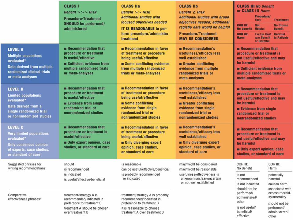
• 2013 ACCF/AHA Guideline for the Management
of Heart Failure: A Report of the American
College of Cardiology Foundation/American
Heart Association Task Force on Practice
Guidelines. Circulation. 2013;128:June.
• Pharmacotherapy; a pathophysiologic approach
(Dipiro 8th edition, 2011)
• Applied Therapeutics 10th Edition (2013)

Contents
• Definition
• Epidemiology
• Etiology
• Pathophysiology
• Clinical manifestation
• Diagnosis
• Classification
• Management of Chronic HF
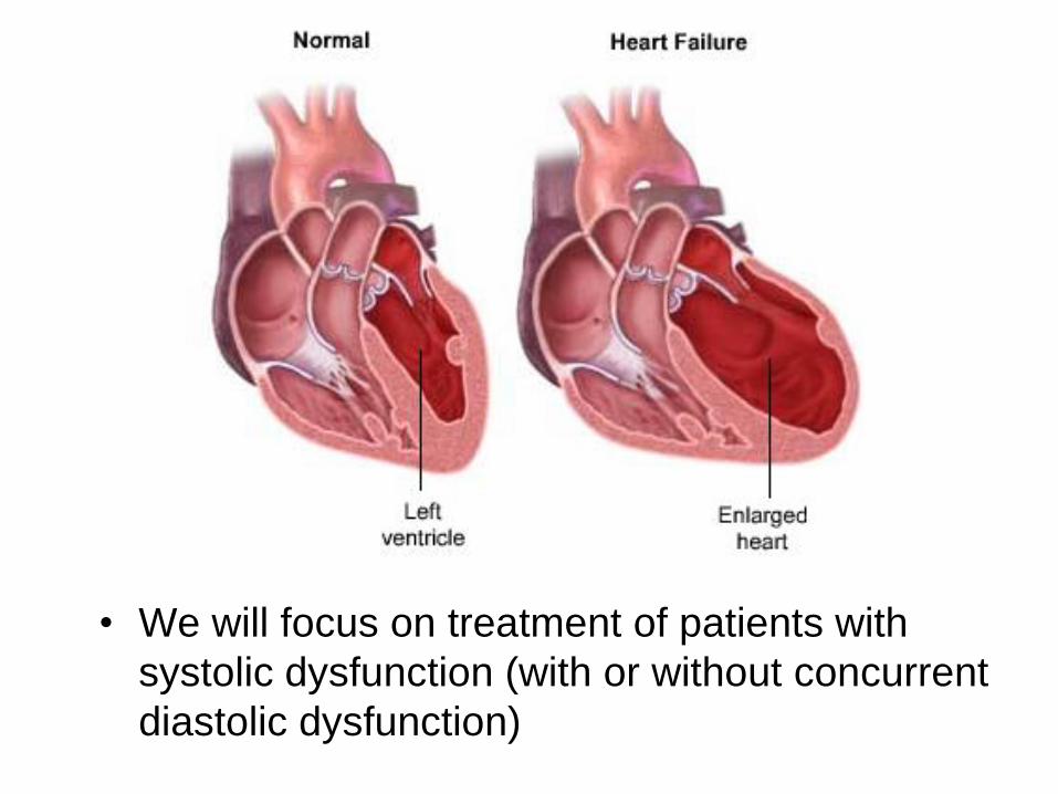
• We will focus on treatment of patients with
systolic dysfunction (with or without concurrent
diastolic dysfunction)
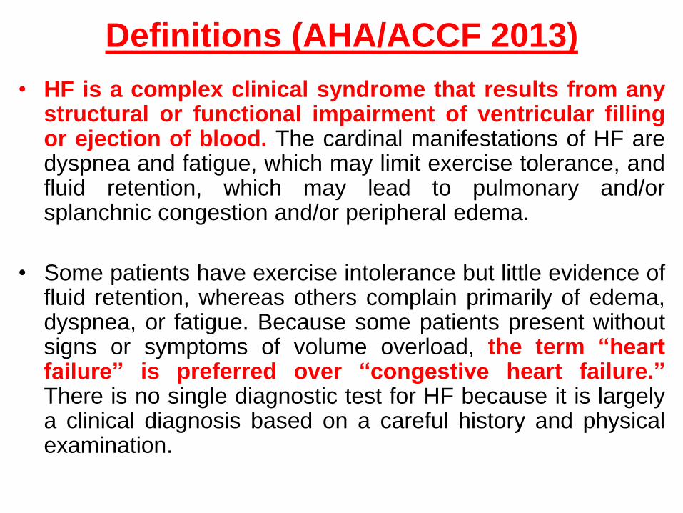
Definitions (AHA/ACCF 2013)
• HF is a complex clinical syndrome that results from anystructural or functional impairment of ventricular fillingor ejection of blood. The cardinal manifestations of HF aredyspnea and fatigue, which may limit exercise tolerance, andfluid retention, which may lead to pulmonary and/orsplanchnic congestion and/or peripheral edema.
• Some patients have exercise intolerance but little evidence offluid retention, whereas others complain primarily of edema,dyspnea, or fatigue. Because some patients present withoutsigns or symptoms of volume overload, the term “heartfailure” is preferred over “congestive heart failure.”There is no single diagnostic test for HF because it is largelya clinical diagnosis based on a careful history and physicalexamination.

The clinical syndrome of HF may result from
disorders of the pericardium, myocardium,
endocardium, heart valves, or great vessels or
from certain metabolic abnormalities, but most
patients with HF have symptoms due to
impaired left ventricular (LV) myocardial
function.
It should be emphasized that HF is not
synonymous with either cardiomyopathy or
LV dysfunction; these latter terms describe
possible structural or functional reasons for the
development of HF.
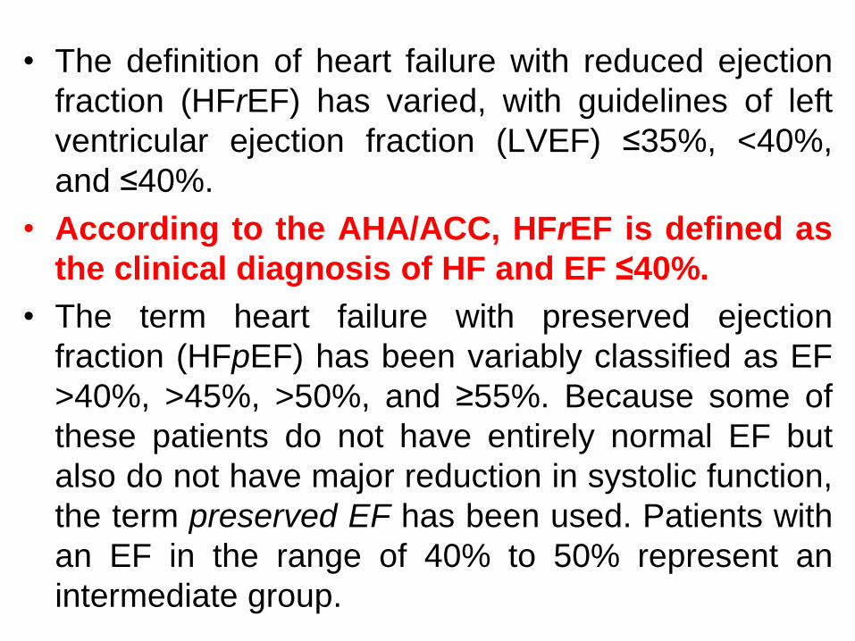
• The definition of heart failure with reduced ejection
fraction (HFrEF) has varied, with guidelines of left
ventricular ejection fraction (LVEF) ≤35%, <40%,
and ≤40%.
• According to the AHA/ACC, HFrEF is defined as
the clinical diagnosis of HF and EF ≤40%.
• The term heart failure with preserved ejection
fraction (HFpEF) has been variably classified as EF
>40%, >45%, >50%, and ≥55%. Because some of
these patients do not have entirely normal EF but
also do not have major reduction in systolic function,
the term preserved EF has been used. Patients with
an EF in the range of 40% to 50% represent an
intermediate group.

AHA/ACC 2013
Epidemiology
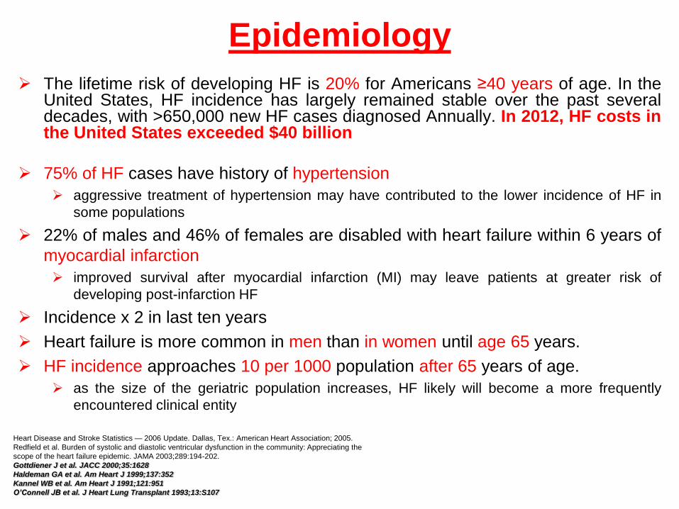
The lifetime risk of developing HF is 20% for Americans ≥40 years of age. In theUnited States, HF incidence has largely remained stable over the past severaldecades, with >650,000 new HF cases diagnosed Annually. In 2012, HF costs inthe United States exceeded $40 billion
75% of HF cases have history of hypertension
aggressive treatment of hypertension may have contributed to the lower incidence of HF in
some populations
22% of males and 46% of females are disabled with heart failure within 6 years of
myocardial infarction
improved survival after myocardial infarction (MI) may leave patients at greater risk of
developing post-infarction HF
Incidence x 2 in last ten years
Heart failure is more common in men than in women until age 65 years.
HF incidence approaches 10 per 1000 population after 65 years of age.
as the size of the geriatric population increases, HF likely will become a more frequently
encountered clinical entity
Heart Disease and Stroke Statistics — 2006 Update. Dallas, Tex.: American Heart Association; 2005.
Redfield et al. Burden of systolic and diastolic ventricular dysfunction in the community: Appreciating the
scope of the heart failure epidemic. JAMA 2003;289:194-202.
Gottdiener J et al. JACC 2000;35:1628
Haldeman GA et al. Am Heart J 1999;137:352
Kannel WB et al. Am Heart J 1991;121:951
O’Connell JB et al. J Heart Lung Transplant 1993;13:S107

Prognosis
• Despite earlier diagnosis and aggressive medical management of
HF, the prognosis is poor.
• Factors affecting the prognosis of patients with heart failure
include, but are not limited to, age, gender, LVEF, renal function,
blood pressure, heart failure etiology, and drug or device therapy.
• The quality of life is adversely affected by progressive functional
disability.
• A greater consequence is the high mortality rate.
– 5 year mortality rate is 50% (AHA/ACC 2013)
– 80% of men and 70% of women under age 65 who have CHF will die
within 8 years
– Median survival following onset is 1.7 years for men and 3.2 years for
women
American Heart Association. 2006 Heart and Stroke Statistical Update. Dallas, Tex.: American Heart
Association, 2005 Massie BM, Shah NB. Am Heart J 1997;133:703-712

Etiologies
• Heart failure can result from any disorder that affects the
ability of the heart to contract (systolic function) and/or relax
(diastolic dysfunction):
– Ischemic heart disease, mostly acute myocardial infarction
• Causes 50-6-% of cases of HF
– Hypertension
• Suspected primary etiology in 30-40% of patients; history of HTN present in 70-80% of HF patients. Long-term treatment of both systolic and diastolic hypertension reduces the risk of HF by approximately 50%
– Idiopathic dilated cardiomyopathy
• Etiology in 5-10% of patients
– Other cardiomyopathies (e.g. alcoholic, viral, hypertrophic)
– Drug induced
• examples
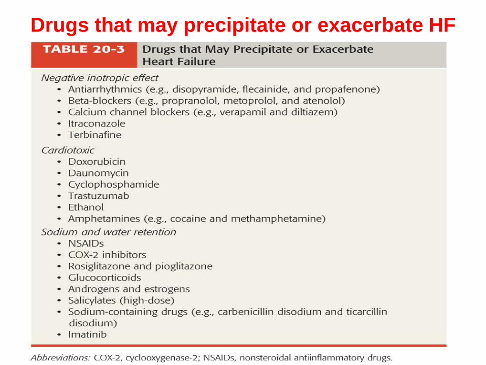
Drugs that may precipitate or exacerbate HF

Descriptive terms in HF
• Low-output versus high-output failure
• Left versus right ventricular heart failure
• Systolic versus diastolic heart failure
• Ischemic versus nonischemic heart failure
• Acute versus chronic heart failure

Classification and Etiology of Left Ventricular Dysfunction
Type of Failure Characteristics Contributing Factors Etiology
Low output,
systolic
dysfunction
(dilated
cardiomyopathy)
(60%–70% of cases)
Hypofunctioning left ventricle;
enlarged heart ;
↑left ventricular end-diastolic
volume;
EF <40%;
↓stroke volume;
↓CO;
S3 heart sound present
1. ↓Contractility
(cardiomyopathy)
2. ↑Afterload (elevated
SVR)
1. Coronary ischemia, MI,
mitral valve stenosis or
regurgitation, alcoholism,
viral syndromes, nutritional
deficiency, calcium and
potassium depletion, drug
induced, idiopathic
2. Hypertension, aortic
stenosis, volume overload
Low output,
diastolic
dysfunction
(30%–40% of cases)
Normal left ventricular
contractility; normal size heart;
stiff left ventricle; impaired left
ventricular relaxation; impaired
left ventricular filling; ↓left
ventricular end-diastolic
volume; normal EF; ↓SV; ↓CO;
exaggerated S4 heart sound
1. Thickened left
ventricle
(hypertrophic
cardiomyopathy)
2. Stiff left ventricle
(restrictive
cardiomyopathy)
3. ↑Preload
1. Coronary ischemia, MI
hypertension, aortic stenosis
and regurgitation,
pericarditis, enlarged left
ventricular septum
(hypertrophic
cardiomyopathy)
2. Amyloidosis, sarcoidosis
3. Sodium and water retention
High-output failure
(uncommon)
Normal or ↑contractility;
normal size heart;
normal left ventricular end-
diastolic volume;
normal or ↑EF; normal or
increased stroke volume; ↑CO
↑Metabolic and oxygen
demands
Anemia and hyperthyroidism
CO, cardiac output; EF, ejection fraction; K, potassium; MI, myocardial infarction; SV, stroke volume; SVR, systemic
vascular resistance.

Cardiomyopathy – types

Right vs. left HF
• Note: Congestion occurs behind the failing ventricle
– Pulmonary congestion results from left ventricular failure
– Systemic congestion results from right ventricular failure

Right vs. left HF (S&S)

Systolic vs. diastolic HF
• Systolic dysfunction– Impaired ejection
– Decreased contractility
• S&S– Low EF (<45%)
– Cardiomegaly
– S3
– Normal wall thickness
– Hypokinesis
– Symptoms primarily those of reduced cardiac output.
• Diastolic dysfunction– Impaired filling
– Depressed relaxation
– Clinical trials lacking
This group of patients
• S&S– Normal to EF
– Normal size heart
– S4
– wall thickness
– Hyperkinesis
– Symptoms primarily those of blood congestion and may include marked dyspnea and fatigue

Causes of Heart Failure
Systolic dysfunction (decreased contractility)
• Reduction in muscle mass (e.g., myocardial infarction)
• Dilated cardiomyopathies
• Ventricular hypertrophy
– Pressure overload (e.g., systemic or pulmonary hypertension, aortic or pulmonic valve
stenosis)
– Volume overload (e.g., valvular regurgitation, shunts, high-output states)
Diastolic dysfunction (restriction in ventricular filling) = heart failure with
preserved left ventricular function)
• Increased ventricular stiffness
– Ventricular hypertrophy (e.g., hypertrophic cardiomyopathy, other examples above)
– Infiltrative myocardial diseases (e.g., amyloidosis, sarcoidosis, endomyocardial
fibrosis)
– Myocardial ischemia and infarction
• Mitral or tricuspid valve stenosis
• Pericardial disease (e.g., pericarditis, pericardial tamponade)

Pathophysiology of HF
• May involve
– The right ventricle,
– The left ventricle
– Or both,
• The majority of patients with HF have symptoms due to an
impairment of left ventricular function.
• Regardless of the etiology of heart failure, the underlying
pathophysiologic process and principal clinical manifestations
(fatigue, dyspnea, and volume overload) are similar and
appear to be independent of the initial cause.

Cardiac Workload
• A common factor to all forms of HF is increased
cardiac workload.
• The major determinants of left ventricular workload
are:
1. preload,
2. afterload,
3. contractility,
4. heart rate (HR),
5. myocardial compliance
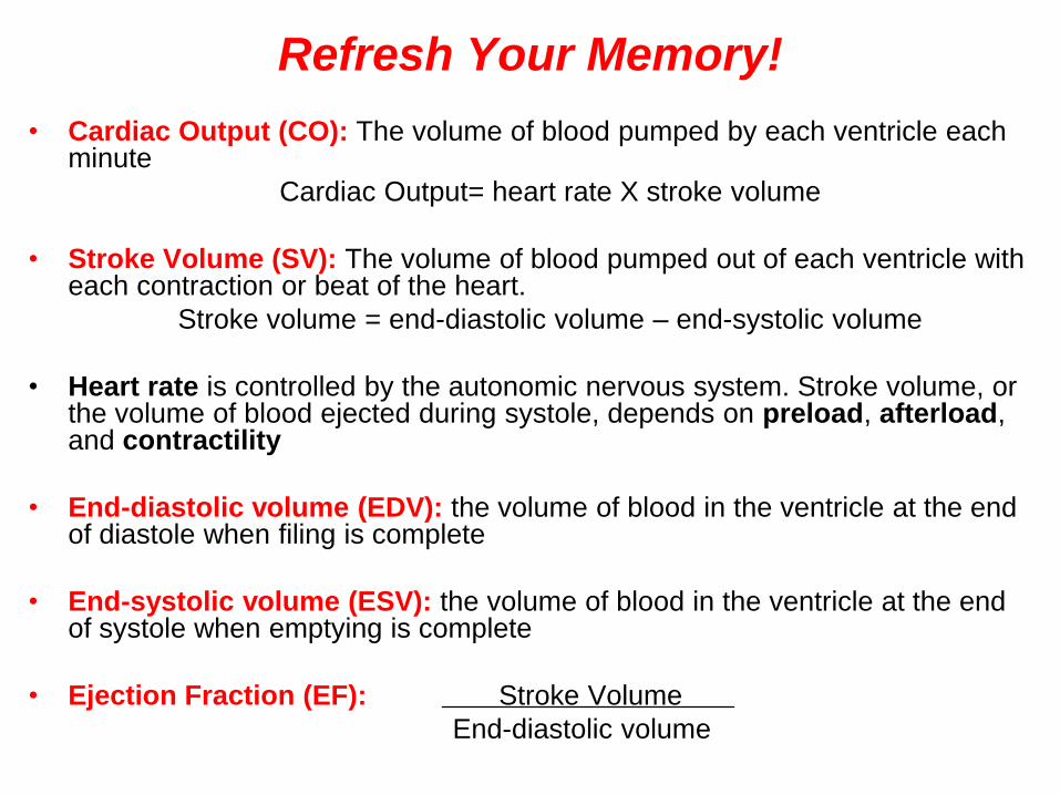
Refresh Your Memory!
• Cardiac Output (CO): The volume of blood pumped by each ventricle each minute
Cardiac Output= heart rate X stroke volume
• Stroke Volume (SV): The volume of blood pumped out of each ventricle with each contraction or beat of the heart.
Stroke volume = end-diastolic volume – end-systolic volume
• Heart rate is controlled by the autonomic nervous system. Stroke volume, or the volume of blood ejected during systole, depends on preload, afterload, and contractility
• End-diastolic volume (EDV): the volume of blood in the ventricle at the end of diastole when filing is complete
• End-systolic volume (ESV): the volume of blood in the ventricle at the end of systole when emptying is complete
• Ejection Fraction (EF): Stroke Volume
End-diastolic volume
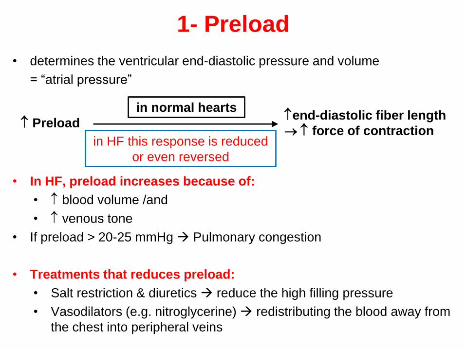
1- Preload
• determines the ventricular end-diastolic pressure and volume
= ―atrial pressure‖
end-diastolic fiber length
force of contraction
in normal hearts
Preload
in HF this response is reduced
or even reversed
• In HF, preload increases because of:
• blood volume /and
• venous tone
• If preload > 20-25 mmHg Pulmonary congestion
• Treatments that reduces preload:
• Salt restriction & diuretics reduce the high filling pressure
• Vasodilators (e.g. nitroglycerine) redistributing the blood away from
the chest into peripheral veins
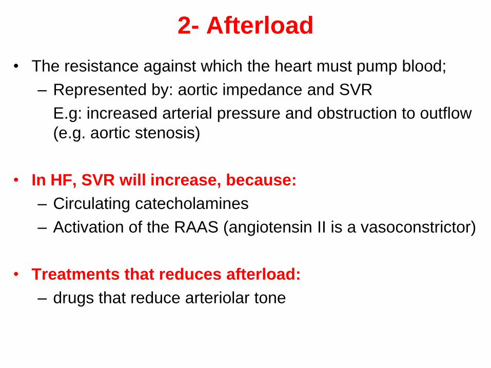
2- Afterload
• The resistance against which the heart must pump blood;
– Represented by: aortic impedance and SVR
E.g: increased arterial pressure and obstruction to outflow
(e.g. aortic stenosis)
• In HF, SVR will increase, because:
– Circulating catecholamines
– Activation of the RAAS (angiotensin II is a vasoconstrictor)
• Treatments that reduces afterload:
– drugs that reduce arteriolar tone

Relationship between stroke volume and systemic vascular resistance. In an individual
with normal left ventricular (LV) function, increasing systemic vascular resistance has
little effect on stroke volume. As the extent of LV dysfunction increases, the
negative, inverse relationship between stroke volume and systemic vascular
resistance becomes more important (B to A).

3- Contractility of the heart
• The terms contractility is used to describe the cardiac muscle's inherent
ability to develop force and shorten its fibers independent of preload or
afterload.
• It is determined largely by the intrinsic strength and integrity of muscle
cells
• In HF: pump performance of the heart.
• Force of heart contraction is by:
1. Ischemic heart disease
MI, chronic severe ischemia
2. Specific disorders affecting the heart muscle
HTN and Myocarditis
3. Disorders of heart muscle of unknown cause
Idiopathic
• Heart increases contractility in response to +ve inotropic drugs.

4- Heart Rate
• Major determinant of CO.
• In HF:
compensatory sympathetic activation of β-adrenoceptors
comes into play to maintain CO
The workload and energy demands of a rapid heart rate
ultimately place undo strain on the heart, however, and can
eventually worsen HF.

5- Myocardial Compliance
• How easy the myocardial fibres can stretch
• An important determinant of ventricular filling and therefore of
CO.
• Compliance can be decreased by:
– Fibrosis
– Hypertrophy
– Ischemia

Compensatory Mechanisms in HF
• The manifestations of CO– The major direct consequence
• exercise tolerance
• Rapid muscular fatigue
– The Other manifestations result from
• The attempts by the body to compensate for the intrinsic cardiac defect in an attempt to maintain CO and oxygenation of vital organs.
• An understanding of the potential benefits and adverse consequences of the compensatory mechanisms is essential to understanding the signs, symptoms, and treatment of HF
• What are the compensatory mechanisms of the body?

Compensatory mechanisms
• These include:
– increased sympathetic tone,
– activation of the renin-
angiotensin-aldosterone
system (RAAS),
– sodium and water retention,
– other neurohormonal
adaptations,
– cardiac ―remodeling‖
(ventricular dilation, cardiac
hypertrophy, and changes in
left ventricular lumen shape).
• The long-term
consequences of these
adaptive mechanisms can
create more harm than good.
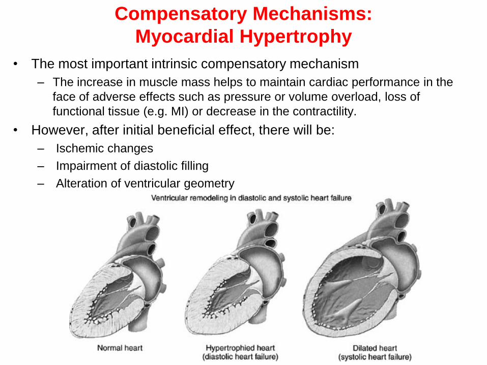
Compensatory Mechanisms:
Myocardial Hypertrophy
• The most important intrinsic compensatory mechanism
– The increase in muscle mass helps to maintain cardiac performance in the
face of adverse effects such as pressure or volume overload, loss of
functional tissue (e.g. MI) or decrease in the contractility.
• However, after initial beneficial effect, there will be:
– Ischemic changes
– Impairment of diastolic filling
– Alteration of ventricular geometry

Beneficial and detrimental effects of the compensatory
responses in heart failure
Compensatory Response Beneficial Effects of Compensation Detrimental Effects of Compensation
Increased preload
(through Na+ and water
retention)
Optimize stroke volume via Frank-
Starling mechanism (whereby an
increase in preload results in an
increase in stroke volume)
Pulmonary and systemic congestion and
edema formation
Increased MVO2
Vasoconstriction Maintain BP in face of reduced CO Increased MVO2
Shunt blood from nonessential organs
to brain and heart
Increased afterload decreases stroke
volume and further activates the
compensatory responses
Tachycardia and
increased contractility
(because of SNS
activation)
Helps maintain CO Increased MVO2
Shortened diastolic filling time
β1-receptor downregulation, decreased
receptor sensitivity
Precipitation of ventricular arrhythmias
Increased risk of myocardial cell death
Ventricular hypertrophy
and remodeling
Helps maintain CO Diastolic dysfunction
Reduces myocardial wall stress Systolic dysfunction
Decreases MVO2 Increased risk of myocardial cell death
Increased risk of myocardial ischemia
Increased arrhythmia risk
Fibrosis

Ventricular
function (Frank-
Starling) curve

Common precipitants of decompensation
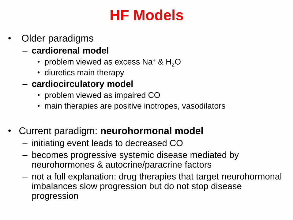
HF Models
• Older paradigms
– cardiorenal model
• problem viewed as excess Na+ & H2O
• diuretics main therapy
– cardiocirculatory model
• problem viewed as impaired CO
• main therapies are positive inotropes, vasodilators
• Current paradigm: neurohormonal model
– initiating event leads to decreased CO
– becomes progressive systemic disease mediated by neurohormones & autocrine/paracrine factors
– not a full explanation: drug therapies that target neurohormonalimbalances slow progression but do not stop disease progression

The neurohormonal model of heart failure and
therapeutic insights it provides

Homework:
What is the role of natriuretic peptides in HF?
• Sources:
1. ACCF/AHA 2009 Guidelines
2. ESC 2008 Guidelines
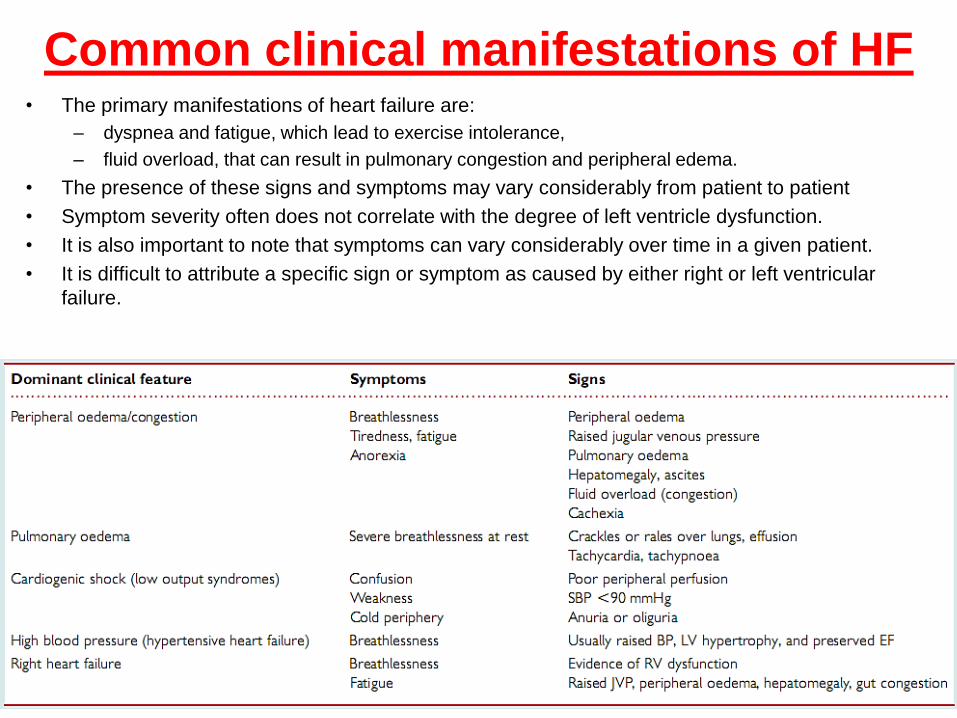
Common clinical manifestations of HF• The primary manifestations of heart failure are:
– dyspnea and fatigue, which lead to exercise intolerance,
– fluid overload, that can result in pulmonary congestion and peripheral edema.
• The presence of these signs and symptoms may vary considerably from patient to patient
• Symptom severity often does not correlate with the degree of left ventricle dysfunction.
• It is also important to note that symptoms can vary considerably over time in a given patient.
• It is difficult to attribute a specific sign or symptom as caused by either right or left ventricular
failure.
Patient Presentation
Non-specific Findings
• LVH on ECG
• Weakness, fatigue, exercise intolerance
• Confusion, lethargy, hallucinations, insomnia,
• Nightmares and headaches
• Pallor, cool extremities, cyanotic digits
• Renal dysfunction
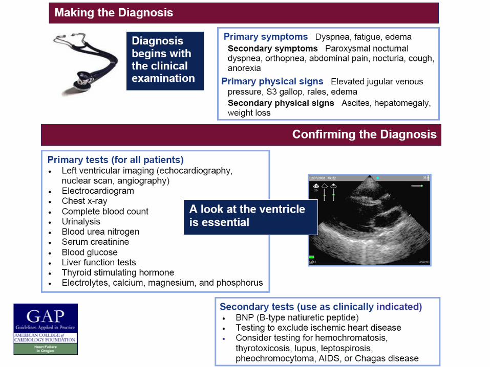
Diagnosis of HF
• Clinical diagnosis is based on careful history and physical
examination
• HF is characterized by
– specific symptoms in medical history
• (dyspnea and fatigue)
– Specific signs on the physical examination
• (edema, rales)
• Remember: Heart failure IS NOT equivalent to • cardiomyopathy
• LV dysfunction;
– these latter terms describe structural or functional reasons for the
development of heart failure
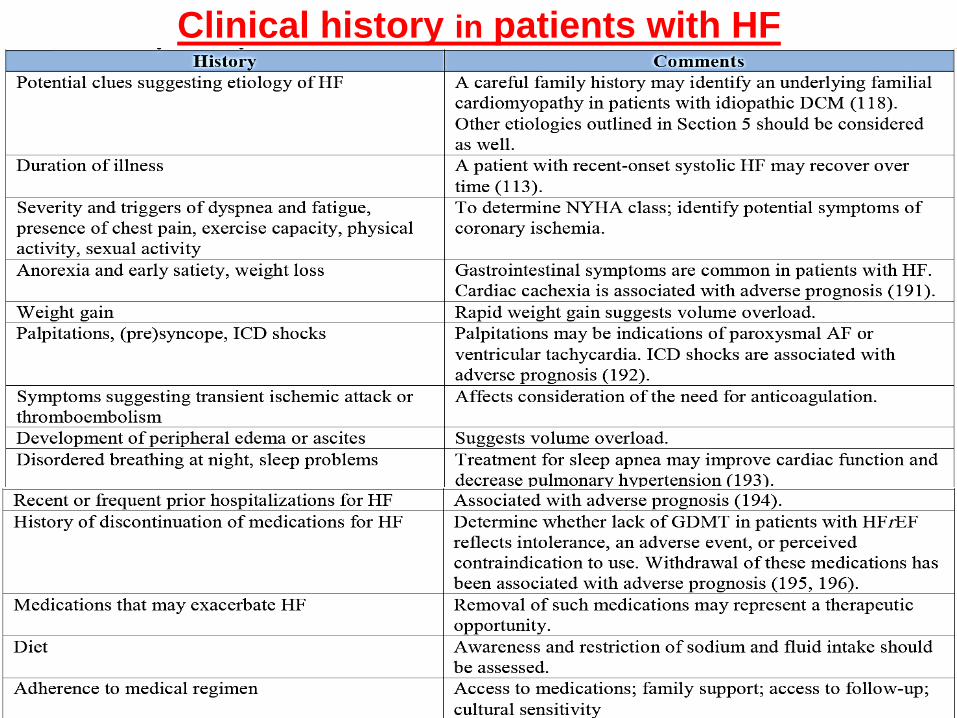
Clinical history in patients with HF


Initial / Ongoing Evaluation
• Identify heart disease
• Assess functional capacity
– NYHA, 6 min walk, …
• Assess volume status:
– edema, rales, jugular, hepatomegaly, body weight
• Lab assessment:
– routine: electrolytes, renal function
– repeat ECHO, RX only if significant changes in
functional status
• Assess prognosis

47
A. Chest x-ray with increased vascular markings (represents interstitial edema, early alveolar
edema)
Arrow: fluid in right lung fissure; cardiomegaly
B. Lateral chest x-ray view.
Arrow: pulmonary effusion
Chest x-ray
48
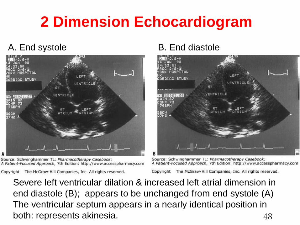
Severe left ventricular dilation & increased left atrial dimension in
end diastole (B); appears to be unchanged from end systole (A)
The ventricular septum appears in a nearly identical position in
both: represents akinesia.
2 Dimension Echocardiogram
A. End systole B. End diastole

Diagnosis of HF

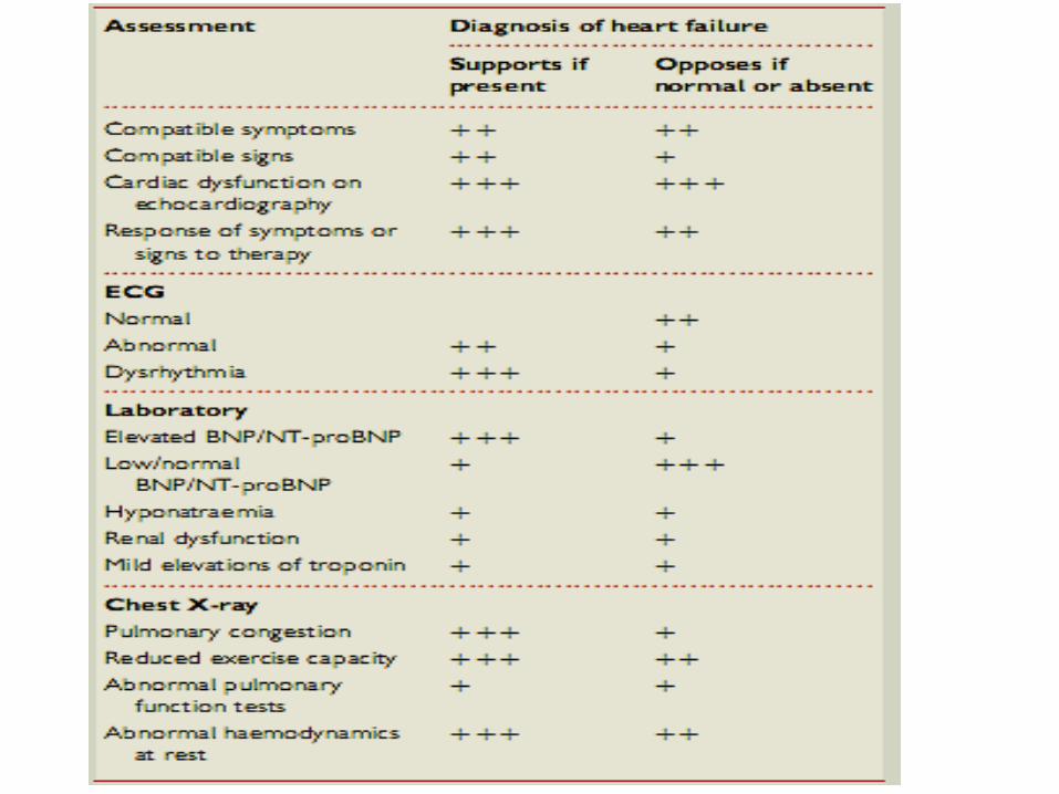

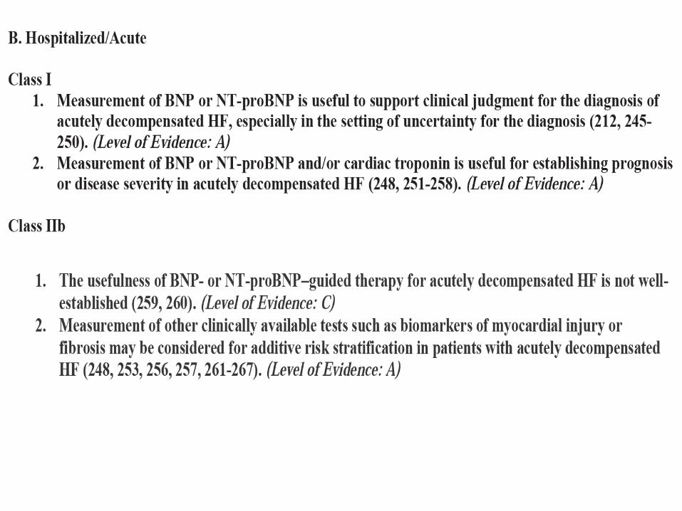
Diagnostic Test Recommendations
(AHA/ACCF 2013)
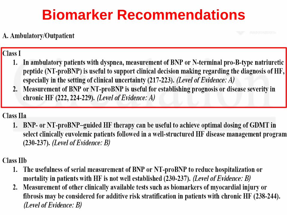
Biomarker Recommendations
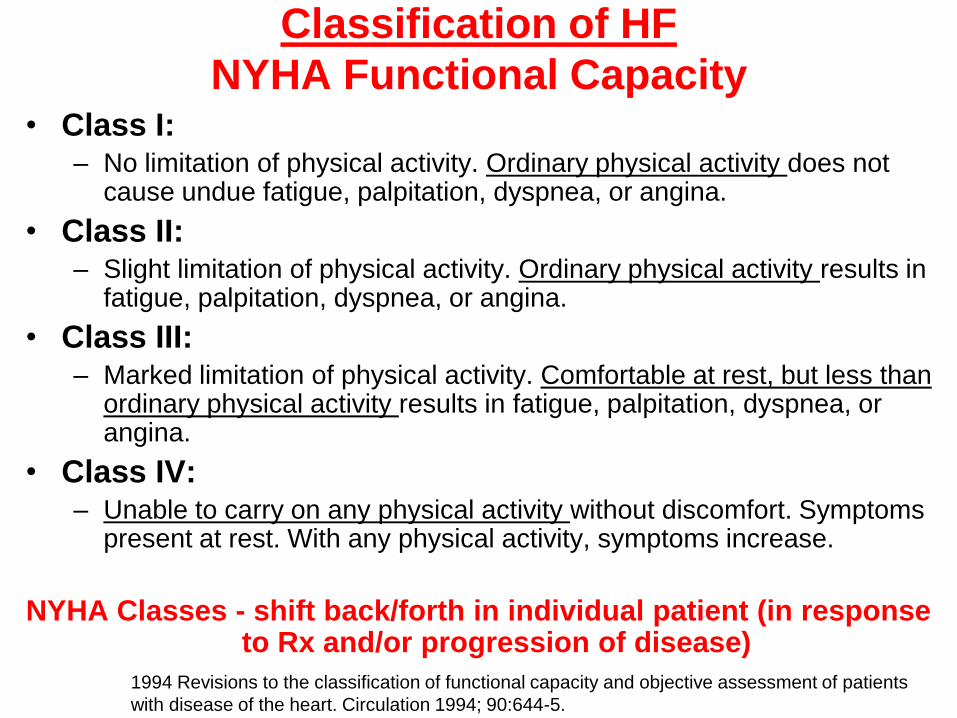
Classification of HF
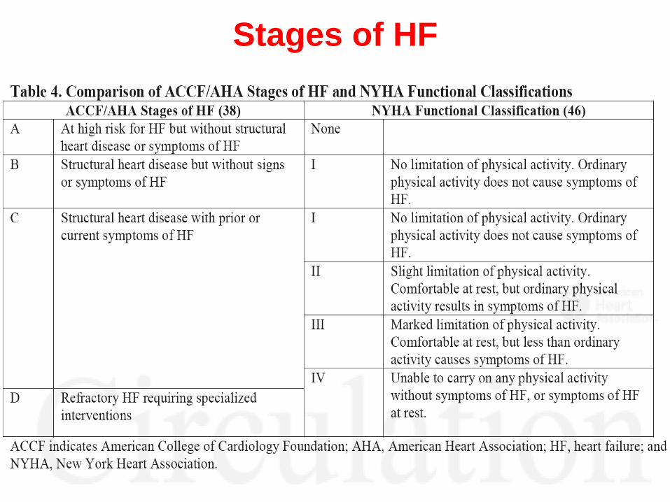
NYHA Functional Capacity• Class I:
– No limitation of physical activity. Ordinary physical activity does not cause undue fatigue, palpitation, dyspnea, or angina.
• Class II: – Slight limitation of physical activity. Ordinary physical activity results in
fatigue, palpitation, dyspnea, or angina.
• Class III: – Marked limitation of physical activity. Comfortable at rest, but less than
ordinary physical activity results in fatigue, palpitation, dyspnea, or angina.
• Class IV: – Unable to carry on any physical activity without discomfort. Symptoms
present at rest. With any physical activity, symptoms increase.
NYHA Classes - shift back/forth in individual patient (in response to Rx and/or progression of disease)
1994 Revisions to the classification of functional capacity and objective assessment of patients
with disease of the heart. Circulation 1994; 90:644-5.
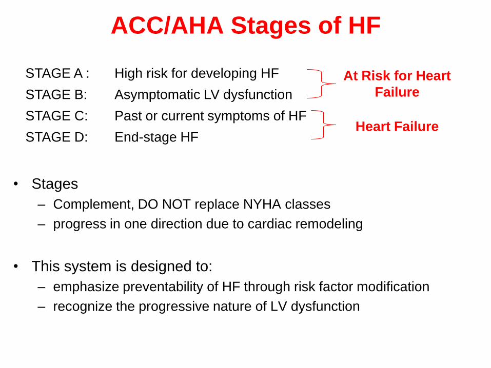
ACC/AHA Stages of HF
• Stages
– Complement, DO NOT replace NYHA classes
– progress in one direction due to cardiac remodeling
• This system is designed to:
– emphasize preventability of HF through risk factor modification
– recognize the progressive nature of LV dysfunction
STAGE A : High risk for developing HF At Risk for Heart
FailureSTAGE B: Asymptomatic LV dysfunction
STAGE C: Past or current symptoms of HFHeart Failure
STAGE D: End-stage HF
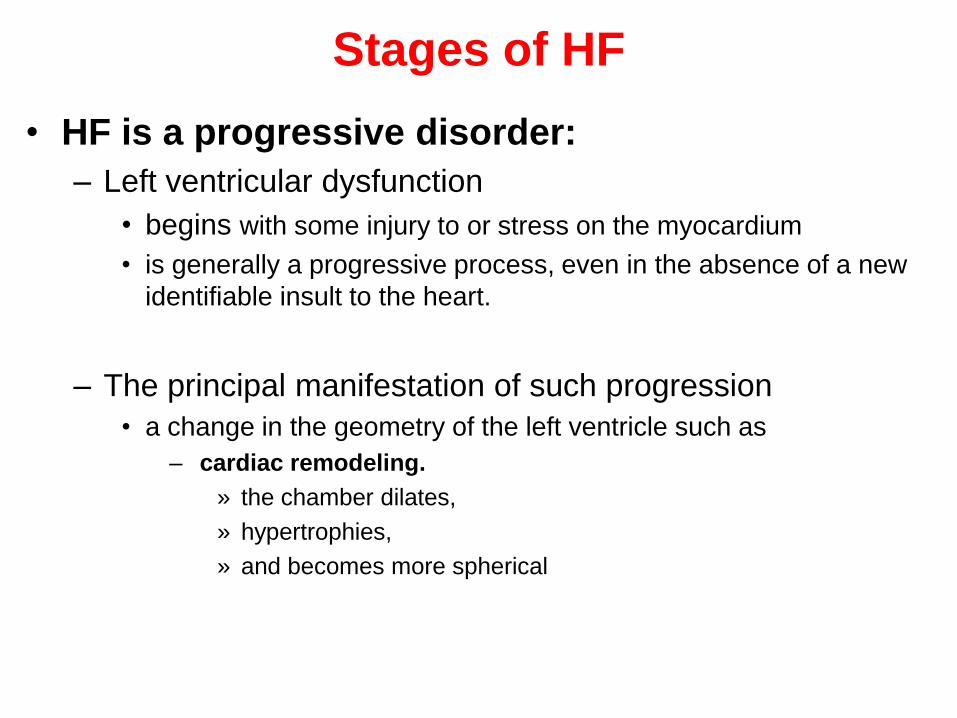
Stages of HF
• HF is a progressive disorder:
– Left ventricular dysfunction
• begins with some injury to or stress on the myocardium
• is generally a progressive process, even in the absence of a new
identifiable insult to the heart.
– The principal manifestation of such progression
• a change in the geometry of the left ventricle such as
– cardiac remodeling.
» the chamber dilates,
» hypertrophies,
» and becomes more spherical
Stages of HF


Treatment of Chronic Heart Failure
Goals of therapy
Pharmacologic management
Nonpharmacologic management

Goals of HF therapy
Survival
Morbidity
Exercise capacity
Quality of life
Neurohormonal changes
Progression of CHF
Symptoms

General measures
• The complexity of the heart failure syndrome necessitates a
comprehensive approach to management that includes:
– accurate diagnosis,
– identification and treatment of risk factors (e.g., diabetes, hypertension,
coronary artery disease),
– elimination or minimization of precipitating factors,
– appropriate pharmacologic and nonpharmacologic therapy,
– close monitoring and followup.
• Patient assessment:
– History and patient examination
• determine etiology
• determine precipitating factors
– Initial and ongoing assessment of activities of daily living (ADL) and volume
status
– Initial CBC, UA, electrolytes (Ca/Mg), BUN/Scr, BG, LFTs, TSH, EKG and
Echo
– Serial electrolytes and renal function
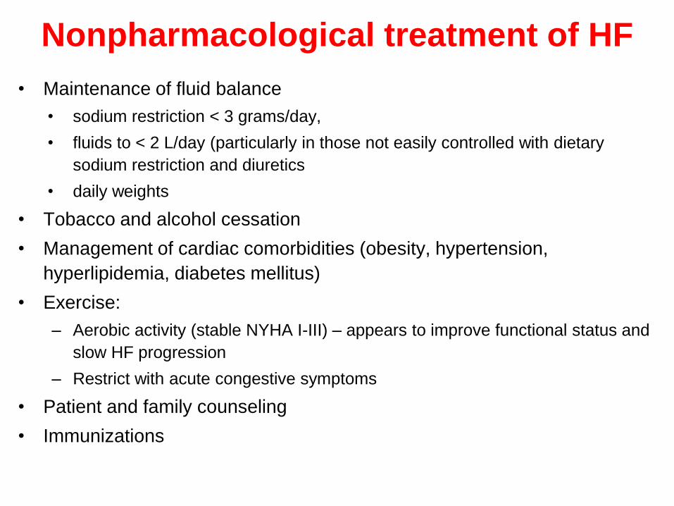
Nonpharmacological treatment of HF
• Maintenance of fluid balance
• sodium restriction < 3 grams/day,
• fluids to < 2 L/day (particularly in those not easily controlled with dietary
sodium restriction and diuretics
• daily weights
• Tobacco and alcohol cessation
• Management of cardiac comorbidities (obesity, hypertension,
hyperlipidemia, diabetes mellitus)
• Exercise:
– Aerobic activity (stable NYHA I-III) – appears to improve functional status and
slow HF progression
– Restrict with acute congestive symptoms
• Patient and family counseling
• Immunizations

Nonpharmacological treatment of HF
• Coronary revascularization
• Biventricular pacing
• Enhanced external counterpulsation therapy
• Surgical ventricular restoration
• Left ventricular assist devices/Heart transplant
• Compassionate end of life care/hospice

Pharmacologic treatment of HF
• Standard drug therapies – Diuretic (if evidence of fluid retention)
– ACE inhibitor or ARB (assuming no CI)
– β-blocker
• Select patients– Digoxin (common)
– Aldosterone antagonist
– Nitrates/Hydralazine
• Other agents– Calcium Channel Blockers
– Anticoagulation
– Antiarrhythmics

Change from
previous
recommendations


Pharmacolotherapeutic agents
1- Diuretics• Benefits:
– Decrease Na and water retention reduce preload
– Decrease signs/symptoms of fluid retention
– Improve exercise tolerance, quality of life
– Improve cardiac function
– Reduce HF hospitalizations
• Do not alter disease progression or prolong survival
• Should not be used alone in patients with HF
• All patients with evidence of fluid retention
• Many patients require chronic diuretic therapy to maintain euvolemia
• Body weight changes: sensitive marker of fluid retention/loss
– daily weights to adjust diuretic therapy
– Report weight gain of > 0.25-0.5 kg/day over several days


Thiazide Diuretics
• Weak diuretics
– more persistent antihypertensive activity than loop
diuretics
– infrequently used alone in HF
• Can use with loop diuretics to promote diuresis
• Preferred in some patients with mild fluid retention &
elevated BP

Loop Diuretics
• Furosemide, bumetanide, toresmide
• Mainstay of HF therapy
• Efficacy reduced by:
– competitors of the organic acid transport pathway,
– excess dietary Na+,
– co-administration with NSAIDs
• Efficacy maintained in impaired renal function
• Once ceiling dose reached, additional diuresis achieved
through increased frequency rather than increased dose.
– Ceiling dose: single dose above which additional response
is unlikely to be observed
• Cause metabolic abnormalities such as hypokalemia,
hypomagnesemia73
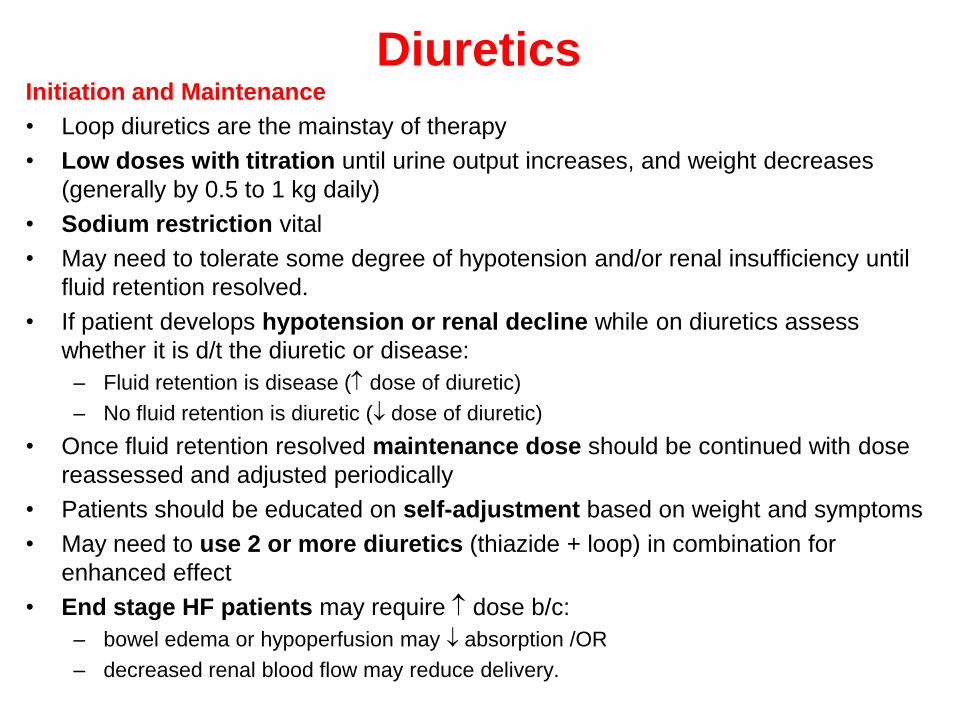
DiureticsInitiation and Maintenance
• Loop diuretics are the mainstay of therapy
• Low doses with titration until urine output increases, and weight decreases
(generally by 0.5 to 1 kg daily)
• Sodium restriction vital
• May need to tolerate some degree of hypotension and/or renal insufficiency until
fluid retention resolved.
• If patient develops hypotension or renal decline while on diuretics assess
whether it is d/t the diuretic or disease:
– Fluid retention is disease (dose of diuretic)
– No fluid retention is diuretic (dose of diuretic)
• Once fluid retention resolved maintenance dose should be continued with dose
reassessed and adjusted periodically
• Patients should be educated on self-adjustment based on weight and symptoms
• May need to use 2 or more diuretics (thiazide + loop) in combination for
enhanced effect
• End stage HF patients may require dose b/c:
– bowel edema or hypoperfusion may absorption /OR
– decreased renal blood flow may reduce delivery.
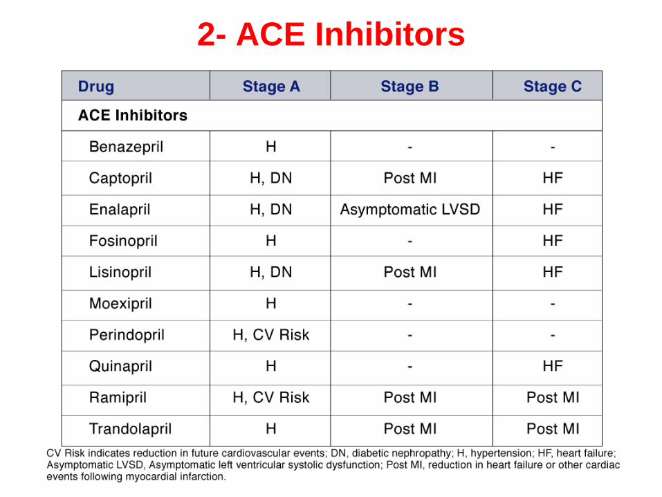
2- ACE Inhibitors
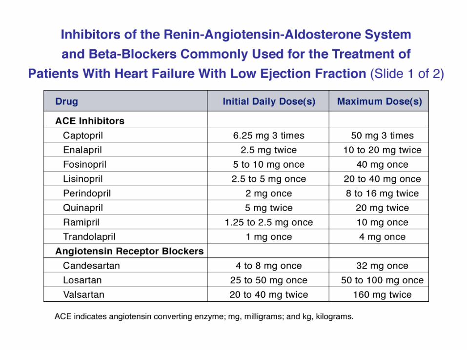


ACE Inhibitors
• Cornerstone of HF pharmacotherapy
– captopril, enalapril, lisinopril, quinapril, ramipril, fosinopril,
trandolapril, perindopril
– not all FDA approved for HF
• Actions:
– Decrease preload
– Decrease afterload
– Decrease sympathetic activation
– Decrease left ventricular hypertrophy, dilation and
remodeling associated with HF slow progression of HF

ACE Inhibitors
• Benefits:
– Effective for preventing HF development, reducing CV risk
– Alleviate symptoms and improve clinical status (prevent RAAS mediated worsening of
myocardial function)
– Enhance overall sense of well being (QOL)
– Mortality (improve survival 20 to 30% compared to placebo)
– Hospitalizations
• Place in therapy
– All patients with left systolic HF should be taking ACE-I for morbidity/mortality benefits
– For patients with C/I or those unable to tolerate ACE-I, alternative therapy with ARBs or
hydralazine/nitrate combination is recommended
• ARBs are appropriate alternatives in those patients in which a cough is troublesome, as
this is a bradykinin mediated effect. Role of ARBs in patients with angioedema
controversial
• Hydralazine/Isosorbide dinitrate should be used if the patients renal dysfunction,
hyperkalemia or hypotension is uncontrollable. This combo can also be used in those
patients who develop angioedema to ACEIs
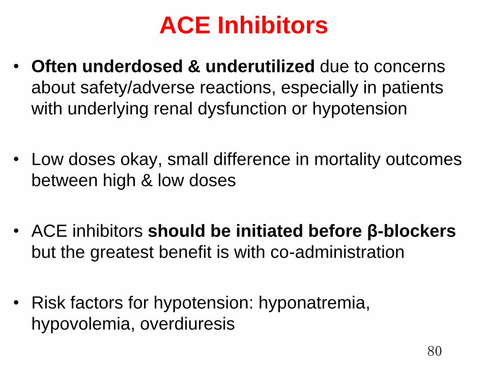
ACE Inhibitors
• Often underdosed & underutilized due to concerns
about safety/adverse reactions, especially in patients
with underlying renal dysfunction or hypotension
• Low doses okay, small difference in mortality outcomes
between high & low doses
• ACE inhibitors should be initiated before β-blockers
but the greatest benefit is with co-administration
• Risk factors for hypotension: hyponatremia,
hypovolemia, overdiuresis
80

ACE Inhibitors
Initiation and Maintenance
• Low doses with K+ and renal function checked within 1 to 2 weeks and
periodically after.
• Titrated as tolerated to doses demonstrated to provide a clinical benefit or
to moderate-high to high doses
– Studies evaluating ACE-I titrated to a target dose NOT therapeutic response
– Studies evaluating other drugs on top of ACE-I usually had at least
intermediate doses of ACE-I given
• Concurrent diuretic therapy may need to be adjusted initially or after
therapy started
• 85 to 90% of patients can tolerate short- and long-term therapy
IMP.
• Do not abruptly withdraw ACE-I’s b/c the patient can acutely deteriorate.
• Decrease dose gradually, unless patient is experiencing a life threatening
reaction.
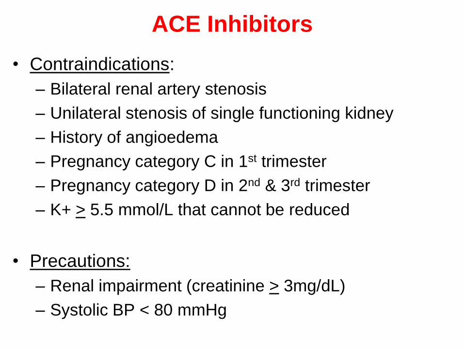
• Contraindications:
– Bilateral renal artery stenosis
– Unilateral stenosis of single functioning kidney
– History of angioedema
– Pregnancy category C in 1st trimester
– Pregnancy category D in 2nd & 3rd trimester
– K+ > 5.5 mmol/L that cannot be reduced
• Precautions:
– Renal impairment (creatinine > 3mg/dL)
– Systolic BP < 80 mmHg
ACE Inhibitors
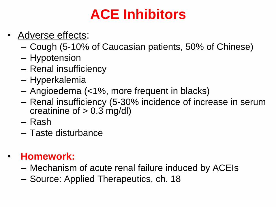
• Adverse effects: – Cough (5-10% of Caucasian patients, 50% of Chinese)
– Hypotension
– Renal insufficiency
– Hyperkalemia
– Angioedema (<1%, more frequent in blacks)
– Renal insufficiency (5-30% incidence of increase in serum creatinine of > 0.3 mg/dl)
– Rash
– Taste disturbance
• Homework:– Mechanism of acute renal failure induced by ACEIs
– Source: Applied Therapeutics, ch. 18
ACE Inhibitors
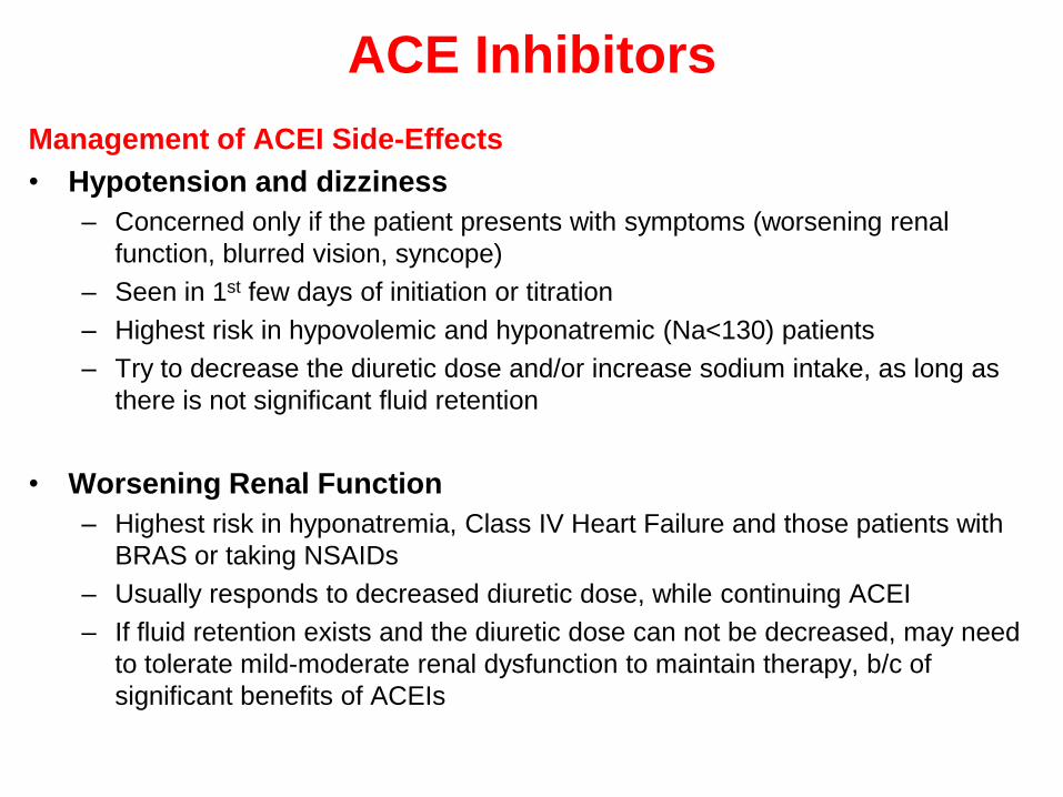
Management of ACEI Side-Effects
• Hypotension and dizziness
– Concerned only if the patient presents with symptoms (worsening renal
function, blurred vision, syncope)
– Seen in 1st few days of initiation or titration
– Highest risk in hypovolemic and hyponatremic (Na<130) patients
– Try to decrease the diuretic dose and/or increase sodium intake, as long as
there is not significant fluid retention
• Worsening Renal Function
– Highest risk in hyponatremia, Class IV Heart Failure and those patients with
BRAS or taking NSAIDs
– Usually responds to decreased diuretic dose, while continuing ACEI
– If fluid retention exists and the diuretic dose can not be decreased, may need
to tolerate mild-moderate renal dysfunction to maintain therapy, b/c of
significant benefits of ACEIs
ACE Inhibitors

ACE Inhibitors
• Cough
– Occurs in 5-10% of Caucasian patients, 50% of Chinese
– Non-productive, persistent tickle in the back of throat that
occurs within 1st month of therapy
– If D/C will disappear within 1-2 wks and reoccur upon
rechallenge with ACEI
– Exclude pulmonary causes of cough
• Hyperkalemia
– Highest risk in patients receiving potassium supplementation or
if renal function is impaired
• Angioedema
– Occurs in <1% of patients, but is life threatening, therefore
clinical suspicion warrants avoidance of ACEIs
– Do not initiate ACEI in any patient with history of angioedema
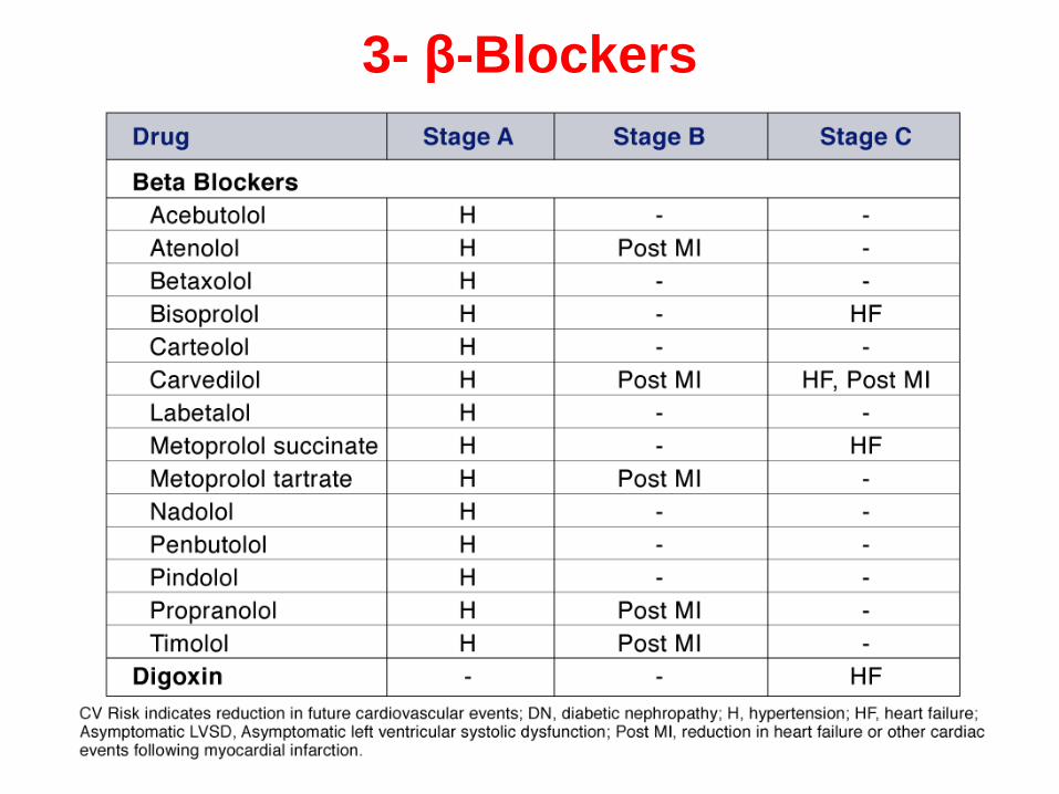
3- β-Blockers

β-Blockers
• Multiple randomized controlled trials show specific β-blockers
reduce morbidity & mortality in HF patients
– carvedilol, metoprolol succinate, bisoprolol
– not a class effect, not all β-blockers show benefit
– several studies stopped early due to overwhelming benefit
• Mechanism of Action
– Cardiac myocyte protection of receptors from catecholamines
– Prevention of binding of auto-antibodies to adrenoceptors
– Heart rate reduction
• Improved (diastolic) coronary artery flow and myocardial
oxygenation
• Improved force-frequency relationship
• Cardiac myocyte energy conservation

β-Blockers
• Benefits:
– Improve Symptoms
– Improve clinical status
– Enhance overall sense of well being (QOL)
– decrease HF progression
– Hospitalizations
– Mortality (improve survival)
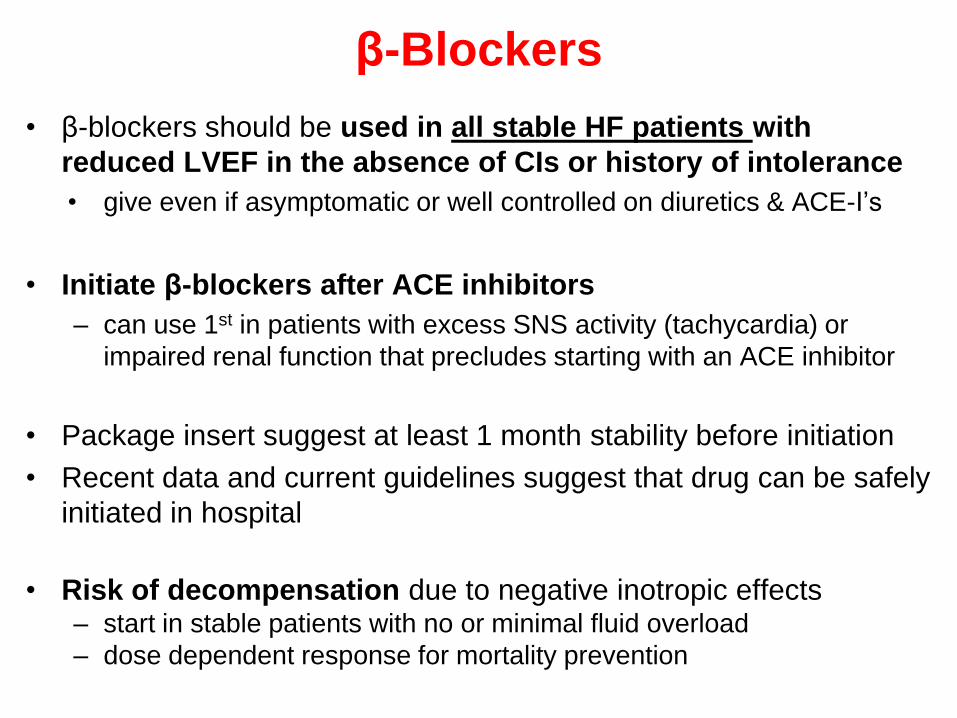
β-Blockers
• β-blockers should be used in all stable HF patients with
reduced LVEF in the absence of CIs or history of intolerance
• give even if asymptomatic or well controlled on diuretics & ACE-I’s
• Initiate β-blockers after ACE inhibitors
– can use 1st in patients with excess SNS activity (tachycardia) or
impaired renal function that precludes starting with an ACE inhibitor
• Package insert suggest at least 1 month stability before initiation
• Recent data and current guidelines suggest that drug can be safely
initiated in hospital
• Risk of decompensation due to negative inotropic effects– start in stable patients with no or minimal fluid overload
– dose dependent response for mortality prevention

β-Blockers
Initiation and Maintenance
• Very low doses with titration (every 2 weeks in trials) after
demonstrated tolerability of dose
• Titrated as tolerated (over 6-8 wks) to doses demonstrated to
provide a clinical benefit
• Studies evaluating beta-blockers titrated to a target or maximally
tolerated dose NOT therapeutic response
Drug Initial Dose Target Dose
Bisoprolol 1.25 mg daily 10 mg daily
Carvedilol 3.125 mg bid 25 mg bida
Carvedilol CR 10 mg daily 80 mg daily
Metoprolol succinate 12.5 – 25 mg daily 200 mg daily
a Target dose for patient > 85 kg is 50 mg bid.

β-Blockers
• Concurrent diuretic therapy may need to be adjusted initially or after
therapy started
• 85% of patients can tolerate short- and long-term therapy
• Patients should be monitored closely for worsening HF symptoms or S/Es
• Clinical responses may not become apparent for 2-3 months
• Should not be abruptly withdrawn b/c the patient can acutely deteriorate.
Decrease dose gradually, unless patient is experiencing a life threatening
reaction.
• If on a B-blocker for >3months and heart failure worsens, it’s probably not
d/t the B-blocker, but rather progression of disease or an exacerbation for
some other reason
– Increase dose of diuretic, do not D/C B-blocker for above reason, unless
hypoperfusion is an issue

Recent update:
• whether beta-blocker dose or degree of heart rate reduction is the optimal endpoint to guide dose-
titration and predict survival remains uncertain.
• In a recent meta-analysis, heart rate reduction and beta-blocker dose were compared as
predictors of survival in patients with heart failure
• The results from this study suggest that the degree of beta-blocker mediated reduction in
resting heart rate, but not beta-blocker dose, is associated with the magnitude of improved
survival.
• Some published reports are consistent with these findings, whereas others have found no
relationship between heart rate reduction and clinical outcomes with beta-blockers. All of these
analyses are limited by their retrospective design, inability to account for other factors affecting
heart rate (e.g., vagal activity, beta-receptor pharmacogenomics) and reliance on resting heart
rate as a surrogate marker for extent of beta-blockade.
• Although resting heart rate is routinely used clinically to evaluate extent of beta-blockade, it
is not as accurate as inhibition of exercise heart rate. Whether magnitude of resting heart
rate reduction or achievement of clinical trial doses is the optimal surrogate marker for
improved outcomes with beta-blockers in heart failure remains uncertain and may only be
definitively determined by prospective trials.
• McAlister FA, Wiebe N, Ezekowitz JA, Leung AA, Armstrong PW. Meta-analysis: β-blocker
dose, heart rate reduction, and death in patients with heart failure. Ann Intern Med
2009;150:784-94
β-Blockers
• Counseling of HF patients started on B-blockers:
– Possibility of worsening of symptoms initially
• Monitor for increased SOB, wt gain…
– Need for slow upward dose titration
– Long term benefit of therapy that make initial
difficulties (if they occur) worth sticking through

• Absolute contraindications:
– uncontrolled bronchospastic disease
– symptomatic bradycardia
– advanced heart block (2nd or 3rd degree) without a
pacemaker
• Precautions:
– Asthma
– Severe peripheral arterial disease
– Uncompensated HF
β-Blockers

• Adverse effects:
– Cold peripheries
– Bronchoconstriction
– Interference with autonomic and metabolic response
to hypoglycemia
– bradycardia
– heart block
– hypotension
– fatigue
– worsening HF
β-Blockers

β-Blockers
Management of Adverse Events
• Fluid retention and worsening heart failure- more likely
to occur during initiation and first several months
– Daily weights and careful adjustment of diuretics
• Hypotension- more likely with carvedilol (administer with
food)
– Administer ACE-I separately or temporarily reduce ACEI
• Bradycardia and heart block- risk of 5-10% as dose
increased
– If symptomatic or > 1st degree block need to reduce dose
• Fatigue/Weakness- may resolve with time or reduction in
dose

4- Digoxin
• Mechanism of action
– Inhibit Na+/K+/ATPase pump in cardiac cells
increased contractility
– Inhibit Na+/K+/ATPase pump in non-cardiac cells
sensitization of cardiac baroreceptors decreasing
sympathetic CNS outflow
– Inhibit Na+/K+/ATPase pump in renal cells
reduction in renal tubular absorption of sodium and
increased presentation to distal tubules
suppression of renin secretion
Digoxin
• Exact mechanism of benefit in HF is unclear but
probably not +ve inotropic effect.
• Benefits likely to be from neurohormonal inhibition
– Decrease sympahetic outflow
– Improved baroreceptor function and increase vagal tone
• Benefits seen with low plasma concentrations; little
added benefit at higher doses
– target 0.5 to 1.0 ng/mL
Digoxin
• Efficacy in heart failure
– Short term studies
– Withdrawal studies
– One long-term prospective, randomized, study (DIG Trial)
• Digitalis Investigational Group (DIG trial)
– no significant difference in mortality
– decreased morbidity
• Other studies show improved
– LVEF
– quality of life
– exercise tolerance
– HF symptoms
• There is no survival benefit
Digoxin
• Place in therapy:
– Clinical studies show no evidence of slowing disease
progression or decreased mortality
– Its primary use is in patients who remain symptomatic
on ACE-I’s (or alternative therapies), diuretics and B-
blockers.
– First line therapy in HF with supraventricular
tachycardia (e.g. atrial fibrillation) for its ventricular
rate control properties
– Beneficial in symptomatic/stage C HF & reduced
LVEF reduce hospitalization
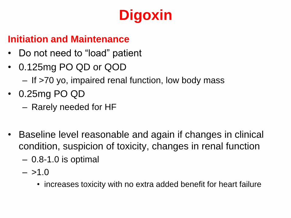
Digoxin
Initiation and Maintenance
• Do not need to ―load‖ patient
• 0.125mg PO QD or QOD
– If >70 yo, impaired renal function, low body mass
• 0.25mg PO QD
– Rarely needed for HF
• Baseline level reasonable and again if changes in clinical
condition, suspicion of toxicity, changes in renal function
– 0.8-1.0 is optimal
– >1.0
• increases toxicity with no extra added benefit for heart failure

Digoxin
• Contraindications
– 2-3rd degree heart block (without PM)
– Wolff-Parkinson-White with Afib
– Ventricular fibrillation
– Hypersensitivity
• Precautions
– Amyloid cardiomyopathy
– Idiopathic hypertrophic subaortic stenosis
– Constrictive pericarditis
– Others…

Digoxin
• Adverse Reactions
– Heart block
– CNS (dizziness, visual disturbances, confusion,
weakness)
– Dermatologic: rash (1.6%)
– Gastrointestinal: nausea, vomiting, diarrhea
– Others: Increased estrogen levels, impotence
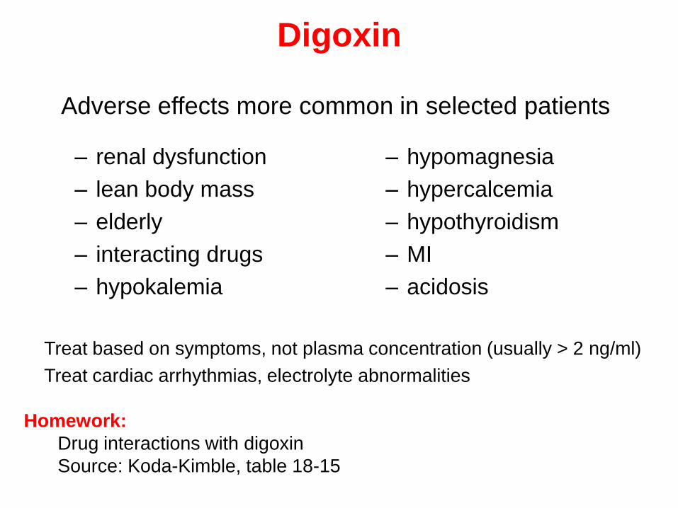
Digoxin
– renal dysfunction
– lean body mass
– elderly
– interacting drugs
– hypokalemia
– hypomagnesia
– hypercalcemia
– hypothyroidism
– MI
– acidosis
Adverse effects more common in selected patients
- Treat based on symptoms, not plasma concentration (usually > 2 ng/ml)
- Treat cardiac arrhythmias, electrolyte abnormalities
Homework:
Drug interactions with digoxin
Source: Koda-Kimble, table 18-15
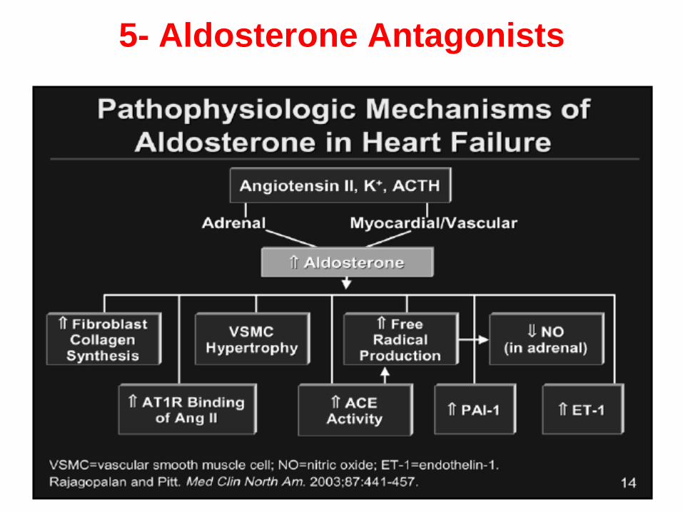
5- Aldosterone Antagonists
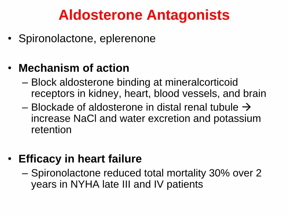
Aldosterone Antagonists
• Spironolactone, eplerenone
• Mechanism of action
– Block aldosterone binding at mineralcorticoidreceptors in kidney, heart, blood vessels, and brain
– Blockade of aldosterone in distal renal tubule increase NaCl and water excretion and potassium retention
• Efficacy in heart failure
– Spironolactone reduced total mortality 30% over 2 years in NYHA late III and IV patients
Aldosterone Antagonists
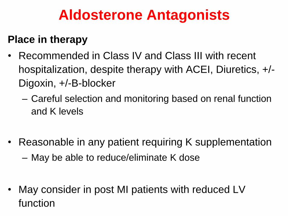
Place in therapy
• Recommended in Class IV and Class III with recent
hospitalization, despite therapy with ACEI, Diuretics, +/-
Digoxin, +/-B-blocker
– Careful selection and monitoring based on renal function
and K levels
• Reasonable in any patient requiring K supplementation
– May be able to reduce/eliminate K dose
• May consider in post MI patients with reduced LV
function
Aldosterone Antagonists
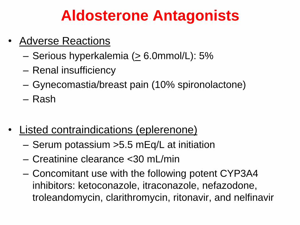
• Adverse Reactions
– Serious hyperkalemia (> 6.0mmol/L): 5%
– Renal insufficiency
– Gynecomastia/breast pain (10% spironolactone)
– Rash
• Listed contraindications (eplerenone)
– Serum potassium >5.5 mEq/L at initiation
– Creatinine clearance <30 mL/min
– Concomitant use with the following potent CYP3A4
inhibitors: ketoconazole, itraconazole, nefazodone,
troleandomycin, clarithromycin, ritonavir, and nelfinavir

Aldosterone Antagonists
Reducing Hyperkalemia Risk
• Avoid aldosterone antagonists in patients with the following:– SCr > 2.0 in women or > 2.5 mg/dL in men
– CrCl < 30 mL/min
– recent worsening of renal function
– serum K+ ≥ 5.0 mEq/L
– history of severe hyperkalemia
• Start with low doses (12.5 mg/day spironolactone, 25 mg/day eplerenone)– especially
• elderly
• DM
• CrCl < 50 mL/min
• Decrease/discontinue K+ supplements when starting an aldosterone antagonist
• If K rises to >5.4, decrease spironolactone dose
• Avoid ACE inhibitor, ARB, aldosterone antagonist triple therapy
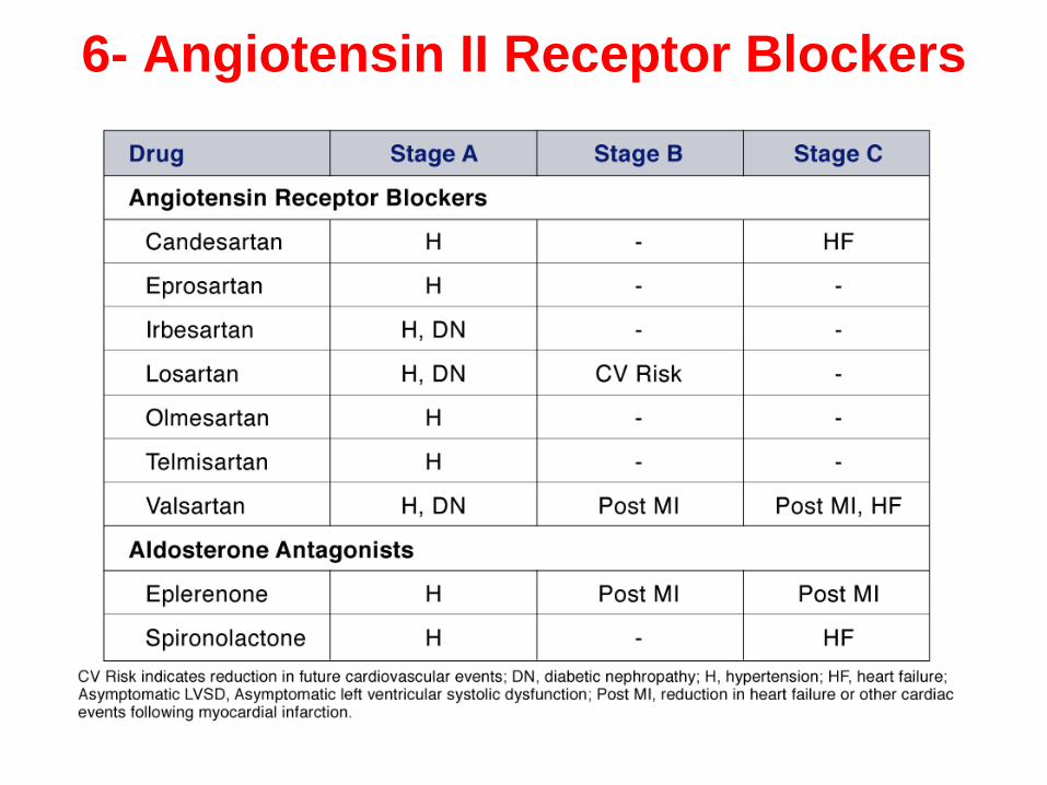
6- Angiotensin II Receptor Blockers

ARBs
• Use controversial in HF
– valsartan, candesartan shown beneficial in trials
• ARBs have theoretical advantage over ACE
inhibitors:
– ACE inhibitors have ACE escape which leads to
increased angiotensin II & aldosterone
– no effect on bradykinin (lower incidence of cough)
– not metabolized by cytochrome P-450; no significant
drug-drug interactions
ARBs
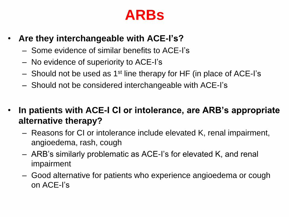
• Are they interchangeable with ACE-I’s?
– Some evidence of similar benefits to ACE-I’s
– No evidence of superiority to ACE-I’s
– Should not be used as 1st line therapy for HF (in place of ACE-I’s
– Should not be considered interchangeable with ACE-I’s
• In patients with ACE-I CI or intolerance, are ARB’s appropriate
alternative therapy?
– Reasons for CI or intolerance include elevated K, renal impairment,
angioedema, rash, cough
– ARB’s similarly problematic as ACE-I’s for elevated K, and renal
impairment
– Good alternative for patients who experience angioedema or cough
on ACE-I’s
ARBs
Place in therapy
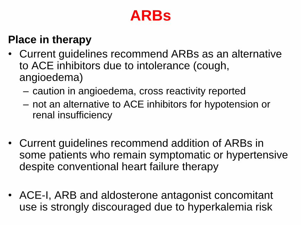
• Current guidelines recommend ARBs as an alternative to ACE inhibitors due to intolerance (cough, angioedema)
– caution in angioedema, cross reactivity reported
– not an alternative to ACE inhibitors for hypotension or renal insufficiency
• Current guidelines recommend addition of ARBs in some patients who remain symptomatic or hypertensive despite conventional heart failure therapy
• ACE-I, ARB and aldosterone antagonist concomitant use is strongly discouraged due to hyperkalemia risk

• Adverse effects:
– hypotension
– decreased renal function
– increased serum K+
• Contraindications:
– pregnancy category C in 1st trimester
– pregnancy category D in 2nd & 3rd trimester
114
ARBs
7- Nitrates & Hydralazine
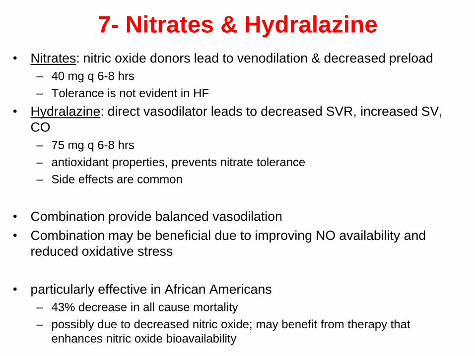
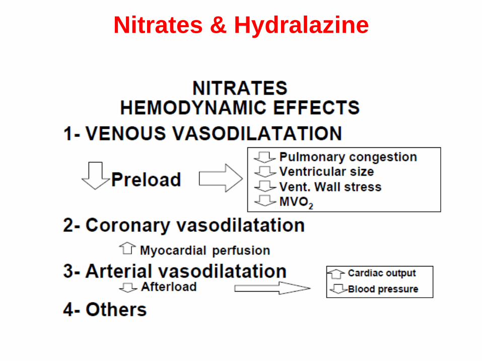
• Nitrates: nitric oxide donors lead to venodilation & decreased preload
– 40 mg q 6-8 hrs
– Tolerance is not evident in HF
• Hydralazine: direct vasodilator leads to decreased SVR, increased SV,
CO
– 75 mg q 6-8 hrs
– antioxidant properties, prevents nitrate tolerance
– Side effects are common
• Combination provide balanced vasodilation
• Combination may be beneficial due to improving NO availability and
reduced oxidative stress
• particularly effective in African Americans
– 43% decrease in all cause mortality
– possibly due to decreased nitric oxide; may benefit from therapy that
enhances nitric oxide bioavailability
Nitrates & Hydralazine
Nitrates & Hydralazine
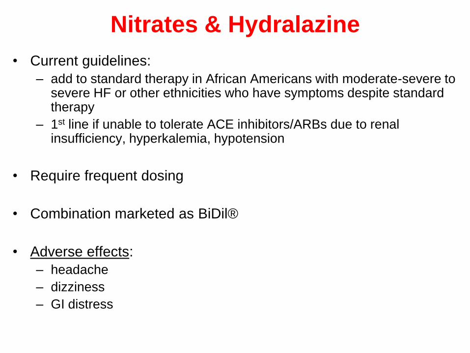
• Current guidelines:
– add to standard therapy in African Americans with moderate-severe to severe HF or other ethnicities who have symptoms despite standard therapy
– 1st line if unable to tolerate ACE inhibitors/ARBs due to renal insufficiency, hyperkalemia, hypotension
• Require frequent dosing
• Combination marketed as BiDil®
• Adverse effects:
– headache
– dizziness
– GI distress
8. Calcium Channel Blockers
• No role in treating chronic heart failure associated
with LV systolic dysfunction
• Newer agents (felodipine ER, amlodipine) may be
used safely for other indications (i.e. angina,
hypertension) in patients with chronic heart failure
Anticoagulants
• Most justified in patients with heart failure who have
had a previous embolic event or are in atrial
fibrillation


9. Antiarrhythmic therapy
• Patients with heart failure may have frequent and complex ventricular arrhythmias and a high risk of sudden death
• Class I or III antiarrhythmic drugs are not recommended in patients with HF for the prevention of ventricular arrhythmias
• The use of antiarrhythmic medication is not indicated as primary treatment for asymptomatic ventricular arrhythmias or to improve survival in patients with HF
• It is reasonable to prescribe amiodarone to decrease recurrence of atrial arrhythmias and to decrease recurrenceof ICD discharge for ventricular arrhythmias
• ICD’s clearly superior to antiarrhythmic drugs in the prevention of sudden cardiac death (SCD)
ICD = implantable cardioverter defibrillator

Stage D HF Treatment
• Stage D patients
– symptoms at rest refractory to maximal medical care
– undergo recurrent hospitalizations
– cannot be discharged from the hospital without
special intervention
• Specialized therapies
– mechanical circulatory support
– continuous IV positive inotrope
– cardiac transplant
– hospice care

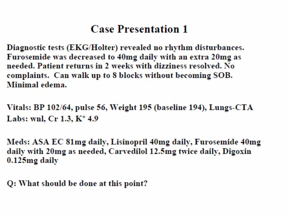
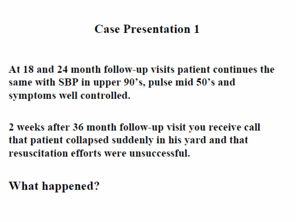
Putting it All Together: Clinical Applications

Putting it all together; continue


Statin therapy has been broadly implicated in prevention of
adverse cardiovascular events, including new-onset HF.
Originally designed to lower cholesterol in patients with
cardiovascular disease, statins are increasingly recognized for
their favorable effects on inflammation, oxidative stress, and
vascular performance. Several observational and post analyses
from large clinical trials have implied that statin therapy may
provide clinical benefit to patients with HF. However, 2 large
RCTs have demonstrated that rosuvastatin has neutral effects
on long-term outcomes in patients with chronic HFrEF when
added to standard GDMT.
At present, statin therapy should not be prescribed
primarily for the treatment of HF to improve clinical
outcomes.
Supplementation with omega-3 PUFA has been evaluated as
an adjunctive therapy for cardiovascular disease and HF. Trials
in primary and secondary prevention of coronary heart disease
showed that omega-3 PUFA supplementation results in a 10%
to 20% risk reduction in fatal and nonfatal cardiovascular
events.
The GISSI (Gruppo Italiano per lo Studio della Sopravvivenza
nell'Infarto miocardico ) Prevenzione trial demonstrated a 21%
reduction in death among post-MI patients taking 1 g of omega-
3 PUFA (850 to 882 mg of eicosapentaenoic acid [EPA] and
docosahexaenoic acid [DHA] as ethyl esters in the ratio of
1:1.2)
The use of omega-3 PUFA supplementation is reasonable
as adjunctive therapy in patients with chronic HF.

Concomitant Disorder Treatment
• Hypertension
– 2/3 HF patients have history of or current
hypertension
– 1st line: ACE inhibitors, β-blockers, diuretics
– 2nd line: ARBs, aldosterone antagonists, isosorbide
dinitrate/hydralazine or 2nd generation CCBs
(amlodipine, felodipine)
– Avoid CCBs with negative inotropic effects & direct
acting vasodilators that cause Na+ retention in
patients with systolic dysfunction

Concomitant Disorder Treatment
• Angina
– coronary artery disease: most common HF etiology
– 1st line: nitrates, β-blockers
– must be fluid controlled for antianginal medications to
be effective
Concomitant Disorder Treatment
• Atrial fibrillation
– 10 to 30% HF patients
– 1st line: ACE inhibitors, ARBs, β-blockers
– digoxin slows ventricular response but not HF
progression; β-blocker + digoxin better than either
alone
– avoid CCBs with negative inotropic function
– amiodarone: preferred antiarrhytmic, dofetilide also
safe & effective; avoid class I antiarrhythmic agents
– increases risk of thromboembolism, decreases CO,
leads to hemodynamic compromise

Concomitant Disorder Treatment
• Antithrombotic therapy for atrial fibrillation
– high risk patients: paroxysmal, persistent, or
permanent atrial fibrillation (target INR range 2 to 3)
at high risk for stroke
• warfarin
– intermediate risk patients (age 65 to 75, no stroke risk
factors)
• warfarin or ASA 325 mg/day depending on risk factors
– low risk patients (age < 65 years, no stroke risk
factors)
• ASA 325 mg/day

Concomitant Disorder Treatment
• DM
– ~1/3 of HF patients; HF risk in diabetic patients is independent of coronary artery disease & HTN
– concerns of adverse effects with glitazones, metformin
– Glitazones: contraindicated in class III & IV HF patients
– metformin labeling: CI in HF
• retrospective analysis > 3000 HF patients shows metformin safe
– decreases mortality & hospitalizations
• no prospective data
• monitor volume & renal status

Treatment of Acute
Decompensated Heart Failure


Treatment
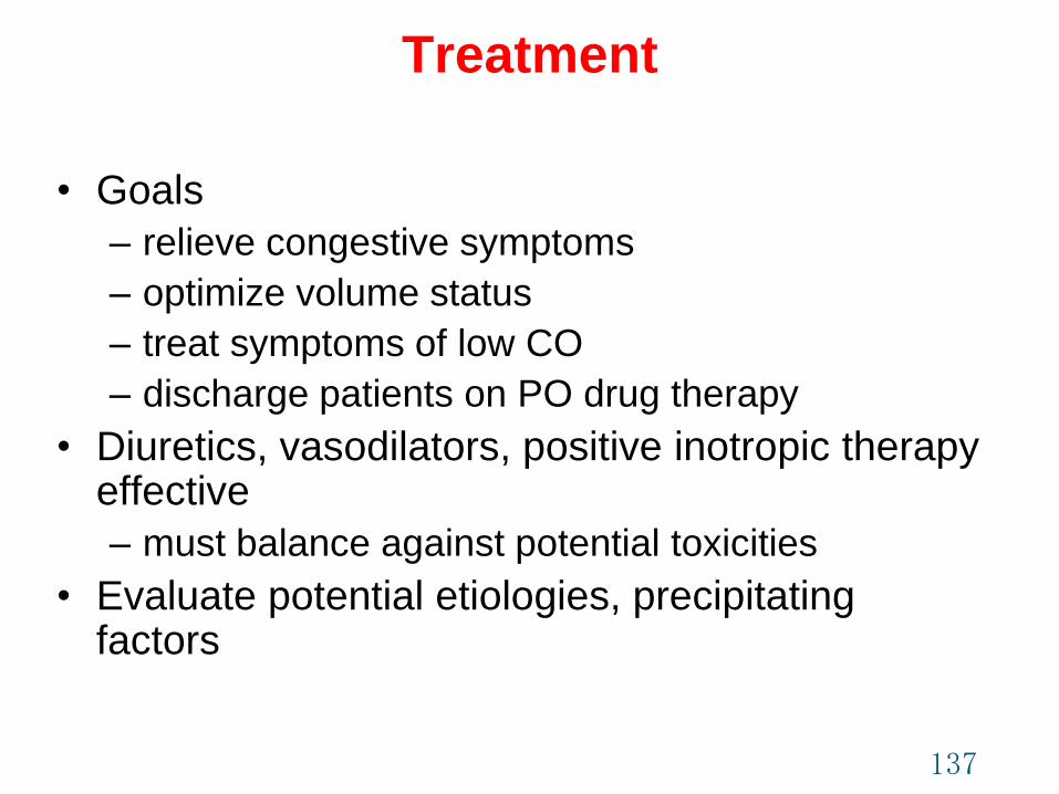
• Goals
– relieve congestive symptoms
– optimize volume status
– treat symptoms of low CO
– discharge patients on PO drug therapy
• Diuretics, vasodilators, positive inotropic therapy effective
– must balance against potential toxicities
• Evaluate potential etiologies, precipitating factors
137
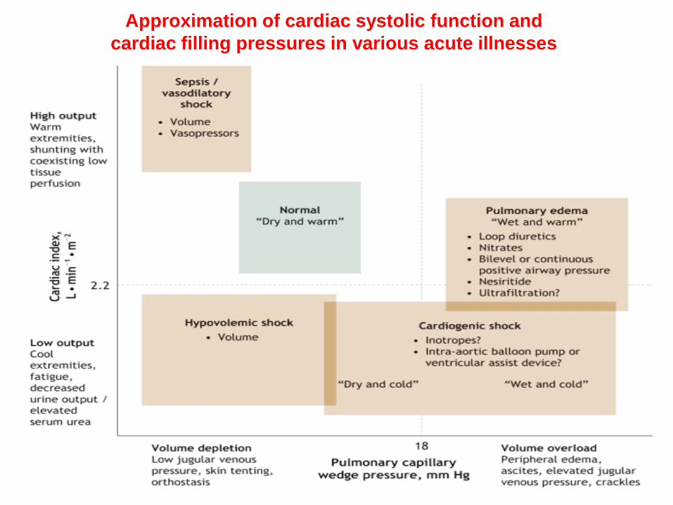
Approximation of cardiac systolic function and
cardiac filling pressures in various acute illnesses
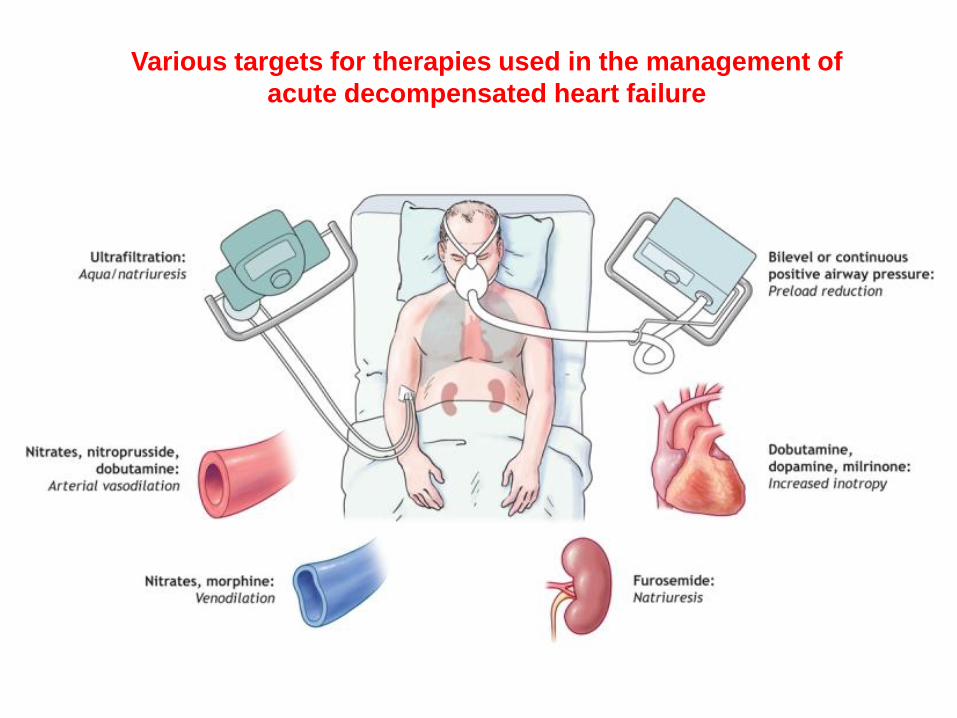
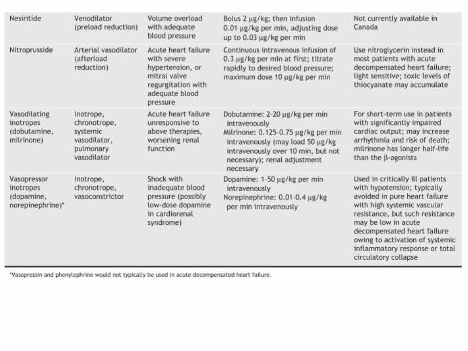
Various targets for therapies used in the management of
acute decompensated heart failure
140

Acute Decompensated HFMonitoring Recommendations
Value Frequency Specifics
Weight At least daily Determine after voiding in the morning
Account for possible increased food intake as a
result of improved appetite
Fluid intake/output At least daily Strict documentation necessary
Vital signs More than daily Including orthostatic blood pressure
Signs At least daily Edema, acites, pulmonary rales, hepatomegaly,
increased jugular venous pressure, hepatojugular
reflux, liver tenderness
Symptoms At least daily Orthopnea, paroxysmal nocturnal dyspnea,
nocturnal cough, dyspnea, fatigue
Electrolytes At least daily Potassium, magnesium, sodium
Renal function At least daily Blood urea nitrogen, serum creatinine
141DiPiro JT, Talbert RL, Yee GC, Matzke GR, Wells BG, Posey LM: Pharmacotherapy: A Pathophysiologic Approach, 7th Edition: http://www.accesspharmacy.com

Outcome evaluation of acute HF
Focus on:
1. acute improvement of symptoms and hemodynamics due to
intravenous therapies;
2. criteria for a safe discharge from the hospital;
3. optimization of oral therapy.
• Initially, monitor patients for rapid relief of symptoms related to the
chief complaint on admission.
– This includes improvement of dyspnea, oxygenation, fatigue, JVD,
and other markers of congestion or distress.
• Monitor for adequate perfusion of vital organs
– through assessment of mental status, creatinine clearance, liver
function tests, and a stable HR between 50 and 100 beats per
minute.
• Adequate skin and muscle blood perfusion and normal pH is
desirable.
Outcome evaluation of acute HF
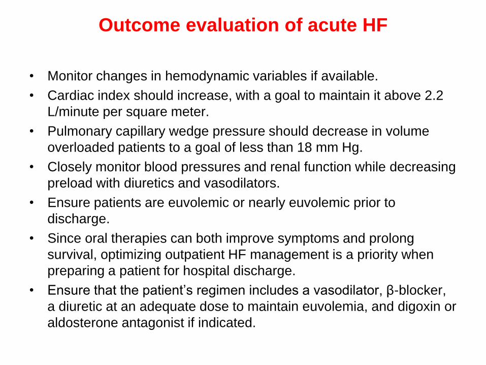
• Monitor changes in hemodynamic variables if available.
• Cardiac index should increase, with a goal to maintain it above 2.2
L/minute per square meter.
• Pulmonary capillary wedge pressure should decrease in volume
overloaded patients to a goal of less than 18 mm Hg.
• Closely monitor blood pressures and renal function while decreasing
preload with diuretics and vasodilators.
• Ensure patients are euvolemic or nearly euvolemic prior to
discharge.
• Since oral therapies can both improve symptoms and prolong
survival, optimizing outpatient HF management is a priority when
preparing a patient for hospital discharge.
• Ensure that the patient’s regimen includes a vasodilator, β-blocker,
a diuretic at an adequate dose to maintain euvolemia, and digoxin or
aldosterone antagonist if indicated.

Heart failure with preserved left
ventricular ejection fraction

Heart failure with preserved left ventricular
ejection fraction
• In the absence of more landmark clinical studies, the
current treatment approach for diastolic dysfunction or
preserved LVEF is:
1. correction or control of underlying etiologies (including
optimal treatment of hypertension and CAD and
maintenance of normal sinus rhythm);
2. Reduction of cardiac filling pressures at rest and during
exertion;
3. increased diastolic filling time.

Heart failure with preserved left ventricular
ejection fraction
• Diuretics, ACE inhibitors, and ARBs are frequently used to
control congestion.
• Angiotensin receptor blockers may also slow disease
progression.
• β-Blockers and calcium channel blockers can theoretically
improve ventricular relaxation through negative inotropic and
chronotropic effects.
• Unlike in systolic HF, nondihydropyridine calcium channel
blockers (diltiazem and verapamil) may be especially useful
in improving diastolic function by limiting the availability of
calcium that mediates contractility.
• The role of digoxin for symptom management and HR control
in these patients is not well established. AHA/ACCF
currently DO NOT recommend digoxin in HFprEF.


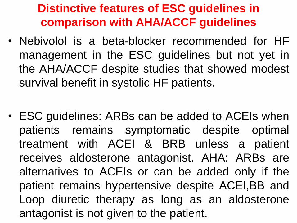
Distinctive features of ESC guidelines in
comparison with AHA/ACCF guidelines
• Nebivolol is a beta-blocker recommended for HF
management in the ESC guidelines but not yet in
the AHA/ACCF despite studies that showed modest
survival benefit in systolic HF patients.
• ESC guidelines: ARBs can be added to ACEIs when
patients remains symptomatic despite optimal
treatment with ACEI & BRB unless a patient
receives aldosterone antagonist. AHA: ARBs are
alternatives to ACEIs or can be added only if the
patient remains hypertensive despite ACEI,BB and
Loop diuretic therapy as long as an aldosterone
antagonist is not given to the patient.