chronic chagasic cardiomyopathy (ccc) with severe...
TRANSCRIPT

Chronic Chagasic Cardiomyopathy (CCC) with severe biventricular involvement and intraventricular dromotropic disturbances
Andrés Ricardo Pérez-Riera, M.D. Ph.D.Design of Studies and Scientific Writing
Laboratory in the ABC School of Medicine,
Santo André, São Paulo, Brazil
https://ekgvcg.wordpress.com
Raimundo Barbosa-Barros, MD
Chief of the Coronary Center of the Hospital de Messejana Dr. Carlos
Alberto Studart Gomes. Fortaleza – CE- Brazil
Portuguese
Relato de caso
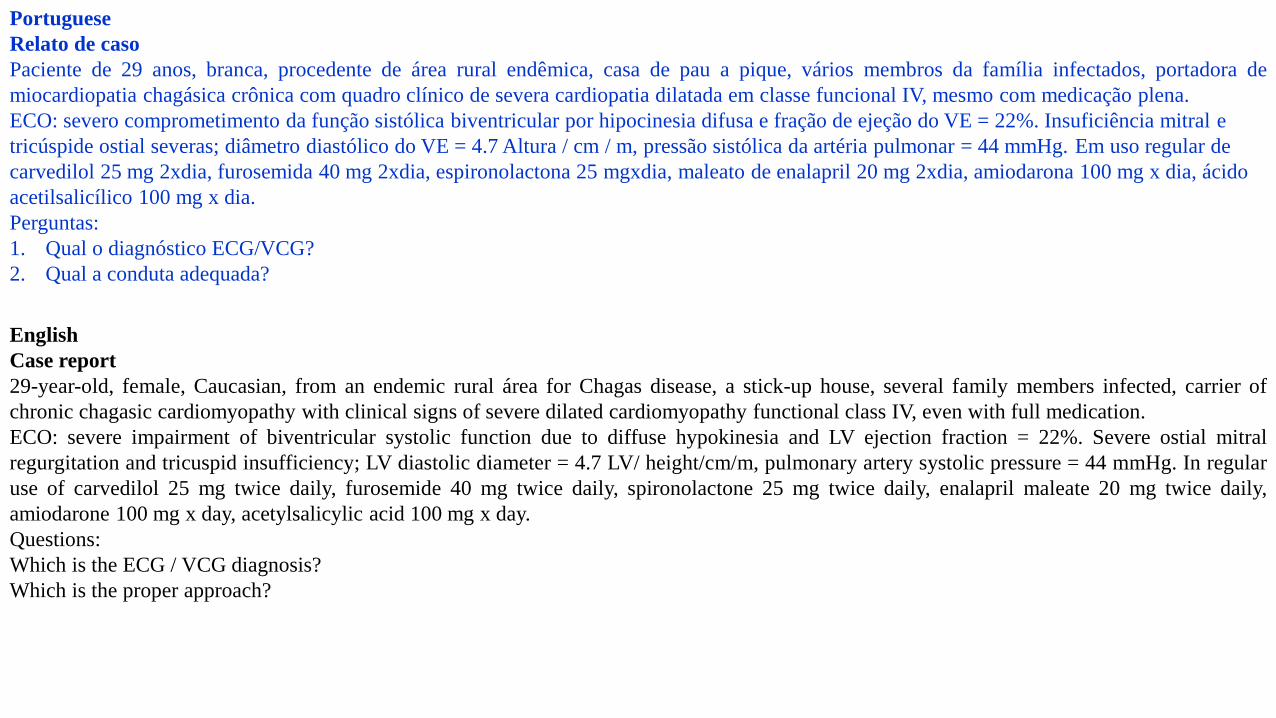
Paciente de 29 anos, branca, procedente de área rural endêmica, casa de pau a pique, vários membros da família infectados, portadora de
miocardiopatia chagásica crônica com quadro clínico de severa cardiopatia dilatada em classe funcional IV, mesmo com medicação plena.
ECO: severo comprometimento da função sistólica biventricular por hipocinesia difusa e fração de ejeção do VE = 22%. Insuficiência mitral e
tricúspide ostial severas; diâmetro diastólico do VE = 4.7 Altura / cm / m, pressão sistólica da artéria pulmonar = 44 mmHg. Em uso regular de
carvedilol 25 mg 2xdia, furosemida 40 mg 2xdia, espironolactona 25 mgxdia, maleato de enalapril 20 mg 2xdia, amiodarona 100 mg x dia, ácido
acetilsalicílico 100 mg x dia.
Perguntas:
1. Qual o diagnóstico ECG/VCG?
2. Qual a conduta adequada?
English
Case report
29-year-old, female, Caucasian, from an endemic rural área for Chagas disease, a stick-up house, several family members infected, carrier of
chronic chagasic cardiomyopathy with clinical signs of severe dilated cardiomyopathy functional class IV, even with full medication.
ECO: severe impairment of biventricular systolic function due to diffuse hypokinesia and LV ejection fraction = 22%. Severe ostial mitral
regurgitation and tricuspid insufficiency; LV diastolic diameter = 4.7 LV/ height/cm/m, pulmonary artery systolic pressure = 44 mmHg. In regular
use of carvedilol 25 mg twice daily, furosemide 40 mg twice daily, spironolactone 25 mg twice daily, enalapril maleate 20 mg twice daily,
amiodarone 100 mg x day, acetylsalicylic acid 100 mg x day.
Questions:
Which is the ECG / VCG diagnosis?
Which is the proper approach?


IIIII
aVF
X I
Y
ECG/VCG correlation in the
frontal plane
Magnified T-loop –
present case
Efferent
Limb
Afferent
Limb
J point
0 pointAfferent
branch
with faster
recording
Slower recording
velocity in its
efferent branch
Normal T-loop/wave
T-wave
J and 0 points are
coincident

X V6
V1
V4
V5
V2
V3
Z
ECG/VCG correlation in the horizontal plane
Magnified T-loop
Efferent
Limb
Afferent
Limb
J point
0 point
J and 0 points are very distant from
each other
0 point
E point
RA
LA
X
Magnified P-loop

YaVF
V2
ECG/VCG correlation in the right sagittal plane
Magnified T-loop
Efferent
Limb
Afferent
Limb
J point
0 point
qR

Colleagues Opinion

ECG : ritmo sinusal, BIA-a; bloqueo tetrafascicular (AV 1°+HASI+BDAM+BCRD).
En VCG (sólo opinaré sobre el BIA-a) creo que se ve el asa hacia adelante y a la derecha para luego dirigirse desde abajo hacia arriba y a la
izquierda, como matráz de erlemmeyer.
Conducta: ante una miocardiopatia dilatada, con HTPulmonar, hipocinesia difusa biventricular, insuficiencia mitro-tricuspídea severa, con muy
baja F Ey , a pesar del tratamiento farmacológico pleno y tan severo trastorno de conducción: Resincronizador + CDI como puente al trasplante
cardíaco.
Afectuosamente, y a la espera de la opinión de los expertos.
Dr Juan Carlos Manzzardo
English
ECG: sinus rhythm, advanced IAB; tetrafascicular block (1º AV+LAFB+LSFB+CRBBB).
In VCG (I will only discuss the A-IAB), I think the loop is seen forward and at the right, to later head from down to up and at the left, as an
“Erlenmeyer flask”.
Management: before dilated cardiomyopathy with pulmonary hypertension, diffuse biventricular hypokinesis, severe mitral-tricuspid
insufficiency, with very low LVEF, in spite of the full pharmacological treatment and such severe conduction disturbance: resynchronizer + ICD as
a bridge into heart transplant.
Warm regards, and I wait for the opinions by the experts,
Juan Carlos Manzzardo, MD, Mendoza, Argentina
Spanish

Hola querido Andrés: refiere severa miocardiopatia dilatada y en el resumen el “diámetro diastólico del VI de 4.7?". Solo puede ser un error de
tipeo Dear Andrés, you say severe dilated cardiomyopathy and in the summary, “LV diastolic diameter = 4.7?" It must be a typing mistake
Un abrazo A hug
Martín Ibarrola
Answer: Dear Martin, I think that the parameter was expressed in LV diastolic diameter/height (cm/m)
1. Lang RM, Bierig M, Devereux RB, et al. American Society of Echocardiography's Nomenclature and Standards Committee., Task Force on
Chamber Quantification., American College of Cardiology Echocardiography Committee., American Heart Association., and European
Association of Echocardiography, European Society of Cardiology.. Recommendations for chamber quantification. Eur J Echocardiogr.
2006;7(2):79-108.
Reference limits and partition values of left ventricular size (1)
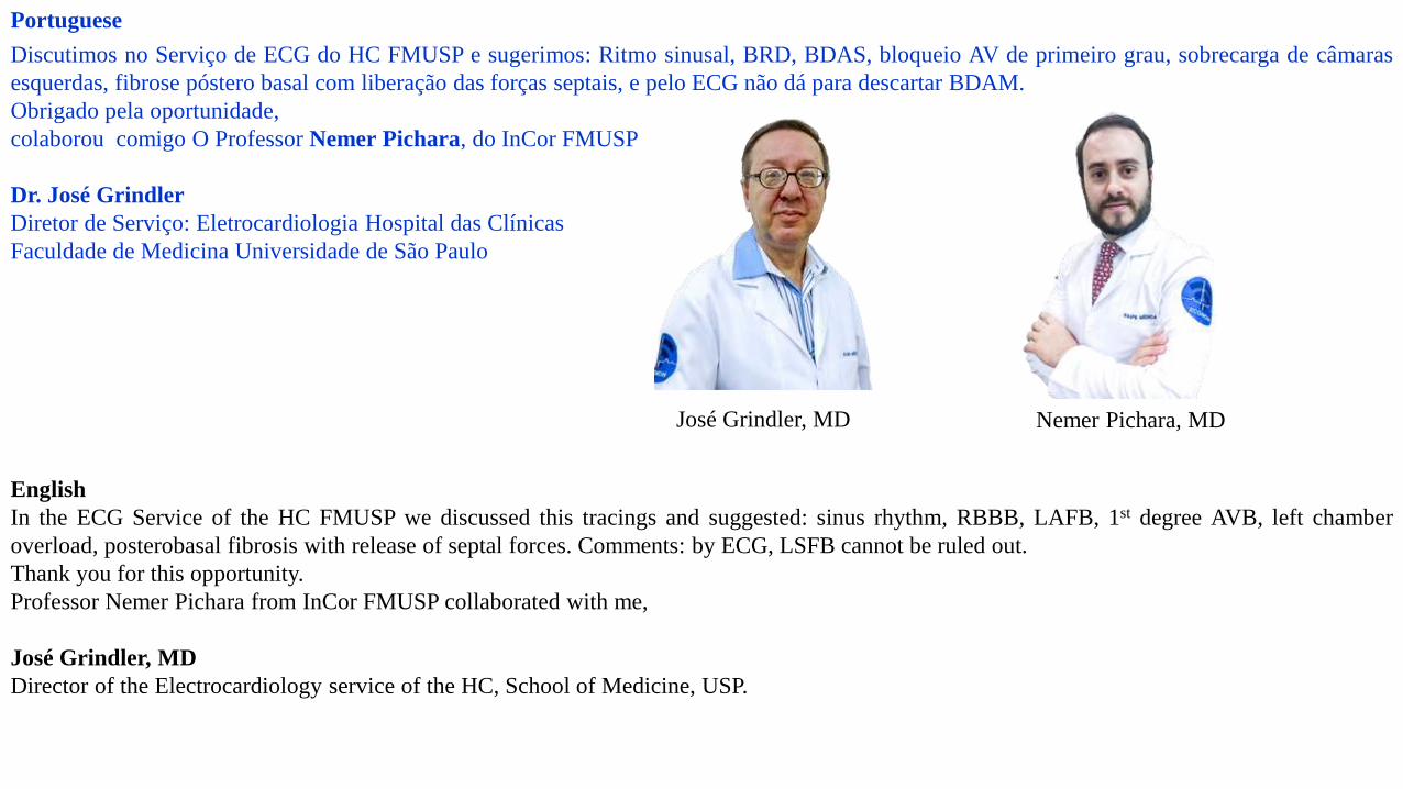
Discutimos no Serviço de ECG do HC FMUSP e sugerimos: Ritmo sinusal, BRD, BDAS, bloqueio AV de primeiro grau, sobrecarga de câmaras
esquerdas, fibrose póstero basal com liberação das forças septais, e pelo ECG não dá para descartar BDAM.
Obrigado pela oportunidade,
colaborou comigo O Professor Nemer Pichara, do InCor FMUSP
Dr. José Grindler
Diretor de Serviço: Eletrocardiologia Hospital das Clínicas
Faculdade de Medicina Universidade de São Paulo
Portuguese
English
In the ECG Service of the HC FMUSP we discussed this tracings and suggested: sinus rhythm, RBBB, LAFB, 1st degree AVB, left chamber
overload, posterobasal fibrosis with release of septal forces. Comments: by ECG, LSFB cannot be ruled out.
Thank you for this opportunity.
Professor Nemer Pichara from InCor FMUSP collaborated with me,
José Grindler, MD
Director of the Electrocardiology service of the HC, School of Medicine, USP.
Nemer Pichara, MDJosé Grindler, MD

Este é o típico paciente que com frequência, interna em nossa enfermaria. IC por disfunção sistólica avançada, CF IV, apesar da terapia otimizada,
acredito que já algum tempo! Apresenta ainda grave distúrbio de condução Hissiano, caracterizado por bloqueio avançado de ramo direito,
BDASE, BDAM e BAV 1 grau que pode traduzir bloqueio nodal AV influenciado pelo betabloqueador e/ou retardo de condução pelo único
fascículo íntegro (póstero inferior esquerdo). Há de fazer diagnóstico diferencia de SVD, associado ou não ao BDAM. Gostaria de saber mais o
vetocardiograma! O principal motivo que escrevo é que nos nossos casos a ressincronização ventricular não teve boa resposta, apesar da largura
do QRS. Motivos: 1) é BRD e não BRE: 2) a CF está muito avançada; 3) presença de fibrose extensa em regiões de implante do eletrodo
ventricular esquerdo. Não existe também indicação para CDI como indicação primária para Cardiomiopatia Chagásica. O estudo está correndo
(Estudo CHAGASICS). Enfim, acho que este é um candidato a transplante de coração. Poderia também, se possível, introduzir hidralazina
associado ao nitrato para o tratamento da IC que permanece sintomática.
Um grande Abraço!
Marcelo Garcia Leal MD, Ribeirão Preto - São Paulo, Brasil
Portuguese
English
This is the typical patient that often, is admitted into our infirmary. HF by advanced systolic dysfunction, FC IV, in spite of
optimized therapy I believe for quite some time! He also presents severe His conduction disorder, characterized by advanced
RBBB, LAFB, LSFB and 1st degree AVB that may be manifesting AV node block influenced by β-blocker and/or conduction
delay by the only whole fascicle (left posteroinferior).
The differential diagnosis is made with RVH, associated or not with LSFB. I would like to know more about the VCG! The
main reason I am writing this is that in our chagasic cases, cardiac resynchronization therapy did not have a good response,
in spite of the QRS width. Reasons: 1) RBBB and not LBBB; 2) Very advanced cardiac failure; 3) Presence of extensive
fibrosis in regions of left ventricular electrode implant.
There is no ICD indication either, as primary indication for Chagasic cardiomyopathy. There is an ongoing study
(CHAGASICS study). Anyway, I think this is a good candidate to heart transplant. Hydralazine could be introduced if
possible, associated with nitrates for the treatment of HF that would remain symptomatic.
Best regards!
Marcelo Garcia-Leal, MD

Ola.
1. Diagnóstico ECG/VCG - RS, BRD+BDAS+BDAM
2. Conduta : Ressincronização . Transplante.
Horácio Gomes Pereira Filho MD
University of São Paulo
Faculty of Medicine (FM)
São Paulo, Estado de Sao Paulo, Brazil
Médico do grupo Fleury São Paulo Brazil
English
Hello
1. ECG/VCG diagnosis – SR, RBBB+LSFB+LAFB.
2. Management: Resynchronization, transplant.
Horácio Gomes Pereira Filho MD

Spanish
Queridos amigos:
Voy a partir de subrayar la triste realidad de esta casi niña de 29 años que presenta tan severa miocardiopatía, que al decir de Juan Carlos, sería
candidata a TRC y transplante. ¡Qué terrible!
Comparto también con Juanca su descripción de las alteraciones dromotrópicas y quiero hacer un agregado: en las derivaciones de la cara inferior
se observa una gran elevación del punto Jota, lo cuál también es muy evidente en el VCG de todos los planos, donde se observa una enorme
separación del Punto 0 y el Punto J.
Me da la sensación que este hallazgo va más allá de la presencia del trastorno de conducción asociado, de la dilatación e hipertrofia de las
cavidades y paredes.
¿Deberemos en este caso aplicar las enseñanzas de Andrés y Raimundo acerca de los síndromes de elevación del Punto Jota?
Un abrazo
Edgardo Schapachnik MD Buenos Aires Argentina
English
Dear Friends,
I will begin by emphasizing the sad reality of this almost child of 29 years of age, who presents such a severe
cardiomyopathy that as Juan Carlos states, would make her a candidate for CRT and transplant. How awful! I also agree
with him on his description of the dromotropic disturbances and I would like to add something: in the inferior leads, a
great J point elevation is observed, which is also very evident in the VCG in all planes, where a great separation between
the 0 and the J points is observed. I have the feeling this finding goes beyond the presence of associated conduction
disorder, dilatation and hypertrophy in chambers and walls. In this case, should we apply the teachings by Andrés and
Raimundo about J point elevation syndromes?
Best regards,
Edgardo Schapachnik MD Buenos Aires Argentina
Hola Amigos
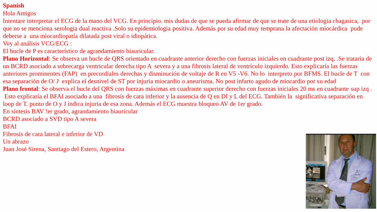
Intentare interpretar el ECG de la mano del VCG. En principio. mis dudas de que se pueda afirmar de que se trate de una etiologia chagasica, por
que no se menciona serologia dual reactiva .Solo su epidemiologia positiva. Además por su edad muy temprana la afectación miocárdica pude
deberse a una miocardiopatía dilatada post viral o idiopática.
Voy al análisis VCG/ECG :
El bucle de P es característico de agrandamiento biauricular.
Plano Horizontal: Se observa un bucle de QRS orientado en cuadrante anterior derecho con fuerzas iniciales en cuadrante post izq. .Se trataria de
un BCRD asociado a sobrecarga ventricular derecha tipo A severa y a una fibrosis lateral de ventrículo izquierdo. Esto explicaría las fuerzas
anteriores prominentes (FAP) en precordiales derechas y disminución de voltaje de R en V5 -V6. No lo interpreto por BFMS. El bucle de T con
esa separación de O/ J explica el desnivel de ST por injuria miocardio o aneurisma. No post infarto agudo de miocardio por su edad
Plano frontal: Se observa el bucle del QRS con fuerzas máximas en cuadrante superior derecho con fuerzas iniciales 20 ms en cuadrante sup izq .
Esto explicaría el BFAI asociado a una fibrosis de cara inferior y la ausencia de Q en DI y L del ECG. También la significativa separación en
loop de T. punto de O y J indica injuria de esa zona. Además el ECG muestra bloqueo AV de 1er grado.
En síntesis BAV !er grado, agrandamiento biauricular
BCRD asociado a SVD tipo A severa
BFAI
Fibrosis de cara lateral e inferior de VD
Un abrazo
Juan José Sirena, Santiago del Estero, Argentina
Spanish
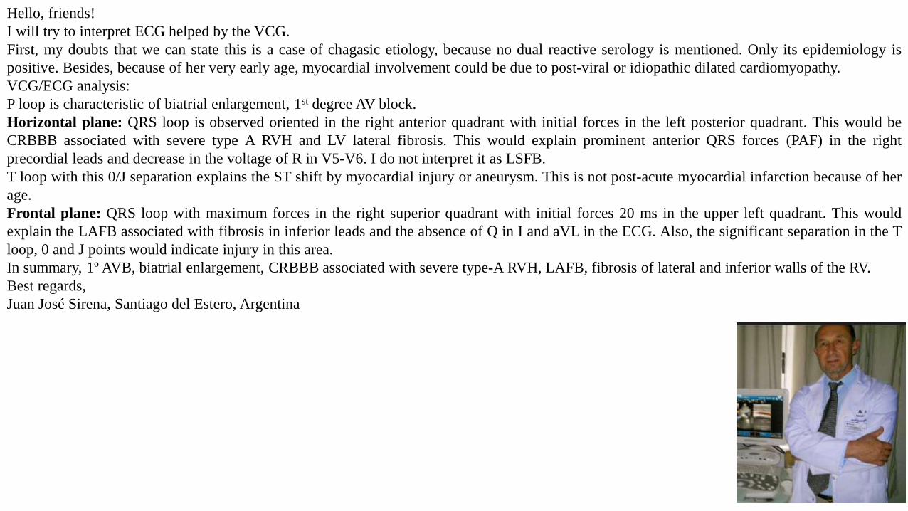
Hello, friends!
I will try to interpret ECG helped by the VCG.
First, my doubts that we can state this is a case of chagasic etiology, because no dual reactive serology is mentioned. Only its epidemiology is
positive. Besides, because of her very early age, myocardial involvement could be due to post-viral or idiopathic dilated cardiomyopathy.
VCG/ECG analysis:
P loop is characteristic of biatrial enlargement, 1st degree AV block.
Horizontal plane: QRS loop is observed oriented in the right anterior quadrant with initial forces in the left posterior quadrant. This would be
CRBBB associated with severe type A RVH and LV lateral fibrosis. This would explain prominent anterior QRS forces (PAF) in the right
precordial leads and decrease in the voltage of R in V5-V6. I do not interpret it as LSFB.
T loop with this 0/J separation explains the ST shift by myocardial injury or aneurysm. This is not post-acute myocardial infarction because of her
age.
Frontal plane: QRS loop with maximum forces in the right superior quadrant with initial forces 20 ms in the upper left quadrant. This would
explain the LAFB associated with fibrosis in inferior leads and the absence of Q in I and aVL in the ECG. Also, the significant separation in the T
loop, 0 and J points would indicate injury in this area.
In summary, 1º AVB, biatrial enlargement, CRBBB associated with severe type-A RVH, LAFB, fibrosis of lateral and inferior walls of the RV.
Best regards,
Juan José Sirena, Santiago del Estero, Argentina

Final conclusions
by
Andrés Ricardo Pérez-Riera and Raimundo Barbosa-Barros
“In medical science there are vast realms of which I have no special
knowledge and, again, no, I am not a great man; I am a happy man.”-
Karel Frederik Wenckebach (1868-1940)
See Wenckebach biography at the end of this presentation

IIIII
aVF
X I
Y
ECG/VCG correlation in the frontal plane
Magnified T-loop –
present case
Efferent
Limb
Afferent
Limb
J point
0 pointAfferent
branch
with faster
recording
Slower recording
velocity in its
efferent branch
Normal T-loop/wave
T-wave
J and 0 points are
coincident
QRS axis - 120°
SÂP axis +25°

P-wave and P-loop analysis
P-wave
1. P-duration: 110ms (normal)
2. P-voltage: 1.3mm(normal)
3. P-axis (SÂP) +25°(normal). PII<PI (unusual). P-axis values between 0° and +75° are considered normal.
4. P-shape: rounded (normal)
5. P-wave polarity in V1: plus-minus with deep P-terminal force (PTF- V1) or deep terminal negativity of P wave in V1 (DTNPV1) exceeding
0.04 mm/s. This is the terminal, negative part of the P wave in lead V1 expressed as the multiplication of its depth in millimeters and width in
seconds (mm/s). The normal PTF- V1 does not exceed 0.04 s wide and 1mm deep, i.e., 0.04 mm/s. Morris index (Morris 1964) DTNPV1 is
predictive of SCD suggesting its potential utility in risk stratification in the general population (Tereshchenko 2014).
V1
I
II

P-loop in the present case PHNormal P-loop in the PH called also the P sÊ loop
Normal P-loop in the PH
Z
P-loop in the PH
0
EX
Maximal
Posterior
Forces
≥ 0,05mV
Maximal
Posterior
Forces
≥ 0,05mV
LAE
Maximal
leftward
forces ≥ 0,1mV
Horizontal Plane
P-loop Rotation CCW or in eight
P-loop Direction Anterior, initial part; and posterior, final part
P-loop Morphology Oval
P-loop Location 1/3 in anterior quadrant and 2/3 in posterior quadrant
Location of P-loop Maximal Vector +50° to –45°
Voltage of P-loop Maximal Vector ≤ 0.1 mV
P-Loop Maximal Anterior Forces Adults up to 0.06 mV; Children up to 0.08 mV
P-Loop Maximal Posterior Forces Up to 0.04 mV
P-Loop Maximal Left Forces Adults up to 0.09 mV; Children up to 0.13 mV
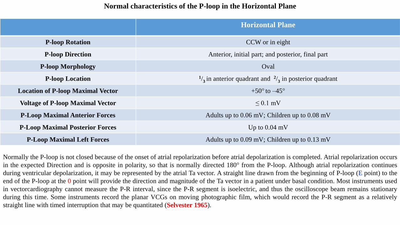
Normal characteristics of the P-loop in the Horizontal Plane
Normally the P-loop is not closed because of the onset of atrial repolarization before atrial depolarization is completed. Atrial repolarization occurs
in the expected Direction and is opposite in polarity, so that is normally directed 180° from the P-loop. Although atrial repolarization continues
during ventricular depolarization, it may be represented by the atrial Ta vector. A straight line drawn from the beginning of P-loop (E point) to the
end of the P-loop at the 0 point will provide the direction and magnitude of the Ta vector in a patient under basal condition. Most instruments used
in vectorcardiography cannot measure the P-R interval, since the P-R segment is isoelectric, and thus the oscilloscope beam remains stationary
during this time. Some instruments record the planar VCGs on moving photographic film, which would record the P-R segment as a relatively
straight line with timed interruption that may be quantitated (Selvester 1965).
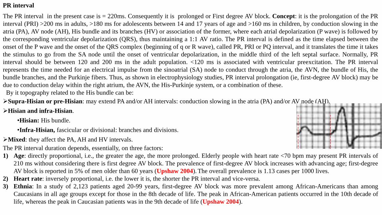
The PR interval in the present case is = 220ms. Consequently it is prolonged or First degree AV block. Concept: it is the prolongation of the PR
interval (PRI) >200 ms in adults, >180 ms for adolescents between 14 and 17 years of age and >160 ms in children, by conduction slowing in the
atria (PA), AV node (AH), His bundle and its branches (HV) or association of the former, where each atrial depolarization (P wave) is followed by
the corresponding ventricular depolarization (QRS), thus maintaining a 1:1 AV ratio. The PR interval is defined as the time elapsed between the
onset of the P wave and the onset of the QRS complex (beginning of q or R wave), called PR, PRI or PQ interval, and it translates the time it takes
the stimulus to go from the SA node until the onset of ventricular depolarization, in the middle third of the left septal surface. Normally, PR
interval should be between 120 and 200 ms in the adult population. <120 ms is associated with ventricular preexcitation. The PR interval
represents the time needed for an electrical impulse from the sinoatrial (SA) node to conduct through the atria, the AVN, the bundle of His, the
bundle branches, and the Purkinje fibers. Thus, as shown in electrophysiology studies, PR interval prolongation (ie, first-degree AV block) may be
due to conduction delay within the right atrium, the AVN, the His-Purkinje system, or a combination of these.
By it topography related to the His bundle can be:
➢Supra-Hisian or pre-Hisian: may extend PA and/or AH intervals: conduction slowing in the atria (PA) and/or AV node (AH).
➢Hisian and infra-Hisian.
•Hisian: His bundle.
•Infra-Hisian, fascicular or divisional: branches and divisions.
➢Mixed: they affect the PA, AH and HV intervals.
The PR interval duration depends, essentially, on three factors:
1) Age: directly proportional, i.e., the greater the age, the more prolonged. Elderly people with heart rate <70 bpm may present PR intervals of
210 ms without considering there is first degree AV block. The prevalence of first-degree AV block increases with advancing age; first-degree
AV block is reported in 5% of men older than 60 years (Upshaw 2004). The overall prevalence is 1.13 cases per 1000 lives.
2) Heart rate: inversely proportional, i.e. the lower it is, the shorter the PR interval and vice-versa.
3) Ethnia: In a study of 2,123 patients aged 20-99 years, first-degree AV block was more prevalent among African-Americans than among
Caucasians in all age groups except for those in the 8th decade of life. The peak in African-American patients occurred in the 10th decade of
life, whereas the peak in Caucasian patients was in the 9th decade of life (Upshaw 2004).
PR interval
AV nodeN
S
Atria H
i
s
RP
P
R
T
PR
ECG
HE
(His Electrogram)A H
V
AN
N
NH
AV
node
SA node
His
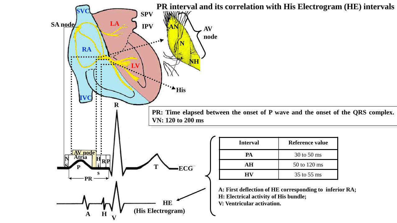
A: First deflection of HE corresponding to inferior RA;
H: Electrical activity of His bundle;
V: Ventricular activation.
PR: Time elapsed between the onset of P wave and the onset of the QRS complex.
VN: 120 to 200 ms
PR interval and its correlation with His Electrogram (HE) intervals
Interval Reference value
PA 30 to 50 ms
AH 50 to 120 ms
HV 35 to 55 ms
LA
LV
RA
SPV
IPV
SVC
IVC

QRS and repolarization analysis

QRS loop in isolated LAFB QRS loop in LAFB + RBBB QRS loop in LAFB + LFB + RBBB + RVH
I
IIIII III
Initial 10-20 ms Downward and rightward Downward and rightward Downward and leftward
QRS location Predominantly left superior
quadrant. QRS axis -45°/-70°
Predominantly left superior quadrant with
RECD in the upper right quadrant
Predominantly right superior quadrant with
middle conduction delay (MCD) and RECD
QRS rotation CCW CCW CCW
I and aVL qR qR Rs or R
aVR QS or Qr qR qR
II/III ratio SIII > SII + QRSd = 110 ms SIII > SII + QRSd ≥120 ms SIII > SII + QRSd >120 ms
RECD: Right End Conduction DelayCCW: Counterclockwise rotation
(The present case)

X V6
V1
V4
V5
V2
V3
Z
ECG/VCG correlation in the horizontal plane
Magnified T-loop
Efferent
Limb
Afferent
Limb
J point
0 point
J and 0 points are very distant from
each other
Magnified P-loop: LAE
RECD: Right End Conduction Delay; CWR: Clock Wise Rotation; Initial 10ms QRS loop directed to back and leftward;
CWR
qR
qR qRs
rS
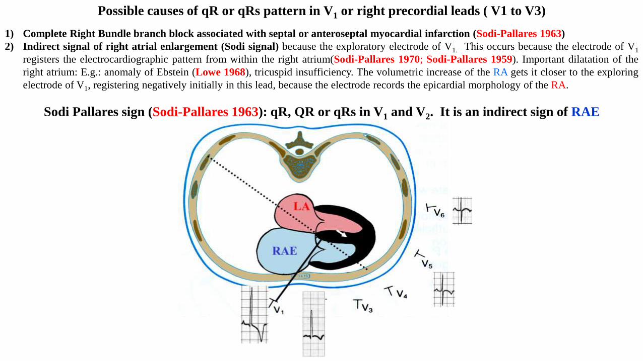
Possible causes of qR or qRs pattern in V1 or right precordial leads ( V1 to V3)
1) Complete Right Bundle branch block associated with septal or anteroseptal myocardial infarction (Sodi-Pallares 1963)
2) Indirect signal of right atrial enlargement (Sodi signal) because the exploratory electrode of V1. This occurs because the electrode of V1
registers the electrocardiographic pattern from within the right atrium(Sodi-Pallares 1970; Sodi-Pallares 1959). Important dilatation of the
right atrium: E.g.: anomaly of Ebstein (Lowe 1968), tricuspid insufficiency. The volumetric increase of the RA gets it closer to the exploring
electrode of V1, registering negatively initially in this lead, because the electrode records the epicardial morphology of the RA.
Sodi Pallares sign (Sodi-Pallares 1963): qR, QR or qRs in V1 and V2. It is an indirect sign of RAE

3. Acute pulmonary embolism (APE).( Kukla 2011)
4. Extreme systolic right ventricular hypertrophy with strain pattern of repolarization and suprasystemic right intraventricular
pressure: Ex critical Pulmonary valve stenosis and int act ventricular septum. The direction of the initial QRS vector on the X axis is helpful
in predicting severity. With X initial vector to the left and in negative hemifield of V1, the right intraventricular pressure is frequently but not
necessarily suprasystemic. (Mehran-Pour 1979)
Extreme Right Ventricular Hypertrophy (suprasystemic intraventricular right ventricle pressure
qRV1
Z
XT
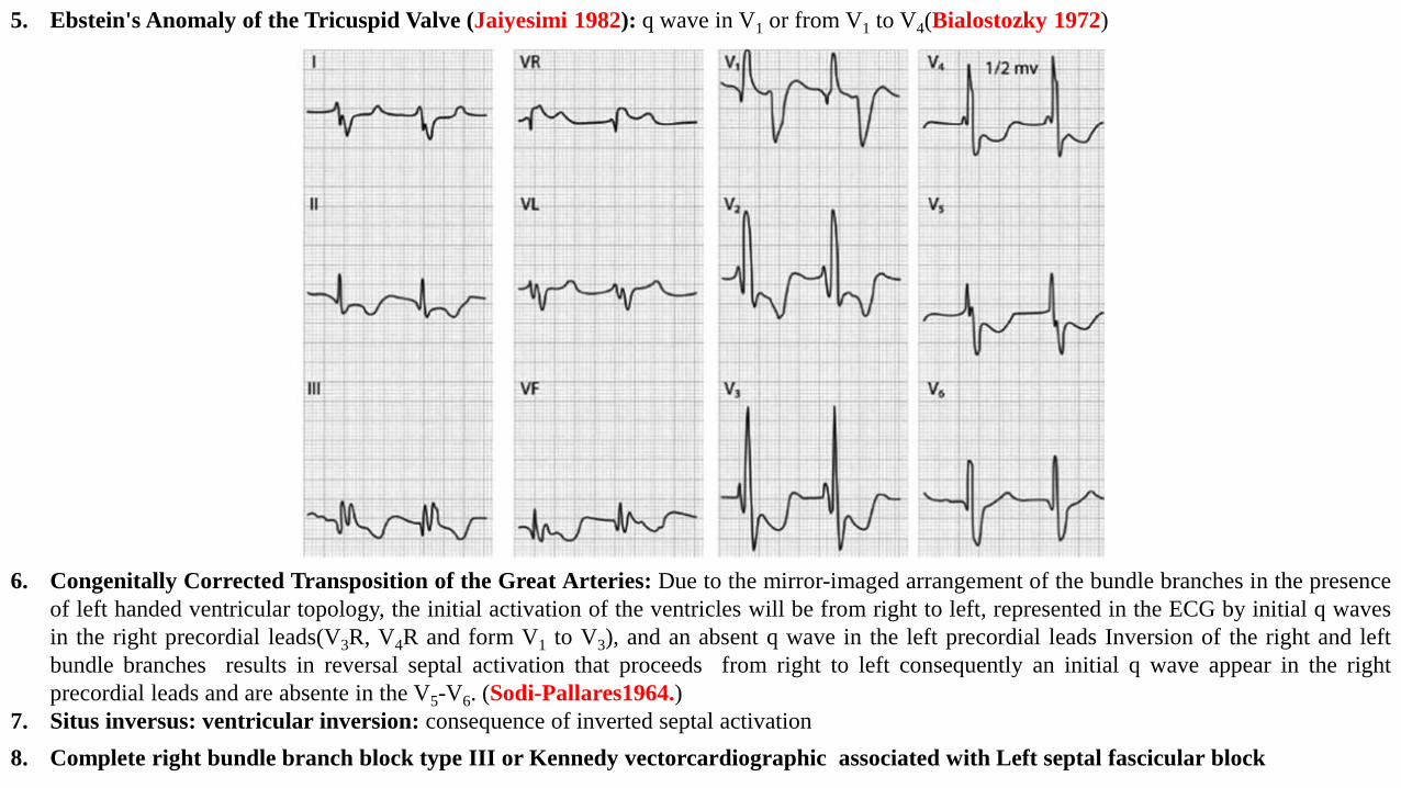
5. Ebstein's Anomaly of the Tricuspid Valve (Jaiyesimi 1982): q wave in V1 or from V1 to V4(Bialostozky 1972)
6. Congenitally Corrected Transposition of the Great Arteries: Due to the mirror-imaged arrangement of the bundle branches in the presence
of left handed ventricular topology, the initial activation of the ventricles will be from right to left, represented in the ECG by initial q waves
in the right precordial leads(V3R, V4R and form V1 to V3), and an absent q wave in the left precordial leads Inversion of the right and left
bundle branches results in reversal septal activation that proceeds from right to left consequently an initial q wave appear in the right
precordial leads and are absente in the V5-V6. (Sodi-Pallares1964.)
7. Situs inversus: ventricular inversion: consequence of inverted septal activation
8. Complete right bundle branch block type III or Kennedy vectorcardiographic associated with Left septal fascicular block

V
6
V1
X
Z
TP
Afferent
limb
RECD: Right End Conduction Delay
V
6
X
Z
V
6
X
Z
T
Afferent
limb
T
Grishman or Kennnedy type I Cabrera or Kennedy type II Kennedy type III or C
V
1
TX
Z V
1
T
We find type II in ASD, PS, in
COPD and more rarely in chronic
Chagasic myocardits.
X
Z
Right Anterior Quadrant
Initial vector to the front, QRS
loop of CW rotation and main
body located in anterior
quadrants.
X
Z
T
VCG classification of isolated Complete Right Bundle Branch Block in the HP

V
6
X
Z
T
RBBB Kennedy type III or C
Kennedy III or C
Initial vectorTo front and rightward or to
the left
QRS duration ≥ 120ms (≥ 60 comets)
QRS-loop rotation CW
Efferent limb To front orthogonal X lead
Afferent limb To front orthogonal X lead
Terminal appendixRight Anterior Quadrant and
slow inscription
Maximal vector
Decreased and with
significative anterior
displacement
Clinical significance Severe RVH
T-loop on HPCW rotation and directed to
back and leftward
CRBBB + Severe RVH
Z
T
V1V2
qR
qR
V5
V6
Rs
Rs
CRBBB + LSFB
Initial 10-20 ms To back and leftward To back and leftward
QRS loop rotation Clockwise Clockwise
QRS loop location Predominantly right anterior
quadrant: type A VCG RVH
Predominantly left anterior quadrant +
RECD
T-loop Directed to back Directed to back

YaVF
V2
ECG/VCG correlation in the right sagittal plane
Magnified T-loop
Efferent
Limb
Afferent
Limb
J point
0 point
qR
The initial 10ms vector (red arrow) with slow inscription is
directed downwards and backwards. The qR pattern in V1 may
indicate the association of LSFB + LAFB. Only the vector
dependent on the left posterior fascicle(LPF) is not blocked,
which justifies the initial activation directed downward and to
back.

Magnified T-loop
Efferent
Limb
Afferent
Limb
J point
0 pointJ point
STSE = 4.5mm. This important
elevation of the J point and the ST
segment is a secondary to the right
bundle branch block that
corresponds to the great distance
found in the T loop between J point
J and 0 point.
VCG/ECG correlation showing J point and ) point distance correspondent with J point and STSE of the ECG
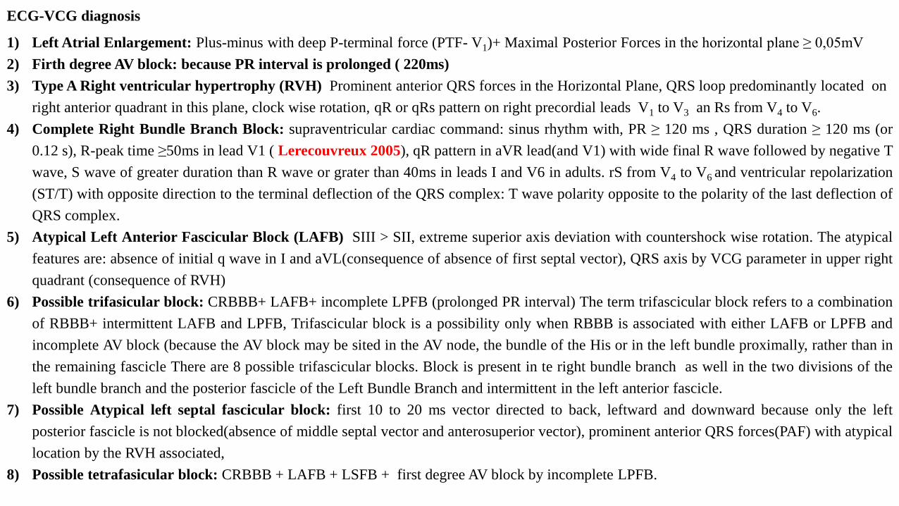
ECG-VCG diagnosis
1) Left Atrial Enlargement: Plus-minus with deep P-terminal force (PTF- V1)+ Maximal Posterior Forces in the horizontal plane ≥ 0,05mV
2) Firth degree AV block: because PR interval is prolonged ( 220ms)
3) Type A Right ventricular hypertrophy (RVH) Prominent anterior QRS forces in the Horizontal Plane, QRS loop predominantly located on
right anterior quadrant in this plane, clock wise rotation, qR or qRs pattern on right precordial leads V1 to V3 an Rs from V4 to V6.
4) Complete Right Bundle Branch Block: supraventricular cardiac command: sinus rhythm with, PR ≥ 120 ms , QRS duration ≥ 120 ms (or
0.12 s), R-peak time ≥50ms in lead V1 ( Lerecouvreux 2005), qR pattern in aVR lead(and V1) with wide final R wave followed by negative T
wave, S wave of greater duration than R wave or grater than 40ms in leads I and V6 in adults. rS from V4 to V6 and ventricular repolarization
(ST/T) with opposite direction to the terminal deflection of the QRS complex: T wave polarity opposite to the polarity of the last deflection of
QRS complex.
5) Atypical Left Anterior Fascicular Block (LAFB) SIII > SII, extreme superior axis deviation with countershock wise rotation. The atypical
features are: absence of initial q wave in I and aVL(consequence of absence of first septal vector), QRS axis by VCG parameter in upper right
quadrant (consequence of RVH)
6) Possible trifasicular block: CRBBB+ LAFB+ incomplete LPFB (prolonged PR interval) The term trifascicular block refers to a combination
of RBBB+ intermittent LAFB and LPFB, Trifascicular block is a possibility only when RBBB is associated with either LAFB or LPFB and
incomplete AV block (because the AV block may be sited in the AV node, the bundle of the His or in the left bundle proximally, rather than in
the remaining fascicle There are 8 possible trifascicular blocks. Block is present in te right bundle branch as well in the two divisions of the
left bundle branch and the posterior fascicle of the Left Bundle Branch and intermittent in the left anterior fascicle.
7) Possible Atypical left septal fascicular block: first 10 to 20 ms vector directed to back, leftward and downward because only the left
posterior fascicle is not blocked(absence of middle septal vector and anterosuperior vector), prominent anterior QRS forces(PAF) with atypical
location by the RVH associated,
8) Possible tetrafasicular block: CRBBB + LAFB + LSFB + first degree AV block by incomplete LPFB.
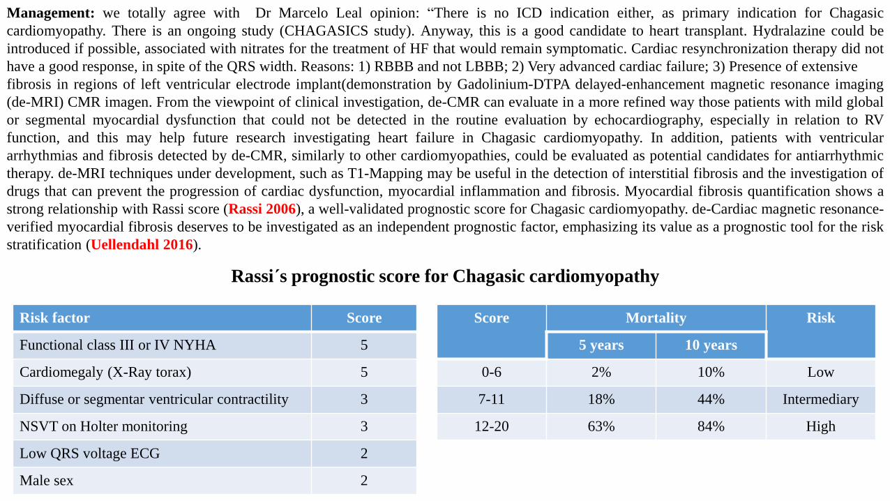
Management: we totally agree with Dr Marcelo Leal opinion: “There is no ICD indication either, as primary indication for Chagasic
cardiomyopathy. There is an ongoing study (CHAGASICS study). Anyway, this is a good candidate to heart transplant. Hydralazine could be
introduced if possible, associated with nitrates for the treatment of HF that would remain symptomatic. Cardiac resynchronization therapy did not
have a good response, in spite of the QRS width. Reasons: 1) RBBB and not LBBB; 2) Very advanced cardiac failure; 3) Presence of extensive
fibrosis in regions of left ventricular electrode implant(demonstration by Gadolinium-DTPA delayed-enhancement magnetic resonance imaging
(de-MRI) CMR imagen. From the viewpoint of clinical investigation, de-CMR can evaluate in a more refined way those patients with mild global
or segmental myocardial dysfunction that could not be detected in the routine evaluation by echocardiography, especially in relation to RV
function, and this may help future research investigating heart failure in Chagasic cardiomyopathy. In addition, patients with ventricular
arrhythmias and fibrosis detected by de-CMR, similarly to other cardiomyopathies, could be evaluated as potential candidates for antiarrhythmic
therapy. de-MRI techniques under development, such as T1-Mapping may be useful in the detection of interstitial fibrosis and the investigation of
drugs that can prevent the progression of cardiac dysfunction, myocardial inflammation and fibrosis. Myocardial fibrosis quantification shows a
strong relationship with Rassi score (Rassi 2006), a well-validated prognostic score for Chagasic cardiomyopathy. de-Cardiac magnetic resonance-
verified myocardial fibrosis deserves to be investigated as an independent prognostic factor, emphasizing its value as a prognostic tool for the risk
stratification (Uellendahl 2016).
Risk factor Score
Functional class III or IV NYHA 5
Cardiomegaly (X-Ray torax) 5
Diffuse or segmentar ventricular contractility 3
NSVT on Holter monitoring 3
Low QRS voltage ECG 2
Male sex 2
Score Mortality Risk
5 years 10 years
0-6 2% 10% Low
7-11 18% 44% Intermediary
12-20 63% 84% High
Rassi´s prognostic score for Chagasic cardiomyopathy

1. Bialostozky D, Horwitz S, Espino-Vela J. Ebstein's malformation of the tricuspid valve. A review of 65 cases. Am J Cardiol. 1972;29(6):826-
36.
2. Jaiyesimi F. Electrocardiographic abnormalities in Ebstein's anomaly. Deductions based on comparison with endomyocardial fibrosis.
Cardiology. 1982;69(2):61-9.
3. Kukla P, Długopolski R, Krupa E, et al. Electrocardiography and prognosis of patients with acute pulmonary embolism. Cardiol J.
2011;18(6):648-53.
4. Lerecouvreux M, Perrier E, Leduc PA, et al. Right bundle branch block: electrocardiographic and prognostic features. Arch Mal Coeur
Vaiss. 2005;98(12):1232-8.
5. Mehran-Pour M, Whitney A, Liebman J, Borkat G. Quantification of the Frank and McFee-Parungao orthogonal electrocardiogram in valvular
pulmonic stenosis. Correlations with hemodynamic measurement. J Electrocardiol. 1979;12(1):69-76.
6. Morris JJ Jr, Estes EH Jr, Whalen RE, et al. P-wave analysis in valvular heart disease. Circulation. 1964;29:242-52.
7. Rassi A Jr, Rassi A, Little WC, et al .Development and validation of a risk score for predicting death in Chagas' heart disease. N Engl J Med.
2006;355(8):799-808.
8. Sodi-Pallares D, Bisteni A, Fishleder BL, Medrano GA. Importance of the unipolar morphologies in the interpretation of the
electrocardiogram: the theoretical basis of the unipolar morphologies and its correlation with vectorial analysis, with cardiac activation, and
with the potential variations at the epicardial surface of the heart. Am Heart J. 1959;57(4):590-605.
9. Sodi-Pallares D, Cisneros F, Medrano GA, et al. Electrocardiographic diagnosis of myocardial infarction in the presence of bundle branch
block (right and left), ventricular premature beats and Wolff-Parkinson-White syndrome. Prog Cardiovasc Dis. 1963;6:107-36.
10. Sodi-Pallares D, Testelli MR. Electrocardiography in the diagnosis of congenital heart disease. Heart Bull. 1964;13:24-30.
11. Sodi-Pallares D. Deductive electrocardiography in congenital heart disease.Am J Cardiol. 1968;21(5):617-8.
12. Sodi-Pallares D, Ponce de León J, Bisteni A, Medrano GA. Polyparametric electrocardiography concerning new information obtained from
clinical electrocardiogram. Prog Cardiovasc Dis. 1970;13(1):97-117.
13. Tereshchenko LG, Henrikson CA, Sotoodehnia N, et al. Electrocardiographic deep terminal negativity of the P wave in V(1) and risk of
sudden cardiac death: the Atherosclerosis Risk in Communities (ARIC) study. J Am Heart Assoc. 2014;3(6):e001387.
14. Uellendahl M, Siqueira M, Calado, et al. Cardiac Magnetic Resonance-Verified Myocardial Fibrosis in Chagas Disease: Clinical Correlates
and Risk Stratification. Arq Bras Cardiol. 2016;107(5):460-6.
References

15. Wallis GA, Debich-Spicer D, Anderson RH. Congenitally corrected transposition. Orphanet J Rare Dis. 2011;6:22.

Karel Frederik Wenckebach was a physician, anatomist and cardiologist, born in The Hague, the seat of the Dutch parliament, government and
Royal Court. Wenckebach had two brothers, Henri Johan Eduard (1861–1924), Director of State Mines and later of the Dutch Ironworks at
Ijmuiden; and Willem Reymert Ludwig (1860–1937), a renowned painter and book illustrator. Wenckebach’s son Oswald became a painter,
sculptor, and metallurgist, best known for his war monuments and for designing the Dutch coins issued between 1948 and 1981. Wenckebach
began his studies in 1881 at Utrecht University Medical School and graduated in 1888 with a thesis entitled ‘About the structure and development
of the bursa of Fabricius’, a thesis on the sac-shaped lymphoid organ in the roof of the cloaca in birds. After graduating in 1888, Wenckebach
worked at a zoological institute, but quickly switched to physiology because his color-blindness proved an insurmountable obstacle to a career in
zoology. Wenckebach started his career as a general physician in 1891 in rural Heerlen, in south-eastern Holland, the region in which his father
Eduard (1813– –1874) had spearheaded the development of telegraphic communication lines between Haarlem and Amsterdam. Here, he gained a
deep respect for the clinical elements of medicine and became fascinated with the rhythms of the beating heart, sometimes listening to patients’
heart sounds for hours on end. Wenckebach returned to academia at Utrecht University in 1896 to study under his mentor, the renowned German
scientist T.W. Engelmann. Nurturing the curiosities born from his clinical experiences, he began to study irregular heart rhythms. He gained
experience in kymographic recordings and rhythm disturbances in frogs. He observed that the irregular pulses with compensatory pauses seen in
animals could also be detected in humans. In 1901, Wenckebach became Professor of Medicine at the University of Groningen, and two years later
he published his most important work: ‘Arrhythmia as an expression of certain functional disorders of the heart’ (‘Die Arrhythmie als Ausdruck
bestimmter funktionsstörungen des Herzens’ [1]) (Fig. 1). Later, he held appointments as a professor at the universities of Strasbourg (1911–1914)
and Vienna (1914– –1929). In Vienna, he studied cardiac function and pathology in soldiers. In his later years, he became intrigued with the
cardiac manifestations of beriberi and visited the West Indies. Karel Frederik Wenckebach died in Vienna in November 11, 1940 after eleven years
of retirement. Why is he still remembered today? Not only are the discoveries he made at the turn of the 20th century fundamental to our current
understanding of cardiac electrophysiology and automaticity, this founding father of modern electrocardiology made these ground-breaking
discoveries without having the electrocardiogram at his disposal and before the discovery of the sino-atrial (SA) and atrio-ventricular (AV) nodes.
His groundbreaking 1899 report ‘On the analysis of irregular pulses’ [1], described the heartbeats of a patient using tracings of the radial pulse of a
woman who complained of an irregular heartbeat. When analyzing her pulse, he noted predictable pauses every three to four beats. These pauses
differed from the pauses coming after extrasystoles, in that they were not followed by small extra pulse waves. Through careful measurement, he
noted that the length of the pause was not twice that of the preceding pulse-pulse interval, as one would expect with an extrasystole, but actually
Pérez-Riera AR, Femenía F, McIntyre WF, Baranchuk A. Karel Frederick Wenckebach (1864-1940): a giant of medicine. Cardiol J.
2011;18(3) 337-9.
less than half the preceding pulse-pulse interval. He concluded from this that the irregularity of the heart rhythm could not be due to extrasystoles.
On further analysis, he noted that the first interval after each pause was longer than the others, and that subsequent intervals were shorter. This
pattern was repeated again and again. Wenckebach called these groups ‘Luciani’s periods’ after the Italian physiologist [2], who while working in
the laboratory of Carl Ludwig in Leipzig in 1873, was the first to show a group of heartbeats that he called ‘periodical rhythm’. While he
recognized the pattern in his data, Wenckebach was unable to postulate a physiological explanation. Progress was made when he analyzed old data
given to him by Engelmann — simultaneous tracings of a dying frog’s atrial and ventricular contractions. Wenckebach observed a gradual
lengthening in the interval between atrial and ventricular contractions until an atrial contraction occurred that was not followed by a ventricular
contraction — resulting in a pause [3]. At this time, before the 1907 description of the SA node, Wenckebach believed that the “rhythmic
excitation” of the heart originated in the mouth of the vena cava. He concluded that as the heart’s action gradually worsens, the interval between
atrial and ventricular systole interval gradually becomes longer due to decreased electrical conductibility in the tissue, and there finally comes a
time when the atrial excitation is no longer conducted. During the subsequent pause, the conductibility has time to recover and the pattern begins
to repeat itself [4]. Comparing the frog and human tracings, Wenckebach realized that: “in both cases, the repeated irregularity was exactly the
same, and was repeated with almost mathematical precision.” He went on to speculate that this phenomenon must be due to damaged heart muscle
at the AV border [5]. The tracing in Figure 2 is an original of a jugular venous pulse recorded by Wenckebach. Note the progressive widening of
the ‘a-c interval’ (corresponding to the PR interval) until the ‘a wave’ is not followed by the ‘c wave’. Following Einthoven’s introduction of the
string galvanometer electrocardiograph, Wenckebach was able in 1906 to demonstrate the progressive prolongation of the PR interval before a
dropped ventricular beat, a phenomenon now known as the Wenckebach phenomenon [6]. In the same paper, Wenckebach described the median
bundle of the intra-atrial conduction system of the heart. This bundle joins the SA node to the AV node. Even now, this bundle is referred to as ‘the
median bundle of Wenckebach’ and it is one of the recognized internodal pathways, along with ‘the posterior internodal tract of Thorel’ and the
two branches of ‘the anterior internodal pathways or Bachmann’s bundle’) (Fig. 3). Wenckebach was also one of the firstproponents of the use of
quinine to treat paroxysmal atrial fibrillation [7]. Cinchona officinalis (family Rubiaceae) is a tree from the Andes whose bark contains the
alkaloids quinine and quinidine. ‘Jesuit’s bark’, as it was called, was discovered in Europe in the 17th century to be valuable in treating malaria. At
Strasbourg University, he administered quinine to several patients. He noted that while only a few patients converted to sinus rhythm, many felt
better. Wenckebach demonstrated in 1914 that quinine (1 g/daily) could halt paroxysms of this arrhythmia [5]. In his brilliant career, Wenckebach
earned numerous awards: The Order of Merit of the Austrian Republic, Honorary Fellow of the Royal College of Physicians and Surgeons of
Glasgow, Honorary Member of the Medico-Chirurgical Society of Edinburgh and the Cardiac Society of Great Britain and Ireland, and a
Corresponding Foreign member of the Societé Française de Cardiologie. Karel Frederik Wenckebach was a man who stood out for his modesty,
and had many famous friends who would visit him in his Vienna home. We would like to finish this brief summary of his life by quoting his own

1. Wenckehach KF. On the analysis of irregular pulses. Z Klin Med, 1899; 37: 475–488.
2. Ritchie WT. Karel Frederik Wenckebach. Br Heart J, 1941; 3: 141–144.
3. Mendoza-Davila N, Varon J. Resuscitation great. Karel Wenckebach: The story behind the block. Resuscitation, 2008; 79: 189–192.
4. Tandon A, Simpson L, Assar MD. Unusual origin of type 1 atrio- -ventricular block with comments on Wenckebach’s contribution. Proc Bayl
Univ Med Cent, 2011; 24: 9–12.
5. Upshaw CB, Silverman ME. The Wenckebach phenomenon: A salute and comment on the centennial of its original description. Ann Intern
Med, 1999; 130: 58–63.
6. Wenckebach KF.Contributions to the knowledge of human cardiac activity. Arch Anat Physiol, 1906; 297–354.
7. Wenckebach K. Cinchona derivatives in the treatment of heart disorders. JAMA, 1923; 81: 472–474.
References
words: “In medical science there are vast realms of which I have no special knowledge and, again, no, I am not a great man; I am a happy man.”
Figure 1. Cover of Wenckebach’s first book on cardiacarrhythmias entitled ‘Arrhythmia of the heart: Aphysiological and clinical study’, published in 1904.
Figure 2. Wenckebach second degree atrio-ventricular block (Mobitz type I) in the jugular
venous pulse.
Karel Frederik Wenckebach