chronic aortic and mitral regurgitation - ucsf cme gregoratos valvular... · 12/19/15 1 chronic...
TRANSCRIPT

12/19/15
1
Chronic Aortic and Mitral Regurgitation
When Should We Intervene?
Gabriel Gregoratos, MD, FACC
32nd Annual Advances in Heart Disease
Disclosure Statement
• Nothing to Disclose

12/19/15
2
Valvular Regurgitation: When Should we Intervene?
A complex Issue 1. Regurgitant lesions are mostly well tolerated and patients
remain asymptomatic for many years while irreparable ventricular dysfunction develops-which may not improve after intervention
2. Limited data comparing interventions vs. medical therapies or timing of intervention and most data obtained from retrospective studies or prospective non-randomized registries. Therefore most guideline recommendations have evidence level “B” or “C” (Not robust evidence)
3. Some conflicting conclusions in different studies 4. Valve replacement exchanges one disease for another
Factors Affecting Decision and Timing of Intervention
• Severity (quantified objectively by TTE, Cath, etc.) • Etiology and chronicity of regurgitation • Natural history of chronic regurgitation • Presence or absence of symptoms • Ventricular function and other predictors of outcome
such as pulmonary hypertension, atrial fibrillation • Patient status (age, co-morbidities, etc) • Type of intervention and durability: • Repair vs. Replacement and Surgical vs. Transcatheter

12/19/15
3
The Heart Valve Team and Heart Valve Centers of Excellence
Recommendations COR LOE Patients with severe VHD should be evaluated by a multidisciplinary Heart Valve Team when intervention is considered
I C
Consultation with or referral to a Heart Valve Center of Excellence is reasonable when discussing treatment options for 1) asymptomatic patients with severe VHD, 2) patients who may benefit from valve repair versus valve replacement, or 3) patients with multiple comorbidities for whom valve intervention is considered
IIa C
Heart Valve Centers of Excellence
• Composed of experienced providers from multiple disciplines
• Offer all available options for diagnosis and management (surgery, valve repair, trans-catheter therapies)
• Participate in outcome registries (regional or national) • Demonstrate adherence to Guidelines • Participate in continual evaluation and QI processes • Publicly report mortality and success rates

12/19/15
4
Stages of Progression of VHD Stage Definition Description
A At risk Patients with risk factors for the development of VHD
B Progressive Patients with progressive VHD (mild-to-moderate severity and asymptomatic)
C
Asymptomatic severe
Asymptomatic patients who have reached the criteria for severe VHD C1: Asymptomatic patients with severe VHD in whom the left or right ventricle remains compensated C2: Asymptomatic patients who have severe VHD, with decompensation of the left or right ventricle
D Symptomatic severe
Patients who have developed symptoms as a result of VHD
**
Chronic Aortic Regurgitation

12/19/15
5
Natural History of Chronic Asymptomatic AR
Bonow, Circulation 1991 and Borer, Circulation 1998
}
Survival without Surgery - Chronic AR Significance of Symptoms
Dujardin et al. Circulation 1999

12/19/15
6
Natural History of Chronic AR Based on 9 Studies / 593 Patients
♥ Asymptomatic patients with normal LV systolic function: • Progression to symptoms and/or LV dysfunction: ≤6%/ year • Progression to asymptomatic LV dysfunction: ≤3.5% /year • Sudden death: < 0.2% per year
♥ Asymptomatic patients with LV systolic dysfunction: • Progression to cardiac symptoms: >25% per year
♥ Symptomatic patients: • Mortality rate: >10% per year
JACC 2008:52:e1-e142
Predictors of Sub-optimal Surgical Outcomes in Chronic AR Patients
• LVEF < 50% • LVESD≥55 mm • LVEDD ≥ 75 mm • LVESV ≥ 200 ml/m2
• Prolonged LV dysfunction (>12 mos.) • Age>80 yrs. • Ventricular response to exercise (??) • Advanced symptoms (class III & IV)
JACC 2008:52:e1-e142 JACC 1997;30(3):746

12/19/15
7
Serial Evaluation in Asymptomatic Chronic AR
• Clinical evaluation: annually • TTE evaluation: ♥ Stage B (progressive, mild AR): every 3-5 years ♥ Stage B (progressive, moderate AR): every 1-2 years ♥ Stage C (severe AR): annually ♥ Stage C (severe AR, dilating LV): more frequently
Quantification of Severe AR by TTE *
• Jet width ≥65% of LVOT • Vena contracta >0.6 cm • Holodiastolic flow reversal in the proximal abdominal
aorta • Reg. Vol ≥60 mL/beat • Reg. Fraction ≥50% • ERO ≥0.3 cm2 • Angiography grade 3+ to 4+ • Plus evidence of LV dilation

12/19/15
8
Severe AR
Role of Exercise Testing in Evaluating Chronic AR *
• Exercise LVEF and change in LVEF from rest to
exercise are often abnormal even in asymptomatic patients, but these changes have not been shown to be of independent prognostic value above that provided by LV function and dimensions
• Sedentary patients and/or those with no or equivocal symptoms to assess true functional capacity – useful
• No specific recommendation in current guideline • Change from prior version where indications for
exercise testing were class IIa (reasonable)
12/19/15
9
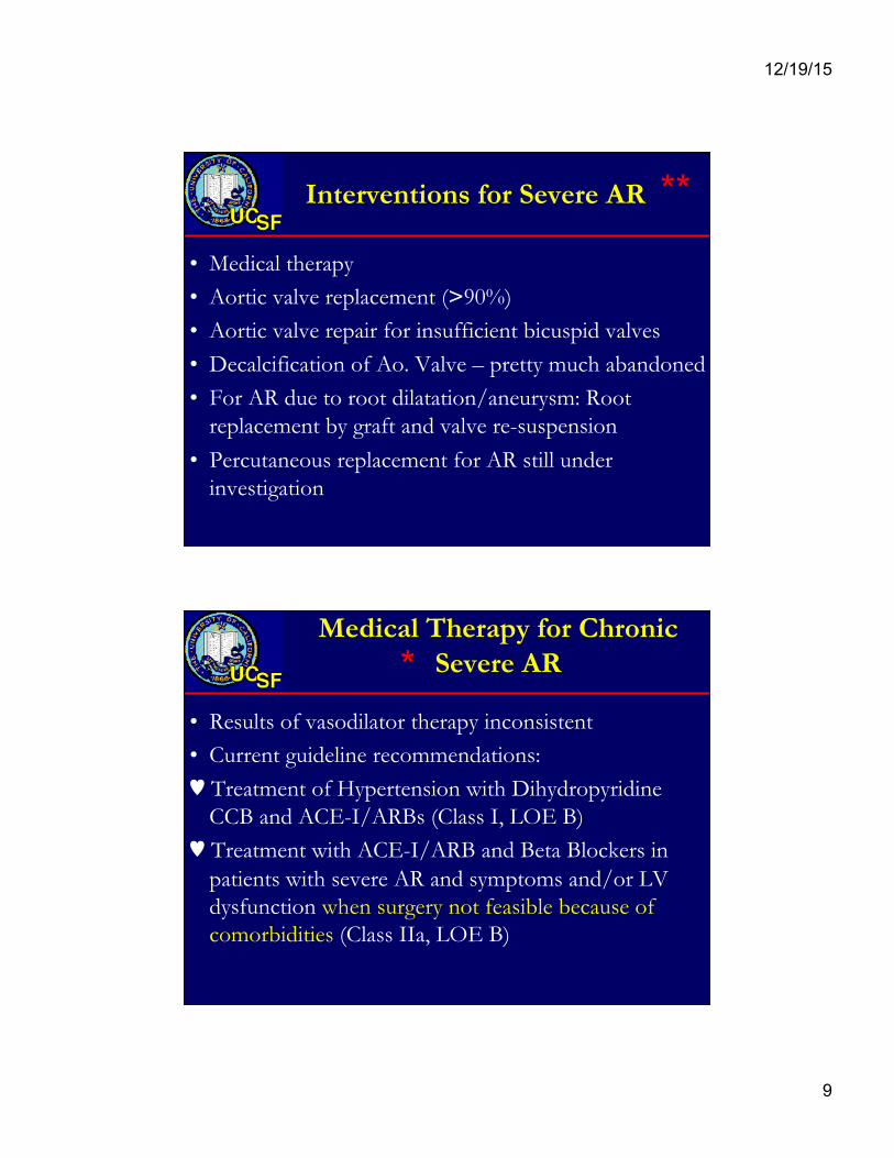
Interventions for Severe AR **
• Medical therapy • Aortic valve replacement (>90%) • Aortic valve repair for insufficient bicuspid valves • Decalcification of Ao. Valve – pretty much abandoned • For AR due to root dilatation/aneurysm: Root
replacement by graft and valve re-suspension • Percutaneous replacement for AR still under
investigation
Medical Therapy for Chronic Severe AR
• Results of vasodilator therapy inconsistent • Current guideline recommendations: ♥ Treatment of Hypertension with Dihydropyridine
CCB and ACE-I/ARBs (Class I, LOE B) ♥ Treatment with ACE-I/ARB and Beta Blockers in
patients with severe AR and symptoms and/or LV dysfunction when surgery not feasible because of comorbidities (Class IIa, LOE B)
*

12/19/15
10
ACC/AHA Class I Recommendations for Aortic Valve Surgery (AVR)
1. AVR is indicated for symptomatic patients with
severe AR (stage D) regardless of LV systolic function. (LOE: B)
2. AVR is indicated for asymptomatic patients with chronic severe AR and LV systolic dysfunction (LVEF<0.50) at rest. (LOE: B)
3. AVR is indicated for patients with chronic severe AR (stage C or D) while undergoing cardiac surgery for other indications (LOE: C)
ACC/AHA Class IIa Recommendations for Aortic Valve Surgery
1. AVR is reasonable for asymptomatic patients with
severe AR with normal LV systolic function (EF≥0.50) but with severe LV dilatation, defined as
LVESD>50 mm (stage C2) (LOE: B) 2. AVR is reasonable in patients with moderate AR
(stage B) undergoing cardiac surgery for other indications. (LOE B)

12/19/15
11
ACC/AHA Class IIb Recommendations for Aortic Valve Surgery
1. AVR may be considered for asymptomatic pts. with severe AR, normal LV systolic function (LVEF≥0.50, stage C1) and progressive severe LV dilatation, defined as LVEDD>65 mm if surgical risk is low (LOE: C)
Intervention for Chronic Severe AR Conclusions
• Optimal outcomes from Aortic valve surgery can be achieved if AVR takes place….. ♥ Before NYHA Class III / IV HF symptoms develop ♥ Before LV dysfunction ( EF<0.50) develops ♥ Before LVESD exceeds 50 mm ♥ Before LVEDD exceeds 65 mm
• Close clinical and echo follow-up (per guidelines) is necessary • Quantification of AR severity by TTE is essential • Exercise testing may be useful in few selected cases

12/19/15
12
Is it Ever too Late for AVR in Aortic Regurgitation?
• Very few patients should be denied AVR even when they have missed the opportunity for best surgical results (even if LVESD>50mm and/or EF severely depressed) because ♥ AVR reduces LV afterload and leads to ↑ LVEF and forward Cardiac Output
• However, preliminary and unconfirmed data suggest that patients in this setting who cannot generate systolic LV pressure>120 mmHg are likely to experience less afterload reduction after AVR and are probably poor surgical candidates (Carabello, JACC 2004)
• Comorbidities and advanced physiologic age must be considered
Bicuspid Aortic Valve & Aortopathy
• Bicuspid Ao. Valves frequently associated with aortic root dilatation
• Result of structural aortic wall changes and abnormal hemodynamics of the bicuspid valve
• Aortopathy more frequent with fusion of Right and Noncoronary cusps and less frequent with the more common fusion phenotype of Right and Left cusps
• Specific genetic cause not identified • Risk of dissection ↑ when Ao. Root Diameter>4.5
cm
*

12/19/15
13
Repair of Aortic Sinuses or Replacement of Ascending Aorta:
• Class I, LOE B: Diameter of ascending aorta or aortic sinuses >5.5 cm
• Class IIa, LOE C: Diameter of ascending aorta or aortic sinuses > 5 cm plus dissection risk (FH of dissection or rate of diameter increase ≥ 0.5 cm/year)
• Class IIa, LOE C: Ascending aorta diameter > 4.5 cm in patient undergoing AVR for severe AS or AR
*
Hot off the Press Clarification JACC December 2015, published ahead of print
• Class IIa, LOE C: Diameter of ascending aorta or aortic sinuses 5.0-5.5 cm plus dissection risk (FH of dissection or rate of diameter increase ≥ 0.5 cm/year) OR if patient is at low risk and surgery is performed by an experienced surgical team
• Low surgical risk is defined as STS mortality risk<4%
*

12/19/15
14
Chronic Mitral Regurgitation
Chronic Mitral Regurgitation *
Primary (degenerative) MR
• MV prolapse • Infective Endocarditis • CT diseases • Rheumatic heart disease • Congenital MV cleft • Radiation heart disease
Secondary (functional) MR
• Severe LV dysfunction • CAD with/without MI
(ischemic secondary MR) • Primary myocardial disease
(non ischemic secondary MR)
These two conditions have more differences than similarities

12/19/15
15
Typical Myxomatous MV
Chronic Primary MR Pathophysiology
• Mild/moderate MR may remain asymptomatic for many years
• Even with severe chronic MR, due to favorable loading conditions the LVEF is maintained in low normal range of 50% to 60% and forward CO is maintained, despite the presence of significant myocardial (contractile) dysfunction
• This is the key consideration in current thinking that intervention should be carried out before overt LV dysfunction develops
*

12/19/15
16
Natural History of Severe Chronic MR (Enriquez-Sarano et al. NEJM 1996 & Rosen et al. AJC 1994)
Survival post MV Surgery as a Function of LVEF
(Enriquez-Sarano, et al. Circulation 1994)

12/19/15
17
Predictors of Sub-optimal Surgical Outcomes in Chronic Severe Primary MR
• Advanced symptoms • Functional status NYHA III-IV • LVEF<60% • LVESD ≥40 mm • RV dysfunction • Pulmonary hypertension • Atrial Fibrillation • LA dimensions/volume • BNP level?
Serial Evaluation of Asymptomatic Patients with Chronic Primary MR
(JACC 2014;63(22):e57-e185)
• Mild MR (with no evidence of LV enlargement, dysfunction, or PH): Annual clinical evaluation. TTE every 2-3 years
• Moderate MR (with no evidence of LV enlargement, dysfunction or PH): Annual clinical and TTE
• Severe MR: Annual or Semiannual clinical and TTE With careful monitoring of LVEF, LVESD, and PA pressure

12/19/15
18
Quantification of Severe MR by TTE * *
• Central jet MR > 40% LA or holosystolic eccentric jet MR • Vena contracta ≥ 0.7 cm • Regurgitant volume ≥ 60 cc • Regurgitant fraction ≥ 50% • ERO ≥ 0.40 cm2 • Angiographic grade 3–4+
Severe MR

12/19/15
19
Exercise Testing Recommendations Class IIa
• Exercise hemodynamics with either Doppler echocardiography or cardiac catheterization is reasonable in symptomatic patients with chronic primary MR where there is a discrepancy between symptoms and the severity of MR at rest (stages B and C)
• Exercise treadmill testing can be useful in patients with chronic primary MR to establish symptom status and exercise tolerance (stages B and C)
Exercise Testing in Evaluating Chronic Primary MR *
• Assess symptomatic status, especially if good history of exercise capacity cannot be obtained - useful
• Functional capacity on Cardiopulmonary testing (<84% of expected for age and sex). Griffin, Circulation 2006
• ECHO parameters to be assessed with exercise: ♥ Severity of MR during exercise - useful ♥ PA pressure during exercise - useful ♥ Changes in LVEF during exercise - not very useful
due to loading conditions
12/19/15
20
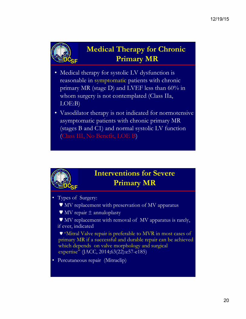
Medical Therapy for Chronic Primary MR
• Medical therapy for systolic LV dysfunction is reasonable in symptomatic patients with chronic primary MR (stage D) and LVEF less than 60% in whom surgery is not contemplated (Class IIa, LOE:B)
• Vasodilator therapy is not indicated for normotensive asymptomatic patients with chronic primary MR (stages B and C1) and normal systolic LV function (Class III, No Benefit, LOE B)
Interventions for Severe Primary MR
• Types of Surgery: ♥ MV replacement with preservation of MV apparatus ♥ MV repair ± annuloplasty ♥ MV replacement with removal of MV apparatus is rarely,
if ever, indicated ♥ “Mitral Valve repair is preferable to MVR in most cases of primary MR if a successful and durable repair can be achieved which depends on valve morphology and surgical expertise” (JACC, 2014;63(22):e57-e185)
• Percutaneous repair (Mitraclip)

12/19/15
21
Interventions for Chronic Primary Severe MR (Class I, LOE:B)
• MV surgery is recommended for symptomatic patients with chronic severe primary MR (stage D) and LVEF >30%
• MV surgery is recommended for asymptomatic patients with chronic severe primary MR and LV dysfunction (LVEF 30%–60% and/or LVESD ≥40 mm, stage C2)
• Concomitant MV repair or replacement is indicated in patients with chronic severe primary MR undergoing other cardiac surgery
Interventions for Chronic Primary MR (Class I, LOE:B)
• MV Repair is recommended in preference to MV Replacement when surgical treatment is indicated for patients with chronic severe primary MR limited to the posterior leaflet
• MV Repair is recommended in preference to MV Replacement when surgical treatment is indicated for patients with chronic severe primary MR involving the anterior leaflet or both leaflets when a successful and durable repair can be accomplished

12/19/15
22
Interventions for Chronic Primary MR (Class IIa, LOE:B)
• MV Repair is reasonable in asymptomatic patients with chronic severe primary MR (stage C1) with preserved LV function (LVEF >60% and LVESD <40 mm) – provided…
♥ the likelihood of a successful and durable repair without residual MR is >95%, with an expected mortality <1% when performed at a Heart Valve Center of Excellence OR ♥ there is a high likelihood of a successful and durable repair
of a non rheumatic MV with 1) new onset of AF or 2) resting pulmonary hypertension (PA systolic arterial pressure >50 mm Hg)
* Interventions for Chronic Primary MR (Class IIa, LOE:B)
• Concomitant MV Repair is reasonable in patients with chronic moderate primary MR (stage B) undergoing other cardiac surgery

12/19/15
23
Interventions for Chronic Primary MR (Class IIb)
• MV surgery may be considered in symptomatic patients with chronic severe primary MR and LVEF ≤30% (stage D)(LOE:C)
• MV repair may be considered in patients with rheumatic mitral valve disease when surgical treatment is indicated if a durable and successful repair is likely or if the reliability of long-term anticoagulation management is questionable (LOE:B)
Interventions for Chronic Primary MR
• Percutaneous MV repair may be considered for severely symptomatic patients (NYHA class III-IV) with chronic severe primary MR (stage D) who have a reasonable life expectancy, prohibitive surgical risk because of severe comorbidities (Class IIb, LOE:B)
• MV Replacement should not be performed for the treatment of isolated severe primary MR limited to less than one half of the posterior leaflet unless MV repair has been attempted and was unsuccessful (Class III-Harm, LOE:B)

12/19/15
24
The Argument for Early Surgery in Asymptomatic Patients *
• Surgery is almost inevitable in severe primary MR • Surgery in patients with advanced symptoms has inferior
outcomes. • Early surgery has low operative mortality of 0.5-1.0% (when
performed by expert surgeons) and restores patients to survival almost identical to that of the general population
• Good preoperative imaging predicts reparability with high degree of confidence
• Severe primary MR has serious consequences if not corrected • No alternative therapies have been established
Long-term Outcomes of Patients with Severe Degenerative (Primary) MR
(Montant P et al. J Thor & CV Surg 2009; 138(6):1339-48)
Follow-up in years
OVE
RA
LL S
UR
VIVA
L (%
)
(n=67)
(n=125)

12/19/15
25
Long-term Outcomes in Severe Primary MR
Suri RM et al. JAMA 2013;310(6):609-616
446 patients
575 patients
(<3m.)
Long-term Outcomes in Severe Primary MR
Suri RM et al. JAMA 2013;310(6):609-616

12/19/15
26
Intervention for Chronic Severe Primary MR - Conclusions
• MV repair superior to replacement • Severity of MR must be quantified objectively • MV surgery for severely symptomatic MR and LV
dysfunction is “Rescue Surgery” necessary but not desirable with inferior long-term outcomes
• MV surgery outcomes best before major symptoms develop, before LVEF<60%, before LVESD>40 mm and before AF and/or PH develop, if likelihood of repair is very high (>90%--98%)
• Close follow-up important (Heart Valve Center)
Interventions for Chronic Secondary Severe MR
• Standard HF therapy for patients with secondary MR, HF and reduced LVEF (Class I, LOE A)
• MV surgery is reasonable for patients with severe secondary MR who are undergoing CABG or AVR (Class IIa, LOE C)
• MV surgery may be considered for severely symptomatic patients (NYHA class III-IV) with chronic severe secondary MR (Class IIb, LOE B)
• MV repair may be considered for patients with chronic moderate secondary MR who are undergoing other cardiac surgery (Class IIb, LOE C)
**
12/19/15
27
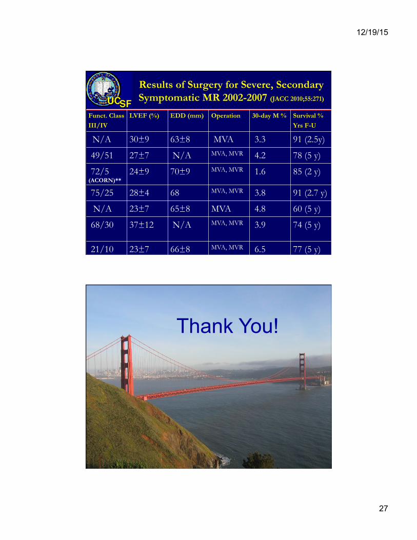
Results of Surgery for Severe, Secondary Symptomatic MR 2002-2007 (JACC 2010;55:271)
Funct. Class III/IV
LVEF (%) EDD (mm) Operation 30-day M % Survival % Yrs F-U
N/A 30±9 63±8 MVA 3.3 91 (2.5y)
49/51 27±7 N/A MVA, MVR 4.2 78 (5 y)
72/5 (ACORN)**
24±9 70±9 MVA, MVR 1.6 85 (2 y)
75/25 28±4 68 MVA, MVR 3.8 91 (2.7 y)
N/A 23±7 65±8 MVA 4.8 60 (5 y)
68/30 37±12 N/A MVA, MVR
3.9 74 (5 y)
21/10 23±7 66±8 MVA, MVR 6.5 77 (5 y)
Thank You!

12/19/15
28
Supplemental Slides
Stages of Chronic Aortic Regurgitation
Stage Definition Valve Anatomy Valve Hemodynamics
Hemodynamic Consequences
Symptoms
A At risk of AR ● Bicuspid aortic valve (or other congenital valve anomaly)
● Aortic valve sclerosis ● Diseases of the aortic
sinuses or ascending aorta
● History of rheumatic fever or known rheumatic heart disease
● IE
● AR severity none or trace
● None ● None

12/19/15
29
Stages of Chronic Aortic Regurgitation (cont.) Stage Definition Valve Anatomy Valve Hemodynamics Hemodynamic
Consequences Symptoms
B Progressive AR
● Mild-to-moderate calcification of a trileaflet valve bicuspid aortic valve (or other congenital valve anomaly)
● Dilated aortic sinuses
● Rheumatic valve changes
● Previous IE
● Mild AR: o Jet width <25% of LVOT o Vena contracta <0.3 cm o RVol <30 mL/beat o RF <30% o ERO <0.10 cm2
o Angiography grade 1+ ● Moderate AR: o Jet width 25%–64% of
LVOT o Vena contracta 0.3–0.6
cm o RVol 30–59 mL/beat o RF 30%–49% o ERO 0.10–0.29 cm2 o Angiography grade 2+
● Normal LV systolic function
● Normal LV volume or mild LV dilation
● None
Stages of Chronic Aortic Regurgitation (cont.) Stage Definition Valve Anatomy Valve Hemodynamics Hemodynamic
Consequences Symptoms
C Asymptomatic severe AR
● Calcific aortic valve disease
● Bicuspid valve (or other congenital abnormality)
● Dilated aortic sinuses or ascending aorta
● Rheumatic valve changes
● IE with abnormal leaflet closure or perforation
● Severe AR: o Jet width ≥65% of
LVOT o Vena contracta >0.6 cm o Holodiastolic flow
reversal in the proximal abdominal aorta
o RVol ≥60 mL/beat o RF ≥50% o ERO ≥0.3 cm2
o Angiography grade 3+ to 4+
o In addition, diagnosis of chronic severe AR requires evidence of LV dilation
C1: Normal LVEF (≥50%) and mild-to-moderate LV dilation (LVESD ≤50 mm) C2: Abnormal LV systolic function with depressed LVEF (<50%) or severe LV dilatation (LVESD >50 mm or indexed LVESD >25 mm/m2)
● None; exercise testing is reasonable to confirm symptom status

12/19/15
30
Stages of Chronic Aortic Regurgitation (cont.) Stage Definition Valve Anatomy Valve Hemodynamics Hemodynamic
Consequences Symptoms
D Symptomatic severe AR
● Calcific valve disease
● Bicuspid valve (or other congenital abnormality)
● Dilated aortic sinuses or ascending aorta
● Rheumatic valve changes
● Previous IE with abnormal leaflet closure or perforation
● Severe AR: o Doppler jet width ≥65%
of LVOT; o Vena contracta >0.6
cm, o Holodiastolic flow
reversal in the proximal abdominal aorta,
o RVol ≥60 mL/beat; o RF ≥50%; o ERO ≥0.3 cm2; o Angiography grade 3+
to 4+ o In addition, diagnosis
of chronic severe AR requires evidence of LV dilation
● Symptomatic severe AR may occur with normal systolic function (LVEF ≥50%), mild-to-moderate LV dysfunction (LVEF 40% to 50%) or severe LV dysfunction (LVEF <40%);
● Moderate-to-severe LV dilation is present.
● Exertional dyspnea or angina, or more severe HF symptoms
Indications for Aortic Valve Replacement for Chronic Aortic Regurgitation
12/19/15
31
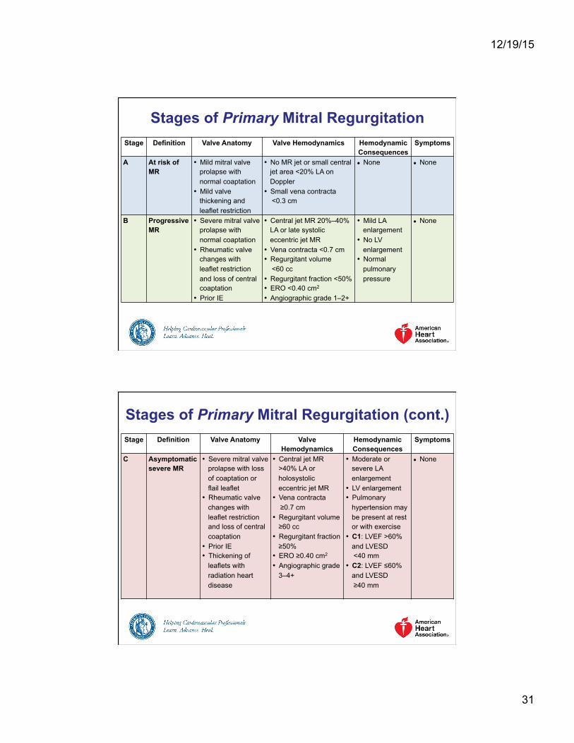
Stages of Primary Mitral Regurgitation Stage Definition Valve Anatomy Valve Hemodynamics Hemodynamic
Consequences Symptoms
A At risk of MR
• Mild mitral valve prolapse with normal coaptation
• Mild valve thickening and leaflet restriction
• No MR jet or small central jet area <20% LA on Doppler
• Small vena contracta <0.3 cm
● None ● None
B Progressive MR
• Severe mitral valve prolapse with normal coaptation
• Rheumatic valve changes with leaflet restriction and loss of central coaptation
• Prior IE
• Central jet MR 20%–40% LA or late systolic eccentric jet MR
• Vena contracta <0.7 cm • Regurgitant volume <60 cc • Regurgitant fraction <50% • ERO <0.40 cm2 • Angiographic grade 1–2+
• Mild LA enlargement
• No LV enlargement
• Normal pulmonary pressure
● None
Stages of Primary Mitral Regurgitation (cont.) Stage Definition Valve Anatomy Valve
Hemodynamics Hemodynamic Consequences
Symptoms
C Asymptomatic severe MR
• Severe mitral valve prolapse with loss of coaptation or flail leaflet
• Rheumatic valve changes with leaflet restriction and loss of central coaptation
• Prior IE • Thickening of
leaflets with radiation heart disease
• Central jet MR >40% LA or holosystolic eccentric jet MR
• Vena contracta ≥0.7 cm • Regurgitant volume ≥60 cc
• Regurgitant fraction ≥50%
• ERO ≥0.40 cm2 • Angiographic grade
3–4+
• Moderate or severe LA enlargement
• LV enlargement • Pulmonary
hypertension may be present at rest or with exercise
• C1: LVEF >60% and LVESD
<40 mm • C2: LVEF ≤60%
and LVESD ≥40 mm
● None

12/19/15
32
Stages of Primary Mitral Regurgitation (cont.) Stage Definition Valve Anatomy Valve
Hemodynamics Hemodynamic Consequences
Symptoms
D Symptomatic severe MR
• Severe mitral valve prolapse with loss of coaptation or flail leaflet
• Rheumatic valve changes with leaflet restriction and loss of central coaptation
• Prior IE • Thickening of
leaflets with radiation heart disease
• Central jet MR >40% LA or holosystolic eccentric jet MR
• Vena contracta ≥0.7 cm • Regurgitant volume ≥60 cc
• Regurgitant fraction ≥50%
• ERO ≥0.40 cm2 • Angiographic grade
3–4+
• Moderate or severe LA enlargement
• LV enlargement • Pulmonary
hypertension present
• Decreased exercise tolerance
• Exertional dyspnea
Indications for Surgery for Mitral Regurgitation

12/19/15
33
Stages of Secondary Mitral Regurgitation (cont.)
Grade Definition Valve Anatomy Valve Hemodynamics
Associated Cardiac Findings
Symptoms
A At risk of MR
● Normal valve leaflets, chords, and annulus in a patient with coronary disease or a cardiomyopathy
• No MR jet or small central jet area <20% LA on Doppler
• Small vena contracta <0.30 cm
• Normal or mildly dilated LV size with fixed (infarction) or inducible (ischemia) regional wall motion abnormalities
• Primary myocardial disease with LV dilation and systolic dysfunction
● Symptoms due to coronary ischemia or HF may be present that respond to revascularization and appropriate medical therapy
Stages of Secondary Mitral Regurgitation (cont.)
Grade Definition Valve Anatomy Valve Hemodynamics
Associated Cardiac Findings
Symptoms
B Progressive MR
• Regional wall motion abnormalities with mild tethering of mitral leaflet
• Annular dilation with mild loss of central coaptation of the mitral leaflets
• ERO <0.20 cm2
• Regurgitant volume <30 cc
• Regional wall motion abnormalities with reduced LV systolic function
• LV dilation and systolic dysfunction due to primary myocardial disease
● Symptoms due to coronary ischemia or HF may be present that respond to revascularization and appropriate medical therapy

12/19/15
34
Stages of Secondary Mitral Regurgitation (cont.)
Grade Definition Valve Anatomy Valve Hemodynamics
Associated Cardiac Findings
Symptoms
C Asymptomatic severe MR
• Regional wall motion abnormalities and/or LV dilation with severe tethering of mitral leaflet
• Annular dilation with severe loss of central coaptation of the mitral leaflets
• ERO ≥0.20 cm2 • Regurgitant
volume ≥30 cc
• Regional wall motion abnormalities with reduced LV systolic function
• LV dilation and systolic dysfunction due to primary myocardial disease
● Symptoms due to coronary ischemia or HF may be present that respond to revascularization and appropriate medical therapy
Stages of Secondary Mitral Regurgitation (cont.) Grade Definition Valve Anatomy Valve
Hemodynamics Associated
Cardiac Findings Symptoms
D Symptomatic severe MR
• Regional wall motion abnormalities and/or LV dilation with severe tethering of mitral leaflet
• Annular dilation with severe loss of central coaptation of the mitral leaflets
• ERO ≥0.20 cm2 • Regurgitant
volume ≥30 cc
• Regional wall motion abnormalities with reduced LV systolic function
• LV dilation and systolic dysfunction due to primary myocardial disease.
• HF symptoms due to MR persist even after revascularization and optimization of medical therapy
• Decreased exercise tolerance
• Exertional dyspnea