chromosome studies in 156 patients with …heart.bmj.com/content/heartjnl/27/5/756.full.pdf ·...
TRANSCRIPT

Brit. Heart J., 1965, 27, 756.
CHROMOSOME STUDIES IN 156 PATIENTS WITH CONGENITALHEART DISEASE
BY
JANET M. ANDERS, ELIZABETH C. MOORES, AND RICHARD EMANUEL
From The Institute of Cardiology and National Heart Hospital, London W.]Received February 26, 1965
Abnormal chromosomes are present in Down's syndrome, where the incidence of congenitalheart disease varies from 22 to 38 per cent (Kaiser and Schmid, 1952; Rowe and Uchida, 1961) andin Turner's syndrome, which may be complicated by coarctation of the aorta (Polani, Hunter, aildLennox, 1954) and pulmonary valve stenosis (Rainier-Pope et al., 1964). In the rare trisomicsyndromes involving chromosomes 13-15 and 17-18 the frequency of ventricular septal defect andpersistent ductus arteriosus has been noted (Townes et al., 1963; Smith et al., 1963). In additionpersistent ductus arteriosus was found in 3 of the 10 cases of XXXXY sex chromosomes reviewedby Joseph, Anders, and Taylor (1964). Chromosome abnormalities have also been reported inpatients with uncomplicated atrial septal defects and other forms of congenital heart disease (Book,Santesson, and Zetterqvist, 1961; Sasaki, Makino, and Kajii, 1963). It was this background thatprompted us to examine the chromosomes in patients with isolated congenital heart disease. Casesin which the cardiac lesion was associated with Down's and Turner's syndromes were excluded asthe cytogenetic abnormalities of these syndromes have already been described.
SUBJECTS AND METHODThe material examined was obtained from 156 patients with congenital heart disease (Table I). The
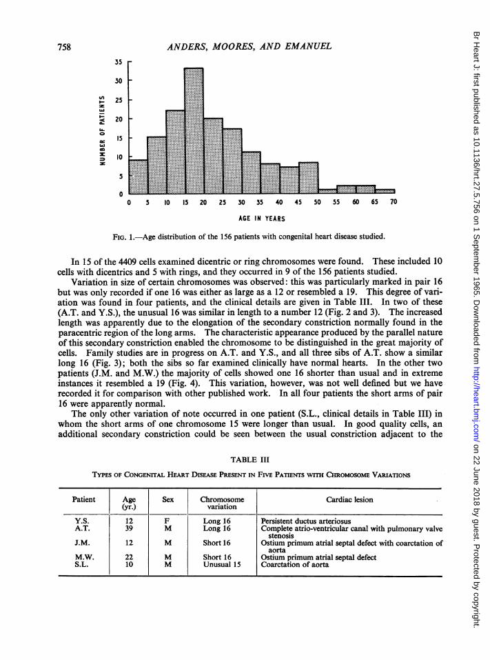
diagnosis in these patients was confirmed anatomically (operation or necropsy) in 82, by angiocardiographyand/or cardiac catheterization in 66, and by clinical, electrocardiographic, and radiological examinationalone in 8. Their ages varied from 3 months to 67 years. The distribution within this age range is shown inFig. 1.
Congenital heart disease was familial in 22 patients, representing 14 different families. In 18 instancesthe defect in the relative was the same as in the propositus.
Chromosome preparations were obtained from leucocyte cultures in all cases. Blood for these studieswas collected either by venepuncture or during cardiac catheterization. The technique used for culture was amodification of that described by Moorhead et al. (1960). Details of the modification are described else-where (Anders, Moores, and Emmanuel, 1965).A minimum of 25 cells from each patient was studied under the microscope except in three cases where the
technical quality of the preparations was poor, and only 12, 13, and 15 suitable cells, respectively, were found.One representative karyotype was prepared from each patient, except where further confirmation of resultswas required.
RESULTS
No major chromosome abnormality was found. In all patients studied the modal number was46. The total counts for each age-group with percentages of hypo- and hypermodal cells is givenin Table II. The increase of hypomodal cells with advancing age was similar to that reported byJacobs et al. (1963) in a group of 247 people with normal karyotypes.
756
on 22 June 2018 by guest. Protected by copyright.
http://heart.bmj.com
/B
r Heart J: first published as 10.1136/hrt.27.5.756 on 1 S
eptember 1965. D
ownloaded from

757CHROMOSOME STUDIES IN CONGENITAL HEART DISEASE
TABLE IANALYSIS OF CONGENITAL HEART DISEASE IN 156 PATIENTS STUDIED
Atrial septal defect (secundum)
Atrio-ventricular defects*
Ventricular septal defect
Fallot's tetralogy
Pulmonary valve stenosis
Coarctation
Patent ductus arteriosus
Aortic valve stenosis
Hypertrophic obstructivecardiomyopathy
Supravalve aortic stenosis
Other lesions
Uncomplicated 14Familial 14With mitral valve disease 2With skeletal deformity of upper limbs 2
Uncomplicated 14Familial 3With pulmonary valve stenosis 2With syndactyly IWith coarctation of aorta IWith secundum ASD 1
Uncomplicated PFamilialWith multiple skeletal deformities I
Uncomplicated IWith patent ductus arteriosus
Uncomplicated 1With dextrocardia
UncomplicatedWith aortic valve disease
UncomplicatedFamilial
Uncomplicated
UncomplicatedFamilial
Transposition (1) Corrected transposition (4)Primary pulmonary hypertensionEisenmenger PDA (3) VSD (1)
VSD with double outflow RVEbstein's diseaseDextrocardia (complex)Pulmonary atresiaAnomalous coronary arteryTwo-chambered right ventricleCor triatriatumIsolated lkvocardia
Total . .
4)32
4322
1J5}17}131117
}15
4}10
4}1010
1
53422311111
* Atrio-ventricular defects include simple ostium primum, partial atrio-ventricular canal, and complete atrio-ven-tricular canal.
TABLE IIDISTRIBUTION OF CHROMOSOME CouNTs IN RELATION TO AGE
Age-group No. of Chromosome counts Per cent Per centpatients hypomodal hypermodal
<45 45 46 47 >47 Total cells cells
0-14 46 7 13 1149 7 0 1176 1-70 0 6015-29 70 27 30 1982 21 3 2063 2-76 1*1630-70 40 13 45 1104 8 0 1170 4-96 0-68
Total 156 47 88 4235 36 3 4409 3 06 0 86
1561
on 22 June 2018 by guest. Protected by copyright.
http://heart.bmj.com
/B
r Heart J: first published as 10.1136/hrt.27.5.756 on 1 S
eptember 1965. D
ownloaded from
ANDERS, MOORES, AND EMANUEL35
30U_:125
I20
50
0 5 10 15 20 25 30 35 40 45 50 55 60 65 70
AGE IN YEARS
FIG. 1.-Age distribution of the 156 patients with congenital heart disease studied.
In 15 of the 4409 cells examined dicentric or ring chromosomes were found. These included 10cells with dicentrics and 5 with rings, and they occurred in 9 of the 156 patients studied.
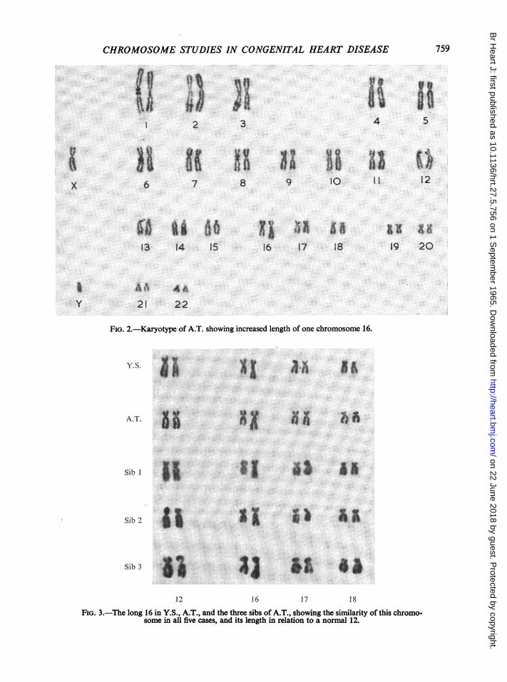
Variation in size of certain chromosomes was observed: this was particularly marked in pair 16but was only recorded if one 16 was either as large as a 12 or resembled a 19. This degree of vari-ation was found in four patients, and the clinical details are given in Table III. In two of these(A.T. and Y.S.), the unusual 16 was similar in length to a number 12 (Fig. 2 and 3). The increasedlength was apparently due to the elongation of the secondary constriction normally found in theparacentric region of the long arms. The characteristic appearance produced by the parallel natureof this secondary constriction enabled the chromosome to be distinguished in the great majority ofcells. Family studies are in progress on A.T. and Y.S., and all three sibs of A.T. show a similarlong 16 (Fig. 3); both the sibs so far examined clinically have normal hearts. In the other twopatients (J.M. and M.W.) the majority of cells showed one 16 shorter than usual and in extremeinstances it resembled a 19 (Fig. 4). This variation, however, was not well defined but we haverecorded it for comparison with other published work. In all four patients the short arms of pair16 were apparently normal.
The only other variation of note occurred in one patient (S.L., clinical details in Table III) inwhom the short arms of one chromosome 15 were longer than usual. In good quality cells, an
additional secondary constriction could be seen between the usual constriction adjacent to the
TABLE III
TYPES OF CONGENITAL HEART DISEASE PRESENT IN FIVE PATTENTs WITH CHROMOSOME VARIATIONS
Patient Age Sex Chromosome Cardiac lesion(yr.) variation
Y.S. 12 F Long 16 Persistent ductus arteriosusA.T. 39 M Long 16 Complete atrio-ventricular canal with pulmonary valve
stenosisJ.M. 12 M Short 16 Ostium primum atrial septal defect with coarctation of
aortaM.W. 22 M Short 16 Ostium primum atrial septal defectS.L. 10 M Unusual 15 Coarctation of aorta
758
on 22 June 2018 by guest. Protected by copyright.
http://heart.bmj.com
/B
r Heart J: first published as 10.1136/hrt.27.5.756 on 1 S
eptember 1965. D
ownloaded from
CHROMOSOME STUDIES IN CONGENITAL HEART DISEASE
I
6
ef
2
7
13- 14 I5
A0 AA21 22
FiG. 2.-Karyotype of A.T. showing increased length of one chromosome 16.
YSUi X1 asAI -A
ATs. 13 51" laA.T. 55R81 6
as iA
I) A
12 16 17 18
FiG. 3.-The long 16 in Y.S., A.T., and the three sibs of A.T., showing the similarity of this chromo-some in all five cases, and its length in relation to a normal 12.
3
x
B4
I I
5
I)9 108
16
Iy
ia417 18
XA Xsll19 20
Sib I
Sib 2
Sib 3
SE3533s
759
on 22 June 2018 by guest. Protected by copyright.
http://heart.bmj.com
/B
r Heart J: first published as 10.1136/hrt.27.5.756 on 1 S
eptember 1965. D
ownloaded from
ANDERS, MOORES, AND EMANUEL
M.W.
16 17 18 19 20
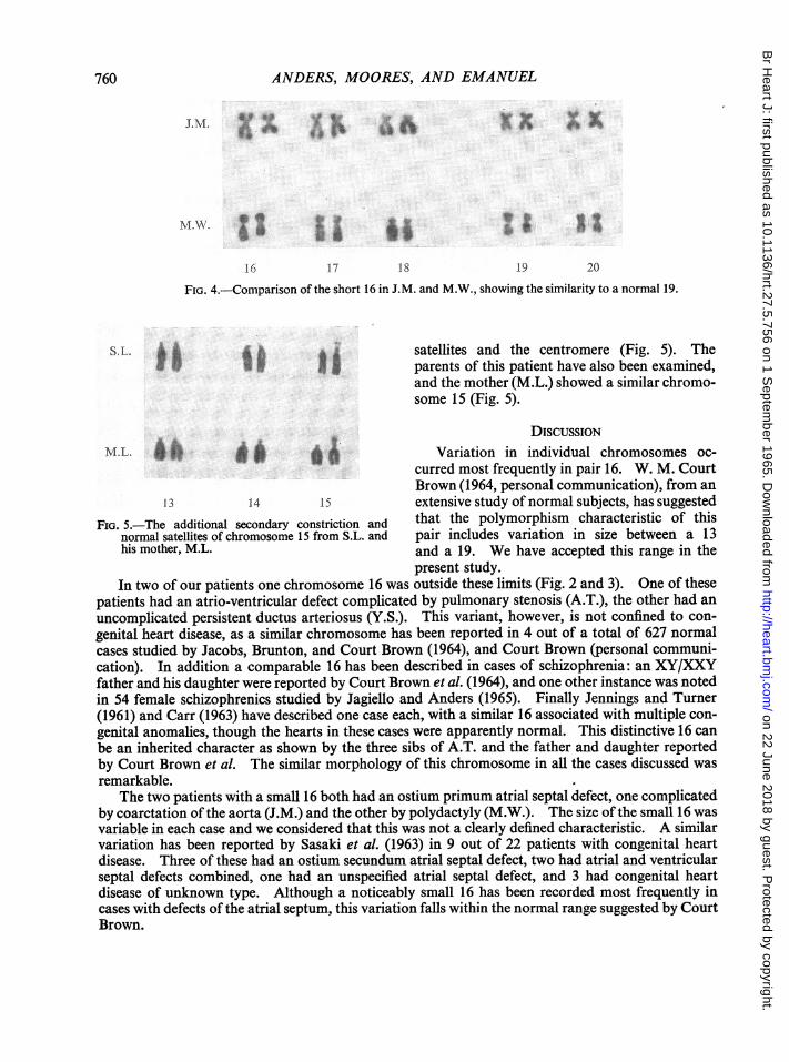
FIG. 4.-Comparison of the short 16 in J.M. and M.W., showing the similarity to a normal 19.
S. L. I 1; Isatellites and the centromere (Fig. 5). Theparents of this patient have also been examined,and the mother (M.L.) showed a similar chromo-some 15 (Fig. 5).
DISCUSSIONM.L. * tF * * * ^ Variation in individual chromosomes oc-
curred most frequently in pair 16. W. M. CourtBrown (1964, personal communication), from an
13 14 15 extensive study of normal subjects, has suggestedFIG. 5.-The additional secondary constriction and that the polymorphism characteristic of this
normal satellites of chromosome 15 from S.L. and pair includes variation in size between a 13his mother, M.L. and a 19. We have accepted this range in the
present study.In two of our patients one chromosome 16 was outside these limits (Fig. 2 and 3). One of these
patients had an atrio-ventricular defect complicated by pulmonary stenosis (A.T.), the other had anuncomplicated persistent ductus arteriosus (Y.S.). This variant, however, is not confined to con-genital heart disease, as a similar chromosome has been reported in 4 out of a total of 627 normalcases studied by Jacobs, Brunton, and Court Brown (1964), and Court Brown (personal communi-cation). In addition a comparable 16 has been described in cases of schizophrenia: an XY/XXYfather and his daughter were reported by Court Brown et al. (1964), and one other instance was notedin 54 female schizophrenics studied by Jagiello and Anders (1965). Finally Jennings and Turner(1961) and Carr (1963) have described one case each, with a similar 16 associated with multiple con-genital anomalies, though the hearts in these cases were apparently normal. This distinctive 16 canbe an inherited character as shown by the three sibs of A.T. and the father and daughter reportedby Court Brown et al. The similar morphology of this chromosome in all the cases discussed wasremarkable.
The two patients with a small 16 both had an ostium primum atrial septal defect, one complicatedby coarctation of the aorta (J.M.) and the other by polydactyly (M.W.). The size of the small 16 wasvariable in each case and we considered that this was not a clearly defined characteristic. A similarvariation has been reported by Sasaki et al. (1963) in 9 out of 22 patients with congenital heartdisease. Three of these had an ostium secundum atrial septal defect, two had atrial and ventricularseptal defects combined, one had an unspecified atrial septal defect, and 3 had congenital heartdisease of unknown type. Although a noticeably small 16 has been recorded most frequently incases with defects of the atrial septum, this variation falls within the normal range suggested by CourtBrown.
760
on 22 June 2018 by guest. Protected by copyright.
http://heart.bmj.com
/B
r Heart J: first published as 10.1136/hrt.27.5.756 on 1 S
eptember 1965. D
ownloaded from

CHROMOSOME STUDIES IN CONGENITAL HEART DISEASE
The other morphological variation of note occurred in a boy (S.L.) with coarctation of the aorta.Both he and his mother (M.L.) had a chromosome 15 with unusually long short arms (Fig. 5),similar to those reported by Jacobs et al. (1964) in one of their normal cases. Chandra and Hunger-ford (1963) observed a variation of this with only terminal satellites in a normal father and daughter,and Jacobs et al. found a further variant in their study of normal subjects, where there was neitherthe additional secondary constriction nor the usual satellites.
In contrast to Book et al. (1961), we found no abnormality in the 22 familial cases examined,though these included 14 instances of secundum atrial septal defect representing 9 different families.
In the single case of supravalve aortic stenosis with characteristic facies (Williams, Barratt-Boyes,and Lowe, 1961) the chromosomes were normal, unlike one of the cases reported by Palmer (1963)but in keeping with the other published findings (Ebeile and Beuren, 1963; Palmer, 1963; Joseph,Polani, and Gold, 1963; de Grouchy and Emerit, 1963).
In conclusion, therefore, we consider it improbable that any of the variations reported are directlyassociated with congenital heart disease.
SUMMARYNo major chromosome abnormalities were found in a series of 156 patients with congenital heart
disease. The modal count was 46 in each instance, and an increase in hypomodal cells with advanc-ing age was noted. A few cells showed unstable structural abnormalities. Variations in size wereobserved in certain chromosomes, particularly number 16. In two patients one of pair 16 was aslarge as a number 12, and in two others it resembled a 19; three of these four patients had an atrio-ventricular defect. In one patient with coarctation of the aorta a number 15 had particularlylong short arms. The long 16 in one of the cases reported and the unusual chromosome 15 wereshown to be inherited features. These results are discussed in relation to normal surveys and otherinstances where similar chromosomes have been reported.
We should like to express our gratitude to the Nuffield Foundation for supporting the Cytogenetics Laboratory atthe Institute of Cardiology and to Professor P. E. Polani for his advice on the original organization. We are alsoindebted to Dr. W. M. Court Brown for his generous help with this paper and to the physicians of the National HeartHospital for their co-operation.
REFERENCESAnders, J. M., Moores, E. C., and Emanuel, R. (1965). Chromosome preparations from leucocyte culture: a sim-
plified method for collecting cultures by post. J. med. Genet. In the press.Book, J. A., Santesson, B., and Zetterqvist, P. (1961). Association between congenital heart malformation and
chromosomal variations. Acta peediat. (Uppsala), 50, 217.Carr, D. H. (1963). Chromosomal abnormalities and their relation to disease. Canad. med. Ass. J., 88, 456.Chandra, H. S., and Hungerford, D. A. (1963). An aberrant autosome (13-15) in a human female and her father, both
apparently normal. Cytogenetics, 2, 34.Court Brown, W. M., Mantle, D. J., Buckton, K. E., and Tough, I. M. (1964). Fertility in an XY/XXY male married
to a translocation heterozygote. J. med. Genet., 1, 35.Eberle, P., and Beuren, A. J. (1963). Chromosome studies in patients with supravalvular aortic stenosis. Lancet,
2, 438.de Grouchy, J., and Emerit, I. (1963). Chromosome studies in patients with supravalvular aortic stenosis. Lancet,
2, 789.Jacobs, P. A., Brunton, M., and Court Brown, W. M. (1964). Cytogenetic studies in leucocytes on the general popu-
lation: subjects of ages 65 years and more. Ann. hum. Genet., 27, 353.-, Doll, R., and Goldstein, H. (1963). Change of human chromosome count distributions with age:
evidence for a sex difference. Nature (Lond.), 197, 1080.Jagiello, G., and Anders, J. M. (1965). In preparation.Jennings, A. N., and Turner, B. (1961). Autosomal chromosome anomalies. Med. J. Aust., 2, 830.Joseph, M. C., Anders, J. M., and Taylor, A. I. (1964). A boy with XXXXY sex chromosomes. J. med. Genet., 1,
95.Polani, P. E., and Gold, R. G. (1963). Chromosome studies in patients with supravalvular aortic stenosis.Lancet, 2, 788.
Kaiser, A., and Schmid, F. (1952). Die Herzfehlbildung beim Mongolismus. Munch. med. Wschr., 94, 2167.Moorhead, P. S., Nowell, P. C., Mellman, W. J., Battips, D. M., and Hungerford, D. A. (1960). Chromosome pre-
parations of leukocytes cultured from human peripheral blood. Exp. Cell Res., 20, 613.
761
on 22 June 2018 by guest. Protected by copyright.
http://heart.bmj.com
/B
r Heart J: first published as 10.1136/hrt.27.5.756 on 1 S
eptember 1965. D
ownloaded from

762 ANDERS, MOORES, AND EMANUEL
Palmer, C. G. (1963). Chromosome studies in patients with supravalvular aortic stenosis. Lancet, 2, 788.Polani, P. E., Hunter, W. F., and Lennox, B. (1954). Chromosomal sex in Turner's syndrome with coarctation of the
aorta. Lancet, 2, 120.Rainier-Pope, C. R., Cunningham, R. D., Nadas, A. S., and Crigler, J. F., Jr. (1964). Cardiovascular malformations
in Turner's syndrome. Pediatrics, 33, 919.Rowe, R. D., and Uchida, I. A. (1961). Cardiac malformation in mongolism. Amer. J. Med., 31, 726.Sasaki, M. S., Makino, S., and Kajii, T. (1963). Chromosomal aberrations in congenital cardiovascular disorders of
man. Proc. Japan Acad., 39, 394.Smith, D. W., Patau, K., Therman, E., Inhorn, S. L., and DeMars, R. I. (1963). The D1 trisomy syndrome. J.
Pediat., 62, 326.Townes, P. L., Kreutner, K. A., Kreutner, A., and Manning, J. (1963). Observations on the pathology of the trisomy
17-18 syndrome. J. Pediat., 62, 703.Williams, J. C. P., Barratt-Boyes, B. G., and Lowe, J. B. (1961). Supravalvular aortic stenosis. Circulation, 24,
1311.
on 22 June 2018 by guest. Protected by copyright.
http://heart.bmj.com
/B
r Heart J: first published as 10.1136/hrt.27.5.756 on 1 S
eptember 1965. D
ownloaded from