chromosomal instability in two siblings with dubowitz syndrome
TRANSCRIPT

124 Case Reports
CHROMOSOMAL INSTABILITY IN TWO SIBLINGS WITH DUBOWITZ SYNDROME
The Dubowitz syndrome is a rare autosomal recessive disorder characterized by intra-uterine and post-natal growth retardation, microcephaly and peculiar facial appear- ance (Kiister & Majewski, 1986). Since its description in 1965, more than 70 cases have been reported in the literature (Ilyana & Lurie, 1990). Patients with Dubowitz syndrome are at increased risk for malignancy and haemato- poietic failure. Immunodeficiency is also a common finding. To date, chromosome breakage studies performed only in a few cases have not found a hypersensitivity to the clastogenic effect of DNA cross-linking agents.
We report here two sisters with Dubowitz syndrome who have leucopenia and dysglobulinaemia. Their chromosome analysis revealed an increase in spontaneous and induced chromosomal breakage rates.
JV and MV. two Caucasian girls respectively aged 7 and 4 . 5 years, were referred to the Hospital in September, 1990. The two sisters had a history of failure to thrive, microcephaly, repeated infectious and recurrent ulcerative stomatitis. Par- ents denied consanguinity but a part of the father’s family originated from a village near the mother’s village.
JV was the first child, born at 38 weeks with a birth weight of 2280 g, and a head circumference of 29 cm. Physical examination revealed a child with a height of 110 cm ( - 1.7 SD). a weight of 16 kg ( - 2 SD) and microcephaly (45 cm, -4 .8 SD). She had mild mental retardation and attended a special education class. Since infancy, recurrent facial eczema was present. The following characteristic features were observed: small facies, telecanthus, epicanthus folds, broad tip of the nose, fine scalp hair, moderate retrognatia and clinodactyly of the fifth fingers. Except for delayed bone age (4.5 years), the skeletal survey was normal. Haematolo- gical data were as follows: Hb 10.6 g/dl, platelets 209 x lo9/ I. WBC 1.7 x 10⁹/1 with 30% neutrophils. A bone marrow aspirate showed reduced mature granulocytes and a pre- dominance of promyelocytes and myelocytes. There was a complete IgA deficiency with normal IgG levels and elevated IgM levels (6 .4 g/l) in the serum. Cytogenetic studies were performed on peripheral blood lymphocytes. The diepoxybu- tane (DEB) test was carried out as previously described (Auerbach et al, 1981). The nitrogen mustard test used mechlormethamine hydrochloride (trade name, Caryolysin) as described by Berger et aJ(l980). The results are given in Table I.
MV. the second child. was born in the 39th gestational week with a birth weight of 2460 g and microcephaly (30 cm). She was hospitalized in 1988 for an agranulocytosis and suffered from recurrent neutropenia. Her appearance was very similar to that of her sister. In addition, she had left palpebral ptosis. Her height and weight were 3 SD below the mean and she had severe microcephaly (42 cm. - 6 SD) with retarded bone age. The peripheral blood counts showed neutropenia (0.2 x loy neutrophils/l) but platelet and eryth- rocyte counts were normal. The bone marrow was depleted of mature granulocytes. IgA (0.1 8 g/l) and IgG ( 1.72 g/l) levels were low but the IgM level was increased (4 .97 g/l). Cytogenetic investigations revealed high breakage rates (Table I).
Table I. Chromosomal breakage in patients MV and JV. parents and control
Mean breaks per cell (number of analysed cells)
Nitrogen mustard Spontaneous test DEB test
Control O(26) 0.06 (30) 0 ( 3 0 ) MV 0.23 (34) 3 (32) 0.82 (29) Jv 0.3 (60) 0.9 ( 3 1 ) 0.55 ( 3 6 ) Mother 0.08 (34) 0.2 ( 3 5 ) 0 (34) Father 0.05 (34) 0.12 (32) 0 (32)
Nitrogen mustard concentration: 0.05 jig/ml. Diepoxybutane concentratlon: 0.1 jiglml.
Three patients with Dubowitz syndrome have been reported who developed a malignant tumour: one lym- phoma, one neuroblastoma (Sauer & Spelger, 19 77) and one acute lymphoblastic leukaemia (Grobe. 1983). Five cases with bone marrow failure were reported: two patients with aplastic anaemia (Walters & Desposito, 1985; Berthold et al. 1987), one affected sib with neutropenia and thrombocyto- penia (Walters & Desposito. 1985), two cases with ‘severe hypoplastic anaemia’ (Ilyana & Lurie, 1990). The Dubowitz syndrome thus shares several major features with Fanconi’s anaemia and the question of chromosomal damage in Dubowitz syndrome is therefore of particular interest. These two siblings are typical cases of Dubowitz syndrome and isolated recurrent neutropenia and dysglobulinaemia are not present in patients with Fanconi’s anaemia. The two sisters have an increase in spontaneous and nitrogen mustard- induced chromosomal breakages similar to those found in patients with Fanconi’s anaemia (Berger & Le Coniat, 1989). The results in the DEB test are also abnormally high. To the best of our knowledge these are the first reported cases of Dubowitz syndrome with chromosomal instability. Chromo- somal studies performed in three patients with bone marrow failure were normal in two cases. In the third one a mild increase in mean DEB-induced breakage rate (0*12/cell) was noted (Naltos & Desposito. 1985: Berthold et al. 1987).
Dubowitz syndrome can be associated with a chromosomal fragility and thus is a differential diagnosis of Fanconi’s anaemia. These two observations emphasize the need for cytological analysis of all patients with Dubowitz syndrome in order to determine the relevance of chromosomal instabi- lity.
Department of Pediatric Haematology, and *Department of Medical Genetics, Children’s Hospital. La Timone, tcytogenetic Laboratory, Medical School, Marseille, France
ISABELLE THIJRET GERARD MICHEL NICOLE PHILIP’ DOMINIQ~JE HAIRlONt ANNE-MARIE CAPODANOt
HENRI PERRIMOND
REFERENCES
Case Reports 12 5 anaemia in a child with features of Dubowitz syndrome. European Iournal of Pediatrics, 146. 605-607.
GrGbe, H. (1983) Dubowitz Syndrom und akute lymphatische Leukamie. Monatsschrijt fur Kinderheilkunde. 131, 467-468.
Ilyana. H. & Lurie, I.W. (1990) Dubowitr syndrome: possible evidence for a clinical subtype. American lournal of Medical Genetics.
Kuster. W. & Majewski. F. ( 1 986) The Dubowitz syndrome. European lournal of Pediatrics. 144, 574-578.
Sauer, o. & Spelger, G. (1977) Dubowitz-syndrom mit immunodefi- zienz und malignem Neoplasma bei nwei Geschwistern. Monatssch- rijt fur Kinderheilkunde. 125, 885-887.
Waiters, T,R, & Desposito, F, (1985) Aplastic anemia in l,ubowitz syndrome. lourrial of Pediatrics. 106. 622-62 3.
Auerbach. A.D.. Adler, B. & Chaganti. R.S.K. (1981) Prenatal and postnatal diagnosis and carrier detection of Fanconi's anaemia by a cytogenetic method. Pediatrics. 67, 128-1 3 5 .
Berger. K.. Rernheim. A., Le Coniat. M., Vecchione. D. & Schaison, C. ( 1980) Nitrogen mustard-induced chroniosome breakage: a tool for Fanconi's anemia diagnosis. Cancrr ( h e t i c s and Cytogenetics. 2 , 269-174.
Berger, K . & Ia Coniat. M. (1989) Cytogenetic studies in Fanconi Anemia: induced chromosomal breakage and cytogenetics of leukemia. E'cir ic~oni Anmiia: clinicul. cytogenetic and experimental aspects (ed. by T. M. Schroeder- A. D. Auerbach and C. Obe). PP. 9 3-99. Springer, Berlin.
Berthold. F.. Fuhrmann. W. & Lampert. F. (1987) Fatal aplastic
3 5 , 561-565.
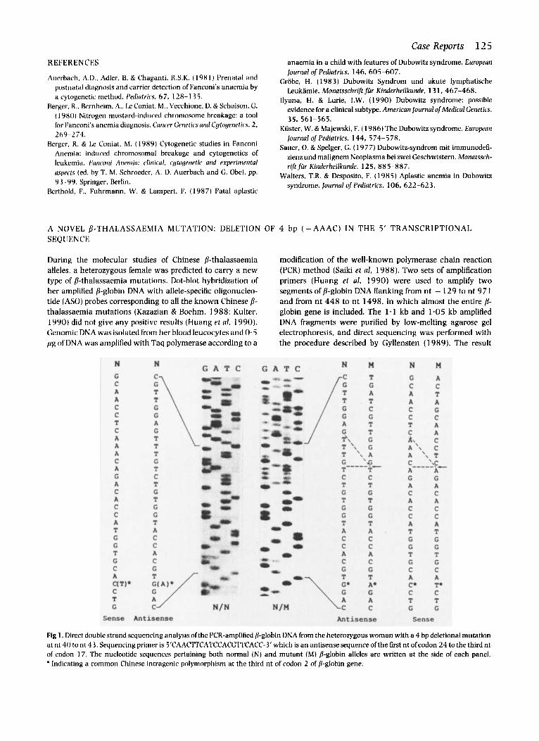
A NOVEL P-THALASSAEMIA MUTATION: DELETION OF 4 bp (-AAAC) IN THE 5' TRANSCKIPTIONAL SEQUENCE
During the molecular studies of Chinese 8-thalassaemia alleles, a heterozygous female was predicted to carry a new type of 8-thalassaemia mutations. Dot-blot hybridization of her amplified 8-globin DNA with allele-specific oligonucleo- tide (ASO) probes corresponding to all the known Chinese 8- thalassaemia mutations (Kazazian & Roehm. 1988: Kulter. 1990) did not give any positive results (Huang et al. 1990). Genomic DNA was isolated from her blood leucocytes and 0.5 pg of DNA was amplified with Taq polymerase according to a
modification of the well-known polymerase chain reaction (PCR) method (Saiki et al. 1988). Two sets of amplification primers (Huang et al. 1990) were used to amplify two segments of b-globin DNA flanking from nt - 129 to nt 971 and from nt 448 to nt 1498. in which almost the entire 8- globin gene is included. The 1 * 1 kb and 1.05 kb amplified DNA fragments were purified by low-melting agarose gel electrophoresis, and direct sequencing was performed with the procedure described by Gyllensten (1 989). The result
Fig 1 . Direct double strand sequencing analysis of the PCR-amplified P-globin DNA from the heterozygous woman with a 4 bp deletional mutation at nt 40 to nt 43. Sequencing primer is S'CAACTTCATCCACGTTCACC-3' which is an antisense sequence ofthe first nt of codon 24 to the third nt of codon 17. The nucleotide sequences pertaining both normal (N) and mutant (M) P-globin alleles are written at the side of each panel. * Indicating a common Chinese intragenic polymorphism at the third nt of codon 2 of fi-globin gene.