chromatin compaction. introduction difference between procaryotic and eucaryotic genome -e. coli: 1x...
TRANSCRIPT

Chromatin Compaction

INTRODUCTION
• Difference between procaryotic and eucaryotic genome
- E. Coli: 1X- Yeast genome: 4X- Fruit fly genome: 40X- Human genome: 1 000X
• How is eucaryotic DNA packaged in cells?

Level of organization of DNA

• DNA tightly bound to a group of small basic proteins histones
• Histones constitute ~1/3 of the total mass of the genetic material
• Chromatin = nucleoproteins + DNA
• 5 types of histones: H1, H2A, H2B, H3 & H4

Histones
• Found in all eukaryotic nuclei
• High content of + charged side chains (lysine and arginine)
• Can exist in different forms due to post-translational modifications
important in packaging DNA

Conservation through time
• H2A, H2B, H3 & H4 highly conserved among species (H4 of calf thymus and pea seedlings)
• H1 more variable in different species
• Unit evolutionary period
the time in which the sequence has changed by 1% after the divergence of two evolutionary lines

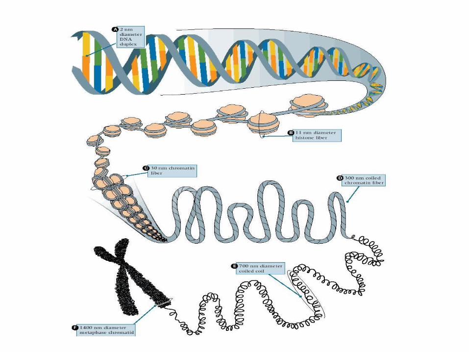
First order of DNA compaction

- Core DNA = 146 bp
- Linker DNA = 8-114 bp (usually 55bp)
- DNA turns 1 and ¾ times around histone octamer.
In 1974 : First model of primary structure of chromatin compaction by Roger Kornberg :
Pieces of evidence that led to his model :
- Protein composition of chromatin
- Electronic micrographs
- X-ray diffraction experiments
- Experiments using nucleases
- Histone octamer formation

Protein composition of chromatin
- High concentration of histones in chromatin- Equal amounts of H2A H2B H3 and H4- Half as many H1
→ H2A H2B H3 and H4 have similar roles ?

Electronic micrography observations
• Beads on a string, the 10nm fiber
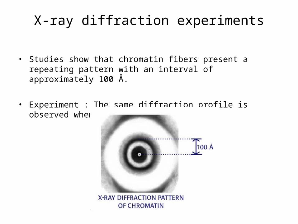
X-ray diffraction experiments
• Studies show that chromatin fibers present a repeating pattern with an interval of approximately 100 Å.
• Experiment : The same diffraction profile is observed when the H1s are removed.

Experiments using nucleases
• Experiment: Digest chromatin with rat liver nuclease at low concentration. (or micrococcal nuclease)
• Electrophoresis of the digested chromatin material.
A regular pattern of bands on the gel, approx. every 200 bp
→ Histones distributed evenly on DNA, and at point which they bind, protect DNA from nuclease digestion. (nuclease digests double stranded DNA)

Measuring the number of nucleosomes:
The results show that the number of spherical particles in a fragment of chromatin is equal to the number of 200 base-pair units. For example, a fragment with 600 base pairs of DNA consists of three 100 Å diameter articles, a trimer.
A

DNase I : Digestion of DNA only on one of its strands
Simple experiment proving DNA is wrapped around the octamer
- DNase I cuts core DNA only on portions of DNA which are not linked to the histones.
- After electrophoresis only 10 bp fragments are found.
DNase I cutting sites

Histone octamer formation
- Two highly conserved histones, H3 and H4, exist in solution as a specific tetramer (H3)2(H4)4, which behaves rather like an ordinary multi-subunit globular protein.
- The same can be said for H2A and H2B.
- The two tetramers form an octamer to which the DNA binds itself.

Summary
But how is the DNA linked to the Histone octamer?

Isolating the Core DNA


Second order of DNA compaction


How did we discovered H1’s position on the chromatin?

How did we discovered its position on the chromatin?A loose zigzag in which
the DNA enters andleaves the nucleosome at sites close together;

How did we discovered its position on the chromatin?
the zigzag
is tighter.

How did we discovered its position on the chromatin?
Nucleosome beads are no longer visible, the structure having openedto produce a fiber of DNA coated with histones;
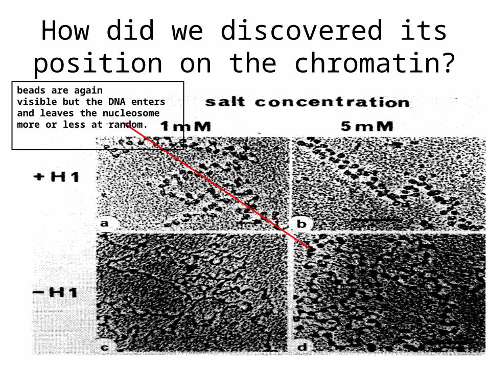
How did we discovered its position on the chromatin?
beads are againvisible but the DNA enters and leaves the nucleosome more or less at random.

Secondary Structure
• H1 : essential for the solenoid structure

Secondary Structure: Essential points
• The Solenoid is stabilized by H1 molecules• H1 has a globular body that binds to the outward
DNA• And 2 terminal arms (N- and C-) contact the
adjacent nucleosomes (actually the correspondent H1 histones that binds to the nucleosomes)
• 1 tour of solenoid = 6 nucleosomes

Third order of DNA compaction

Tertiary structure
• 300 nm coiled chromatin fibers
radial loops
• Non histone proteins (~30% of chromosomal proteins) would be implicated in the process
Non histones proteins
• Diverse group of protein not well understood
• Form a structural scaffolding to which loops of chromatin are attached
nuclear matrix (or chromosome scaffold)
• 2 scaffold proteins are found
Topoisomerase II
• Cleave and seal double stranded DNA
• Scaffolding located in the long axis of the metaphase chromosome
• DNA is tightly bound to this internal scaffold at S/MARs locus (scaffold/matrix attachment regions)

Histone-depleted metaphase chromosome
Protein scaffold
Loops of DNA

Histone-depleted metaphase chromosome
Scaffold/Matrix attachment regions

- THE END -