cholinesterase activity of lamellated ...cholinesterase activity of lamellated sensory corpuscles in...
TRANSCRIPT
CHOLINESTERASE ACTIVITY OF LAMELLATED SENSORY CORPUSCLES IN THE RAT LIP
II-SEI WATANABE *
CHIZUKA IDE **
Sensory nerve endings in the lip of mamais have been studied by light (Winkelmann 2 8 ; S e t o 2 3 ) , and electron microscopy (Munger 1 9 ; Halata and Munger 5 ) . Halata and Munger5 have described two different types of sensory corpuscles in the primate lip: the Meissner corpuscle and the simple coiled corpuscle, the latter may also be called Krause's end-bulb (Krause 1 6 ) , or mucocutaneous end-organs (Winkelmann 2 8 ) . The lamellated sensory corpuscle dealt with in the present study were small in size and have minimal terminal coiling of the axon, as described by Halata and Munger 5 , and were similar in organization to the simple coiled corpuscle.
On the other hand non-specific cholinesterase (ChE) activity has been demonstrated by light microscopy in Pacinian corpuscles (Hebb and Hill 6 ) , Meissner corpuscles (Hurley and Mescon 8 ; Cauna 2 ) and genital sensory corpuscles (Hurley 7 ) as well as by electron microscopy in Pacinian and digital corpuscles of the mouse (Ide and S a i t o 1 0 - 1 1 ) . The present study was performed to examine whether or not there is also ChE activity in lamellated corpuscles, which are presumed to be mechanoreceptors in the oral regions. Lamellated corpuscles were histochemically identified in the rat lip. The ChE activity of these corpuscles was examined by light and electron microscopy, and the ultrastructural localization of the enzyme reaction products was described.
MATERIAL A N D METHODS
Lips of fifteen Wistar rats were used in the present study. The animals were anesthetized by intraperitoneal injection of Nembutal (sodium pentobarbital, 50mg/kg body weight) .
Histochemicai procedures — Animals were fixed by intracardiac perfusion with 50ml of fixative containing 1.25% glutaraldehyde and 4% paraformaldehyde in a 0.065M phosphate buffer (pH 7.4) at room temperature. Lip t issues were dissected and immersed in the same fixative for 1-2 hrs at room temperature. After fixation, the
* Professor Titular de Anatomia do Departamento de Anatomia, Instituto de Ciências Biomédicas da Universidade de São Paulo.
** Professor Titular de Anatomia do Departamento de Anatomia, "Iwate Medicai University School of Medicine, Morioka, Japan>>. Acknowledgements — This study was supported by Japan International Cooperation Agency (JICA, Grant Nº 8205491).

specimens were washed overnight in a 0.065M phosphate buffer containing 8% sucrose at 4°C. Then 40 ^m-thick frozen sections were cut on a freezing microtome. Sections were incubated in the medium described by Karnovsky and Roots 15. Acetylthiocholine iodide (AThCh, Sigma), or butyrylthiocholine iodide (BuThCh, Sigma) was used as a substrate. The incubation medium was prepared as fol lows: 5mg AThCh or 6mg of BuThCh, 6.5ml of 0.1M sodium hydrogen maleate buffer (pH 6.0), 0.5ml of lOOmM sodium citrate, 1.0ml of 30mM copper sulfate, 1.0ml of distilled water, 1.0ml of 5 mM potassium ferricyanide and 1.5g sucrose. Incubation was carried out for 30-40 min at ice cold temperature with intermittent agitation. After incubation, the sections were washed three times in a 0.065M sodium hydrogen maleate buffer (pH 6.0) containing 8% sucrose. For light microscopy, sections were mounted with glycerin jelly on slide glass. For electron microscopy, sections were postfixed in a 1% osmium tetroxide solution adjusted to pH 7.4 with 0.1M phosphate buffer and then dehydrated through a graded series of alcohol and embedded in Epon 812. Epon sections, 1 ^m-thick were stained with toluidine blue to locate the corpuscles by l ight microscopy. Thin sections were cut with a diamond knife on an LKB Ultrotome and observed in a Hitachi H-700 electron microscope after staining with only uranyl acetate or double--staining with uranyl acetate and lead citrate.
Control and inhibitor experiments — A substrate-free medium was employed as
the control. Eserine (Sigma) 10 3 to 10" M in concentration was used to differentiate the cholinesterase activity from aliesterase activity. Iso-OMPA (tetraisopropyl pyro-
phosphoramidade, Sigma) 10 3 to 10 "J M was used for the inhibition of non-specific cholinesterase activity.
RESULTS
General morphology — N o organized sensory corpuscles other than lamellated corpuscles were found in the rat lip. Lamellated corpuscles were mainly located in

the dermal papillae of the lip at the transitional area from skin to mucosa (Fig. 1). Almost no or very few lamellated corpuscles were found in the skin region or in the mucosa area of the rat lip. The density of lamellated corpuscles in the transitional area was not so high: usually 1-2 corpuscles were found in a 40 ^m-thick frozen section. These lamellated corpuscles, measuring approximate ly 18-30 ^m in diameter, had several layers of asymmetrically arranged lamellae of the lamellar cells around axon terminals (Fig. 2), suggest ing that they were not so-called «Golgi-Mazzoni», but quite similar in organization to «glomerular terminals* (Seto 24; Munger 18). These corpuscles were somewhat coiled in the terminals of the axons (Fig. 3), indicating that they were in the same category of the simple coiled corpuscles (Halata and Munger 5). The lamellae were approximate ly 0.5-1 ^m in width (Fig. 2) and contained abundant intermediate filaments, occasional microtubules and mitochondria. Usually no endoplasmic reticulum was found in the cytoplasm of the lamellae. The lamellae were characterized by numerous caveolae on the plasma membrane. The periaxonal space between the axon terminals and their adjacent lamellae, or the interlamellar spaces between neighbouring lamellae were from 0.1 to 0.3 ^m in width. There were fine filaments or amorphous materials in the interlamellar spaces. Such amorphous materials appeared more or less electron opaque at the region near the plasma membrane of the lamellae, indicating that they were in nature basal laminalike materials.
Axon terminals were usually oval in profile and measured about 2.5-3 ^m in diameter. It is notable that axon terminals in the sensory corpuscles contained many mitochondria. Axon terminals contained also neurofilaments and some clear vesicles of about 50 ^m in diameter. Some finger-like cytoplasmic processes occasionally extend from the axon terminals.
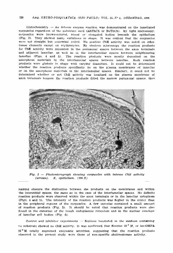
Histochemistry — An intense enzyme reaction was demonstrated on the lamellated corpuscles regardless of the substrate used (AhThCh or BuThCh). By l ight microscopy, corpuscles were brown-colored, round or elongated bodies beneath the epithelium (Fig. 3). They showed many variations in shape. It was evident that the corpuscles were not straight but somewhat coiled. No positive ChE activity was noted on othei t issue elements except on erythrocytes. By electron microscopy the reaction products for ChE activity were deposited in the periaxonal spaces between the axon terminals and adjacent lamellae, as well as in the interlamellar spaces between neighbouring lamellae (Figs. 4 and 5). The reaction products were mostly deposited on the amorphous materials in the interlamellar spaces between lamellae. Such reaction products were globule in shape with varying diameters. It could not be determined whether the reaction products specifically lie on the plasma membranes of lamellae or on the amorphous materials in the interlamellar spaces. Similary, it could not be determined whether or not ChE activity was localized on the plasma membrane of axon terminals because the reaction products filled the narrow periaxonal spaces, thus
making obscure the distinction between the products on the membranes and within the interstitial spaces, the same as in the case of the interlamellar spaces. No definite reaction products were observed within the axon terminals or in the lamellar cytoplasm (Figs. 4 and 5). The intensity of the reaction products was higher in the center than in the peripheral regions of the corpuscles. A few caveolae contained a small amount of reaction products (Fig. 5). It should be noted that reaction products were also found in the cisternae of the rough endoplasmic reticulum and in the nuclear envelope of lamellar cell bodies (Fig. 6).
Control and inhibitor experiments — Sections incubated in the medium containing
no substrate showed no ChE activity. It was confirmed that Eserine 10"4 M, or iso-OMPA
10 4 M totally supressed enzymatic activities, suggest ing that the reaction products observed in the present study were those of non-specific cholinesterase activity.
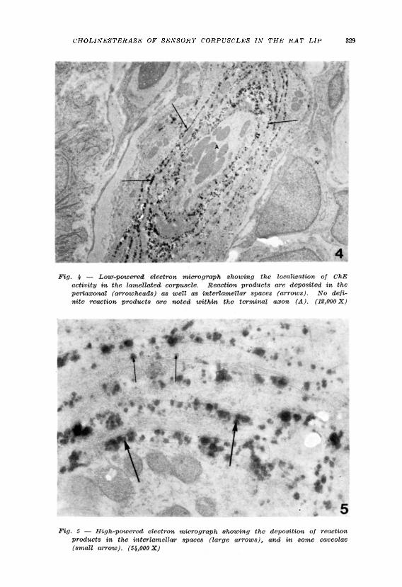
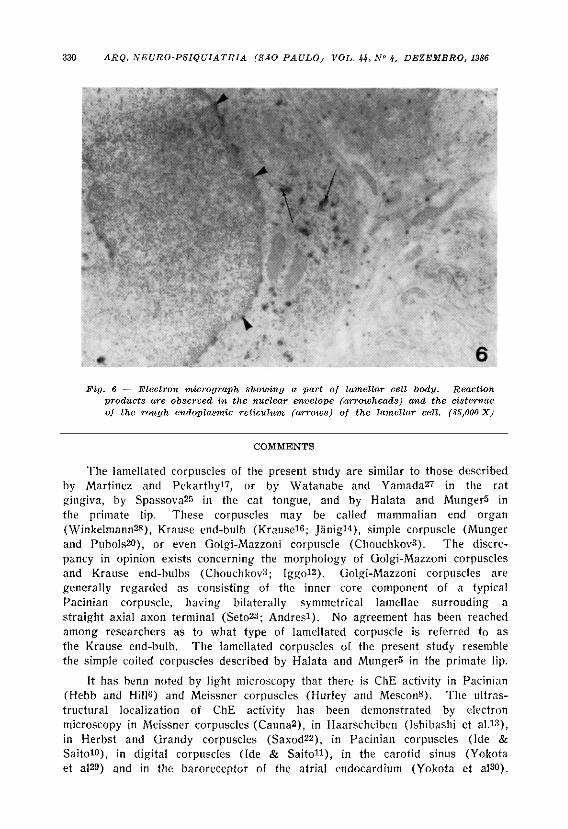
Fig. 6 — Electron micrograph showing a part of lamellar cell body. Reaction products are observed in the nuclear envelope (arrowheads) and the cisternae of the rough endoplasmic reticulum (arrows) of the lamellar cell. (35,000 X)
COMMENTS
The lamellated corpuscles of the present study are similar to those described by Martinez and Pekarthy 1?, or by Watanabe and Yamada 2? in the rat gingiva, by Spassova 2^ in the cat tongue, and by Halata and MungerS in the primate lip. These corpuscles may be called mammalian end organ (Winkelmann 2»), Krause end-bulb (Krause 1 6 ; Jãn ig 1 4 ) , simple corpuscle (Munger and Pubols2**), or even Golgi-Mazzoni corpuscle (Chouchkov 3 ). The discrepancy in opinion exists concerning the morphology of Golgi-Mazzoni corpuscles and Krause end-bulbs (Chouchkov 3; I g g o 1 2 ) . Golgi-Mazzoni corpuscles are generally regarded as consisting of the inner core component of a typical Pacinian corpuscle, having bilaterally symmetrical lamellae surrouding a straight axial axon terminal ( S e t o 2 3 ; Andres 1 ) . No agreement has been reached among researchers as to what type of lamellated corpuscle is referred to as the Krause end-bulb. The lamellated corpuscles of the present study resemble the simple coiled corpuscles described by Halata and Munger5 in the primate lip.
It has benn noted by light microscopy that there is ChE activity in Pacinian (Hebb and Hills) and Meissner corpuscles (Hurley and MesconS). The ultras-tructural localization of ChE activity has been demonstrated by electron microscopy in Meissner corpuscles (Cauna 2 ) , in Haarscheiben (Ishibashi et a l . 1 3 ) , in Herbst and Grandy corpuscles ( S a x o d 2 2 ) , in Pacinian corpuscles (Ide & Saito 1 (>), in digital corpuscles (Ide & S a i t o 1 1 ) , in the carotid sinus (Yokota et a l 2 9 ) and in the baroreceptor of the atrial endocardium (Yokota et a l 3 0) .

Ruffini corpuscles in the articular capsule of the cat elbow joint also were shown to have ChE activity (Toyoma26). The lamellated corpuscles of the present study exhibited ChE activity as in the case of the above cited mecha-noreceptors. It can be said from the findings of these previous studies that mechanoreceptors generally show ChE activity. This implies that histochemical demonstration of ChE activity is useful for detection of mechanoreceptors at the light microscopic level.
The fine localization of ChE activity in the mechanoreceptors at the electron microscopic level is not generally agreed upon. However, it can at least be said that ChE activity is localized not only on the lamellae plasma membrane, but also in the interlamellar as well as periaxonal spaces of the corpuscle, Dubovy4 claims that ChE activity is confined only to the inner core lamellae plasma membrane of vibratome sections of the cat mesentery Pacinian corpuscles. However, it has been demonstrated that the acetylcholinesterase activity is localized on the basal lamina of the motor end plate (Sanes et a l . 2 1 ) . ChE activity in the mouse digital corpuscles remains on the amorphous materials left after lamellar cells undergo atrophy following denervation (Ide9). These studies suggest that ChE activity can actually be located on the amorphous materials in the interlamellar as well as periaxonal spaces of the mechanoreceptors.
The functional significance of ChE in mechanoreceptors remains to be investigated. It may be that ChE is related to metabolic or nutritional aspects of the lamellar cells and/or axon terminals. Further it is possible that ChE might be associated with the trophic effects of lamellar cells on the receptors. Besides it is tempting to consider that ChE might have some relationship to the processes of stimuli-transduction in mechanoreceptors. However, there has been no evidence that ChE or lamellar cells are involved in such specific mechanisms of the mechanoreceptors.
It is not likely that the ChE is transported from the nerve cell bodies to the terminals, because no definite reaction is seen within the corpuscle's axon terminals. Considering that lamellar cell bodies have ChE activity in the nuclear envelope and the cisternae of the rough endoplasmic reticulum, it is probable that lamellar cells themselves can synthesize ChE.
SUMMARY
Non-specific cholinesterase (ChE) activity was demonstrated in lamellated sensory corpuscles of the rat lip by light and electron microscopy using Karnovsky and Root's method. ChE activity was present in the interlamellar spaces between neighbouring lamellae as well as in the periaxonal space between axon terminals and their adjacent lamellae. Reaction products of ChE activity were also deposited in some caveolae of the lamellar cell plasma membrane, and in the cisternae of the rough endoplasmic reticulum as well as in the nuclear envelope of lamellar cell bodies. No definite reaction products were detected within the axon terminals. These findings show that the lamellated corpuscles in the rat lip, like other mechanoreceptors, have an intense ChE

activity which is mainly associated with lamellar cells. It can be said that ChE histochemistry is useful to detect mechanoreceptors. The functional significance of ChE in mechanoreceptors is discussed.
RESUMO
Atividade de colinesterase em corpúsculos lamelares sensitivos no lábio de ratos.
A atividade de colinesterase inespecífica foi demonstrada em corpúsculos lamelares sensitivos do lábio de ratos através da microscopia óptica e eletrônica de transmissão, utilizando o método de Karnovsky & Roots. A atividade colinesterásica estava presente no espaço interlamelar entre as lamelas sucessivas, bem como no espaço entre o axônio terminal e a lamela adjacente. Produtos de reação da atividade colinesterásica eram bem evidentes em algumas cavéolas da membrana plasmática da célula lamelar e em cisternas do retículo endoplasmático granular, bem como na membrana nuclear. Produtos inespe¬ cíficos de reação foram detectados dentro do axônio terminal. Esses dados mostram que os corpúsculos lamelares do lábio de ratos, como outros mecanorre¬ ceptores, apresentam intensa atividade colinesterásica, a qual é associada principalmente com as células lamelares. Pode-se dizer que a histoquímica de colinesterase é útil para detectar os mecanorreceptores. A significância funcional da colinesterase em mecanorreceptores é discutida.
REFERENCES
1. ANDRES, K.H. — Uber die Feinstruktur der Rezeptoren an Sinushaaren. Z. Zellforsch. 75:339, 1966.
2. CAUNA, N. — The distribution of Cholinesterase in the cutaneous receptor organs, specially touch corpuscles of the human finger. J. Histochem. Cytochem. 8:367, 1960.
3. CHOUCHKOV, Ch. — Cutaneous receptors. Adv. Anat. Embryol. Cell Biol. 54:5, 1978.
4. DUBOVY, P. — The problem of the localization of Cholinesterase activity in Pacinian corpuscles by electron microscopy. Histochem. J. 15:287, 1983.
5. HALATA, Z. & MUNGER, B.L. — The sensory innervation of primate facial skin. II. Vermilion border and mucosa of lip. Brain Res. Rev. 5:81, 1983.
6. HEBB, C. & HILL, K.J. — PseudoCholinesterase in Pacinian corpuscles. Nature 175:597, 1955.
7. HURLEY, H.J. — Non-specific Cholinesterase in specialized sensory nerve endings of human genital skin. Brit. J. Derm. 70:284, 1958.
8. HURLEY, H.J., Jr. & MESCON, H. — Localization of non-specific Cholinesterase in Meissner's corpuscle in human skin. Brit. J. Derm. 68:290, 1956.
9. IDE, C. — Degeneration of mouse digital corpuscles. Amer. J. Anat. 163:59, 1982.
10. IDE, C. & SAITO, T. — Electron microscopic histochemistry of Cholinesterase activity of Vater-Pacini corpuscle. Acta histochem. cytochem. 13:298, 1980.
11. IDE, C. & SAITO, T. — Electron microscopic cytochemistry of cholinesterase activity of mouse digital corpuscle. Acta histochem. cytochem. 13:218, 1980.
12. IGGO, A. — Cutaneous receptors. In J.I. Hubbard (ed.): The Peripheral Nervous System. Plenum Press, New York, 1974, pg. 347.
13. ISHIBASHI, Y.; NIIMURA, M. & KAWAMURA, T. — Ultrastructural localization of cholinesterase activity in human Haarscheiben. Acta histocfiem. cytochem. 4:191, 1971.
14. JANIG, W. — Morphology of rapidly and slowly adapting mechanoreceptors in the hairless skin of the cat's hindfoot. Brain Res. 28:217, 1971.
15. KARNOVSXY, M.J. & ROOTS, L. — A <<irect-coloring>> thiocholine method for cholinesterase. J. Histochem. Cytochem. 12:219, 1964.
16. KRAUSE, W. — On the termination of nerves in the conjuctiva. J. Anat. 1:346, 1867.
17. MARTINEZ, I.R., Jr. & PEKARTHY, J.M. — Ultrastructure of encapsulated nerve endings in rat gingiva. Amer. J. Anat. 140:135, 1974.
18. MUNG-ER, B.D. — Cytology and ultrastructure of sensory receptors in the adult and newborn or mate tongue (development in the fetus and infant). In J.F. Bosma (ed.): Fourth Symposium on Oral Sensation and Perception, DHEW Publication No. (NIH) 73-546, 1973, pg. 75.
19. MUNGER, B.L.. — Cytology of mechanoreceptors in oral mucosa and facial skin of the rhesus monkey. In D.B. Tower & R.O. Brady (eds.): The Nervous System. The Basic Neuroscience. Raven Press, New York, 1975, vol. 1, pg. 71.
20. MUNGER, B.L.. & PUBOLS, L..M. — The sensorineural organization of the digital skin of the racoon. Brain Behav. Evol. 5:367, 1972.
21. SANES, J.R.; MARCHALL, L.M. & McMAHAN, U.J. — Reinnervation of muscle fiber basa_ lamina after removal of myofibers. J. Cell Biol. 78:176, 1978.
22. SAXOD, R. — Activité cholinestérasique des corpuscles sensoriels cutanés de Herbst et de Grandry. Histochemie 34:43, 1973.
23. SETO, H. — Studies on the Sensory Innervation — Human Sensibility. Igaku Shoin, Tokyo, 1963.
24. SETO, H. — Color Atlas on Sensory Innervation. Published by Department of Anatomy, Tohoku University School of Medicine. Sendai, Japan, 1977, pg. 20.
25. SPASSOVA, I. — Ultrastuctural relationships between the receptor nerve fiber and surrounding lamellae in Krause end-bulbs. Acta anat. 109:360, 1981.
26. TOYOMA, Y. — The morphology of sensory corpuscles in joint capsule: ultrastructural and histochemical rtudy. J. orthop. Assoc. 59:1985, in press.
27. WATANABE, I. & YAMAJ A, E. — The fine structure of lamellated nerve endings found in the rat gingiva. Arch. Hist. jpn. 46:173, 1983.
28. WINKELMANN, R.K. — Nerve Endings in Normal and Pathologic Skin. Charles C. Thomas, Springfield, 1960, pg. 81.
29. YOKOTA, R. ; IDE, C. : NITATORI, T. & ONODERA, S. — Cholinesterase activity in the carotid sinus baroreceptor. Acta histochem. cytochem. 15:537, 1982.
30. YOKOTA, R. ; IDE, C. ; NITATORI, T. & ONODERA, S. — Electron microscopy histochemistry of cholinesterase activity in the baroreceptor of the cat atrial endocardium. Acta histochem. cytochem. 16:129, 1983.
Departamento de Anatomia, Instituto de Ciências Biomédicas - Universidade de S. Paulo — Caixa Postal 4365 - 01000 - São Paulo. SP - Brasil.